© 2019 by the Serbian Biological Society How to cite this article: Čairović AD, Stanimirović DM, Krajnović TT, Dojčinović 347 BP, Maksimović VM, Cvijović-Alagić ILj. Recasting as a booster of Ag-Pd alloy
cytotoxicity: Induction of cell senescence prior to mass cell death. Arch Biol Sci. 2019;71(2):347-56.
Recasting as a booster of Ag-Pd alloy cytotoxicity: induction of cell senescence prior to
mass cell death
Aleksandra D. Čairović1,*, Dragan M. Stanimirović2, Tamara T. Krajnović3, Biljana P. Dojčinović4, Vesna M.
Maksimović5 and Ivana Lj. Cvijović-Alagić5
1Clinic for Prosthodontics, University of Belgrade, School of Dental Medicine 4, Rankeova 11 000 Belgrade, Serbia
2Department for Periodontology and Oral Medicine, University of Belgrade, School of Dental Medicine, Doktora Subotića 4, 11000 Belgrade, Serbia
3Department of Immunology, University of Belgrade, Institute for Biological Research “Siniša Stanković”, Bulevar despota Stefana 142, 11060 Belgrade, Serbia
4Institute of Chemistry, Technology and Metallurgy, University of Belgrade, Njegoševa 12, 11000 Belgrade, Serbia
5“Vinča” Institute of Nuclear Sciences, University of Belgrade, Mike Petrovića Alasa 12-14, 11351, Belgrade, Serbia
*Corresponding author: [email protected]
Received: March 5, 2019; Revised: March 15, 2019; Accepted: March 21, 2019; Published online: March 25, 2019
Abstract: The biological quality and chemical composition of alloys used in dental practice change during heat treatment. Often the residues of the previous cast are not disposed of but are reused and recycled until consumed. Thus, manufactured dental restorations have modified biological quality and chemical composition, and compromised biocompatibility. The aim of this study was to investigate the influence of repeated casting on the cytotoxicity of the silver-palladium (Ag-Pd) alloy. Our results showed that repeated casting of the Ag-Pd dental alloy affected its biocompatibility by promoting toxic-ity against transformed fibroblasts in a contact-independent manner. A strong decrease in cell proliferation, induction of senescence and massive cell death were observed in cultures exposed only to a medium previously incubated with dental alloy samples. The obtained data indicated that toxicity mediated by the accumulation of the Ag, Pd, Cu and Zn cations released from the Ag-Pd material was enhanced by recasting. The induction of cell senescence and subsequent apoptotic and necrotic death were accompanied by amplified intracellular production of reactive oxygen and nitrogen species, sug-gesting their involvement in the cell destruction process. Therefore, compromised biocompatibility after recasting with the Ag-Pd alloy can be the cause of serious local cell destruction, as observed in clinical practice.
Keywords: dental alloys; cytotoxicity; necrosis; recasting; reactive oxygen species
INTRODUCTION
The biocompatibility of dental alloys used in fixed prosthodontics is a fundamental property in view of the close and permanent contact with oral tissue. The term biocompatibility refers to the material features with respect to their application in medical practice, which must avoid any undesirable local or systemic effects and enable clinically optimal performance [1]. To reach this goal and the most appropriate cellular and tissue response, the potential cytotoxicity as the main element of biocompatibility, needs to be assessed.
Corrosion of dental alloys in the oral environment is followed by ion release [4,5], which dissolves in saliva and eventually affects the surrounding tissues and can be ingested and absorbed by enterocytes. The toxic effects depend on the rate of solvation, the destructive potential of the cation, reactions with the surrounding tissue and its concentration [6,7].
The microstructure of a dental alloy determines its mechanical, chemical and other properties. During the laboratory manufacturing process of dental work, dental alloys undergo changes. These changes, whether they are spontaneous or induced deliberately in order to improve the physical/mechanical properties, can cause decomposition of materials [8].
In everyday prosthetic practice, a silver-palladium (Ag-Pd) dental alloy is frequently used. This dental alloy has a very long history of use and has its basic application in fixed prosthetic dentistry. However, a dark-blue-colored area can often be found in the surrounding gingival tissue of a devitalized tooth that has been reinforced with a cast upgrade made of Ag-Pd dental alloy [9,10]. This gingival discoloration is the result of ion release [11]. The oral cavity is a very corrosive environment due to the electrolytic nature of saliva. In everyday practice, dental technicians do not dispose of the casting residue, but reuse the alloy remains and recast them [12]. It is very common for dental technicians to add about 50% of a new, not-cast-before dental alloy to improve the quality of the recast remains. By melting the dental alloy in a dental laboratory, the alloy absorbs some elements from the air and also releases elements through evaporation. Therefore, recast and recycled dental restorations possess altered biological qualities and consequently, changed biocompatibility [13,14].
This study investigated the influence of multiple recasting on Ag-Pd alloy cytotoxicity. The obtained results revealed the key role of toxic molecules released from the alloy in cell culture medium [15,16], which promoted an intracellular oxidative burst and massive necrosis. The observed effects were in clear correlation with recasting and the duration of exposure to dental materials. This paper provides important guidelines for dental practitioners on how to use dental alloys and avoid the toxic effects caused by recasting.
MATERIALS AND METHODS
Preparation of dental material
The tested dental alloy is a commercially available Ag-Pd dental alloy composed of silver (65%), palladium (21%), gold (4%), copper (9%) and zinc (1%). The samples were prepared at the Dental Laboratory of the Clinic for Prosthetic Dentistry, School of Dental Medicine, University of Belgrade. They were formed in the in-duction furnace as disks, 5 mm in diameter and 1 mm thick. Three groups of samples were used: P1 samples that were melted once and cast, then the surplus was melted three more times and cast (P4), and samples in which the surplus was melted four more times and cast (P8). The casting procedure was performed according to the manufacturer’s instructions. After casting, the samples were ground, polished and cleaned with alcohol and distilled water. The alloy samples were tested after the first, fourth and eight recasting cycles. The number of samples for every sample group was 6. As a negative control, glass disks (5 mm in diameter, 1 mm-thick) were used, being a material that is not cytotoxic.
Reagents and cell culture
Cell culture medium RPMI-1640 was obtained from Biowest (Riverside, MO, USA). Fetal calf serum (FCS), trypan blue (TB), phosphate-buffered saline (PBS), trypsin, dimethyl sulfoxide (DMSO) and 3-(4, 5-di-methylthiazol-2-yl)-2, 5-diphenyltetrazolium bromide (MTT) were purchased from Sigma Aldrich (St. Louis, MO, USA). 3H-timidin was obtained from Amersham
(Bucks, UK). Annexin V-FITC (AnnV) was from Bio-Legend (San Diego, CA, USA) and propidium iodide (PI) was obtained from BD Pharmingen (San Diego, CA, USA). Dihydrorhodamine-123 (DHR-123) was from Molecular Probes (Eugene, OR, USA). Murine fibro-sarcoma cell line L929 was obtained from the European Collection of Animal Cell Cultures (Salisbury, UK).
Trypan blue (TB) assay
Dental alloy samples as well as their glass equivalent that served as a negative control (dimensions 5×5×1 mm) were placed in the center of each well in 24-well plate, and L929 cells were seeded at densities of 5×10⁴/
respectively. To determine the cytotoxicity of the dental alloy samples by TB staining, the cells were trypsinized, washed and resuspended in PBS. The cell suspension was mixed with TB solution in a 1:1 ratio and cyto-toxicity was calculated using the following formula: TB+ cells×100/total number of cells [17].
Light microscopy
For microscopic assessment of cells in the presence of dental alloy/glass disks, L929 cells (2.5×10⁴/well) were
cultivated in 24-well plates in the presence of the alloy (5×5×1 mm). After 72 h of incubation, the morphol-ogy of cells in the surrounding area of the applied material was evaluated under an inverted microscope at 620× magnification. To evaluate the cytotoxicity of the medium preincubated with dental alloy samples, disks made from dental alloys or glass were incubated for 3 days in culture medium and conditioned medium was collected. L929 cells were seeded in 24-well plates until they reached 40-50% of confluence and then ex-posed to a mixed medium made from the conditioned medium and fresh culture medium at a 4:1 ratio. After 12 h and 24 h of incubation, the morphology of the cells was assessed by a Nikon inverted microscope TS2 (Tokyo, Japan) at 100× magnification.
MTT assay for detection of cell viability
Ag-Pg alloy samples/glass disks were placed in the cen-ter of each well in 96-cell plate. L929 cells were seeded (1×10⁴/well) and incubated for 72 h. The cell number
was determined by the MTT test, based on the reduction of MTT into formazan crystals by active mitochondria in viable cells [17]. After the cultivation period, 50 μL of MTT solution (0.5 mg MTT/mL culture medium) were added in each well and cells were further incubated at 37°C for 1 h. DMSO was added to dissolve the formazan and the absorbance was quantified at 540 nm using an automatic microplate reader (LKB5060-006).
[3H]-thymidine proliferation assay
[3H]-thymidine is a labeled DNA precursor that
incor-porates into the DNA of proliferating cells. The level of the radioactive signal reflects the proliferation rate. After a 3-day incubation with dental alloy samples
or glass, the medium was collected and mixed with fresh RPMI at a 4:1 ratio. L929 cells (1×10⁴/well) were
cultured in a 96-well plate in a medium prepared as described above and cultivated for 48 h. [3H]-thymidine
was added during the last 8 h of incubation at a final concentration 5µCi/mL. Cell cultures were harvested on an automatic harvester (Titertek Cell Harvester, Flow Laboratories) and [3H]-thymidine incorporation was
measured in a liquid scintillation counter (LKB-1219, Rackbeta, Helsinki, Finland).
Instrumental analysis
The concentrations of metals (Ag, Pd, Au, Cu, and Zn (µg/L)) in solution samples were determined by inductively-coupled plasma optical emission spectrom-etry (ICP-OES,iCAP 6500 Duo ICP, Thermo Fisher Scientific, Cambridge, UK). Multi-Element plasma standard solution 4, Specpure®, 1000 µg/mL, and Pre-cious Metals plasma standard solution, Specpure®, 100 µg/mL (Alfa Aesar GmbH & Co KG, Germany) were used to prepare the calibration solutions for ICP-OES measurement. The metals in the solutions were measured at the following emission wavelengths: Ag, I 328.068 nm; Pd, I 340.458 nm; Au, II 208.209 nm; Cu, I 324.754 nm, and Zn, I 213.856 nm. Quality control was carried out using blank samples, matrix-matched calibration solutions and triplicate analyses (n=3) of each sample. The reliability of measurements was accepted if the relative standard deviation was lower than 0.5%. The detection limits (LOD) for Ag, Pd, Au, Cu and Zn were 0.059,0.081, 0.064, 0.095 and 0.071 µg/L, respectively.
Detection of cell senescence
Dental alloy samples were incubated in the cell culture medium for 72 h (1 disk/1 mL of medium). After the indicated time interval, the conditioned medium was aspirated and mixed with fresh RPMI-1640 at a ratio of 4:1. L929 cells (1.5×10⁵/well) were seeded in
Annexin V-FITC/PI staining for cell death detection
Dental alloy samples were incubated in the cell cul-ture medium for 72 h. After the indicated time, the conditioned medium was aspirated and mixed with fresh RPMI-1640 at a 4:1 ratio. L929 cells (1.5×10⁵/
well) were seeded in 6-well plates, left overnight and exposed to medium prepared as described. After 12 and 24 h of incubation, the cells were trypsinized, collected, washed and stained according to the manufacturer’s instructions [18]. The presence of viable, early apop-totic and late apopapop-totic/necrotic cells was analyzed with CyFlow® Space Partec employing PartecFloMax® software (Partec GmbH, Münster, Germany).
DHR staining for the detection of reactive oxygen (ROS) and reactive nitrogen (RNS) species
L929 cells were stained with 1 μM dihydrorhoda-mine-123 for 20 min before seeding (1.5×10⁵/well)
in 6-well plates and exposing to conditioned medium mixed with fresh RPMI-1640 at a 4:1 ratio. After 24 h of incubation, the cells were trypsinized, collected, washed and resuspended in PBS as described previ-ously [19]. The presence of ROS/RNS was analyzed by CyFlow® Space Partec using PartecFloMax® software (Partec GmbH, Münster, Germany).
Statistical analysis
To analyze the significance of the differences between the controls and the Ag-Pd alloy P1 and corresponding recasts (P4 and P8), analysis of variance (ANOVA) was used, followed by the Student-Newman-Keuls test. A p value less than 0.05 was considered significant.
RESULTS
The presence of Ag-Pd alloy affected L929 cell viability and changed their morphology: correlation with duration of exposure and number of alloy recastings
To explore the cytotoxic influence of recasting of a fre-quently used Ag-Pd dental alloy, the mouse fibroblast cell line L929 was selected because these cells simulate
the appropriate microenvironment for dental mate-rial application and, according to their proliferative potential, provide a valuable setting for the assess-ment of eventual cytotoxicity. Cells were exposed to the dental alloy samples for 24 h and 72 h, and after the indicated time intervals they were collected and the TB assay was performed. Results clearly showed that an increased percentage of TB+ cells in the
pres-ence of material samples depended on recasting and the duration of cell exposure to dental alloys. The effect was moderate after 24 h of incubation while an additional 48-h incubation period resulted in the accumulation of nonviable cells in cultures exposed to P4 and P8 samples, reaching about 15% of the cell population (Fig. 1A). Internalized TB, which reflects compromised membrane permeability, indicated that the cells underwent necrosis. Furthermore, microscopic evaluation of L929 cells cultivated 72 h in the pres-ence of Ag-Pd alloys recast 1, 4 and 8 times, revealed a remarkable change in cell morphology and density in the alloy-surrounding area, which was in correlation with recasting number. As can be seen in Fig. 1B, 72 h of cultivation in the presence of P4 led to the atypical shape of neighboring cells. This effect became more profound in the presence of alloys that were recast 8 times. The cells acquired polygonal, flattened and/or elongated morphology with long processes. The ob-served change in phenotype when compared to cells cultivated only in culture medium or in the presence of glass clearly indicated that the presence of certain dental materials promoted the change in cell phenotype typical for differentiation or rapid aging accompanied by the loss of dividing potential. The appearance of sporadic orbicular cells that started to detach from the plastic bottom was in agreement with the data obtained by the TB assay.
Ag-Pd alloys abrogated L929 cell proliferation in correlation with the number of recastings: the effect was contact-independent
that recast dental material affected cell proliferation. Blocked division is a common cell reaction in response to moderate intoxication [20]. It represents a period of mitotic arrest that the cell requires in order to re-cycle damaged organelles, proteins and to repair DNA defects. If the damage overcomes the capacity of the cell to repair itself, the cell dies.
To explore the influence of recast-ing of the Ag-Pd alloy on cell prolif-eration, L929 cells were incubated in the presence of recasts (P1, P4, and P8) and the MTT test was performed after 72 h. The number of viable cells in this assay was quantified according to the total intensity of mitochon-drial respiration in culture. As seen in Fig. 2A, the number of viable cells was reduced by about 40% and was detected in cultures with dental alloy samples, but the number was not in significant correlation with repeated casting. Importantly, the presence of glass disks also affected the cell number by decreasing it by about 20%. This result suggested that the contact of inert material with cells also influenced the capacity of cells to divide.
In view of literature data about dental material toxicity based on the release of metal ions in the microen-vironment [3,21], we wanted to de-termine if the antiproliferative effect of recasting was connected, at least in part, to mediators released in the culture medium, or whether it hap-pened exclusively in the presence of the alloy. Fluctuation of respiration per cell in response to toxic stimuli from hyper- to hyporespiration can compromise the interpretation of the obtained data. The proliferation rate was evaluated using a more sensi-tive assay based on the incorporation of radioactive thymidine into DNA chains of newly-formed cells. The cells were cultivated in medium con-taining 80% of conditioned medium and collected after a 3-day-long incubation with dental alloy. After a 48-h-long incubation, cell proliferation was determined by [3H]-thymidine incorporation.
Inhibition of proliferation was observed, starting from cultures exposed to medium collected from P1. Posi-tive correlation between the number of recastings and
Fig. 2. Cell proliferation was disturbed in the presence of conditioned medium generated by incubation of the alloy samples with respect to recasting. L929 cells (1×10⁴/well) were
seeded in the presence of alloys and cultivated for 3 days when the MTT assay was per-formed (A); medium collected after a 3-day-long incubation of dental alloy samples was transferred to the L929 cultures and mixed with fresh RPMI in 4:1 ratio. Proliferation rate was assessed by the [3H]-thymidine assay after 48 h (B). The results were calculated from
triplicate values and are presented as the percentage of the control±SD from one representa-tive of three independent experiments. *#p<0.05; * – non-treated control; # – glass sample.
Fig. 1. Multiple recasting of Ag-Pd alloy potentiated its cytotoxicity. L929 cells were cultivated in the presence of Ag-Pd for 24 and 72 h. After the indicated time points, the TB assay was performed. The data are presented as the percentage of TB+cells±SD,
calculated from triplicates of one representative from three independent experiments. *#p<0.05, * – non-treated control, # – previous number of recastings (A). B – Changes in
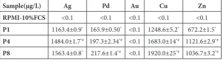
the antiproliferative effect of the conditioned medium was observed. However, the difference was not statisti-cally significant (Fig. 2B). Importantly, the measured inhibition of proliferation was more profound than the one detected by the MTT assay. While the MTT assay revealed the reduction in cell number by 22% in the presence of glass disks, the medium collected from glass disks after a 3-day-long incubation did not affect cell proliferation. These results clearly indicated that the carriers of dental alloy toxicity were the cations released in the medium, and subsequently transferred to the cells. To prove this, the concentrations of all metals present in the Ag-Pd alloy (in µg/L) in the conditioned medium were determined by ICP-OES and the results are presented in Table 1. Although the quantity of Ag was approximately 9-10 times higher than that of Pd, the concentrations of both metals were significantly increased by recasting. Having in mind that the amounts of Cu and Zn in the Ag-Pd alloy are minor in comparison to Ag and Pd, the rate of their release in conditioned medium surpassed the most abundant metals. Their concentration, as expected, was also amplified by casting. Only the presence of the Au cation in conditioned medium was very low (<0.1µg/L). We speculated that all released metals contributed to the enhancement of cytotoxicity provoked by repeated casting of the tested dental material.
Table 1. Metal ion content in the medium collected from the al-loys after a 3-day-long incubation determined using ICP-OES.
Sample(µg/L) Ag Pd Au Cu Zn
RPMI-10%FCS <0.1 <0.1 <0.1 <0.1 <0.1
P1 1163.4±0.9* 165.9±0.50* <0.1 1248.6±5.2* 672.2±1.5*
P4 1484.0±1.7*# 197.3±2.34*# <0.1 1683.0±14*# 1121.6±2.9*#
P8 1563.4±0.8* 217.6±1.4*# <0.1 1920.0±25*# 1036.7±3.2*#
P1, P4, P8 - alloys recast 1, 4 and 8 times, respectively;
p<0.05 in comparison to *control or #P1; calculated from 3 repeated measurements.
Mediators released from Ag-Pd surface in medium provoked cell senescence and dramatic cell death in L929 cultures: the effect was in correlation with the number of recastings
To precisely define how the conditioned medium in which Ag-Pd alloys were incubated affected cell vi-ability, L929 cells were exposed to medium collected from dental alloy samples after a 72-h incubation and diluted with fresh medium at a 4:1 ratio. Microscopic
evaluation of cell cultures exposed to medium col-lected from P1 and P4 recasts revealed the presence of enlarged atypical cells together with rounded cells displaying a tendency to detach from the plastic bottom. Almost all L929 cells exposed to P8 medium assumed the shape of dying cells (Fig. 3A).
The presence of enlarged cells in cultures exposed to P1 and P4 media indicated that cells had entered the process of senescence prior to cell death. To examine this further, we performed the B galactosidase assay, a specific test for detecting cell senescence. As can be seen in Fig. 3B, increasing B galactosidase expression was observed in all cultures exposed to conditioned media, as follows: P1>P4>P8. Enlarged cells corre-sponding to the senescent phenotype were replaced with a dominant dying phenotype in cultures exposed to the P8 medium.
To further define the type of cell death, Ann V/ PI double staining of cells cultivated in the presence of medium obtained from P1 to P8 was performed after 12 and 24 h of incubation. As can be seen in Fig. 3C, repeated casting of Ag-Pd alloys triggered cell death, which was in clear correlation with the number of repeated castings. The process was rapid and even after 12 h considerable amounts of Ann+/
PI- (early apoptotic) and double positive cells (late
apoptotic/necrotic) were observed. Clearly, repeated casting amplified the toxicity of the medium and accordingly potentiated the presence of cells with damaged cell membrane permeability. This process was even more pronounced after an additional 12 h of incubation, when in the culture exposed to P8 medium overt necrosis was detected. According to the presence of Ann+/PI- cells, which are considered as
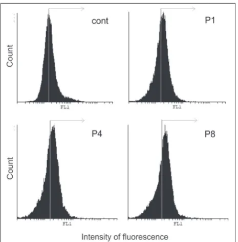
Mediators accumulated in the medium upon incubation with the Ag-Pd alloy triggered ROS/RNS production in L929 cells and the effect was potentiated by recasting
To explore the involvement of an oxidative burst in response to molecules released from the alloy samples in the medium, intracel-lular ROS/RNS were assessed using DHR-123 dye, which is specific for the detection of hydrogen peroxide, peroxynitrite and hypochlorous acid produced in cells. In-cubation of L929 cells in media containing toxic cations released from the recast alloys revealed significantly increased ROS/RNS production (Fig. 4). The observed effect correlated with the number of recastings. It can be expected that reactive molecules produced by cells in response to toxic me-diators eluted from casts were at least in part responsible for the rapid dying of cells, observed as mass cell necrosis.
DISCUSSION
In the course of designing different dental alloys, more than 25 elements were used, making the evaluation of their biocompat-ibility extremely complex. According to many attractive features as well as a more acceptable price in comparison to gold, Ag-Pd alloys have been increasingly used in prosthetic dentistry. The safety of their application is still under investigation, especially because of reports of increased incidence of allergic reactions to palladium, as well as its toxicity in cell cultures [22,23]. In everyday dental practice, it is not a rare occurrence to observe a dark blue tattoo on the gingiva surrounding a tooth with a cast upgrade made from Ag-Pd dental alloy. Unlike developed countries
Fig. 3. Medium containing molecules released from recast Ag-Pd alloy triggered senescence and mass cell death. L929 cells were exposed to conditioned medium when confluence of the culture was about 50%. Changes in cell morphology were visualized by inversion microscopy (magnification 100×) (A); L929 cells (1.5×10⁵/
well) were seeded, left overnight and then exposed to conditioned medium for 24 h for senescence detection or 12 h and 24 h for detection of cell death. After the indicated time points, β-galactosidase assay (B) and Ann/PI double staining (C) were performed and samples were analyzed by flow cytometry.
Fig. 4. Mass cell death of cells exposed to Ag-Pd medium cells was followed by enhanced production of ROS/RNS relative to the number of recasting. L929 cells (1.5×10⁵/well) were stained
where noble alloys as well as all-ceramic materials are preferentially used, in undeveloped countries, base and less frequently noble metal alloys are the most prevalent types. Moreover, in everyday practice, the surplus left after casting of dental alloys is not disposed of but is recast until subsequent reuse. This recycling of dental alloys leads to chemical and biological changes in ma-terials brought about by repeated heating and melting. Such use of dental alloys leads to the production of fixed dentures with unknown chemical composition and changed biocompatibility.
In this study, the toxicity of Ag-Pd alloy dependent on recasting was investigated, using L929 fibrosarcoma cells of mouse origin. The selected cell line is the most widely used model for this type of study [24] as the cells simulate the appropriate microenvironment and present a good model for the assessment of material influence on cell proliferation rate and viability. On the other hand, the tumorigenic potential of L929 cells is relatively low [25]. The assessment of cytotoxicity covers two key aspects of dental alloy toxicity: contact-mediated and contact-mediated by cations eluted from the alloy surface in a liquid microenvironment [26,27]. Toxicity is indicated by changes in cell morphology, decreased capacity of a cell to proliferate and/or the presence of different types of cell death. These parameters are usually causally connected and dependent on the level and duration of exposure to toxins.
Cultivation of L929 cells in the presence of Ag-Pd alloy disks influenced cell morphology that was de-pendent on the duration of exposure and the number of recastings. An obvious positive correlation between cell phenotype changes in areas around the samples was detected in the presence of the alloy recast 4 times, with more profound changes detected in the presence of alloy recast 8 times after a 72 h-long incubation. The observed flattened appearance of cells with long processes pointed to the development of a more dif-ferentiated and less proliferative phenotype under the influence of dental material. In parallel, the appearance of rounded, shrunken cells that started to detach from the bottom of the wells, the number of which was in correlation with the number of recastings, indicated loss of cell vitality. This was additionally confirmed by the TB assay which revealed a remarkable accumula-tion of cells in the terminal phase of destrucaccumula-tion, with diminished membrane permeability and internalized
TB dye. Examination of cell viability in the presence of the dental alloy showed a significant decrease in the number of cells, but without an obvious correla-tion with repeated recasting. This was ascribed to a deficiency of the test procedure used to assess cell proliferation. Namely, mitochondrial respiration, which is very sensitive to toxic influence, is an unreliable parameter for cell number quantification since it can oscillate even in viable, functionally-preserved cells. Further assessment of contact-independent toxicity of the alloys was performed with a more reliable assay for cell proliferation, using determination based on the incorporation of radioactively-labeled [3H]-thymidine
in daughter cell DNA.
In agreement with literature data about the release of toxic cations from the surface of dental materials into the microenvironment, this paper showed that the highly toxic potential of the Ag-Pd alloy was trans-ferred to the medium, and that it exerted a powerful cell destructive potential independent of the intimate contact of the cells and the dental alloy. Analysis of the metal content of the medium collected from the alloys following the 3-day-long incubation revealed a dramatic accumulation of all metals, except Au, as a result of release from the tested material after each recasting. Apoptotic cell death was followed by pro-gressive failure of cell membrane function, leading to massive necrosis of cells incubated in this medium, in clear correlation with the metal content. Intoxication was proportional to the number of recastings and duration of exposure. In cultures exposed to medium with the lowest metal content, the highest fraction of cells with the senescent phenotype was found, indicat-ing that cells underwent cell senescence. These cells are non-proliferative and their appearance decreased overall with the increase of the metal ion content in the medium, and were replaced with dead cells. The presence of cell senescence triggered by intoxication upon exposure to the specific content of the medium collected from casts explained the result of the pro-liferation assay, confirming a radical decrease in cell division rate even in the presence of less toxic metals.
presence of these molecules in intracellular compart-ments was connected with the apoptotic cell death promoted by the repeated recasting of materials [6]. However, the transfer of Co-Ni alloy toxicity against cells by the conditioned medium, and its influence on the endogenous level of ROS/RNS was not evaluated. Hornez et al. [29] showed that gold, palladium, plati-num and indium ions had no cytotoxic effect, while chromium, copper and silver ions exerted medium toxicity. According to the same authors, nickel, zinc and cobalt ions are extremely toxic [29]. Also, Wataha et al. [30] claimed that silver and copper, or molecules generated from their interactions with gold present in the alloy, appear to be carriers of cytotoxicity [30]. Wa-taha et al. [30] found that the release of silver, copper, palladium and zinc was higher in saline-bovine serum albumin (BSA) solution compared to saline alone [30]. Accordingly, the accumulation of ions released from dental materials in the medium used for cell cultiva-tion was potentiated by medium supplementacultiva-tion with BSA or FCS. This further implies that the process of metal ion accumulation is accelerated in the presence of plasma proteins in vivo. On the other hand, several studies mentioned different triggers for the release of metal ions from the alloy in the context of the com-plex microenvironment in the oral cavity. This data is extremely important in the context of dental alloy recycling [31].
The results presented in this paper underline the change in Ag-Pd alloy biocompatibility following repeated casting, confirming the cytotoxic effect of mediators released from the dental alloy, which is cumulative and amplified by recasting. This result opens up questions about how changes in biocompat-ibility as a consequence of recasting are reflected on Ag-Pd alloy behavior in the oral cavity in response to numerous microenvironmental challenges.
CONCLUSION
This study showed that the cytotoxicity of the Ag-Pd dental alloy significantly increased with recast-ing. This phenomenon was in correlation with the duration of exposure and is completely transferred in the conditioned medium containing the molecules released from the alloy during the incubation period. Cytotoxicity was manifested through the induction
of senescence prior to cell death accompanied by in-creased production of ROS and RNS. The presented data show that recasting of the Ag-Pd alloy affects its biocompatibility and can cause serious complications in clinical practice.
Acknowledgments: This work was supported by the Serbian Min-istry of Education, Science and Technological Development (Grant Nos. 45012 and 173013). We thank to Mrs. Jasmina Ninkov, B.A. in English Language and Literature, a Commissioned Scientific and Technical Translator, for proofreading the entire manuscript. Author contributions: All authors have made significant contri-butions to this study and in the preparation of the manuscript; A. Čairović, T. Krajnović and B. Dojčinović preformed the experi-ments; D. Stanimirović wrote the draft manuscript; V. Maksimović and I. Cvijović-Alagić prepared the figures. All authors have made substantial contributions to the concept and design of the study and revision of the manuscript. All authors have read and approved the final manuscript.
Conflict of interest disclosure: The authors declare no conflict of interest.
REFERENCES
1. Cortizo MC, De Mele MF, Cortizo AM. Metallic den-tal material biocompatibility in osteoblastlike cells: cor-relation with metal ion release. Biol Trace Elem Res. 2004;100(2):151-68.
2. Wataha JC, Nelson SK, Lockwood PE. Elemental release from dental casting alloys into biological media with and without protein. Dent Mater. 2001;17(5):409-14.
3. Zhao LB, Si J, Wei Y, Li SR, Jiang YJ, Zhou R, Liu B, Zhang H. Toxicity of porcelain-fused-to-metal substrate to zebrafish
(Danio rerio) embryos and larvae. Life Sci. 2018;203:66-71.
4. Čairović A, Maksimović V, Radović K, Đurišić S. The effect of recasting on biological properties of Ni-Cr dental alloy. Srp Arh Celok Lek. 2016;144(11-12):574-9.
5. Vaillant-Corroy A-S, Corne P, De March P, Fleutot S, Cley-mand F. Influence of recasting on the quality of dental alloys: A systematic review. J Prosth Dent. 2015;114(2):205-11. 6. Čairović A, Đorđević I, Bulatović M, Mojić M, Momčilović
M, Stošić-Grujičić S, Maksimović V, Maksimović-Ivanić D, Mijatović S, Stamenković D. In vitro assessment of Ni-Cr and Co-Cr dental alloys upon recasting: Cellular compat-ibility. Dig J Nanomater Bios. 2013;8(2):877-86.
7. Zhang CY, Cheng H, Lin DH, Zheng M, Ozcan M, Zhao W, Yu H. Effects of recasting on the biocompatibility of a Ni-Cr alloy. Chin J Dent Res 2012;15(2):105-13.
8. Imirzalioglu P, Alaaddinoglu E, Yilmaz Z, Oduncuoglu B, Yilmaz B, Rosenstiel S. Influence of recasting different types of dental alloys on gingival fibroblast cytotoxicity. J Prosth Dent. 2012;107(1):24-33.
10. Al-Hiyasat, AS, Darmani, H. The effects of recasting on the cytotoxicity of base metal alloys. J Prosth Dent. 2005;93(2):158-63.
11. Venclikova Z, Benada O, Bartova J, Joska L, Mrklas L. Metal-lic pigmentation of human tooth and gingiva: morphological and immunological aspects. Dent Mater J. 2007;26(1):96-104.
12. Ristić Lj, Miljković Ž, Ilić S, Đurić T. Prebojenost gingive u prisustvu fiksnih zubnih nadoknada.Vojnosanit Pregl. 2005;62(5):371-6.
13. Ristić Lj, Ilić S, Živanović A. Influence of metal-ceramic fixed dental restorations on the occurence of discoloration of gingiva. Vojnosanit Pregl. 2006;63(4):409-13.
14. Garcia-Contreras R, Sakagami H, Nakajima H, Shimada J. Type of cell death induced by various metal cations in cul-tured human gingival fibroblasts. In vivo. 2010;24(4):513-7. 15. Yamazaki T, Zamayaki A, Hibino Y, Chowdhury SA, Yakote
Y, Kanda Y, Sakagami H, Nakajima H, Shimada J. Bio-logical impact of contact with metals on the cells. In vivo. 2006;20(5):605-11.
16. Horasawa N, Marek M. The effect of recasting on corrosion of a silver-palladium alloy. Dent Mater. 2004;20(4):352-7. 17. Harhaji Lj, Mijatović S, Maksimović-Ivanić D, Stojanović I,
Momcilović M, Maksimović V, Tufegdzić S, Marjanović Z, Mostarica-Stojković M, Vucinić Z, Stosić-Grujicić S. Anti-tumor effect of Coriolusversicolor methanol extract against mouse B16 melanoma cells: in vitro and in vivo study. Food ChemToxicol. 2008;46(5):1825-33.
18. Maksimović-Ivanić D, Bulatović M, Edeler D, Bens-ing C, Golić I, Korać A, Kaluđerović GN, MijatovićS. The interaction between SBA-15 derivative loaded with Ph3Sn(CH2)6OH and human melanoma A375 cell line: uptake and stem phenotype loss. J BiolInorg Chem. 2019; 24(2):223-34.
19. Paskas S, Mazzon E, Basile MS, Cavalli E, Al-Abed Y, He M, Rakocevic S, Nicoletti F, Mijatovic S, Maksimovic-IvanicD. Lopinavir-NO. A nitric oxide-releasing HIV protease inhibi-tor, suppresses the growth of melanoma cells in vitro and
in vivo. Invest New Drugs. 2019;DOI:10.1007/s10637-019-00733-3.
20. Schmalz G, Garhammer P. Biological interactions of dental cast alloys with oral tissues. Dent Mater. 2002;18(5):396-406. 21. Jia XY, Wang QA, Meng H, Sun H, Zhan DS. Effects of dif-ferent dental alloys on cytotoxic and apoptosis related genes expression in L929 cells. J Hard Tissue Biol. 2010;19(2):95-100.
22. Maksimović-Ivanić D, Mijatović S, Miljković Đ, Harhaji-Trajković Lj, Timotijević G, Mojić M, Dabideen D, Fan Cheng K, McCubrey JA, Mangano K, Al-Abed Y, Libra M, Garotta G, Stošić-Grujičić S, Nicoletti F. The antitumor properties of a nontoxic, nitric oxide–modified version of saquinavir are independent of Akt. Mol Cancer Ther. 2009;8(5):1169-78.
23. Wataha JC. Biocompatibility of dental casting alloys: a review. J Prosthet Dent. 2000;83(2):223-34.
24. Williams DF. On the mechanisms of biocompatibility. Bio-materials. 2008;29(20):2941-53.
25. Wataha JC, Messer RL. Casting alloys. Dent Clin North Am. 2004;48(2):499-512.
26. Aberer W, Holub H, Strohal R, Slavicek R. Palladium in dental alloys - the dermatologists’ responsibility to warn? Contact Dermatitis. 1993;28(3):163-5.
27. Mareci D, Sutiman D, Cailean A, Bolat G. Comparative cor-rosion study of Ag-Pd and Co-Cr alloys used in dental appli-cations. Bull Mater Sci. 2010;33 (4):491-500.
28. Colavitti R, Finkel T. Reactive oxygen species as mediators of cellular senescence. IUBMB Life. 2005;57(4-5):277-81. 29. Hornez JC, Lefevre D, Joly D, Hildebrand HF. Multiple
parameter cytotoxicity index on dental alloys and pure met-als. Biomol Eng. 2002;19(2-6):103-17.
30. Wataha JC, Lockwood PE, Nelson SK, Bouillaguet S. Long-term cytotoxicity of dental casting alloys. Int J Prosthodont. 1999;12(3):242-8.