R E S E A R C H
Open Access
Temporal regulation of mRNAs for select bone
morphogenetic proteins (BMP), BMP receptors
and their associated SMAD proteins during
bovine early embryonic development: effects of
exogenous BMP2 on embryo developmental
progression
Kyung-Bon Lee
1,2,4, Joseph K Folger
1,2, Sandeep K Rajput
1,2and George W Smith
1,2,3*Abstract
Background:We previously demonstrated embryotrophic actions of maternal (oocyte-derived) follistatin during bovine early embryogenesis. Classical actions of follistatin are attributed to inhibition of activity of growth factors including activins and bone morphogenetic proteins (BMP). However, temporal changes in BMP mRNA in early bovine embryos and the effects of exogenous BMP on embryo developmental progression are not understood. The objectives of present studies were to characterize mRNA abundance for select BMP, BMP receptors and BMP receptor associated SMADs during bovine oocyte maturation and early embryogenesis and determine effects of addition of exogenous BMP protein on early development.
Methods:Relative abundance of mRNA forBMP2,BMP3,BMP7,BMP10,SMAD1,SMAD5,ALK3,ALK6,ALK2,BMPR2, ACVR2AandACVR2Bwas determined by RT-qPCR analysis of germinal vesicle (GV) and in vitro matured metaphase II (MII) oocytes and in vitro produced embryos collected at pronuclear, 2-cell (C), 4C, 8C, 16C, morula and blastocyst stages. Effects of addition of recombinant human BMP2 (0, 1, 10 and 100 ng/ml) during initial 72 h of embryo culture on early cleavage (within 30 h post insemination), total cleavage, development to 8C-16C and blastocyst stages and blastocyst mRNA abundance for markers of inner cell mass (NANOG) and trophectoderm (CDX2) were also determined.
Results:Abundance of mRNA forBMP2,BMP10,SMAD1,SMAD5,ALK3, ALK2,BMPR2andACVR2Bwas elevated in MII oocytes and/or pronuclear stage embryos (relative to GV) and remained elevated through the 8C -16C stages,
whereasBMP3,BMP7andALK2mRNAs were transiently elevated. Culture of embryos to the 8C stage in the
presence ofα-amanitin resulted in increased abundance for all of above transcripts examined relative to untreated 8C embryos. Effects of addition of exogenous BMP2 on early cleavage rates and rates of development to 8C-16C
and blastocyst stages were not observed, but BMP2 treatment increased blastocyst mRNA forCDX2andNANOG.
(Continued on next page)
* Correspondence:[email protected]
1
Department of Laboratory of Mammalian Reproductive Biology and Genomics, Michigan State University, East Lansing, MI 48824, USA 2
Department of Animal Science, Michigan State University, East Lansing, MI 48824, USA
Full list of author information is available at the end of the article
(Continued from previous page)
Conclusions:Abundance of maternally derived mRNAs for above BMP system components are dynamically regulated during oocyte maturation and early embryogenesis. Exogenous BMP2 treatment does not influence progression to various developmental endpoints, but impacts characteristics of resulting blastocysts. Results support a potential role for BMPs in bovine early embryogenesis.
Keywords:Ovary, Bovine, Oocyte, Embryo, Blastocyst, BMP, BMP receptor, SMAD
Background
Oocyte developmental competence was defined by Sirard
et al.[1] as the capacity of the oocyte to resume meiosis, cleave after fertilization, help promote embryonic develop-ment and implantation, and bring a pregnancy to term in good health [1]. Our previous studies support a positive functional role for maternal (oocyte-derived) follistatin in bovine oocyte competence. Follistatin mRNA is positively associated with developmental competence in two distinct bovine models of egg quality [2,3]. Furthermore, follistatin supplementation during the first 72 h of bovine embryo culture (until embryonic genome activation) enhanced proportion of embryos that cleaved early and proportion of embryos developing to the blastocyst stage in a dose dependent fashion [3]. Follistatin treatment also increased total blastocyst cell numbers specifically through an in-crease in trophectoderm (TE) cells, with no effect on numbers of inner cell mass (ICM) cells and increased blastocyst mRNA for the TE specific transcription factor
CDX2 [3]. We also observed similar effects of follistatin treatment on early cleavage and rates of development to blastocyst stage for rhesus monkey embryos [4], demon-strating potential translational relevance of results from the bovine model system. However, the mechanisms re-sponsible for stimulatory effects of follistatin on multiple indices of bovine early embryonic development to date still remain elusive.
Follistatin was initially classified as a high affinity bind-ing protein inhibitbind-ing activin action [5]. However, stimu-latory effects of exogenous activin treatment on early cleavage and blastocyst rates for bovine embryos were also observed suggesting a potential alternative mechan-ism of follistatin action [3]. Follistatin can also bind and regulate activity of multiple additional TGFβsuperfamily members including BMPs [6-8]. A prominent role for BMPs in regulation of patterning of early embryos has been described [9]. Furthermore, BMPs have been impli-cated in regulation of trophoblast differentiation in hu-man embryonic stem cells [10]. However, less is known about their role during preimplantation embryonic de-velopment, particularly in farm species.
In this study, we investigated temporal changes in mRNA abundance for multiple BMP, BMP receptors and their associated SMADs in bovine oocytes and
embryos and the source of such transcripts (maternal versus embryonic) in bovine embryos coincident with em-bryonic genome activation. We also determined the effect of exogenous BMP2 on multiple endpoints relevant to bo-vine early embryonic developmental progression. Results demonstrate dynamic regulation of maternal mRNAs for BMP system components during bovine early embryonic development and effects of exogenous BMP2 treatment on indices of cell lineage determination in bovine blasto-cysts. Results also suggest embryotropic actions of follista-tin on bovine early embryogenesis are likely not mediated by antagonism of BMP2 signaling.
Methods
Oocyte collection, in vitro maturation, in vitro fertilization and embryo culture
Bovine oocytes used for all described experiments were obtained from ovaries harvested at a local abattoir. As-piration and in vitro maturation of oocytes, in vitro fertilization and culture of embryos was performed as previously described [3]. Cumulus-oocyte complexes (aspirated from 2–7 mm visible follicles) with > 4 compact cumulus cell layers and homogeneous cyto-plasm were matured in TCM 199 [supplemented with 0.2 mM sodium pyruvate, 5 mg/ml gentamicin sulfate, 6.5 mM L-glutamine, 156 nM bovine LH (Sioux Bio-chemical, Sioux Center, Iowa), 15.6 nM bovine FSH (Sioux Biochemical), 3.67 nM 17β-estradiol and 10% v/v defined FBS (Hyclone, Logan, UT)] for 24 h at 38.5°C, 5% CO2 in air with maximum humidity. For in vitro fertilization, matured oocytes were washed and co-incubated with sperm for 20 h in fertilization medium (114 mM NaCl, 25 mM NaHCO3, 3.2 mM KCl, 0.34 mM NaH2PO4, 0.183 mM penicillin-G, 16.6 mM sodium lac-tate, 0.5 mM MgCl2:6H2O, 2.7 mM CaCl2:2H2O, 0.2 mM
Temporal changes in BMP system mRNA abundance in bovine oocytes and early embryos
Germinal vesicle (GV) and metaphase (MII) stage (oo-cytes), pronuclear (PN), 2-cell (C), 4C, 8C, 16C, morula and blastocyst stage embryos (n = 4 pools of 10 each), were collected as we described previously [3,11]. For GV and MII stage oocyte RNA samples, cumulus cells were completely removed by hyaluronidase (0.1%) digestion and repeated pipetting and denuded oocytes in groups of 10 each were snap frozen in 100 μl lysis solution (RNAqueous Micro Kit, Ambion Inc, Austin, TX) and stored at −80°C until RNA isolation. Embryo samples were also processed as described above in groups of 10 per sample per stage with PN embryos harvested at 20 h post insemination (hpi), 2C embryos collected 33 hpi, 4C embryos 44 hpi, 8C embryos 52 hpi, 16C embryos 72 hpi, and morulas and blastocysts at 5 and 7 d post in-semination respectively. 250 femtograms of polyadeny-lated GFP RNA were added to each sample prior to RNA extraction. After finishing RNA isolation and cDNA synthesis, cDNA was diluted 1:5 and mRNA quantification for BMP2, BMP3, BMP7, BMP10, ALK2,
ALK3, ALK6, BMPR2, ACVR2A, ACVR2B, SMAD1 and
SMAD5 mRNAs was performed in duplicate for each sample by qPCR using our procedures reported previ-ously [11]. Relative expression levels were calculated using theΔΔCT method with RPS18as the housekeep-ing gene [12], RPS18 mRNA abundance is very stable across MII through 16C stages and then is increased in later stages coincident with increase in cell numbers (Additional file 1: Figure S1). Abundance of exogenous control (GFP) RNA was also measured to account for variation in RNA recovery and cDNA synthesis across samples and exogenous GFP mRNA abundance was similar (P > 0.2) across samples. Primer sequences and fragment sizes for all transcripts measured are included in Table 1 and PCR efficiencies for all primer sets were between 90 and 103%.
Effect of transcriptional inhibition on BMP system mRNA abundance
To determine the effects of inhibition of transcription on mRNA abundance in in vitro derived embryos, pre-sumptive zygotes were cultured in serum free KSOM with 0.3% BSA with or without the addition of 50μg/ml α-amanitin (Sigma, St. Louis, MO). The 8C embryos were then collected at 52 h post insemination (n = 4 pools of 10 embryos per group) and placed in lysis buffer and snap frozen and stored as above until RNA isolation. Half the RNA was subjected to reverse transcription using oligo(dT) primers as described above for use in quantification of polyadenylated transcripts for genes of interest. The remaining RNA was transcribed using ran-dom hexamers for quantification of adenylated and
deadenylated (total) transcripts for genes of interest. The cDNA produced was subjected to qPCR for the BMP system components as described above. Data were nor-malized relative to abundance of endogenous control (RPS18).
Effect of BMP2 supplementation on bovine embryo developmental progression
To examine the effect of BMP2 supplementation on early cleavage rate (assessed 30 hpi), rate of development to 8C-16C stage (assessed 72 hpi), blastocyst rate (assessed 7 d post insemination) and the abundance of mRNA for the TE marker (CDX2) and ICM marker (NANOG) in resulting blastocysts, presumptive zygotes were cultured in KSOM medium supplemented with 0.3% BSA containing 0, 1, 10 or 100 ng/ml BMP 2 (30 presumptive zygotes per group, 4 replicates). The 8C-16C stage embryos were then separated 72 h post fertilization and cultured in fresh KSOM medium (minus exogenous BMP2) supplemented with 0.3% BSA and 10% FBS until d 7. Blastocysts were harvested at d 7 post fertilization (n = 4 pools of 5 blastocysts each per treatment) and lysed and frozen as above until RNA iso-lation and RT-qPCR analysis as described above.
Statistical analysis
All data were analyzed using one way ANOVA in SAS followed by Fishers Protected Least Significant Differ-ence Test to determine differDiffer-ences between means. For embryo culture experiments, % data were arc-sin trans-formed prior to analysis. Data are presented as mean ± SEM.
Results and discussion
Temporal regulation of BMP mRNA abundance during oocyte maturation and early embryogenesis
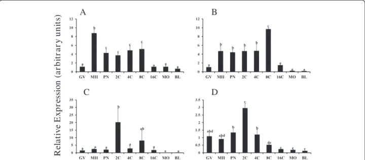
Transcriptome analysis of human oocytes indicates that multiple key components of the TGFβ superfamily sig-naling pathway are potentially active [13] and previous studies support a functional role for TGFβ superfamily members during bovine oocyte maturation and early embryogenesis [3,14,15]. Over 20 members of the BMP subfamily have been described [16], and expression of BMP in the bovine ovary has been extensively studied [17-19]. Abundance of specific mRNA transcripts during oocyte maturation and early embryonic development is under complex regulation and influenced by post tran-scriptional and trantran-scriptional mechanisms in a stage specific fashion [20]. Results of present studies revealed unique temporal changes in mRNA abundance for above BMP examined during oocyte maturation and early em-bryogenesis (Figure 1). Relative abundance of mRNA for
BMP3(Figure 1C) andBMP7(Figure 1D) mRNA abun-dance did not change during meiotic maturation. For
BMP2 and BMP10, mRNA abundance remained ele-vated in early embryos after fertilization until declining by the 16C stage (P < 0.05) and remained low in morula and blastocyst stage embryos (Figure 1A and B). In con-trast, relative mRNA abundance for BMP3 was transi-ently elevated (>15 fold) in 2C embryos (P < 0.05) and did not differ at other time points examined (Figure 1C).
BMP7 mRNA (Figure 1D) was also transiently elevated at the 2C stage and was lowest at 16C, morula and blastocyst stages (P < 0.05). Results demonstrate dy-namic, ligand specific temporal regulation of mRNA abundance for BMP2,BMP3, BMP7 and BMP10during bovine oocyte maturation and early embryogenesis.
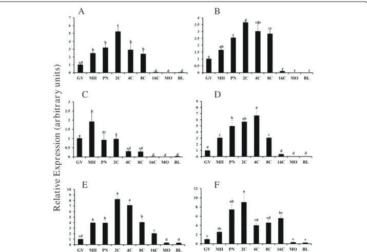
The BMPs bind to multiple TGFβ superfamily recep-tors [16]. Expression of BMP receprecep-tors in bovine ovarian sections and or isolated oocytes has been reported
previously [19], but temporal regulation of mRNA abun-dance for BMP receptors during oocyte maturation and early embryogenesis is not understood. In the current study, temporal changes in abundance of mRNA in bo-vine oocytes and early embryos for the prominent Type I (ALK3, ALK6, ALK2) and Type II receptors (BMPR2,
ACVR2A, ACVR2B) that bind BMPs was examined (Figure 2). Abundance of mRNA for ALK3 (Figure 2) was increased in MII oocytes (relative to GV stage), fur-ther increased, albeit transiently at the 2C stage, de-creased in 4C and 8C embryos and further dede-creased in 16C, morula and blastocyst stage embryos (P < 0.05). In contrast,ALK6mRNA (Figure 2B) is elevated at the PN stage, further increased, albeit transiently at the 2C stage and decreased at 16C, morula and blastocyst stages (P < 0.05).ALK2mRNA (Figure 2C) is transiently elevated in MII oocytes relative to oocytes collected at the GV stage, and reduced even further in embryos
Table 1 Sequence of primers for real time RT-PCR for TGFβsuperfamily members and receptors,CDX2andNANOG
Gene Genbank accession number Primer sequence Size (bp)
BMP2 BC134682 F: 5′-AAGGCCCTTGCTTGTCACTTT-3′ 72
R: 5′-TGCTTGCCGCTTTTCTCTTC-3′
BMP3 XM_587912 F: 5′-ATCTGTGGCTGAGCTGCTTGT-3′ 62
R: 5′-GGAAGGGCTGCCTGAGTCT-3′
BMP7 XM_612246 F: 5′-TGCCACTAGCTCTTCCTGGAA-3′ 65
R: 5′-TGAGAGACCCAGGATCCAGAA-3′
BMP10 XM_583418 F: 5′-CGCCCACGAGCAATTCC-3′ 66
R: 5′-TCCCCAGGTCCGTTGGA-3′
SMAD1 BC116117 F: 5′-CACCATGAACTGAAACCATTGG-3′ 68
R: 5′-GATGCACACCTCCTTCTGCTT-3′
SMAD5 DV821574 F: 5′-GCAACGTTTCCTGATTCTTTCC-3′ 74
R: 5′-GGCGGGTAGGGACTATTTGG-3′
ALK3 NM_001076800 F: 5′-TCAGCGAACTATTGCCAAACAG-3′ 75
R: 5′-CCCATCCACACTTCTCCGTATC-3′
ALK6 NM_001105328 F: 5′-CCCACCCCTCGTCCAAAG-3′ 63
R: 5′-GACCGAGTCTTCTGGACAATGG-3′
ALK2 BC133311 F: 5′-TTGGCCTCATCATTTTGTCTGT-3′ 70
R: 5′-CGGAGAGCAACTCCCAATAGG-3′
ACVR2A NM_174227 F: 5′- CCACAAACCCGCCATATCTC -3′ 185
R: 5′- TAGCACCCTCTAACACCTCTG -3′
ACVR2B NM_174495 F: 5′-GGAGCCATCAACTTCCAGAG-3′ 121
R: 5′-GCATGTACTCATCCACAGGTC-3′
BMPR2 XM_002685492 F: 5′-AACACCACTCAGTCCACCTC-3′ 120
R: 5′-GTCAGCATCCTATATCCAAAGCA-3′
NANOG NM_001025344 F: 5′-AAAGTTACGTGTCCTTGCAAACG-3′ 73
R: 5′-GAGGAGGGAAGAGGAGAGACAGT-3′
CDX2 AM293662 F: 5′-FCGTCTGGAGCTGGAGAAGGA-3′ 70
collected at 4C stage and beyond (P < 0.05). For the type II receptors,BMPR2(Figure 2D) is increased in MII oo-cytes relative to GV stage, further increased post fertilization through the 4C stage and subsequently de-clines through 16C stage (P < 0.05). While mRNA for
ACVR2B (Figure 2E) but notACVR2A (Figure 2F) was increased in MII oocytes relative to GV stage, mRNA for both ACVR2B and ACVR2A was elevated at PN stage and remained elevated through the 16C stage, and was further reduced at morula and blastocyst stages (P < 0.05; Figure 2E and F). Observed temporal regulation of type I and Type II BMP receptor mRNAs supports a potential intrinsic role for BMPs in meiotic maturation and early embryogenesis.
Signal transduction by members of the TGFβ super-family is mediated primarily by the SMAD pathways, with BMP signal transduction linked to SMAD1, SMAD5 and SMAD8 [16]. Thus changes in mRNA for BMP receptor associated SMADs during oocyte matur-ation and early embryogenesis were also investigated (Figure 3). Messenger RNA for SMAD8 was undetect-able in bovine oocytes and early embryos. Messenger RNA for SMAD1 (Figure 3A) and SMAD5 (Figure 3B) were increased in MII oocytes relative to GV stage (P < 0.05). For SMAD1, mRNA remained elevated after fertilization until the 4C stage and was further decreased in 16C, morula and blastocyst stage embryos (P < 0.05; Figure 3A). For SMAD5, mRNA was further increased after fertilization at PN, 2C and 4C stages and lowest at 16C, morula and blastocyst stages (P < 0.05; Figure 3B).
Effect of embryo culture in the presence of the
transcriptional inhibitorα-amanitin on BMP, BMP receptor and SMAD1 and SMAD5 mRNA abundance
Messenger RNA transcript adenylation and deadenyla-tion are prominent mechanisms of reguladeadenyla-tion of mRNA abundance during oocyte maturation and early develop-ment until initiation of embryonic genome activation [20,21]. Given abundance of above BMP, BMP type I re-ceptor and SMAD transcripts was elevated in early em-bryos prior to or through the 8C stage, studies were done to determine the source (maternal versus embry-onic) ofBMP2,BMP3,BMP7,BMP10,SMAD1,SMAD5,
ALK2,ALK3andALK6, transcripts detected in early bo-vine embryos. When presumptive zygotes were treated with 50 μg/ml of the RNA polymerase II inhibitor α-amanitin for 72 h, relative abundance of mRNA for all BMP system components examined was increased at the 8C stage compared to control embryos (Table 2). Results suggest that such transcripts present in early embryos at 8C stage are maternal in origin and that post transcrip-tional regulation of mRNA abundance for such genes may in fact be transcription dependent. Furthermore ele-vated transcript abundance was observed when reverse transcription of mRNA was conducted using oligo dT primers and not when random hexamers were utilized (Table 2), suggesting that transcript deadenylation may help contribute to the stage specific decrease in tran-script abundance for BMP, BMP receptors and their re-ceptor associated SMAD in the current studies. Since removal of the poly-A tail inhibits translation and is a
B
A
D
C
R
ela
tive E
xp
ression
(a
rb
it
ra
ry u
n
it
s)
starting point for RNA degradation [22], results suggests that down regulation of above transcripts may be im-portant during stages of embryo development after em-bryonic genome activation.
Effect of BMP 2 supplementation on embryo
development and trophectoderm and inner cell mass marker mRNA abundance in resulting blastocysts
Previous studies demonstrated a positive association of oocyte follistatin expression with developmental compe-tence [2] and potent embryotrophic actions of follistatin during early embryogenesis including enhanced propor-tion of embryos cleaving early, increased numbers of embryos developing to the blastocyst stage, and elevated mRNA for the TE cell markerCDX2in resulting blasto-cysts [3]. Classical actions of follistatin are commonly at-tributed to high affinity binding and inhibition of activity of the TGFβ superfamily member activin [5]. However, previous studies showed stimulatory effects of activin treatment on rates of early cleavage and blastocyst
development, suggesting that actions of follistatin on bo-vine embryos are likely nonclassical [3]. However, follis-tatin can also bind at a lower affinity and inhibit activity of BMPs [7,17]. The current studies demonstrated dy-namic temporal changes in maternal mRNA for several BMP, type I and II BMP receptors and their receptor as-sociated SMADs in bovine embryos prior to genome ac-tivation. Therefore to further elucidate the potential role of BMPs in bovine early embryogenesis, effects of BMP2 supplementation during first 72 h of in vitro embryo cul-ture (through 8C-16C stage) on above developmental endpoints were investigated. The addition of 1, 10 or 100 ng/ml of recombinant human BMP2 did not alter percentage of early cleaving embryos, total cleavage rates, percent embryos developing to 8C-16C stage or blastocyst rate (Figure 4A-D). However supplementation with 100 ng/ml of BMP2 increased mRNA for the TE marker CDX2 and the ICM marker NANOGrelative to embryos cultured without BMP2 (Figure 4E-F). Thus, while BMP2 supplementation did not impact efficiency
R
ela
tive E
xp
ression
(a
rb
it
ra
ry u
n
it
s)
B
A
D
C
F
E
of bovine in vitro embryonic development as measured by numbers of embryos reaching a transferable (blastocyst stage), BMP2 treatment did however impact characteristics of resulting blastocysts as measured by abundance of mRNA for CDX2and NANOG. These re-sults are in contrast to rere-sults obtained following treat-ment with exogenous follistatin [3] whereby stimulatory effects onCDX2 mRNA, but notNANOG mRNA were observed and suggesting biological actions of follistatin on blastocyst cell allocation are manifest specifically on the TE lineage.
A growing body of evidence suggests that oocyte secreted factors can enhance oocyte developmental competence and embryo developmental progression [14,15,23]. Our previous studies support a role for en-dogenous, oocyte-derived follistatin in promoting bovine embryo developmental progression and cell allocation to the TE lineage [3]. Results were obtained with follistatin treatment during initial stages of embryo culture (d 1–3) up to embryonic genome activation. To our knowledge, effects of exogenous BMP2 treatment during culture of bovine embryos have not been previously reported. Addition of BMP2 or BMP4 during in vitro maturation
did not impact rates of meiotic maturation and cumulus expansion or rates of embryonic development to blasto-cyst stage following in vitro fertilization [19]. However, evidence supports a role for oocyte-derived GDF9 and BMP15 in promoting oocyte developmental competence as addition of these growth factors exogenously during in vitro maturation can enhance rates of development to the blastocyst stage and blastocyst cell allocation to TE [14,15]. Thus, levels of endogenous BMP2 available dur-ing in vitro maturation and embryo culture may not be limiting to embryo developmental capacity in vitro as reflected by rates of development to the blastocyst stage. However, the current studies do demonstrate that bovine embryos can respond to BMP2 stimulation during initial 3 d of culture in vitro with an increase in blastocyst mRNA for NANOG and CDX2 measured 4 d later and demonstrate effects of BMP2 treatment on indices of cell allocation mediated well after treatment administration. The mechanisms responsible for increased blastocyst
NANOGandCDX2mRNA in response to BMP2 stimu-lation are not known. However, BMP4 mediated induc-tion of CDX2 mRNA expression [24] and promoter activity [25] have been described in other cell lines and
Table 2 Effects ofα-Amanitin treatment on mRNA abundance in bovine 8C embryos
Gene RT with oligo dT RT with random hexamers
Control α-Amanitin treated Control α-Amanitin treated
SMAD1 1.15 ± 0.27a 9.08 ± 1.19b 1.03 ± 0.16 1.77 ± 0.17
SMAD5 1.17 ± 0.35a 7.33 ± 1.27b 1.02 ± 0.12 1.45 ± 0.17
BMP2 1.10 ± 0.27a 3.19 ± 0.76b 1.25 ± 0.49 1.30 ± 0.21
BMP3 1.02 ± 0.11a 2.85 ± 0.60b 1.15 ± 0.36 3.26 ± 0.88
BMP7 1.02 ± 0.12a 3.36 ± 0.66b 1.17 ± 0.40 2.96 ± 1.31
BMP10 1.04 ± 0.19a 3.82 ± 0.25b 1.07 ± 0.23 1.59 ± 0.31
ALK3 1.23 ± 0.37a 9.41 ± 0.72b 1.06 ± 0.22 1.42 ± 0.31
ALK6 1.12 ± 0.25a 5.43 ± 0.71b 1.05 ± 0.20 1.38 ± 0.16
ALK2 1.27 ± 0.50a 5.73 ± 1.34b 1.02 ± 0.13 0.86 ± 0.09
Note.Different letters (a and b) represent significantly different value (P≤0.05).
B
A
R
ela
tive E
xp
ression
(a
rb
it
ra
ry
u
n
it
s)
in human ES cells and signaling for both BMP2 and BMP4 is mediated via SMAD1/5 [16]. However, SMAD2/ 3 pathways are linked toNANOG promoter regulation in embryonic stem cells and antagonistic to BMP signaling and differentiation to TE fate [26]. While the addition of BMP2 had no effect on the proportion of embryos that cleaved early or developed to the 16C or blastocyst stages, the highest dose of BMP2 did increase bothNANOGand
CDX2mRNA compared to control embryos at the cysts stage. NANOG is a marker of the ICM of the blasto-cysts and is important for maintaining pluripotency of the cells in the ICM [27]. In contrast CDX2 is a marker for TE cells and is in fact required for the establishment of the TE in the blastocysts of mice and cattle [28,29]. NANOG and CDX2 also cross regulate each other to pro-mote proper blastocyst formation [30]. As BMP2 supple-mentation increases mRNA for both markers it is possible that BMP2 enhances the differentiation of the two cell types, rather than promoting one cell type over the other.
Conclusions
In summary, results of present studies demonstrate pro-nounced temporal regulation of mRNA for select BMP ligands, type I and II receptors and cognate intracellular signaling molecules during bovine early embryonic de-velopment. This study was limited in that it only exam-ined changes in abundance rather than accompanying changes in protein abundance. However, to our know-ledge, temporal changes in abundance of mRNA for spe-cific BMP includingBMP2,BMP3,BMP7andBMP10 at specific stages of bovine early embryonic development have not been examined previously. While stimulatory effects of BMP2 on early cleavage and development to 8C to 16C and blastocyst stages were not noted in re-sponse to BMP2 supplementation of culture media (at doses tested) during first 72 h of development, increased mRNA forCDX2andNANOGwas detected in resulting blastocysts, demonstrating a functional BMP2 signaling system in early bovine embryos. Furthermore, distinct
A
F
E
D
C
B
results observed in present studies suggest that embryo-trophic actions of follistatin reported in previous studies [3] likely are not linked to inhibition of endogenous BMP2 activity.
Additional file
Additional file 1: Figure S1.Temporal changes in RPS18 mRNA during oocyte maturation and early embryogenesis in vitro. Quantitative real time RT-PCR analysis was performed on samples of germinal vesicle (GV) and metaphase II (MII) stage oocytes and in vitro derived embryos collected at the pronuclear (PN), 2-cell (2C), 4-cell (4C), 8-cell (8C), 16-cell (16C), morula (MO) and blastocyst (BL) stages (n = 4 pools of 10 oocytes/ embryos per pool). Data are shown as mean ± SEM. Values with different superscripts across time points denote significant differences (P < 0.05).
Competing interests
The authors declare that they have no competing interests.
Authors’contributions
KBL collected samples, isolated RNA, performed RT-qPCR analysis, performed embryo culture experiments and contributed to design of studies and inter-pretation of results. JKF analyzed data and contributed to design of studies, interpretation of results and helped draft the manuscript. SR performed RT-qPCR analysis and contributed to design of studies and interpretation of re-sults. GWS conceived the study, contributed to design of experiments and interpretation of results and helped draft the manuscript. All authors read and approved the final manuscript.
Acknowledgements
This project was supported by the National Institute of Child Health and Human Development of the National Institutes of Health under award number R01HD072972.
Author details
1
Department of Laboratory of Mammalian Reproductive Biology and Genomics, Michigan State University, East Lansing, MI 48824, USA. 2
Department of Animal Science, Michigan State University, East Lansing, MI 48824, USA.3Department of Physiology, Michigan State University, East Lansing, MI 48824, USA.4Department of Biology Education, College of Education, Chonnam National University, Gwangju 500-757, Republic of Korea.
Received: 25 February 2014 Accepted: 7 July 2014 Published: 15 July 2014
References
1. Sirard MA, Richard F, Blondin P, Robert C:Contribution of the oocyte to embryo quality.Theriogenology2006,65:126–136.
2. Patel OV, Bettegowda A, Ireland JJ, Coussens PM, Lonergan P, Smith GW:
Functional genomics studies of oocyte competence: evidence that reduced transcript abundance for follistatin is associated with poor developmental competence of bovine oocytes.Reproduction2007,
133:95–106.
3. Lee KB, Bettegowda A, Wee G, Ireland JJ, Smith GW:Molecular determinants of oocyte competence: potential functional role for maternal (oocyte-derived) follistatin in promoting bovine early embryogenesis.Endocrinology2009,150:2463–2471.
4. VandeVoort CA, Mtango NR, Lee YS, Smith GW, Latham KE:Differential effects of follistatin on nonhuman primate oocyte maturation and pre-implantation embryo development in vitro.Biol Reprod2009,
81:1139–1146.
5. Nakamura T, Takio K, Eto Y, Shibai H, Titani K, Sugino H:Activin-binding protein from rat ovary is follistatin.Science1990,247:836–838. 6. Balemans W, Van Hul W:Extracellular regulation of BMP signaling in
vertebrates: a cocktail of modulators.Dev Biol2002,250:231–250. 7. Lin SY, Morrison JR, Phillips DJ, de Kretser DM:Regulation of ovarian function
by the TGF-beta superfamily and follistatin.Reproduction2003,126:133–148.
8. Otsuka F, Moore RK, Iemura S, Ueno N, Shimasaki S:Follistatin inhibits the function of the oocyte-derived factor BMP-15.Biochem Biophys Res Commun 2001,289:961–966.
9. Kimelman D, Pyati UJ:Bmp signaling: turning a half into a whole.
Cell2005,123:982–984.
10. Giakoumopoulos M, Golos TG:Embryonic stem cell-derived trophoblast differentiation: a comparative review of the biology, function, and signal-ing mechanisms.J Endocrinol2013,216:R33–R45.
11. Bettegowda A, Patel OV, Ireland JJ, Smith GW:Quantitative analysis of messenger RNA abundance for ribosomal protein L-15, cyclophilin-A, phos-phoglycerokinase, beta-glucuronidase, glyceraldehyde 3-phosphate dehydro-genase, beta-actin, and histone H2A during bovine oocyte maturation and early embryogenesis in vitro.Mol Reprod Dev2006,73:267–278.
12. Livak KJ, Schmittgen TD:Analysis of relative gene expression data using real-time quantitative PCR and the 2(−Delta Delta C(T)) Method.
Methods2001,25:402–408.
13. Kocabas AM, Crosby J, Ross PJ, Otu HH, Beyhan Z, Can H, Tam WL, Rosa GJ, Halgren RG, Lim B, Fernandez E, Cibelli JB:The transcriptome of human oocytes.Proc Natl Acad Sci U S A2006,103:14027–14032.
14. Hussein TS, Sutton-McDowall ML, Gilchrist RB, Thompson JG:Temporal effects of exogenous oocyte-secreted factors on bovine oocyte develop-mental competence during IVM.Reprod Fertil Dev2011,23:576–584. 15. Hussein TS, Thompson JG, Gilchrist RB:Oocyte-secreted factors enhance
oocyte developmental competence.Dev Biol2006,296:514–521. 16. Cao X, Chen D:The BMP signaling and in vivo bone formation.
Gene2005,357:1–8.
17. Glister C, Kemp CF, Knight PG:Bone morphogenetic protein (BMP) ligands and receptors in bovine ovarian follicle cells: actions of BMP-4,−6 and−7 on granulosa cells and differential modulation of Smad-1 phosphorylation by follistatin.Reproduction2004,127:239–254.
18. Glister C, Satchell L, Knight PG:Changes in expression of bone morphogenetic proteins (BMPs), their receptors and inhibin co-receptor betaglycan during bovine antral follicle development: inhibin can antagonize the suppressive effect of BMPs on thecal androgen production.Reproduction2010,140:699–712.
19. Fatehi AN, van den Hurk R, Colenbrander B, Daemen AJ, van Tol HT, Monteiro RM, Roelen BA, Bevers MM:Expression of bone morphogenetic protein2 (BMP2), BMP4 and BMP receptors in the bovine ovary but absence of effects of BMP2 and BMP4 during IVM on bovine oocyte nuclear maturation and subsequent embryo development.
Theriogenology2005,63:872–889.
20. Bettegowda A, Smith GW:Mechanisms of maternal mRNA regulation: implications for mammalian early embryonic development.Front Biosci 2007,12:3713–3726.
21. Bettegowda A, Lee KB, Smith GW:Cytoplasmic and nuclear determinants of the maternal-to-embryonic transition.Reprod Fertil Dev2008,20:45–53. 22. Guhaniyogi J, Brewer G:Regulation of mRNA stability in mammalian cells.
Gene2001,265:11–23.
23. Lee KB, Wee G, Zhang K, Folger JK, Knott JG, Smith GW:Functional Role of the Bovine Oocyte-Specific Protein JY-1 in Meiotic Maturation, Cumulus Expansion, and Subsequent Embryonic Development.Biol Reprod2014,
90(3):Article 69 (1-7).
24. Barros R, Pereira B, Duluc I, Azevedo M, Mendes N, Camilo V, Jacobs RJ, Paulo P, Santos-Silva F, van Seuningen I, van den Brink GR, David L, Freund JN, Almeida R:Key elements of the BMP/SMAD pathway co-localize with CDX2 in intestinal metaplasia and regulate CDX2 expression in human gastric cell lines.J Pathol2008,215:411–420.
25. Hayashi Y, Furue MK, Tanaka S, Hirose M, Wakisaka N, Danno H, Ohnuma K, Oeda S, Aihara Y, Shiota K, Ogura A, Ishiura S, Asashima M:BMP4 induction of trophoblast from mouse embryonic stem cells in defined culture conditions on laminin.In Vitro Cell Dev Biol Anim2010,46:416–430. 26. Wu Z, Zhang W, Chen G, Cheng L, Liao J, Jia N, Gao Y, Dai H, Yuan J, Xiao L:
Combinatorial signals of activin/nodal and bone morphogenic protein regulate the early lineage segregation of human embryonic stem cells.
J Biol Chem2008,283:24991–25002.
27. Silva J, Nichols J, Theunissen TW, Guo G, van Oosten AL, Barrandon O, Wray J, Yamanaka S, Chambers I, Smith A:Nanog is the gateway to the pluripotent ground state.Cell2009,138:722–737.
28. Berg DK, Smith CS, Pearton DJ, Wells DN, Broadhurst R, Donnison M, Pfeffer PL:Trophectoderm lineage determination in cattle.Dev Cell2011,
29. Deb K, Sivaguru M, Yong HY, Roberts RM:Cdx2 gene expression and trophectoderm lineage specification in mouse embryos.Science2006,
311:992–996.
30. Chen L, Yabuuchi A, Eminli S, Takeuchi A, Lu CW, Hochedlinger K, Daley GQ:
Cross-regulation of the Nanog and Cdx2 promoters.Cell Res2009,
19:1052–1061.
doi:10.1186/1477-7827-12-67
Cite this article as:Leeet al.:Temporal regulation of mRNAs for select bone morphogenetic proteins (BMP), BMP receptors and their associated SMAD proteins during bovine early embryonic development: effects of exogenous BMP2 on embryo developmental progression.
Reproductive Biology and Endocrinology201412:67.
Submit your next manuscript to BioMed Central and take full advantage of:
• Convenient online submission • Thorough peer review
• No space constraints or color figure charges • Immediate publication on acceptance
• Inclusion in PubMed, CAS, Scopus and Google Scholar
• Research which is freely available for redistribution