Study of the Inclusion Complexation
of Piroxicam -
- Cyclodextrin and
Determination of the Stability Constant
(
K) by UV-Visible Spectroscopy
N. Gharib Naseri
, A. Ashnagar 1
and F. Husseini 1
Over the past four decades, interest in the physical and chemical properties of inclusion complexes has grown considerably. One of the most important reasons for this is the relevance that inclusion complexes have to enzyme-substrate and drug-receptor interactions. Inclusion complexation between the drug piroxicam and -cyclodextrin was investigated by using the simple and easily accessible UV-visible spectroscopy technique, and the stability constants of the inclusion complexes at two dierent concentrations of the guest molecule were calculated. The stability constant of the inclusion complex with a diluted solution of the piroxicam drug was K = 24:755:89 mol 1.L atmax= 352 nm and that of the saturated solution of piroxicam was calculated to be: K = 69:355:65 mol 1.L at max = 285 nm andK = 56:348:34 mol 1.L atmax = 251 nm.
INTRODUCTION
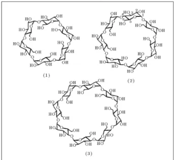
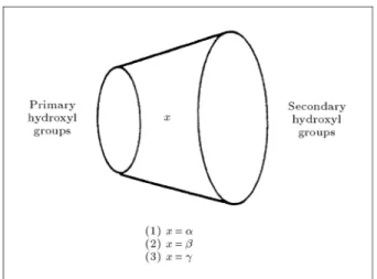
Cyclodextrins are oligosaccharides produced by the action of the amylase of Bacillus macerans on starch related compounds. They consist of (+)-D-glucopyranose units joined to each other by- (1! 4)-linkages. Cyclodextrins are toroidal in shape with all the glucose units in substantially undistorted C1 (D) chair conformations [1]. The interior of the torus consists only of a ring of C-H groups, a ring of glucosidic oxygens and another ring of C-H groups, rendering the interior of the torus relatively non-polar compared to water. There are three predominant types of cyclodextrins; - cyclodextrin (-CD), which consists of six,-cyclodextrin (-CD), which consists of seven and -cyclodextrin (-CD), which has eight D-(+)-glucopyanose units, respectively (Figure 1). Cyclodex-trins are conveniently depicted by a truncated cone where the narrow end represents the primary hydroxyl groups attached at C-6 positions and the wide end
*. Corresponding Author, Ahwaz Faculty of Petroleum En-gineering, Petroleum University of Technology, Ahwas, I.R. Iran.
1. School of Pharmacy, Ahwas Jundi Shapour University of Medical Sciences, Ahwas, I.R. Iran.
Figure 1.
Natural (native) cyclodextrins.represents the secondary hydroxyl groups attached at C-2 and C-3 positions (Figure 2).
The most interesting property of cyclodextrins is that they contain a hydrophobic cavity, which can encapsulate a guest molecule to form an inclusion complex (Figure 3).
Figure 2.
Truncated cone representing cyclodextrins.Figure 3.
Schematic of inclusion complex formation.Cyclodextrins are known to form inclusion com-plexes with a variety of guest molecules in solution and in a solid state, since their inherent annular structure exists stably in both phases. Inclusion complexes are chemical species consisting of two or more associated molecules, in which one of the molecules, the \host", forms or possesses a cavity into which it can admit a \guest" molecule, resulting in a stable association without formation of any covalent bonds. Secondary forces are alone responsible for maintenance of the integrity of all inclusion complexes. The molecular ratio of guest to cyclodextrin is usually found to be 1:1 [2,3], however, this can change, depending on the shape and geometry of the guest and the cyclodextrin. The minimum requirement for an inclusion complex formation is a size compatibility between host and guest molecules, i.e., guest molecules must t, entirely or at least partially, into the cyclodextrin cavity. In general, hydrophobic molecules or residues, rather than hydrophilic ones, have a higher anity to the cyclodex-trin cavity in aqueous solution, because the cavity provides a microheterogeneous matrix in such a polar solvent. Inclusion complex formation in aqueous media usually involves the displacement of water molecules
from the cavity of the cyclodextrin and subsequent replacement with the guest molecule. A vast array of guest molecules has been seen to be included in the cavities of cyclodextrins, ranging from polar amines, acids and ions to hydrophobic non-polar aliphatic and aromatic hydrocarbons [4,5]. An inclusion complex can be thermodynamically quite stable and each is characterized by a thermodynamic stability constant or association constant, K, dened by the position of equilibrium between the host, the guest and the complex (equation in Figure 3).
In the pharmaceutical eld, the number of papers and patents dealing with the practical applications of cylodextrins has also shown an explosive increase during the past three decades [6-12]. This is probably due to the following facts:
1. Fundamental research furnishes information about host-guest interactions;
2. Pure cyclodextrins can be obtained on a large scale; 3. Cyclodextrins can scarcely be considered as having
toxic action.
Although the natural cyclodextrins have been extensively used in pharmaceutical formulation, they have some undesirable properties as drug carri-ers [13,14]. The limited application of natural cy-clodextrins in the pharmaceutical formulation seems to be related to their relatively low aqueous solubility, particularly in the case of-cyclodextrin (1.8 w/v% at 25C).
The narrow end represents the primary hydroxyl groups attached at C-6 positions.
The wide end represents the secondary hydroxyl groups attached at C-2 and C-3 positions.
EXPERIMENTS
The experimental part of this research work in com-prised of three dierent steps:
(i) Determination of the proper pH,
(ii) Determination of the maximum absorbance wave-length (max),
(iii) Determination of the stability constant of the inclusion complex.
Preparation of Phosphate Buer Solution with
pH 7.4
Potassium dihydrogen phosphate (1.2 g, 8.8 mmol) and disodium hydrogen phosphate dodecahydrate (10.89 g, 30.4 mmol) were dissolved in deionized water in a 2000 mL volumetric ask and the volume was made up to 2000 mL by adding more deionized water. The pH was measured and conrmed to be 7.4
Preparation of Phosphate Buer Solution with
pH 7
Solution A
Potassium dihydrogen phosphate (0.908 g, 6.67 mmol) was dissolved in deionized water in a 100 mL volumetric ask and the volume was made up to 100 mL by adding more deionized water.
Solution B
Disodium hydrogen phosphate dodecahydrate (2.38 g, 6.65 mmol) was dissolved in deionized water in a 100 mL volumetric ask and the volume was made up to 100 mL by adding more deionized water.
Then, 38.9 mL of Solution A was added to 61.1 mL of Solution B and stirred well. The pH was mea-sured and conrmed to be 7.
Preparation of Citrate Buer Solution with
pH 6.5
Disodium hydrogen phosphate dodecahydrate (7.16 g, 19.98 mmol) and sodium dihydrogen phosphate (2.43 g, 20.25 mmol) were dissolved in deionized water in a 1000 mL volumetric ask and the volume was made up to 1000 mL by adding more deionized water. The pH was measured and conrmed to be 6.5.
Preparation of Diluted Piroxicam Solution (I)
[1
:024
10
3mol.L
1]
Piroxicam (17 mg, 51.2L) was dissolved in the desired buer solution in a 50 mL volumetric ask and the volume was made up to 50 mL by adding more of the buer solution used. This procedure was carried out for each of the three buer solutions (pH 7.4, 7, 6.5).
Preparation of Piroxicam Solution (II)
[3
:62
10
3mol.L
1] (Almost Saturated
Solution)
Piroxicam (60 mg, 181L) was dissolved in the desired buer solution in a 50 mL volumetric ask and the volume was made up to 50 mL by adding more of the buer solution used. This procedure was carried out for each of the three buer solutions (pH 7.4, 7, 6.5).
Preparation of
-Cyclodextrin Stock Solution
-cyclodextrin (0.4494 g, 0.396 mmol) was dissolved in the desired buer solution in a 25 mL volumetric ask and the volume was made up to 25 mL by adding more of the buer solution used. This procedure was carried out for each of the three buer solutions (pH 7.4, 7, 6.5).Determination of the Proper pH
(i) pH 7.4
5 mL of piroxicam solution (II) [3:6210 3 mol.L 1] was added to each of the ve various concentrations of - cyclodextrin (Table 1) in 5 mL volumetric asks and the nal volume was made up to 10 mL by adding more of the buer solution (pH 7.4). The solutions were kept at room temperature for 2 hours and occasionally were shaken, then ltered, through lter paper. Finally, 2.95 mL of the buer solution with pH 7.4 was added to 50 L of each ltrate. The UV{visible spectrum of each of the solutions was taken at wavelengths 200{ 400 nm. This procedure was repeated 3 times at 15 minute intervals.
(ii) pH 7 and (iii) pH 6.5
Exactly the same amounts were used and the same procedure was carried out as described in (i).
Determination of
maxThe measurements were made with pH 7.4 buer solution. 166L of the piroxicam solution (I) [1:024 10 3mol.L 1] was added to each of the-cyclodextrin concentrations (Table 2) in a 5 mL volumetric ask and the nal volume was made up to 5 mL by adding more of the buer solution (pH 7.4). The UV-visible spectrum of each of the solutions was taken at wavelength 200 - 400 nm) at 25C. From the recorded spectra, it was found thatmaxis 352 nm.
Determination of the Stability Constant (
K)
by Using Diluted Piroxicam Solution (I).
Piroxicam solution (I) [166L, 1:02410 3mol.L 1] was added to each of the diluted solutions of -cyclodextrin given in Table 2 in 5 mL volumetric asks and the volume was made up to 5 mL by adding more of the buer solution with pH 7.4. The asks were kept at room temperature for 2 hours, then ltered on lter paper. Each time, the ltrate was placed
Table 1.
Solutions of the host (-cyclodextrin) forcomplexation with almost saturated piroxicam solution (II).
Volume of Stock
Solution Taken to
Make 10 mL
Final Solution (mL)
Molar Concentration
of Diluted
-Cyclodextrin
(mol.L
1)
0.632 0.0010 1.264 0.0020 1.582 0.0025 3.164 0.0050 4.746 0.0075
Table 2.
Solutions of the host (-cyclodextrin) forcomplexation with diluted piroxicam solution (I).
Volume of Stock
Solution Taken to
Make 5 mL
Final Solution (mL)
Molar Concentration
of Diluted
-cyclodextrin
(mol.L
1)
0.347 0.0011 0.662 0.0021 1.010 0.0032 1.325 0.0042 1.672 0.0053 1.980 0.0063 2.330 0.0074 2.650 0.0084 2.990 0.0095 3.314 0.0105 3.500 0.0111 3.810 0.0121 4.160 0.0132 4.640 0.0147 4.820 0.0153
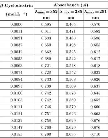
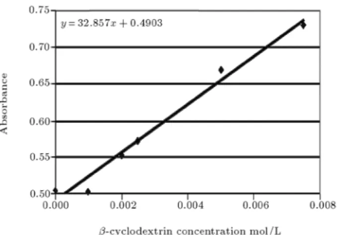
in a UV-visible cell at 25C and, nally, the UV-visible spectrum was taken at wavelengths 200-400 nm. The absorbance at max = 352 nm of each of the various concentrations of -cyclodextrin added to the piroxicam solution was recorded (Table 3) and then plotted versus -cyclodextrin (Figure 4). For each entry of Table 2, the measurements were repeated 3 times at 15 minute intervals. On the basis of max = 352 nm of piroxicam solution and various concentrations of-cyclodextrin, the stability constant was calculated, as given in the results and discussion section of this article (Table 4).
Figure 4.
Observed linear absorption diagram of inclusion complexation of beta cyclopextrin and diluted solution of Piroxicam (I).Table 3.
Absorbance of the inclusion complex formed by addition of various concentrations of-cyclodextrin todiluted piroxicam solution (I) with pH 7.4.
-Cyclodextrin
Absorbance (
A)
(mol.L
1)
max=352
nm
max=285
nm
max=251
nm
0.0000 0.595 0.465 0.570 0.0011 0.611 0.471 0.582 0.0021 0.633 0.483 0.586 0.0032 0.650 0.498 0.605 0.0042 0.662 0.525 0.612 0.0053 0.680 0.542 0.617 0.0063 0.721 0.548 0.618 0.0074 0.728 0.552 0.622 0.0084 0.733 0.568 0.626 0.0095 0.738 0.569 0.637 0.0100 0.742 0.578 0.645 0.0105 0.742 0.589 0.652 0.0111 0.746 0.579 0.660 0.0121 0.751 0.626 0.663 0.0132 0.758 0.629 0.676 0.0147 0.760 0.629 0.676 0.0153 0.790 0.635 0.710
Determination of the Stability Constant (
K)
by Using an Almost Saturated Solution of
Piroxicam (II)
Piroxicam solution (II) [5 mL, with a concentration of 3:6210 3 mol.L 1] (almost saturated solution) was added to each of the diluted solutions of-cyclodextrin given in Table 1 in 10 mL volumetric asks and the volume was made up to 10 mL by adding more of the buer solution with pH 7.4. The concentration of diluted piroxicam solution (II) became [1:8110 3 mol.L 1]. The asks were kept at room temperature for 2 hours, then, ltered on lter paper. 50L of the inclusion complex solution was added to 2.95 mL of the buer solution with pH 7.4 (concentration of more diluted piroxicam solution (II) became [3.0210 5 mol.L 1] ). Each time, the ltrate was placed in a UV-visible cell at 25C and, nally, the UV-visible spectrum was taken at wavelength 200-400 nm. The absorbances atmax= 352, 285 and 251 nm, of each of the various concentrations of -cyclodextrin added to the piroxicam solution, were recorded (Tables 5 to 7) and then plotted versus -cyclodextrin concentration (Figures 5 to 7). For each entry of Table 1, the measurements were repeated 3 times at 15-minute intervals. On the basis ofmax= 352, 285 and 251 nm of piroxicam solution and various concentrations of -cyclodextrin, the stability constants were calculated, as
Figure 5.
Observed linear absorption diagram of inclusion complexation of-cyclodextrin and saturatedsolution of piroxicam (II) at pH 7.4 and maximum absorption wavelength 352 nm.
Figure 6.
Observed linear absorption diagram of inclusion complexation of-cyclodextrin and saturatedsolution of piroxicam (II) at pH 7.4 and maximum absorbance wavelength 285 nm.
Figure 7.
Observed linear absorption diagram of inclusion complexation of-cyclodextrin and saturatedsolution of piroxicam (II) at pH 7.4 and maximum absorption wavelength 251 nm.
Table 4.
Calculated stability constant (K) of theinclusion complex between-cyclodextrin and diluted
piroxicam solution (I) [3:3910 5mol.L 1] at pH 7.4 and max= 352 nm.
Molar Concentration of
Diluted
-cyclodextrin
(mol.L
1)
Constant (
Calculated Stability
K) mol
1.L
0.0011 24.5
0.0021 30.64
0.0032 28.94
0.0042 26.90
0.0053 26.98
0.0063 33.60
0.0074 30.20
0.0084 27.65
0.0095 25.35
0.0105 23.50
0.0111 22.90
0.0121 21.69
0.0132 20.79
0.0147 18.87
0.0153 21.40
Table 5.
Absorbance of the inclusion complex formed by addition of various concentrations of-cyclodextrin tosaturated piroxicam solution (II) with pH 7.4.
-Cyclodextrin
Absorbance (
A)
(mol.L
1)
max=352
nm
max=285
nm
max=251
nm
0.0000 0.525 0.392 0.505 0.0010 0.560 0.420 0.503 0.0020 0.589 0.443 0.553 0.0025 0.616 0.460 0.573 0.0050 0.748 0.540 0.668 0.0075 0.802 0.590 0.731
given in the results and discussion section of this article (Table 8).
RESULTS AND DISCUSSION
Because of the therapeutic importance of the piroxicam drug (IV), [4-hydroxy-2-methyl-N-(2-pyridyl)-2H-1,2-benzothiazine-3-carboxamide 1,1,-dioxide] (Scheme 1) and its widespread use, it was decided to study its inclusion complexation with-cyclodextrin and deter-mine the stability constant (K) of the complex by UV-visible spectroscopy at two dierent concentrations; i.e. diluted and almost saturated solutions of piroxicam.
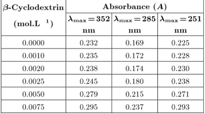
Table 6.
Absorbance of the inclusion complex formed by addition of various concentrations of-cyclodextrin tosaturated piroxicam solution (II) with pH 7.
-Cyclodextrin
Absorbance (
A)
(mol.L
1)
max=352
nm
max=285
nm
max=251
nm
0.0000 0.232 0.169 0.225 0.0010 0.235 0.172 0.228 0.0020 0.238 0.174 0.230 0.0025 0.245 0.180 0.238 0.0050 0.279 0.215 0.271 0.0075 0.295 0.237 0.293
Table 7.
Absorbance of the inclusion complex formed by addition of various concentrations of-cyclodextrin tosaturated piroxicam solution (II) with pH 6.5.
-Cyclodextrin
Absorbance (
A)
(mol.L
1)
max=352
nm
max=285
nm
max=251
nm
0.0000 0.116 0.124 0.142 0.0010 0.118 0.127 0.156 0.0020 0.119 0.129 0.162 0.0025 0.148 0.137 0.178 0.0050 0.166 0.165 0.208 0.0075 0.175 0.169 0.211
Table 8.
Calculated stability constant (K) of theinclusion complex between-cyclodextrin and saturated
piroxicam solution (II) [3:0210 5 mol.L 1] at pH 7.4
andmax= 352, 285 and 251 nm.
-Cyclodextrin
K
(mol
1.L)
(mol.L
1)
max=352
nm
max=285
nm
max=251
nm
0.0010 66.9 68.8 49.6 0.0020 61.5 63.7 48.0 0.0025 70 68.36 53.96 0.0050 85.76 75.0 65.2 0.0075 70.66 67.0 59.78
The work was carried out at three dierent pHs of 6.5, 7 and 7.4. At each of the pHs, various concen-trations of - cyclodextrin (Table 2) were added to a constant amount of the piroxicam drug and then the UV-visible spectrum was recorded at 200{400 nm at 25C. The results are given in Table 4. Based on these results, it was concluded that the absorbance for the complex formation was highest in the phosphate buer solution with pH 7.4, which is the pH of biological uids. Therefore, it can be concluded that the proper
Scheme 1.
Piroxicam, [4-hydroxy-2-methyl-N -(2-pyridyl)-2H-1,2- benzothiazine-3-carboxamide1,1,-dioxide].pH for the inclusion complex formation between -cyclodextrin and piroxicam was pH 7.4.
The second task in this research was to nd out the optimum max. Since the proper pH was found to be 7.4, all the experiments for the determination of max were carried out in buer solution with pH 7.4. After adding various concentrations of -cyclodextrin solutions (Table 2) to a constant amount of piroxicam, the UV{visible spectra were recorded at 200-400 nm. On the basis of the results obtained, it is concluded that themax is 352,285 and 251 nm.
The third and main objective of this research was to determine the stability constant or association constant, thermodynamic constant (K) of the inclu-sion complex between-cyclodextrin and piroxicam at max = 352 nm in a phosphate buer solution with pH 7.4. Since two dierent concentrations of piroxicam were used, dierent values ofKwere determined, which are explained below.
i) Calculation of the Stability Constant
(
K)
by
Using Diluted Solution of Piroxicam (I)
As explained in the previous section (Determination of max), the stability constant can be calculated based on the results given in Table 3 and also from the graph in Figure 3. The (K) value can be calculated, as follows.
In accordance with the Beer-Lambert law: A=":l:c:
Then, the following steps were considered:
1. The initial concentration of piroxicam used was 1:02410 3 mol.L 1, after dilution in the UV-visible cell:
[Piroxicam] =C= 3:3910
5mol.L 1
:
2. For calculating ", by using Figure 4, when no - cyclodextrin was used, i.e. [ - cyclodextrin]=
0.000, the absorbance could be read to be 0.595, therefore:
A= 0:595; l= 1 cm; [Piroxicam] =C= 3:3910
5mol.L 1
; "=
0:595
(1 cm3:3910 5 mol.L
1); "= 17551:62 mol
1
:cm 1:L:
3. The stability constant can be determined from the following formula;
K=
Acomplex
Apure Guest[ cyclodextrin]Free : From Figure 4, Acomplex, for various concentrations of -cyclodextrin, can be calculated, e.g., for the second entry of Table 3 atmax= 352 nm, one will have:
[ cyclodextrin] = 0:0011 mol.L
1
; A(guest+
cyclodextrin)= 0 :611: Therefore:
Acomplex=A(guest+
cyclodextrin)
Apure Guest = 0:611 0:595 = 0:016;
Acomplex= 0:016:
4. Now, the concentration of the complex can be calculated:
Ccomplex=
0:016 17551:62 mol
1
:cm 1:L1cm ; So:
Ccomplex= 9:1110
7mol.L 1
: 5.
[ cyclodextrin]Free= [ cyclodextrin]initial [ cyclodextrin]used: On the other hand:
[ cyclodextrin]used = [Ccomplex]: Therefore:
[ cyclodextrin]Free= [ cyclodextrin]initial [Ccomplex];
[ cyclodextrin]Free= 0:0011 9:1110
7 = 1:0910
3 mol.L 1
:
6. Now,Kcan be calculated by substituting the values into the equation given in Step 3:
K=
0:016
0:5951:0910 3mol.L
1:
Therefore, one would have: K= 24:5 mol 1.L. Then, the K value for each entry of Table 2 and Figure 4, was calculated as above and the results summarized in Table 4.
According to the data given in Table 4, the range of the stability constant is:
30:64 18:87 = 11:77: Therefore:
K= 24:755:89 mol
1
:L:
ii) Calculation of the Stability Constant (
K)
by Using Saturated Solution of Piroxicam (II)
It is well known that when the absorption spectrum of a guest molecule does not change signicantly, due to inclusion complexation, then, stirring the host molecule into an excess amount of the guest molecule results in the enhancement of the absorption of the nal solution relative to the saturated solution of the guest molecule. Therefore, it was decided to investigate this fact and calculate the stability constant of the inclusion complex at a higher concentration of piroxicam.
Exactly the same procedure as in the previous section was carried out for calculating the new stability constant:
[piroxicam]initial = 3:6210
3mol.L 1almost saturated solution. After diluting twice:
[piroxicam]diluted= 3:0210 5 mol.L
1
; [ cyclodextrin] = 0:000 mol.L
1
; A= 0:525; l= 1 cm;
"= 17384:1 mol
1
:cm 1:L:
For example, for the second entry, when:
[ cyclodextrin]Free= 0:0011 atmax= 352 nm; A= 0:560; K= 66:8 mol.L
1
:
TheK values are given in Table 5. The range was: 85:12 61:26 = 23:86 mol.L
1
; K= 73:1911:93 mol
1
K values at othermax = 285 and 251 nm in pH 7.4 buer solution were calculated as well and the results are given in Table 8.
K= 69:355:65 mol
1
:L atmax= 285 nm; K= 56:348:34 mol
1
:L atmax= 251 nm: On the basis of the small values of the stability constant of the inclusion complex, it can be suggested that the interaction between -cyclodextrin and piroxicam molecule is weak. This may be due to either the cavity size of -cyclodextrin annulus, which cannot admit the piroxicam molecule properly, or the UV-visible spectroscopy technique, which may not be an accurate technique for this purpose. Therefore, it is suggested that the inclusion complex between the piroxicam molecule and modied -cyclodextrins, such as those having polar and ionic functional groups attached to the -cyclodextrin molecule or with the linked -cyclodextrin molecules, be investigated. Also, other more accurate techniques, such as High Performance Liquid Chromatography (HPLC), HNMR or CNMR should be used for the determination of the stability constant of the inclusion complexation. The results show that the stability constant (K) at a saturated solution of Piroxicam is almost two to three times greater than at low concentrations of piroxicam and, also, the amount of K at max = 352 nm is the highest.
REFERENCES
1. Bender, M.L. and Komiyama, M. \Cyclodextrin Chemistry",Springer-Verlag, Berlin (1978).
2. Lammers, J.N.J.J. and van Diemen, A.J.G.,Rec. Trav. Chim. Pays-Bas,
91
, p 733 (1972).3. Lammers, J.N.J.J.,Rec. Trav. Chim. Pays-Bas,
91
, p 1163 (1972).4. Wojcik, J.F. and Rohrbach, R.P.,J. Phys. Chem.,
79
, p 225 (1975).5. Cramer, F. and Henglein, F.M.,Angew. Chem.,
68
, p 649 (1956).6. Frank, S.G. \Inclusion compounds", J. Pharm. Sci.,
64
, p 1585 (1975).7. Saenger, W. \Cyclodextrin inclusion compounds in research and industry", Angw. Chem. Int. Ed. Engl.,
19
, p 344 (1980).8. Szejtli, J. \Molecular entrapment and release prop-erties of drugs by cyclodextrins" in Controlled Drug Bioavailability, V.F. Smolen and L.A. Ball, Eds., John Wiley and Sons,
3
, New York, USA, p 365 (1985). 9. Mifune, A. and Shima A. \Cyclodextrins and theirapplications",Yuki Gousei Kagaku Kyokai Shi,
35
, p 116 (1977).10. Uekama, K. \Inclusion complexes of cyclodextrins with organic drug molecules",Jpn. J. Antibiot.,
32
, p S103 (1979).11. Uekama, K. \Pharmaceutical applications of cyclodex-trin complexations",
101
, p 857 (1981).12. Uekama, K. \Pharmaceutical applications of cyclodex-trins",J. Jpn. Soc. Starch Sci.,
30
, p 247 (1983). 13. Uekama, K. \Pharmaceutical applications ofcyclodex-trins and some of their problems",Fragrance J.,
63
, p 68 (1983).14. Uekama, K. and Hirayama, F. \Utilization of cyclodex-trins in the pharmaceutical eld", Kagaku to Kogyo,