Modeling human spinal muscular atrophy mutations in
Drosophila melanogaster
KAVITA PRAVEEN
A dissertation submitted to the faculty of the University of North Carolina at Chapel Hill in partial fulfilment of the requirements for the degree of Doctor of Philosophy in the Curriculum in Genetics and Molecular Biology.
Chapel Hill 2012
Approved By:
A. Gregory Matera, Ph.D. Robert Duronio, Ph.D.
ii
Abstract
KAVITA PRAVEEN: Modeling human spinal muscular atrophy mutations in Drosophila melanogaster
(Under the direction of A. Gregory Matera)
Spinal Muscular Atrophy (SMA) is a common neuromuscular disease that affects one in 6,000-8,000 young children; most of whom die before reaching the age of two years. One in fifty Americans is a carrier for SMA, making this genetic disease a serious health concern. More than 95% of patients with SMA carry deletions in the survival motor neuron 1 (SMN1) gene. The SMN protein is essential for survival and has a
well-characterized role in the biogenesis of small nuclear ribonucleoproteins (snRNPs), which are core components of the spliceosome. Numerous additional functions, both housekeeping and tissue-specific, have been put forth in the literature, however, no convincing link has been made between any putative SMN function and the disease etiology.
To address this question, we are studying the consequences of SMN loss in the Drosophila model system. We have generated a series of transgenic flies that exclusively
construct does not restore snRNA levels, but does rescue the motility defects and lethality of the Smn null flies. In addition, the majority of flies expressing an SMA point mutation, T205I, die as pupae but have a similar snRNA profile to the WT transgenic animals. These data suggest that the reduction in snRNAs in Smn mutants is not a major contributor to their lethality, and indicate that non-snRNP related functions of SMN may be critical to SMA pathology. We have generated twelve additional point mutations in Smn that mimic mutations identified in SMA patients. The phenotypes of these mutations
iv
Acknowledgments
I want to acknowledge my mentor, Greg Matera, for his direction and encouragement, especially through those times when it seemed every approach was failing. I also want to thank Greg for the many opportunities he gave me to work with others on different projects. These opportunities were instrumental to my development as a scientist. I also learnt much about how research is done from other members of the Matera lab, particularly T.K. Rajendra and Graydon Gonsalvez. I am also especially thankful to our lab manager, Ying Wen, who helped set up and manage fly crosses, and spent many hours in front of the microscope to harvest larvae for experiments.
I cannot express enough gratitude towards my close friends, Srikripa and Lukasz, whose support and companionship got me through the difficult two and a half years that I was alone in NC.
My parents taught me to persevere and I am incredibly thankful for that today. I am deeply grateful for the love and support that my parents and my sister have given me.
vi
Table of Contents
List of Tables ... ix
List of Figures ... x
List of Abbreviations ... xii
CHAPTER I ... 1
Spinal Muscular Atrophy ... 1
SMA Etiology ... 2
SMN Function ... 4
snRNP independent functions of SMN ... 9
SMN protein and the SMN complex ... 11
Modeling SMA in Drosophila ... 15
Drosophila SMA models ... 16
Other animal models of SMA ... 18
The conundrum of SMA pathology ... 18
SMA and snRNP biogenesis ... 19
CHAPTER II ... 22
Introduction ... 22
Experimental Procedures ... 24
Fly stocks and genetics ... 24
Rescue constructs ... 25
Northern blotting ... 25
Larval motility assays ... 26
Real-time PCR ... 26
Results ... 27
Characterization of Smn null flies ... 27
Transgenic expression of an SMA patient-derived mutation in dSMN rescues Smn-/- larval lethality ... 33
dSMNT205I is moderately defective in self-oligmerization ... 34
Transgenic expression of SmnT205I rescues larval locomotion ... 38
SmnT205I and SmnWT transgenic larvae are functional in snRNP biogenesis ... 38
Discussion ... 42
SmnT205I is a good model for intermediate SMA ... 42
SMN, snRNA levels and SMA ... 42
Supplementary Data ... 46
CHAPTER III ... 50
Introduction ... 50
Experimental Procedures ... 51
Fly stocks ... 51
Rescue constructs ... 51
Antibodies and Western blotting... 51
Results ... 52
Creating SMA patient-derived point mutations in Drosophila SMN ... 52
Characterizing interactions of SMA point mutations in dSMN ... 56
viii
Discussion ... 69
SMA mutations in humans and flies ... 69
Drosophila models of SMA point mutations ... 71
Incomplete dominance of Smn mutations ... 72
Supplementary Data ... 78
CHAPTER IV ... 81
Smn and Gemins ... 88
SMN, ecdysone and insulin signalling ... 92
Summary ... 98
List of Tables
Table S2.4. Descriptions of 18 minor-class intron containing genes
in the Drosophila genome that were tested via qRT-PCR in figure 2-2.………...49 Table S3.1. Summary of known human SMN binding proteins tested for
x
List of Figures
Figure 1.1. SMN2 cannot fully compensate for loss of SMN1 ... 3
Figure 1.2. Illustration of the major steps in the mammalian snRNP biogenesis pathway. ... 8
Figure 1.3. A summary of the different functions ascribed to SMN ... 11
Figure 1.4. Major domains in the SMN protein. ... 12
Figure 1.5. Cartoon depiction of the cytoplasmic mammalian SMN complex ... 13
Figure 1.6. Mutations in the Drosophila Smn gene ... 17
Figure 2.1. Characterization of Smn null flies ... 29
Figure 2.2. Analysis of minor-class intron splicing defects in Smn and U6atac mutants.. ... 32
Figure 2.3. Transgenic rescue of Smn null animals with an SMA patient-derived point mutation, T205I ... 36
Figure 2.4. SmnWT and SmnT205I animals have similar snRNA profiles ... 40
Figure S2.1. Total snRNA levels in Drosophila larvae reflect snRNP levels ... 46
Figure S2.2. SmnD mutants express a non-canonical U11 snRNA ... 47
Figure 3.1. An alignment of SMN protein from human and various
species including Drosophila ... 54
Figure 3.2. SMA patient mutations in Drosophila Smn... 55
Figure 3.3. Interaction of SMA patient derived mutations in dSMN
with dGemin2 and dGemin3 ... 57
Figure 3.4. Expression of SMA patient derived missense mutations in flies ... 61
Figure 3.5. Viability analyses of SMA patient derived point mutations in flies ... 65
Figure 3.6. Model of incomplete dominance of Smn point mutations
when co-expressed with SmnWT. ... 74
Figure S3.1. dSMN mutations defective in self-oligomerization still retain
the ability to bind WT dSMN ... 79
Figure S3.2. Design of crosses used to bring SmnX7 mutation, the 86Fb attP insertion site (depicted as 86Fattp) and the PhiC31
Integrase (Int) on the X chromosome into one background.. ... 80
Figure S3.3. Levels of dSMN expressed from WT transgene inserted
at 86Fb compared with insertion at 68A4 (attP2) site. ... 80
Figure 4.1. A model for how dGemin3 could influence the oligomerization
and stability of dSMN ... 90
Figure 4.2. A summary of the interconnectivity of the insulin signalling
xii
List of Abbreviations
ATP Adenosine triphosphate
CB Cajal Body
DPE Days post egg laying
dSMN Drosophila SMN
EDTA Ethylenediaminetetraacetic acid
EMS Ethyl methane sulphonate
FLAG Polypeptide epitope
FMRP Fragile X
G3 Gemin3
GFP Green fluorescent protein
GTP Guanosine triphosphate
Impβ Importinβ
IP Immunoprecipitation
m7G 7-methylguanosine
m3G Trimethylguanosine
MOPD1 Microcephalic osteodysplastic primordial dwarfism type I
Myc c-Myc epitope
NP-40 Nonident P-40
mRNA messenger RNA
mRNP messenger RNP
OR Oregon-R
PBS Phosphate buffered saline
PCR Polymerase chain reaction
PHAX Phosphorylated adaptor for snRNA export
Phb2 Prohibitin 2
PRMT5 Protein arginine methyltransferase
REA Repressor of Estrogen Receptor Activity
RIG Rigor Mortis
RNA Ribonucleic acid
RNAi RNA interference
RNP Ribonucleoprotein
RT-PCR Reverse transcriptase PCR
qRT-PCR Quantitative RT-PCR
S2 Schneider 2
SIP1 Survival motor neuron interacting protein 1
SMA Spinal muscular atrophy
SMN Survival motor neuron
scaRNA small cajal body specific RNA
snRNA small nuclear RNA
snRNP small nuclear RNP
snoRNP small nucleolar RNP
SPN Snurportin
xiv
TMG 2,2,7-trimethylguanosine
UAS Upstream activating sequence
Unrip unr-interacting protein
U-snRNA Uridine rich snRNA
CHAPTER I
Introduction
Spinal Muscular Atrophy
Proximal spinal muscular atrophy, also called spinal muscular atrophy (SMA), is the most prevalent genetic cause of infant mortality. With a carrier frequency of 1 in 50 and an incidence rate of 1 in 6000-10,000, it is also the most common autosomal recessive disorder in the population after Cystic Fibrosis (Ogino et al., 2002). The disease is characterized by the degeneration of motor neurons in the anterior horn of the lower spinal cord, leading to symmetrical paralysis, atrophy of the proximal muscles, and loss of motor function. SMA can be classified into three types based on the severity of the phenotype, which is determined by the age of onset and the level of maximum motor function achieved by the patient (Ogino S, 2004). Type I SMA, also known as Werdnig-Hoffman disease, is the most severe form where symptoms can be apparent as early as in utero. Affected infants experience progressive muscle weakness and hypotonia (reduced
2
age of onset after 2 years. Most type III patients are able to stand and walk, but can become wheelchair bound in adulthood. The disease progress is slow and many type III patients have a normal life expectancy. Type I SMA is the most common form of the disease, affecting ~60% of SMA patients (Nicole et al., 2002; Ogino S, 2004).
SMA Etiology
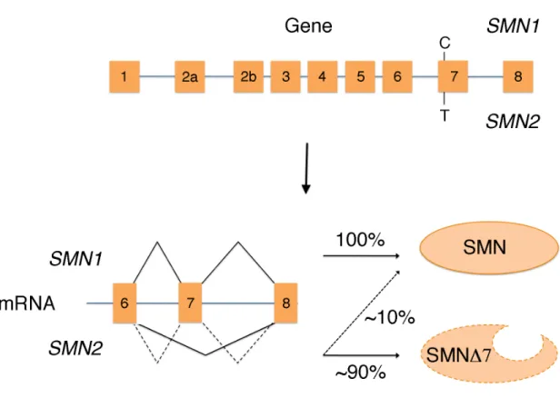
In 1990, linkage analysis was used to map the SMA causing gene to chromosome 5q11.2-13.3 in several SMA affected families (Brzustowicz et al., 1990; Melki et al., 1990). Five years later, Lefebvre et al. (1995) identified a novel gene that lies in the critical region, survival of motor neuron 1 (SMN1), as the causative gene in SMA (Bussaglia et al., 1995; Rodrigues et al., 1995; van der Steege et al., 1995; Chang et al., 1997). SMN1 produces a 1,536 bp mRNA from 9 exons that code for a 294 amino acid protein. Over 95% of SMA patients have deletions involving exon 7 of SMN1 (Lefebvre et al., 1995; Campbell et al., 1997).
Further characterization of the 5q region revealed the existence of at least two copies of the SMN gene in most people. The telomere-proximal copy (SMN1) and the centromere-proximal copy (SMN2) arose as a result of a 500 kb inverted duplication located at 5q11-13 (Lefebvre et al., 1995). There are five base pair differences that distinguish the two SMN copies at the nucleotide level, but they leave the sequence of the protein unchanged. One of these is a change from C to T in exon 7 of the SMN2 gene, which results in the exclusion of exon 7 (SMN∆7) in ~90% of the SMN2 transcripts
that is functional and indistinguishable from that produced by Monani et al., 1999; Lorson et al.,
Figure 1.1. SMN2 cannot fully compensate for loss of
in the SMN2 gene results in the exclusion of exon 7 and production of a truncated and unstable protein ~90% of the time.
Studies in SMA patients of the phenotype and the amount
1997). These findings, coupled with the existence of two non humans, led to the formulation of a gene
SMA is now thought to be a gene dosag
phenotype. There is an inverse correlation between the number of
genome and disease severity. Mildly affected patients generally have more copies of that is functional and indistinguishable from that produced by SMN1 (Lorson et al., 1999
Lorson et al., 2000).
cannot fully compensate for loss of SMN1. A base change from C to T gene results in the exclusion of exon 7 and production of a truncated and unstable protein ~90% of the time.
Studies in SMA patients have revealed an inverse correlation between the severity amount of functional, full length SMN protein (Lefebvre et al., coupled with the existence of two non-redundant SMN
led to the formulation of a gene-disease relationship between SMN and SMA. SMA is now thought to be a gene dosage disorder, and SMN2 is a modifier of the
There is an inverse correlation between the number of SMN2
genome and disease severity. Mildly affected patients generally have more copies of Lorson et al., 1999;
A base change from C to T gene results in the exclusion of exon 7 and production of a truncated and
revealed an inverse correlation between the severity Lefebvre et al., SMN paralogs in
disease relationship between SMN and SMA. a modifier of the SMN2 copies in the
4
SMN2 than patients with more severe phenotypes (Vitali et al., 1999). Consistent with
this observation, earlier studies had found that the levels of SMN protein in cells from SMA type I patients were reduced to 5-20% of levels in controls, while in type III SMA patients, SMN levels were comparable to controls (Lefebvre et al., 1997; Vitali et al., 1999). It is hypothesized that type I SMA is caused by deletions and/or mutations in the SMN1 gene, whereas type III SMA generally results from gene conversion events that
convert SMN1 to SMN2 (Campbell et al., 1997). In the latter case, there are more copies of SMN2 and thus more functional SMN protein, resulting in a milder SMA phenotype. Missense mutations in SMN1 have also been identified in SMA patients in all three categories of SMA, however, these are rare compared to gross deletions of the gene (Burghes et al., 2009).
SMN Function
snRNP biogenesis
Uridine-rich snRNAs are non-coding RNAs that perform diverse cellular functions (Kiss, 2004). In particular, the Sm-class U-snRNAs that are assembled by the SMN complex form core components of the spliceosome. Eukaryotic genomes encode RNAs that form two distinct spliceosomes. The so-called “major” spliceosome, is responsible for splicing of over 99% of introns in the human genome, whereas the “minor” spliceosome catalyzes removal of the remaining <1% of introns (Levine et al., 2001). The major spliceosome is comprised of the snRNAs U1, U2, U4 and U6, which are referred to as major-class snRNAs. The minor spliceosome is formed by the minor-class snRNAs U11, U12, U4atac and U6atac. Another spliceosomal snRNA, U5, is a component of both spliceosomes (Patel et al., 2003). Consistent with a greater requirement for the major spliceosome, the major-class snRNAs are ~100 fold more abundant than the minor-class snRNAs (Zieve et al., 1990).
6
2000). The SMN complex then facilitates the binding of seven proteins, SmB/B’, SmD1, SmD2, SmD3, SmE, SmF and SmG (the Sm proteins), onto a conserved motif called the ‘Sm site’ on the pre-snRNA (Meister et al., 2001a; Yong et al., 2002; Yong et al., 2004; Golembe et al., 2005) in an adenosine triphosphate (ATP) dependent reaction. The SMN complex provides specificity and speed to this reaction, which can also occur spontaneously and non-specifically in vitro (Pellizzoni et al., 2002b). Another complex called the protein arginine methyltransferase 5 (PRMT5) complex, consisting of PRMT5, pICln and WD45 (Mep50), symmetrically dimethylates SmB, SmD1 and SmD3 on their RG rich C-terminal domains (Brahms et al., 2000; Brahms et al., 2001; Friesen et al., 2001; Meister et al., 2001b; Friesen et al., 2002). This methylation enhances the binding between these Sm proteins and SMN, and is thought to be important for the transfer of Sm proteins onto snRNAs. However, in Drosophila, the Sm protein methylation activity carried out by the PRMT5 ortholog, Dart5, is not necessary for the snRNP assembly process (Gonsalvez et al., 2008).
After assembly with Sm proteins, the pre-snRNA is trimmed at the 3’ end (Kleinschmidt et al., 1987; Seipelt et al., 1999; Will et al., 2001) and the m7G cap is hypermethylated to a trimethylguanosine (m3G) cap by the enzyme TGS1 (Mattaj, 1986;
Figure 1.2. Illustration of the major steps in
After transcription by RNA polymerase II, 3’
and being bound by the cap binding complex (CBC) the pre
en route to the Cajal body (CB). PHAX then recruits CRM1 and Ran to export the pre snRNA out to the cytoplasm. In the cytoplasm, PHAX,
from the pre-snRNA, which gets loaded with a seven
the SMN complex. A subset of Sm proteins are thought to be symmetrically methylated by the PRMT5 complex to facilitate their recognition by SM
pre-snRNA. The pre-snRNA is further modified by methylation of the 7 guanosine (m7G) cap to a trimethylguanosine (m
the snRNA by an exonulcease (EXO)
with the SMN complex by Snurportin (SPN) and Importin the CB. In the CB, the snRNA
before localizing to speckles for storage or to
The two spliceosomal snRNAs, U6 and U6atac, do not follow the same assembly pathway as the others. U6 and U6atac are RNA polymerase III transcripts that acquire a
8
Illustration of the major steps in the mammalian snRNP biogenesis pathway ription by RNA polymerase II, 3’ end processing by the integrator complex and being bound by the cap binding complex (CBC) the pre-snRNA is bound by PHAX en route to the Cajal body (CB). PHAX then recruits CRM1 and Ran to export the pre snRNA out to the cytoplasm. In the cytoplasm, PHAX, CRM1, Ran and the CBC release
nRNA, which gets loaded with a seven membered ring of Sm proteins by A subset of Sm proteins are thought to be symmetrically methylated by the PRMT5 complex to facilitate their recognition by SMN and their transfer onto the
snRNA is further modified by methylation of the 7 G) cap to a trimethylguanosine (m3G) cap and trimming of the 3’
by an exonulcease (EXO). The snRNA is imported into the nucleus along plex by Snurportin (SPN) and Importinβ (Impβ), where it localizes to , the snRNA binds other proteins and acquires further modifications g to speckles for storage or to the perichromatin fibrils for active splicing
The two spliceosomal snRNAs, U6 and U6atac, do not follow the same assembly pathway as the others. U6 and U6atac are RNA polymerase III transcripts that acquire a NP biogenesis pathway. end processing by the integrator complex snRNA is bound by PHAX en route to the Cajal body (CB). PHAX then recruits CRM1 and Ran to export the
pre-CRM1, Ran and the CBC release membered ring of Sm proteins by A subset of Sm proteins are thought to be symmetrically methylated N and their transfer onto the snRNA is further modified by methylation of the 7-methyl G) cap and trimming of the 3’ end of nucleus along ), where it localizes to modifications for active splicing.
γ-monomethyl cap after transcription. They are then bound by seven Sm-like (Lsm) proteins (Lsm2-Lsm8) (Achsel et al., 1999), and are referred to as “Lsm-class” snRNAs. Their assembly is thought to occur entirely within the nucleus, and appears to be independent of SMN.
snRNP independent functions of SMN
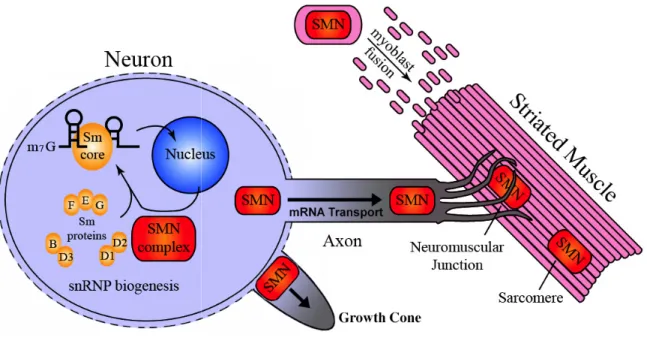
While snRNP biogenesis remains by far the most well established function of SMN, the protein has also been implicated in other global and tissue-specific roles in the cell (Fig. 1.3). SMN is reported to interact with and influence the activity of E2, a viral transcription activator (Strasswimmer et al., 1999), as well as with RNA helicase A and RNA polymerase II (Pellizzoni et al., 2001), invoking the possibility that it may function in transcriptional regulation.
10
Figure 1.3. A summary of the different functions ascribed to S
neuronal cell body where the SMN complex is involved in assembly of spliceosomal snRNPs. SMN has also been
along the neuronal axon, in axonal growth, in neuromuscular junction formati myoblast fusion and maintenance of muscle architecture. Figure
of Karl Shpargel (Shpargel, 2006
SMN protein and the SMN complex
The SMN1 gene and protein are highly conserved in evolution, with homologs in all major eukaryotic model organisms investigated except
primates are the only species that have Furthermore, only humans have the
determined by the inability to detect any chimpanzee (DiDonato et al., 1997
294 amino acids long and has two well described domains: found in many RNA-binding proteins,
Tudor domain is thought to be involved
A summary of the different functions ascribed to SMN. The drawing shows a neuronal cell body where the SMN complex is involved in assembly of
spliceosomal snRNPs. SMN has also been implicated in transport of β -along the neuronal axon, in axonal growth, in neuromuscular junction formati
myoblast fusion and maintenance of muscle architecture. Figure adapted from the thesis l, 2006).
SMN protein and the SMN complex
gene and protein are highly conserved in evolution, with homologs in all major eukaryotic model organisms investigated except S. cerevisiae
primates are the only species that have more than one copy of the Furthermore, only humans have the C to T base change that defines the SMN2
determined by the inability to detect any SMN∆7 mRNA in our closest relative, the DiDonato et al., 1997; Rochette et al., 2001). The human SMN prote 294 amino acids long and has two well described domains: the Tudor domain,
binding proteins, and a region called the YG box (Fig. thought to be involved in binding of SMN to Sm proteins (
MN. The drawing shows a neuronal cell body where the SMN complex is involved in assembly of Sm-class -actin mRNA along the neuronal axon, in axonal growth, in neuromuscular junction formation and in from the thesis
gene and protein are highly conserved in evolution, with homologs in cerevisiae. However,
of the SMN gene. SMN2 gene, as
1999) and the YG box has been characterized as a most well conserved region
reported some self-association activity at the N Gemin2 binding site (Young et al., 2000
Figure 1.4. Major domains in the SMN protein. The SMN protein has an N
domain that is important for binding to Gemin2. This region may also contribute to the self-association of SMN. The
proteins, interacts with Sm proteins, and the YG box mediates SMN oligomerization.
Consistent with its function in the housekeeping process of snRNP biogenesis, SMN is ubiquitously express
the cytoplasm as well as in the cell whereas in the nuclei of most tissues (Carvalho et al., 1999). CBs
silver chromate staining (SRy, 1903
nucleolar ribonucleoproteins (snoRNPs), small
(scaRNPs) as well as other proteins involved in RNP metabolism al., 2006). The marker used in
localizes specifically to these structures
Studies over the last decade have shown that SMN is multimeric complex consisting of eight additional proteins
12
x has been characterized as a self-oligomerization domain and is the most well conserved region in the protein (Lorson et al., 1998). Two studies have also association activity at the N-terminal end of SMN, overlapping the
Young et al., 2000; Ogawa et al., 2007).
Major domains in the SMN protein. The SMN protein has an N
domain that is important for binding to Gemin2. This region may also contribute to the association of SMN. The tudor domain, which is present in many RNA binding proteins, interacts with Sm proteins, and the YG box mediates SMN oligomerization.
Consistent with its function in the housekeeping process of snRNP biogenesis, expressed (Coovert et al., 1997; Burlet et al., 1998), and localizes
in the cell nucleus. In the cytoplasm, the SMN signal is diffuse in the nuclei of most tissues it is concentrated in foci corresponding to
CBs are nuclear structures initially characterized by their intense SRy, 1903). They contain high levels of snRNPs, small nucleolar ribonucleoproteins (snoRNPs), small Cajal body specific ribonucleoproteins
proteins involved in RNP metabolism (reviewed in
. The marker used in more recent times for CBs is the protein coilin, which localizes specifically to these structures (Andrade et al., 1991).
t decade have shown that SMN is found as part of a large consisting of eight additional proteins: Gemin2, Gemin3, Gemin4, oligomerization domain and is the Two studies have also terminal end of SMN, overlapping the
Major domains in the SMN protein. The SMN protein has an N-terminal domain that is important for binding to Gemin2. This region may also contribute to the tudor domain, which is present in many RNA binding proteins, interacts with Sm proteins, and the YG box mediates SMN oligomerization.
Consistent with its function in the housekeeping process of snRNP biogenesis, , and localizes to nucleus. In the cytoplasm, the SMN signal is diffuse, in foci corresponding to CBs r structures initially characterized by their intense . They contain high levels of snRNPs, small ajal body specific ribonucleoproteins (reviewed in Matera et is the protein coilin, which
Gemin5, Gemin6, Gemin7, Gemin8 and Unrip (Charroux et al., 1999; Charroux et al., 2000 Pellizzoni et al., 2002a; Grimmler et al., 2005
Importantly, the SMN complex, as a whole, is required for snRNP assembly. Addition of SMN and Gemin2 alone were not sufficient to restore RNP assembly activity in
egg extracts immunodepleted for these proteins
studies have further confirmed this requirement for the other members of the SMN complex in snRNP assembly
2005; Battle et al., 2006; Ogawa et al., 2007 complex members may also be involved in snRNP has been implicated (Zhang et al., 2006
Figure 1.5. Cartoon depiction of the cytoplasmic mammalian SMN complex
numbered ovals in the complex represent Gemins2
contacts with SMN. Only one subunit of the oligomeric SMN complex is depicted here. Based on Otter et al., 2007.
While orthologs of Gemin2 can be identified in all species investigated, the other Gemins are not as well conserved. A comprehensive bioinformatic investigation to Gemin5, Gemin6, Gemin7, Gemin8 and Unrip (a cytoplasm specific complex member
Charroux et al., 2000; Baccon et al., 2002; Gubitz et al., 2002 Grimmler et al., 2005; Carissimi et al., 2006
Importantly, the SMN complex, as a whole, is required for snRNP assembly. Addition of SMN and Gemin2 alone were not sufficient to restore RNP assembly activity in
egg extracts immunodepleted for these proteins (Meister et al., 2001a). A number of studies have further confirmed this requirement for the other members of the SMN in snRNP assembly (Meister et al., 2001a; Feng et al., 2005; Shpargel et al., Ogawa et al., 2007). Additionally, it appears that the SMN y also be involved in snRNP-independent functions in which SMN Zhang et al., 2006; Walker et al., 2008; Todd et al., 2010
Cartoon depiction of the cytoplasmic mammalian SMN complex numbered ovals in the complex represent Gemins2-8. Gemins2, 3, 7 and 8 make direct contacts with SMN. Only one subunit of the oligomeric SMN complex is depicted here.
While orthologs of Gemin2 can be identified in all species investigated, the other Gemins are not as well conserved. A comprehensive bioinformatic investigation to a cytoplasm specific complex member) Gubitz et al., 2002; Carissimi et al., 2006) (Fig. 1.5). Importantly, the SMN complex, as a whole, is required for snRNP assembly. Addition of SMN and Gemin2 alone were not sufficient to restore RNP assembly activity in Xenopus . A number of studies have further confirmed this requirement for the other members of the SMN Shpargel et al., . Additionally, it appears that the SMN independent functions in which SMN
Todd et al., 2010).
Cartoon depiction of the cytoplasmic mammalian SMN complex. The 8. Gemins2, 3, 7 and 8 make direct contacts with SMN. Only one subunit of the oligomeric SMN complex is depicted here.
14
identify Gemin homologs showed that Gemins 5 and 3 are the most ancestral Gemins in the complex (Kroiss et al., 2008). Putative homologs of Gemins 6, 7, 8 and 4 were only found in metazoans, suggesting that they are newer additions to the complex. Interestingly, both Dipterans that were analyzed (D.melanogaster and A.gambiae) seem to have only Gemins 2, 3 and 5.
In particular, Gemins 2, 3, 4 and 5 are required, in addition to SMN, for snRNP assembly in HeLa cells (Feng et al., 2005; Shpargel et al., 2005). However, it was recently shown in vitro that a minimal SMN complex, consisting of SMN and Gemin2 only, was sufficient for assembly of Sm proteins onto snRNAs (Kroiss et al, 2008). This finding suggests that while the other SMN complex members may not be essential for the assembly reaction per se, they play ‘supportive’ roles to enhance snRNP assembly in vivo. It is known that the SMN complex is associated with the Sm-snRNA complex
throughout the cytoplasmic phase of assembly (Massenet et al., 2002) and is also important for re-import of the immature snRNP into the nucleus (Narayanan et al., 2002; Narayanan et al., 2004). Hence, it is possible that the Gemins have critical functions in steps downstream to the assembly of Sm proteins onto snRNAs. Notably, although Drosophila seems to lack many of the components of the SMN complex, its genome does
contain orthologs for most of the Gemins that are known to be important for snRNP biogenesis in vivo.
directly, and is implicated in distinguishing them from other RNAs, thus, providing specificity to the assembly reaction (Battle et al., 2006). Gemin2 has been reported to help stabilize SMN by enhancing SMN’s N-terminal domain mediated self-association (Ogawa et al., 2007). Recently, a crystal structure of Gemin2 with the Gemin2-binding domain of SMN revealed that Gemin2 bound a pentamer of the Sm proteins D1, D2, E, F and G directly (Zhang et al., 2011). Gemins 6 and 7 form a heterodimer that resembles the structure created by heterodimers of SmB/SmD3 and SmD1/SmD2 (Ma et al., 2005), and also to interact with a subset of Sm proteins. Thus, it has been proposed that Gemins 6 and 7 may act as “place holders” for SmB/SmD3 in the Sm protein pentamer that binds Gemin2 (Zhang et al., 2011) before assembly onto the snRNA. Gemin3 is a DEAD box domain containing protein with potential helicase activity, and may perform the ATP dependent step of the assembly reaction (Charroux et al., 1999). No functions have been assigned to the remaining members of the SMN complex.
Modeling SMA in Drosophila
16
The Drosophila Smn ortholog is an 828 nucleotide, intron-less gene that codes for a 226 amino acid protein (dSMN). The human and fly homologs share 23.5% identity and 36.7% similarity. The regions showing greatest conservation correspond to the Gemin2 binding site near the N-terminus, the Tudor domain and the YG box (Miguel-Aliaga et al., 2000). The Drosophila SMN complex participates in the assembly of Sm proteins onto snRNAs, indicating that the function of human SMN in snRNP biogenesis is conserved in the fly (Rajendra et al., 2007).
Drosophila SMA models
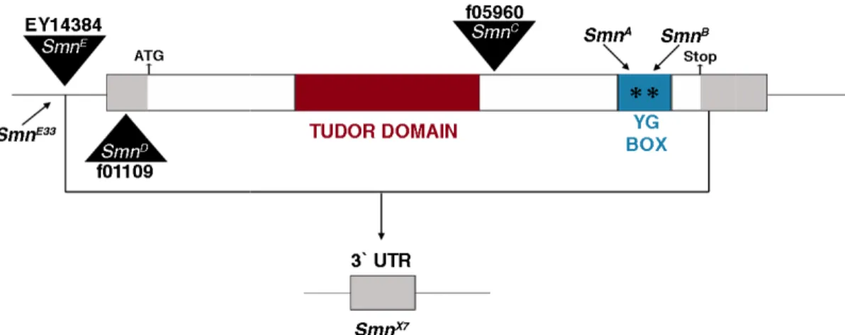
Figure 1.6. Mutations in the
with gray boxes on either side representing 3` and 5` UTRs insertions are shown as black triangles.
+407 bp (coding region) and
SmnE is a P-element insertion upstream of the transcription start site at an imprecise excision mutation derived from
from excision of SmnE33. This deletion
start site, the whole of the 5` UTR, coding region and the 5` most 45 bases of the 3` UTR of Smn. SmnA and SmnB are both point mutations in the oligomerization domain (YG box) of Smn.
Previously, our lab generated addresses the aforementioned issues.
imprecise excision of a P element insertion located upstream to the for adult flies with neuromusc
(excision 33), are viable and fertile, but unable to fly or jump. Severe atrophy of the flight muscles was observed in the mutants, indicated by
the flight muscles. This was accompanied by routing and branching defects in the dorsal longitudinal motor neurons of the flight muscles
utations in the Drosophila Smn gene. The Drosophila Smn gene is shown with gray boxes on either side representing 3` and 5` UTRs, and locations of transposon insertions are shown as black triangles. SmnC and SmnD are both piggyBac insertions at +407 bp (coding region) and -71 bp (5` UTR) of the translation start site, respectively.
element insertion upstream of the transcription start site at -94 bp.
an imprecise excision mutation derived from SmnE. SmnX7 is a deletion line also derived . This deletion is missing 93 bases upstream of the transcription start site, the whole of the 5` UTR, coding region and the 5` most 45 bases of the 3` UTR are both point mutations in the oligomerization domain (YG box)
generated a Drosophila melanogaster model of SMA that addresses the aforementioned issues. An SMN hypomorphic mutant was isolated
element insertion located upstream to the Smn gene for adult flies with neuromuscular phenotypes. These mutants, referred to as
xcision 33), are viable and fertile, but unable to fly or jump. Severe atrophy of the flight muscles was observed in the mutants, indicated by a complete disorganization of
was accompanied by routing and branching defects in the dorsal neurons of the flight muscles (Rajendra et al., 2007).
gene is shown and locations of transposon are both piggyBac insertions at ranslation start site, respectively. 94 bp. SmnE33 is is a deletion line also derived is missing 93 bases upstream of the transcription start site, the whole of the 5` UTR, coding region and the 5` most 45 bases of the 3` UTR are both point mutations in the oligomerization domain (YG box)
model of SMA that was isolated by the gene in a screen ular phenotypes. These mutants, referred to as SmnE33 xcision 33), are viable and fertile, but unable to fly or jump. Severe atrophy of the
18 Other animal models of SMA
SMA has also been modeled in mice, fish, worms and fission yeast (Miguel-Aliaga et al., 1999; Hsieh-Li et al., 2000; Monani et al., 2000a; Owen et al., 2000; McWhorter et al., 2003). Severe reduction or complete knockout of Smn in these organisms is lethal. Moderate reductions in SMN levels in C.elegans and zebrafish, resulted in uncoordination, paralysis and lack of muscle tone in the former, and motor axon pathfinding defects in the latter (without any defects in muscles or overt movement defects). To achieve a milder reduction in SMN levels and thus obtain a mouse model that more closely resembled the human disorder, two groups generated mice that expressed the human SMN2 gene in the background of a homozygous mouse Smn mutation (Smn-/-; SMN2) (Hsieh-Li et al., 2000; Monani et al., 2000b). This model rescued the embryonic lethality of Smn-/- mice and recapitulated many of the pathological features seen in SMA patients. These include a much shorter lifespan, degeneration of spinal motor neurons and progressive muscle weakness (Monani et al, 2000). Furthermore, similar to the human disease, varying the number of copies of SMN2 varied the severity of the phenotype from that resembling type I (1 or 2 copies)
patients to types II and III, and complete rescue (8-10 copies).
The conundrum of SMA pathology
snRNP biogenesis, or that disruptions to the tissue-specific functions of SMN lead to SMA. These hypotheses need not be mutually exclusive.
SMA and snRNP biogenesis
Several groups have attempted to find a correlation between snRNA levels in SMA models and the disease phenotype. The results appear to be conflicting. Studies in mouse models of SMA showed a decrease in the levels of some minor-class spliceosomal snRNAs in homozygous mutant mice compared to heterozygous littermates (Gabanella et al., 2007; Zhang et al., 2008). This decrease varied tissue-specifically in both the degree of reduction as well as the type of snRNA affected (Zhang et al, 2008). Fibroblasts from SMA patients, however, did not show any change in snRNA levels despite a severe reduction in Sm protein assembly activity in vitro (Gabanella et al, 2007). One study in zebrafish failed to find a correlation between snRNP assembly competency of select SMA-causing point mutations and resulting severity of motor neuron defects (Carrel et al., 2006). However, another study in mice found a positive correlation between snRNA levels and severity of the phenotype of SMA-causing mutations (Workman et al., 2009).
20
90% of minor-class intron containing genes that were tested was unaffected in these cells. Collectively, these studies do not establish a convincing link between snRNP biogenesis defects and SMA pathophysiology.
Research Objectives
CHAPTER II
A Drosophila model of Spinal Muscular Atrophy uncouples the snRNP
biogenesis functions of survival motor neuron from locomotion
and viability defects
Introduction
Spinal muscular atrophy (SMA) is the most prevalent genetic cause of early childhood mortality, resulting from recessive loss-of-function mutations in the survival motor neuron 1 (SMN1) gene (Lefebvre et al., 1995). The disease is characterized by
degeneration of motor neurons in the anterior horn of the lower spinal cord, leading to progressive symmetrical paralysis, accompanied by atrophy of the proximal muscles and loss of motor function (Crawford et al., 1996). SMA symptoms vary in severity, and are classified by types I to IV, ranked in decreasing order (Ogino et al., 2004). In the most severe form of the disease (type I), which is also the most common, death usually occurs by 2 years of age.
named SMN1 and SMN2, both of which contribute to the total cellular levels of SMN protein. SMN2, however, contains a single base change that primarily leads to production of a truncated, unstable protein product (Lorson et al., 1999). Current estimates suggest that SMN2 produces only 10-15% of the level of full-length protein produced by SMN1 (Lorson et al., 2010). Whilst this amount is not enough to completely compensate for loss of SMN1, SMN2 is sufficient to rescue embryonic lethality (Monani et al., 2000b).
A causative link between SMN1 and SMA was established over 15 years ago, however, the mechanism of disease pathology remains unclear. A causative link between SMN1 and SMA was established over 15 years ago, however, the mechanism of disease
pathology remains unclear. In addition to roles in snRNP biogenesis, SMN has been implicated in a number of tissue-specific processes, including: axonal pathfinding (Fan et al., 2002; McWhorter et al., 2003; Sharma et al., 2005), axonal transport of β-actin mRNP (Rossoll et al., 2003), neuromuscular junction formation and function (Chan et al., 2003; Kariya et al., 2008; Kong et al., 2009; Voigt et al., 2010), myoblast fusion (Shafey et al., 2005) and maintenance of muscle architecture (Rajendra et al., 2007; Walker et al., 2008; Bowerman et al., 2009). Thus it is not surprising that therapeutic approaches targeting SMA are primarily focused on increasing production of full-length SMN protein from the SMN2 gene in patients (Lorson et al., 2010). While these approaches are extremely important, it is clear that a deeper understanding of the molecular etiology of SMA will be essential to develop effective treatments and minimize side-effects.
24
in larval locomotion. Transgenic expression of FLAG-tagged wild-type (WT) dSMN rescues larval locomotion and organismal viability but, surprisingly, fails to rescue snRNA levels. Expression of an SmnT205I construct (which mimics SMNT274I in humans) also rescues the larval motility and viability defects, but the majority of these animals die as pupae with an snRNA profile similar to that of the wild-type transgenics. These data show that the observed decreases in snRNA levels in Smn null animals are not major contributors to organismal phenotype, and indicate that non-snRNP biogenesis functions of SMN play important roles in SMA pathology.
Experimental Procedures
Fly stocks and genetics
Oregon-R was used as the wild-type allele. The SmnX7 microdeletion allele
(Chang et al., 2008) was a gift from S. Artavanis-Tsakonis (Harvard University,
Cambridge, USA). The SmnD (f01109) transposon insertion allele was obtained from the
Exelixis collection at Harvard Medical School and isogenized to remove non-Smn flies
contaminating this stock (Rajendra et al., 2007). Other fly lines were obtained from the
Bloomington Stock Center. All stocks were cultured on molasses and agar at room
temperature (24 ± 1°C) in half-pint bottles. The WT and T205I transgene constructs were
injected into embryos by BestGene Inc. (Chino Hills, CA). To rescue the Smn null
phenotype, the WT and T205I transgenic lines were recombined with the SmnD line.
Recombinants were identified by genotyping, verified by western analysis for
Rescue constructs
A ~3kb fragment containing the entire Smn coding region was cloned from the Drosophila genome into the pAttB vector (Bischof et al., 2007). A 3X FLAG tag was
inserted downstream of the start codon of dSMN. The T205I point mutation was introduced into this construct using Quickchange (Invitrogen) site-directed mutagenesis according to manufacturer’s instructions.
Antibodies and Western blotting
Larval, pupal and adult lysates were prepared by crushing the animals in lysis
buffer (50mM Tris-HCl, pH 7.5, 150 mM NaCl, 1mM EDTA, 1% NP-40) with 1X
protease inhibitor cocktail (Invitrogen) and clearing the lysate by centrifugation at 13,000
RPM for 10 min at 4ºC. Western blotting on lysates was performed using standard
protocols. Rabbit anti-dSMN serum was generated by injecting rabbits with purified
full-length dSMN protein (Pacific Immunology Corp, CA), and was subsequently affinity
purified. For Western blotting, dilutions of 1 in 2,500 for the affinity purified anti-dSMN,
1 in 10,000 for anti-α tubulin (Sigma) and 1 in 10,000 for polyclonal anti-Myc (Santa
Cruz) were used. FLAG antibody crosslinked to agarose beads (EZview Red
Anti-FLAG M2 affinity gel, Sigma) was used to immunoprecipitate Anti-FLAG tagged proteins
from cells.
Northern blotting
Larvae, pupae and adult Drosophila were homogenized in TRIZOL (Invitrogen)
and total RNA was extracted following manufacturer’s instructions. RNA was run on a
standard 10% polyacrylamide-urea gel (Invitrogen), transferred to a nylon membrane,
26
U4, U5, U6, U11, U12, U4atac, U6atac, and 7SK cDNAs. The blots were visualized
using a Typhoon phosphorimager (Molecular Dynamics) and quantification was
performed by densitometry using ImageQuant software.
Larval motility assays
Larvae used for the motility assays were ~76 hours old. For the righting assay,
larvae were placed on a 3% agarose plate for 120 sec to allow them to equilibrate to the
new environment. They were then placed on their ventral surfaces and the time taken to
return to a crawling position was noted. The assay was terminated at 120 sec. For the
burrowing assay, thirty larvae were placed on a molasses plate with 1.5% agarose. The
plates were kept in the dark and the number of larvae remaining on the surface was
counted after 2 hours. Statistical significance for the righting and burrowing assays were
determined using the Student’s T-test and chi-squared test, respectively.
Real-time PCR
One microgram of total RNA from ~76 hour larvae was used in a 20µl reverse
transcriptase reaction with random hexamer primers (Superscript III first strand synthesis
system, Invitrogen), and ~50-100ng of the cDNA was used for qRT-PCR. Real-time PCR
reactions were carried out on an Applied Biosystems 7900HT Fast Real-time PCR
machine using Maxima® SYBR Green/Rox qPCR master mix (Fermentas). Three
technical replicates were run for each reaction and three biological replicates were tested
for each genotype. A 2-step PCR reaction was used and the data were analyzed using the
Results
Characterization of Smn null flies
In order to minimize genetic background effects arising from recessive second-site mutations, we used flies bearing two different Smn null alleles for phenotypic characterization. SmnD/+ flies were crossed with SmnX7/+ to produce SmnD/X7 trans-heterozygous flies (Rajendra et al., 2010), herein referred to as Smn-/-. Smn-/- larvae begin to die in early third instar, ~80 hrs (day 4) post egg laying (Fig. 2.1A). The primary lethal phase continues through day 5, after which time the number of larvae reduces gradually. Consistent with previous observations (Shpargel et al., 2009), 20-30% of Smn null larvae are ‘long lived’ and survive for several more days without undergoing metamorphosis or exhibiting the wandering behavior typical of the late third instar. Larval dSMN levels are highest at 1 day post egg laying (DPE), then decrease and remain constant. In Smn -/-mutants, dSMN levels are already reduced on day 1 and are almost undetectable by 2 DPE (Fig. 2.1B). Thus, a significant fraction of animals are able to survive for many days with very low levels of dSMN.
28
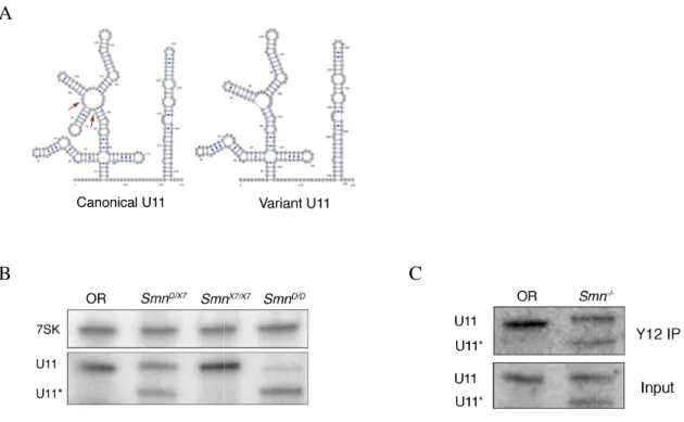
In addition to the canonical forms, we observed expression of U5 and U11 snRNA variants. Consistent with a previous report, the variant forms of U5 are predominantly expressed early in larval development (Chen et al., 2005). The Smn -/-larvae express a variant form of U11, that contains a 22 bp deletion in stem loop II, a region that is not well conserved among Drosophilid species (Fig. S2.2A). The U11 and Smn genes are both on the same chromosome, and we note that this U11 variant tracks
with the SmnD allele and is not present on the homolog containing SmnX7 (Fig. S2.2B). We also note that the variant U11 snRNA can be immunoprecipitated by anti-Sm antibodies (Fig. S2.2C) and that SmnD homozygotes can be rescued by transgenic expression of dSMN (Rajendra et al., 2007, this work). Thus, this variant U11 allele appears to be fully functional.
Figure 2.2. Analysis of minor
qRT-PCR was used to measure the relative levels of spliced mRNA for eighteen genes containing putative U12-dependent introns in ~76 hour OR,
using Rpl32 for normalization. Primers for qRT exon-exon junction flanking the U12
be detected (inset). Levels of spliced mRNA in the mutants have been normalized to those of OR larvae. The U6atac
introns for all the mRNAs tested (group avg. = 0.32). Nine of the eighteen mRNAs were reduced in U6atac-/- animals with p
one mRNA approached, but did not reach, significance (CG7892, p ~ 0.08). In contrast, splicing of these same introns was relatively unaffected in
1.06). Although a majority of the mRNAs were not affected, we note that the four mRNAs showing the greatest decrease in the
affected mRNAs in U6atac
-/-CG11839) had p-values < 0.05, although none were below 0.01.
32
Analysis of minor-class intron splicing defects in Smn and U6atac
PCR was used to measure the relative levels of spliced mRNA for eighteen genes dependent introns in ~76 hour OR, Smn-/- and U6atac
for normalization. Primers for qRT-PCR were designed to anneal across the exon junction flanking the U12-type intron, such that only spliced mRNAs would be detected (inset). Levels of spliced mRNA in the mutants have been normalized to U6atac-/- larvae showed robust defects in splicing of these for all the mRNAs tested (group avg. = 0.32). Nine of the eighteen mRNAs were animals with p-values < 0.01, eight had p < 0.05 and the levels of one mRNA approached, but did not reach, significance (CG7892, p ~ 0.08). In contrast, icing of these same introns was relatively unaffected in Smn-/- animals (group avg. = 1.06). Although a majority of the mRNAs were not affected, we note that the four mRNAs showing the greatest decrease in the Smn-/- mutants were also the most severely
mutants. Each of these (CG18177, CG33108, CG15081 values < 0.05, although none were below 0.01.
Transgenic expression of an SMA patient-derived mutation in dSMN rescues Smn-/- larval
lethality
Although analysis of Smn null mutants is informative, the many putative functions of SMN make it difficult to parse out the precise cause of lethality in these animals. Thus we wanted to develop Drosophila SMA models that perturb a subset of SMN functions, providing increased resolution to the analysis. As a proof of principle, we created flies expressing an SMA patient-derived point mutation, T205I. This mutation corresponds to an SMA type II/III patient mutation, T274I, which is located in the YG box domain of SMN (Hahnen et al., 1997; Sun et al., 2005). We used the PhiC31 integrase system (Bischof et al., 2007) to generate transgenic flies carrying either a wild-type (WT) or a T205I mutant transgene, inserted at site 86Fb on chromosome 3. To preserve endogenous expression patterns, the constructs are driven by the native Smn promoter, and contain native 3' and 5' flanking sequences. The transgenic proteins can be distinguished from the endogenous ones by the presence of an N-terminal 3X FLAG tag.
Expression of the wild-type construct (SmnWT) in the Smn-/- background results in >70% rescue of lethality (Fig. 2.3A). The surviving adults are fertile, with no apparent defects in flight or motility. In contrast with the null phenotype, expression of the
SmnT205I, construct rescues larval lethality; ~25% of these animals eclose as adults, but
34
dSMNT205I is moderately defective in self-oligmerization
A number of human SMA-derived mutations in the YG box region of SMN, including SMNT274I, have been shown to be defective in oligomerization (Lorson et al., 1998). We tested a panel of dSMN proteins, including T205I, for similar defects. FLAG- and Myc-tagged versions of each construct were expressed in Drosophila S2 cells and the co-immunoprecipitation profiles were compared (Fig. 2.3B). Immunoprecipitation was performed with anti-FLAG antibody and the amount of Myc-tagged protein that co-precipitated was measured. The amount of Myc-dSMN(T205I) co-precipitated by FLAG-dSMN(T205I) was reduced compared to the amount of Myc-dSMN(WT) precipitated by FLAG-dSMN(WT), indicating a deficiency in its ability to self-oligomerize. This conservation between the human and fly SMN mutants also extends to the following SMA-derived point mutations. Both SmnG206S (corresponding to human SMNG275S) and
SmnY203C (SMNY272C) mutants were defective in oligomerization, similar to reports of their
mammalian counterparts (Lorson et al., 1998). Furthermore, the severity of these defects is also conserved, as Y203C was less effective at binding to itself than either T205I or G206S (Fig. 2.3D). Likewise, the oligomerization defect of Y272C is known to be more severe than that of T274I (Lorson et al., 1998). Thus, T205I displays a relatively mild defect in self-oligomerization.
finding indicates that the relatively mild defect in T205I oligomerization does not significantly affect dSMN levels. In addition, this suggests that the lethality of the T205I point mutant is not due to insufficient protein levels, but rather due to a molecular defect in dSMN.
36
Figure 2.3. Transgenic rescue of Smn null animals with an SMA patient-derived point
38
Transgenic expression of SmnT205I rescues larval locomotion
Consistent with previous reports (Chan et al., 2003; Rajendra et al., 2007; Shpargel et al., 2009), we found that Smn-/- animals display significant defects in larval locomotion. To further characterize these locomotor defects, we employed two previously described assays (Wu et al., 2003; Ubhi et al., 2007). In the ‘righting’ assay, each larva was placed on its dorsal surface and the time taken to return to its ventral surface (the crawling position) was measured (Fig. 2.3E). The Smn-/- larvae took a significantly longer time to return to the crawling position compared to OR. Larval motility was rescued in animals carrying the WT transgene, and to a large extent in the T205I transgenic animals, which showed a relatively mild defect. In the ‘burrowing’ assay, ~30 larvae were placed atop a dish containing 1.5% agar/molasses media and incubated at room temperature in the dark. After 2 hours, the number of larvae remaining on top of the medium was recorded. The assay was repeated 3 times per genotype. We found that the Smn-/- mutants do not burrow into the medium at all (Fig. 2.3F). However, we also noticed that if there were surface defects (cuts) present in the media, some of these larvae worked themselves into the crevices. Importantly, the burrowing behavior was restored in both the WT and T205I transgenic animals (Fig. 2.3F). We conclude that
SmnT205I rescues the larval viability and locomotor defects observed in the null mutants.
SmnT205I and SmnWT transgenic larvae are functional in snRNP biogenesis
just prior to the beginning of the lethal phase (i.e. at ~76 hours) and snRNA levels were analyzed by northern blotting (Fig. 2.4A). As noted earlier, U1, U4, U5, U11, U12 and U4atac snRNAs showed a reduction in the null animals (Fig. 2.4B). Again, U12 and U4atac levels were the most severely affected (~60% reduction). The SmnWT and SmnT205I animals showed very similar snRNA profiles, despite differences in adult viability. Both transgenes failed to rescue U1, U4 and U4atac snRNA levels and only partially rescued U12 and U5 snRNAs (Fig. 2.4B). Thus, we conclude that the SmnT205I mutant is functional in snRNP biogenesis. In support of this interpretation, the human counterpart to this mutation, SMNT274I, was shown to be active in a HeLa cell Sm-core snRNP assembly assay (Shpargel et al., 2005).
40
A
42
Discussion
SmnT205I is a good model for intermediate SMA
An analysis of SMN point mutations that potentially disrupt only a subset of interactions should be informative in elucidating the contributions of individual SMN functions to the overall phenotype. The human SMNT274I mutation was identified in patients with milder forms of SMA, types II/III (Hahnen et al., 1997; Sun et al., 2005). These patients show a later age of onset, decreased severity of symptoms and longer life expectancy than type I patients (Ogino et al., 2004). The Drosophila equivalent of this mutation, SmnT205I, recapitulates several key features of intermediate SMA, including a slight deficiency in motor function and a much longer lifespan than the null mutant. We also observed a mild defect in oligomerization of SmnT205I, suggesting that on a molecular level, this Drosophila protein behaves similarly to the human mutation. We conclude that the SmnT205I flies represent a good model for intermediate SMA.
SMN, snRNA levels and SMA
As shown above, transgenic expression of SmnWT was able to rescue the locomotion and viability defects observed in Smn null mutants, however, a majority of the snRNAs were largely unrestored to wild-type levels. Given the considerable degree of phenotypic rescue, this was an unexpected finding, suggesting that the observed reduction in snRNAs in the null animals is not a major contributor to larval locomotion and viability.
splicing changes in both minor- and major-class introns in late-symptomatic SMA mice (Zhang et al., 2008). However, subsequent studies revealed that these splicing defects are likely a secondary consequence of severe SMN loss, as pre- and early-symptomatic SMA mice did not show an enrichment of unspliced introns (Baumer et al., 2009).
In our Drosophila system, we note that U12 and (to a lesser extent) U5 levels were partially rescued by transgenic expression of SmnWT and SmnT205I, although U4atac levels remained low (Fig. 2.4B). Thus, one explanation for our results could be that splicing of minor-class (U12-specific) introns is sensitive to small changes in snRNA concentrations and that U12 and U5 are limiting factors, whereas U4atac is not. We tested this hypothesis by using qRT-PCR to measure mRNA levels in 18 of the 20 predicted minor-class (U12-specific) introns in the Drosophila transcriptome (Lin et al., 2010). As shown in Figure 2-2, a mutation in the U6atac gene resulted in pronounced defects in splicing of minor-class introns, however, splicing of these same mRNAs was largely unaffected in Smn null mutants. Among the minor-intron transcripts we analyzed, the steady-state levels of two mRNAs, CG15081 (Phb2 in mice) and CG33108 (ortholog unknown), were reduced by roughly 50% in Smn-/- larvae. It is possible that reduction in levels of one or more of these mRNAs could contribute to the phenotype of Smn null mutants, however, CG15081 heterozygotes are completely viable (Flybase). A similar decrease in levels of the Phb2 mRNA in mice was shown to affect neither organismal viability nor motor function (Park et al., 2011), strongly suggesting that haploinsufficiency for CG15081 is not the cause of the Smn phenotype.
44
encoding human U4atac snRNA do not phenocopy SMA. Instead, loss of U4atac function causes a disease known as microcephalic osteodysplastic primordial dwarfism type I (MOPD I), which is characterized by severe intrauterine growth retardation and multiple organ abnormalities (Edery et al., 2011; He et al., 2011). Similar to the fruitfly U6atac mutants (Pessa et al., 2010; this work), cells derived from MOPD I patients display marked defects in splicing of minor-class (U12-type) introns, whereas splicing of major-class (U2-type) introns is unaffected (Edery et al., 2011; He et al., 2011). Although we cannot entirely exclude the possibility that tissue-specific defects in minor-intron splicing may be causative for SMA, our finding that transgenic expression of SmnWT can rescue viability and fertility without restoring U4atac levels effectively uncouples the observed global snRNA deficits from the organismal phenotype. Moreover, it is important to note that the Smn and U6atac mutants were analyzed just prior to onset of the lethal phase. At this developmental timepoint, Smn null animals already display significant motor function defects. We therefore conclude that perturbations in minor spliceosome levels are not likely to be causative for the larval locomotion and viability defects observed in Smn null mutants.
feedback loop wherein low levels of SMN protein exacerbate exon skipping of human SMN2, leading to a further reduction in SMN expression (Jodelka et al., 2010; Ruggiu et
Supplementary Data
Figure S2.1. Total snRNA levels in
from ~76 hour OR and Sm
antibody, Y12. The bound snRNAs were visualized via northern blotting with probes specific to the indicated snRNAs. The levels of snRNAs co
SmnX7/D were similar to the total lev
46
Total snRNA levels in Drosophila larvae reflect snRNP levels. Lysates SmnX7/D larvae were immunoprecipitated (IP) with anti antibody, Y12. The bound snRNAs were visualized via northern blotting with probes specific to the indicated snRNAs. The levels of snRNAs co-precipitated for OR and
were similar to the total level of snRNAs in the input.
A
B
Figure S2.2. SmnD mutants express a non
the secondary structures of the canonical and variant forms of U11 based on those of (Schneider et al., 2004
2009). The arrows on the canonical U11 indicate genomic sequence of the variant
SmnX7/X7 and SmnD/D larvae with U11 specific probes. The asterisk marks the vari
that migrates as a shorter band in
In SmnD/D larvae, which are homozygous for the U11 contributed canonical U11 can be seen
proteins. Lysates from OR and
antibody, Y12, and RNA was extracted and analyzed by northern blotting with a U11 probe. Both forms of U11
co-C
mutants express a non-canonical U11 snRNA. A. An illustration of the secondary structures of the canonical and variant forms of U11. The structures are
Schneider et al., 2004) and redrawn using VARNA ( . The arrows on the canonical U11 indicate a 22 base stem loop that is
variant allele. B. Northern blot of RNA from OR, larvae with U11 specific probes. The asterisk marks the vari migrates as a shorter band in SmnD/X7 and SmnD/D, but is absent in OR and
larvae, which are homozygous for the U11 variant, remnants of the maternally contributed canonical U11 can be seen. C. The variant U11 is complexed with Sm proteins. Lysates from OR and SmnD/X7 larvae were immunoprecipiated (IP) with anti antibody, Y12, and RNA was extracted and analyzed by northern blotting with a U11
-purified with the Y12 antibody.
Figure S2.3. Real-time qRT
rescue lines. The four mRNAs
mRNA that was not reduced (CG11984)
values among all three genotypes are not significantly different from each other. For CG11839 and CG33108, values are significant to p ~ 0.05 level when comparing OR vs.
SmnT205I and OR vs. SmnWT, respectively.
48
time qRT-PCR analysis of minor-class introns in Smn
rescue lines. The four mRNAs showing the greatest reduction in Fig. 2.2 and a control that was not reduced (CG11984) were analyzed. For CG18177 and CG15081, the values among all three genotypes are not significantly different from each other. For CG11839 and CG33108, values are significant to p ~ 0.05 level when comparing OR vs.
, respectively.
Table S2.1. Descriptions of 18 minor-class intron containing genes in the Drosophila
CHAPTER III
The development and characterization of new Drosophila models of
Spinal Muscular Atrophy
Introduction
The common childhood disorder, spinal muscular atrophy (SMA), is caused by mutations in survival motor neuron 1 (SMN1). Approximately 95% of SMA patients have deletions in SMN1, whereas the remaining 5% have point mutations in SMN1 (Burghes and Beattie, 2009). Despite much progress in elucidating the functions of SMN, the etiology of SMA remains poorly understood. To date, SMN’s most well understood function is in the biogenesis of spliceosomal uridine-rich small nuclear ribonucleoproteins (snRNPs). Several putative neuron and muscle-specific functions for SMN have also been described (Fan et al., 2002; McWhorter et al., 2003; Sharma et al., 2005). However, no convincing link between any function of SMN and SMA etiology has been shown. Thus the key question in SMA research is to understand which function or functions of SMN are critical in causing the disease.
In Chapter II, we showed evidence suggesting that non-snRNP related functions of SMN are important in SMA etiology using an SMA patient-derived point mutation,
SmnT205I, in Drosophila Smn. Here, we extend the analysis to an entire series of