Sharif University of Technology
Scientia IranicaTransactions B: Mechanical Engineering www.scientiairanica.com
Eect of Cu
2+
ion on biological performance of
nanostructured uorapatite doped with copper
R. Nikonam Mofrad
a, S.K. Sadrnezhaad
b;and J. Vahdati Khaki
ca. Department of Mechanical and Industrial Engineering, Concordia University, 1455 De Maisonneuve Blvd. W., Montreal, Quebec, Canada H3G 1M8.
b. Department of Materials Science and Engineering, Sharif University of Technology, Tehran, P.O. Box 11155-9466, Iran. c. Department of Material Engineering, Faculty of Engineering, Ferdowsi University of Mashhad, Mashhad, P.O. Box 91775-1111,
Iran.
Received 8 June 2015; received in revised form 22 July 2016; accepted 7 January 2017
KEYWORDS Nanostructured uorapatite; Ball milling; Biological performance.
Abstract. Nanostructured copper-doped uorapatite (Cux.Ca(10 x).(PO4)6.F2) having
crystallite sizes of 19, 29, and 34 nm at x = 0:9, 0.4, and 0.0, respectively, was synthesized by planetary ball milling of CaO, P2O5, CaF2, and CuO powders. Specications of
the products were determined by Fourier-transform infrared spectroscopy, eld emission scanning electron microscopy, transmission electron microscopy, and X-ray diraction analyses. In-vitro studies and Mossman's Tetrazole Test (MTT) assays were also conducted by incubating Cux.Ca(10 x).(PO4)6.F2powder into Kokubo's Simulated Body Fluid (SBF)
and against BT-20 cell, respectively, to determine bioactivity and biocompatibility of the materials. Antibacterial eects on Staphylococcus aureus were assessed by the disc diusion test method. Measurements showed that the rate of formation of uorapatite was lowered by Cu content. Besides, in-vitro experiments showed the same SBF interacted apatite precipitation for all samples. In contrast, MTT assays revealed dierent behavior for pure uorapatite and apatite with x = 0:9 Cu against BT-20 cell after 24 h of incubation. This highlights the increase of uorapatite cytotoxicity when Cu ion is present in the apatite structure. Copper-doped uorapatite was, however, desirably antibacterial. This stemmed from copper ions interactions with the bacterial metabolism which resulted in enzymes neutralization and copper-doped uorapatite antibacterial behavior.
© 2017 Sharif University of Technology. All rights reserved.
1. Introduction
The well-known biocompatible Calcium Phosphate (CP) uorapatite (Ca10(PO4)6F2) has obtained a wide
reputation for its ample applications in clinical use [1]. However, infections associated with implanted surgi-cal devices may cause signicant problems, such as
*. Corresponding author. Tel.: +98 21 66165215; Fax: +98 21 66005717
E-mail addresses: rahele [email protected] (R. Nikonam Mofrad); [email protected] (S.K. Sadrnezhaad); [email protected] (J. Vahdati Khaki)
doi: 10.24200/sci.2017.4314
implant failure considered to be a common clinical complication [2]. Therefore, synthesis of the metal ion-substituted bioceramics with antimicrobial properties can be a good strategy to decrease the rate of bioma-terials infections. Copper and its alloys have been reg-istered in the U.S. Environmental Protection Agency (EPA) as the rst solid antimicrobial material capable of deactivating microbes upon contact [3,4]. Copper-doped uorapatite (Cux.Ca(10 x).(PO4)6. F2), known
as an eective, eco-friendly and heterogeneous catalyst in recent studies [5,6], is an interesting example of metal ion-substituted ceramic, which is to be discussed in this paper.
produc-tion, the most common way of producing ne uo-rapatite (FA) particles is mechanochemical synthesis method, which is considered as a subarea to Mechanical Alloying (MA) [7,8]. It has the advantages of simplicity, reproducibility, low cost, and ease of producing (even in bulk quantities) of nanostructured materials [9,10]. The outstanding biological potentials of nanostruc-tured biomaterials, which have not been observed in the conventional polycrystalline coarse grained materials, have been reported by previous studies [11-13].
In this study, the eect of doping copper in nanostructured uorapatite with substitution of Cu2+
for Ca2+ on biological performances of uorapatite
is discussed. To study the biological properties of the material, both in-vitro and in-vivo conditions were taken into account. In-vitro evaluation of the samples produced under simulated physiological con-ditions indicated the change of bioactivity due to the incorporation of copper into the uorapatite. MTT assay was also used to determine biocompatibility of the samples. The human breast cancer BT-20 cell line was cultured and treated with prepared samples at various concentrations. The antibacterial activities of the apatite samples against the microorganism of Staphylococcus aureus (as an aerobic Gram-positive bacterium) were investigated via disc diusion test.
2. Materials and methods 2.1. Powder preparation
The starting precursors included calcium oxide (CaO, BDH, catalog no. 11021-462, min. 95% pu-rity), phosphorous oxide (P2O5, BDH, catalog no.
21411, min. 98.5% purity), calcium uoride (Ca F2,MERCK,product no. 102840, precipitated pure, a
mean particle size of about 100 m), and copper oxide (CuO, MERCK, product no. 102671, 96% purity,
particle size 160 m). The planned degree of substitution of Ca2+ by Cu2+ in the general formula
Cux.Ca(10 x).(PO4)6.F2 was x = 0, 0.4, and 0.9 for
which the product was designated as FA, 4CuFA, and 9CuFA, respectively. The overall reaction leading to the formation of the nanostructured copper-doped uorapatite is as follows:
3P2O5(s) + (9 x)CaO(s) + xCuO(s) + CaF2(s)
! Cux.Ca(10 x).(PO4)6.F2(s): (1)
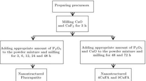
Milling of the mixture was carried out at rotating speed of 260 rpm in a planetary ball mill using stainless still balls of 1 cm diameter and ball-to-powder ratio of 40:1. Synthesis consisted of three stages. First, appropriate amounts of CaO and CaF2 were loaded into the vial
and mechanically activated for 3 h. The milled powder was labeled CF3. Second, CF3 was mixed with P2O5
and milled for 3, 6, 12, 24, and 48 h. The products were labeled CF3P3, CF3P6, CF3P12, CF3P24, and
CF3P48, respectively. Third, appropriate amounts of CuO and P2O5 were added to CF3 and milled
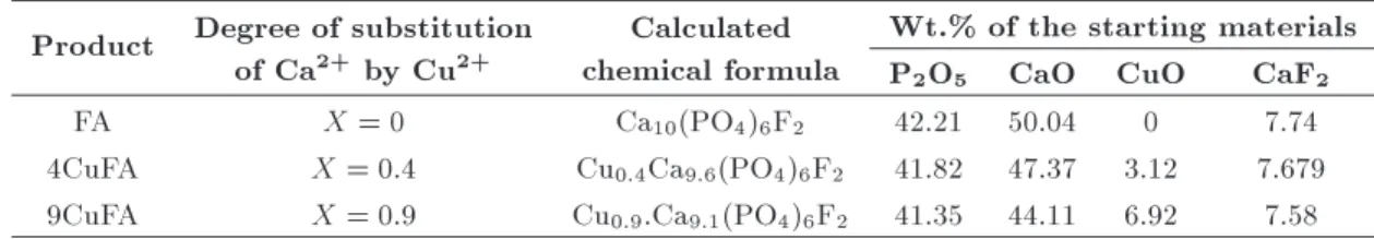
long enough to form 4CuFA and 9CuFA. The detailed sequences of the procedure used are illustrated in the owchart given in Figure 1, and the weight percent of the starting materials is presented in Table 1.
2.2. Characterization
X-Ray Diraction (XRD) by Philips Analytical X-Ray B.V diractometer with Ni-ltered Cu k radiation was used for phase analysis, size determination, and cell parameter calculation. Diraction patterns were taken in 2 range of 10 to 60 at a step size of 0:02 with
a generating voltage of 40 kV and an electric current of 40 mA. Identication of the apatite phase was achieved by comparing the diraction pattern of the powders with JCPDS standard of card No: #02-1088
Figure 1. Flowchart indicating preparation procedure for FA, 4CuFA, and 9CuFA powders through mechanochemical route.
Table 1. Wt.% of the starting materials. Product Degree of substitution
of Ca2+ by Cu2+
Calculated chemical formula
Wt.% of the starting materials P2O5 CaO CuO CaF2
FA X = 0 Ca10(PO4)6F2 42.21 50.04 0 7.74
4CuFA X = 0:4 Cu0:4Ca9:6(PO4)6F2 41.82 47.37 3.12 7.679
9CuFA X = 0:9 Cu0:9.Ca9:1(PO4)6F2 41.35 44.11 6.92 7.58
for CaO, #35-0816 for CaF2, #85-1120 for P2O5,
#74-1021 for CuO, #81-2040 for Ca(OH)2, #85-0846 for
CaCO3, #83-0688 for H3PO4, #03-0604 for Ca2P2O7
and #15-0876 for FA. The peak broadening of the XRD reection was used to estimate the crystallite size of the powder based on Williamson-Hall formula (Eq. (2)) [14]:
cos = 0:89(=d) + 4" sin ; (2) where d is crystallite size (nm), is wavelength of the X-rays, is Full-Width at Half Maximum (FWHM) of each peak (rad), and is Bragg's diraction angle ().
FWHM of (002), (222), and (213) reections was taken to measure the crystallite sizes of the apatite powder.
Apatite lattice parameters (a and c) were calcu-lated using the XRD data of (002) and (222) planes and the standard formula for the hexagonal (HCP) system (Eq. (3)) [15]:
1 d2 =
4 3
h2+ hk + k2
a2
+ l2
c2; (3)
where d is the spacing between adjacent planes in the set of Miller indices (hkl).
The functional groups of the powders were ana-lyzed by using Fourier-transform infrared spectroscopy (FT-IR, ABB Bomen, MB 100) in the frequency range of 400-4000 cm 1with the resolution of 4.0 cm 1using
KBr pellet technique.
Field emission scanning electron microscopy (FE-SEM, Hitachi, S-4160, Japan) operating at 15 kV was utilized to study the morphology and parti-cle/agglomerate size distribution of the powders before and after soaking in the SBF solutions. Prior to this analysis, the as-prepared samples were coated with gold lm using PVD method.
The powder morphology was studied via trans-mission electron microscopy analysis (TEM, LEO 912 AB, Germany) operated at an accelerating voltage of 120 kV. Before TEM observations, the samples were
dispersed ultrasonically in ethanol and suspended on holey-carbon lm-coated copper grids and allowed to air-dry.
Based on the results of Kokubo [16,17], the in-vitro bioactivity of materials can be investigated by the formation of bone-like apatite on the surface of the biomaterials in simulated body uid. In order to evaluate the bioactivity of the prepared powders in the physiological conditions, we used, therefore, Kokubo's SBF and ion concentrations to be nearly equal to the human blood plasma, as shown in Table 2 [17]. The samples washed ultrasonically in acetone for 15 min were immersed into SBF and maintained there at 37.0C for 14 days at a liquid/solid ratio of 1 ml/mg
without refreshing the soaking medium. After the desired immersion time, the powders were ltered, gently washed with deionized water to remove the SBF, and dried at the room temperature. Variations in Ca+2ions concentration and pH of SBF due to soaking
of the apatite powders were measured by Inductivity Coupled Plasma-Optical Emission Spectroscopy (ICP-OES, Model OES-730, Varian), and pH meter. Mor-phology changes of the samples due to desolation and precipitation of bone-like apatite during the immersion were then determined by FE-SEM.
Biocompatibility of pure uorapatite and uo-rapatite with relatively high amount of Cu (9CuFA) on human breast cancer cell BT-20 was evaluated by MTT assay. The colorimetric assay based on the use of the tetrazolium salt MTT is relatively a rapid method for the quantitative measurement of surviving and proliferating cells. This method is based on the capacity of mitochondrial enzymes of viable cells to transform the yellow tetrazolium salt MTT into an insoluble, blue formazan product, which is measured spectrophotometrically. Since the reduction of MTT can only occur in metabolically active cells, the level of activity is a measure of the viability of the cells. The degree of the living cells can be measured through reading the results on a multi-well scanning
Table 2. Ionic concentrations of simulated body uid [21].
Description Ionic concentration (mM)
Na+ K+ Mg2+ Ca2+ Cl HCO3 HPO2
4 SO24
Human plasma 142.0 5.0 1.5 2.5 103.0 27.0 1.0 0.5
spectrophotometer (ELISA reader) [18,19]. For this test, FA and 9CuFA concentrated elutes (not diluted) were prepared by adding culture medium supplemented with penicillin and streptomycin to FA and 9CuFA powder (1 mL to 100 mg of powder), according to the literature [20]. These elutes were then diluted by culture medium to the test concentrations (100%, 75%, 50%, 25%, 10%, 5%, and 1%) of FA and 9CuFA. Then, 200 l cell suspension containing 104cell was added to
multi-well plate. After 24 h of incubation at 37C, the
medium was replaced with the fresh medium containing test concentration of elutes of FA and 9CuFA. Then, cells were exposed to the test concentration of elutes for 24 h. Four wells were used for each concentration. Blank control wells contained an appropriate volume of media, MTT and apatite powders without the presence of cells. Eight separate wells were lled with cells without the addition of elutes which acted as untreated control.
After the incubation time, 50% of media was removed, 10% V/V of MTT at 5 mg/ml was added to the remaining media to all wells of assay, and plates were further incubated at 37C for 4 h. We carefully
re-moved the medium and added (for 24-well plate) 500 l DMSO to each well and mixed thoroughly to dissolve the dark blue crystals of formazan. We measured the absorbance by scanning multi-well spectrophotometer (ELAISA reader, BioTek model ELx800) using 570 nm test wavelength and 630 nm reference wavelength. The tests were performed on three replicate samples.
The percentage of cell survival rate was calculated using the following formula [19]:
Survival rate (%) =Ac AbAt Ab 100; (4) where At is average absorbance of the test compound, Ab is absorbance of blank, and Ac is average ab-sorbance of the control material.
In order to evaluate the antibacterial eect of the samples, the disk diusion method of Kirby and Bauer and the microorganism of Staphylococcus aureus (PTTC No. 1431) were used. About 0.085 g of each synthetic powder was pressed uniaxially to make discs of 6 mm diameter. The compressed disks were sterilized in an autoclave having temperature of 121C
for 20 min. Muller-Hinton Agar (MHA) was used as a growing nutrient medium for the microorganisms. MHA was cast into a plate. Tetracycline and Clo-ramphenicol was used as control material. The MHA containing plate was incubated with the test organism of 3108cells/ml. Three apatites and two antibiogram
discs were placed on the surface of the agar. The plate was inverted and then placed in air incubator of 37C for 20 h and 45 h. After these periods, the
antimicrobial activities of the samples were examined by measuring zone of the inhibition of the bacterial
growth around the discs. The results were then studied by Interpretative Chart of Zone Sizes as the standard guideline for the determination of the susceptibility level of microorganism to the antibiotics used [21] and to estimate the eect of copper ions on the bacterial metabolism.
3. Results and discussion 3.1. X-ray diraction
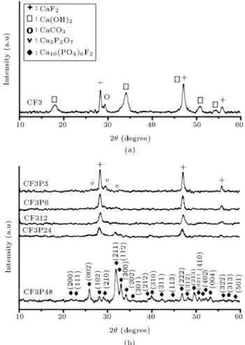
The XRD pattern of the CaO-CaF2 mixture,
mechan-ically activated for 3 h (CF3), is shown in Figure 2(a). In this pattern, in addition to peaks attributed to CaF2, some sharp peaks, assigned to calcium hydroxide
(Ca(OH)2), can also be detected. Since calcium oxide
is unstable, a majority of calcium oxides were trans-formed into calcium hydroxide upon ambient water vapor adsorption. On the other hand, the presence of peak, which was assigned to calcium carbonate (CaCO3), shows that amount of CaO was transformed
to CaCO3during the milling process or originated from
the starting CaO.
XRD patterns of CaO-CaF2-P2O5 powder
mix-ture (CF3P) milled for various durations are shown in
Figure 2. XRD pattern of the mechanically treated mixture of (a) CaO-CaF2 and (b) CaO-CaF2-P2O5.
Figure 2(b). As seen in the gure, sharp characteristic peaks of CaF2 are present in the X-ray diraction
pattern of the 3 h milled samples (CF3P3). This pattern shows one peak at 2 = 29 which can
correspond to CaCO3. This peak can also be attributed
to calcium phosphate (Ca2P2O7) while the formation
of this intermediate product is expectable when P2O5
is present. When mechanical activation extends to 12 h (CF3P12), this peak disappears and those correspond-ing to CaF2 broaden. This conrms the continuation
of the mechanochemical process. By rising the milling time up to 24 h (CF3P24), both broad and weak peaks around 31:8 emerge. This indicates the formation
of poorly crystallized apatite phase. As milling time increased up to 48 h (CF3P48), a further increase in crystalline order of the apatite phase and further sharpening of the principal diraction peaks occurred. Based on the XRD pattern, after 48 h milling, no peaks, except those assigned to FA, can be observed, which shows crystalline apatite phase achievement.
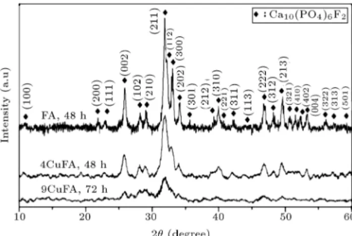
Figure 3 plots the XRD patterns of the FA powders doped with dierent amounts of copper. All peaks of the patterns correspond to the FA structure (JCPDS le card #15-0876). Only apatite phases are detected in the powder. This shows the complete reaction between precursors and suggests the formation of relatively well crystalline uorapatite for all three samples during milling operations. Based on Figure 3, relatively well crystalline uorapatite is synthesized after 48 h milling 4CuFA, while at higher Cu content (9CuFA), 72 h milling is required. Besides, this gure shows that with higher Cu content in the FA structure, less intensive apatite peaks form. Since the ionic radius of Ca2+ (0.99 A) is larger than that of Cu2+ (0.72 A),
substitution of Cu2+into Ca2+position distorts the FA
structure. The result shows the decrease of stability of the FA structure and subsequent lowering of the height of the apatite peaks [22].
The XRD patterns of the 9CuFA sample milled
Figure 3. XRD patterns of synthesized FA, 4CuFA, and 9CuFA powders.
Figure 4. XRD patterns of mechanically treated mixture of CaO-CaF2-P2O5-CuO (x = 0:9) after milling for
various durations.
for 48 h and 72 h are shown in Figure 4. Two identied phases, including poorly crystallized apatite and CaF2,
are observed in the XRD patterns of the powder milled for 48 h. This is due to the incomplete reaction between the precursors. Prolonged milling up to 72 h causes an increase in crystalline order of the apatite phase, while the corresponding CaF2peaks completely
disappear. Based on these observations, the relatively well-crystalline 9CuFA is synthesized after milling for 72 h. As a result, upon the addition of copper oxide to the powder mixture, CuO plays as an obstacle between the reactants to slow down the reaction at higher Cu content.
From XRD data and Williamson-Hall formula, the average crystallite size of FA and 4CuFA (after 48 h milling) and 9CuFA (after 72 h milling) are calculated to be 34, 29, and 19 nm, respectively. Measurements of the lattice parameters of the apatite powders indicate: a = 0:9385 nm and c = 0:6870 nm for FA, a = 0:9366 nm and c = 0:6866 nm for 4CuFA, and a = 0:9361 nm and c = 0:6847 nm for 9CuFA. The observed decreasing sizes of a and c parameters by increasing copper contents in the apatite structure refer to the substitution of Ca2+ by Cu2+. Since the ionic
radius of Cu is lower than Ca, incorporation of more Cu into the FA structure lowers the lattice parameters slightly. However, these values are close to the lattice parameters of the standard FA powder.
3.2. FTIR
Figure 5 records the IR spectra of FA powders with dierent copper concentrations. The FT-IR spectra for all of the three synthesized powders indicate nearly the same characteristic peaks for OH , PO3
4 , and F...OH
groups. These spectra show the existence of phosphate groups (PO3
4 ) at 471 cm 1 corresponding to
asym-metrical 2 stretching vibration [23,24]. Besides, a
doublet appears at 568 and 599 cm 1 corresponding
to 4 bending vibrations [25,26]. As stated in the
literature [27], the presence of the band at 743 cm 1
is due to the increase of F content in the (OH , F ) apatite chain, while the conguration of OH...F is predominant in F rich chains containing only a few
Figure 5. The IR spectra of the synthesized powders: (a) FA, (b) 4CuFA, and (c) 9CuFA.
(OH ) and almost pure uorapatite obtained. The band at 964 cm 1 relates to
1 bending vibrations
of the phosphate group [25], while the most intense band in the spectra at 1050 cm 1 corresponds to
asymmetrical 3 stretching vibration of the phosphate
group [23,25]. According to this graph, substitution of Cu ion with shorter ionic radius than Ca brings about an increase in the P-O bonding strength [28], which shifts 1 and 3 vibrations to the higher frequencies.
The presence of a peak at 873 cm 1 can be related to
2 stretching vibrations of the carbonate group which
suggests substitution of CO2
3 groups into PO34 sites
of apatite lattice (B-type substitution) [29]. By increas-ing the millincreas-ing time from 48 h to 72 h, the amount of adsorbed carbonate group increases which enhances the intensity of the assigned peak at 873 cm 1. Based
on the fact that carbonates are constituents of hard tissue structures, the presence of low content of CO2
3
groups in PO34 sites could be benecial to improve the bioactivity of FA and make it more similar to biological appetites [15]. The sharp band at 1645 cm 1
and the broad band over 3800-2500 cm 1 are mainly
originated from the vibrations of the hydroxyl (OH 1)
group attributed to the presence of the absorbed water in the samples or the KBr pellet [23,24].
3.3. FE-SEM and TEM observations
Figure 6 shows microstructure and morphology of nanopowders after 48 h milling. From FE-SEM micro-graphs, it is revealed that both samples are composed of agglomerates/particles of irregular shapes with wide size distributions. No signicant change in size distri-bution and morphology of the agglomerates/particles of samples can be inferred from these pictures. Also, higher magnications show that the surface of particles is rather smooth before immersion. TEM micrographs of nanopowders after milling are shown in Figure 7. As illustrated, agglomerated particles are composed of smaller nanoparticles averaging from 20 to 60 nm mean diameter for FA and 4CuFa and 15 nm for 9CuFA. Prolonged milling of the latest sample results in the formation of ne particles, which are uniform in size and shape. Unlike 9CuFA, FA and 4CuFA are composed of relatively larger and irregular particles;
Figure 7. TEM images of (a) FA, (b) 4CuFA, and (c) 9CuFA powders at dierent magnications.
however, both are similar in shape and morphology, typically semicircular crystals. These gures are in agreement with the crystallite sizes measured by XRD patterns through Williamson Hall equations.
3.4. In-vitro test
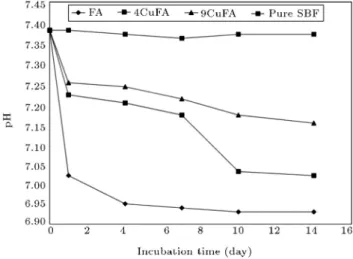
Variations of [Ca2+] and pH of SBF solutions as a
func-tion of soaking time are shown in Figures 8 and 9, re-spectively. Figure 8 indicates that the calcium ions con-centration of SBF decreases as the incubation time in-creases. Besides, based on Figure 9, during this period,
Figure 8. [Ca+2] prole of SBF solution containing
prepared powders during the soaking time.
Figure 9. pH prole of SBF solution containing prepared powders during the soaking time.
the pH of pure SBF is relatively constant, whereas, in the case of 9CuFA, 4CuFA, and FA, the pH curves show a sudden drop during the rst day. After that period, the pH values decrease slightly throughout the incuba-tion time while reaching a minimum at the 14th day.
The observed trend of [Ca2+] and pH can be
justied by the Kim's theory [30], which reveals that the process of bone-like apatite formation on the surface of biomaterials in the SBF is divided into the following stages. First, dissolution of apatite powder occurs, resulting in an increase in [Ca2+] and pH of
SBF and appearance of negative charge on the surface of apatite particles. In the second step, the negative surface charge attracts Ca2+ ions to form the
Ca-rich Amorphous Calcium Phosphate (ACP) layer. The resultant positive surface charge attracts the negative phosphate ion in the uid to form Ca-poor ACP layers. [Ca2+] and pH of SBF decrease during this step.
Finally, this layer gradually stabilizes and crystalizes into bone-like apatite in SBF. Indeed, when samples are immersed in SBF, simultaneous dissolution of the apatite particles and precipitation of the new apatite layer both occur. When precipitation is dominant, new apatite layers were formed, and tiny sediments precipitate on the surface of the immersed samples [31]. However, in this research, since no pH and [Ca2+] data
are collected during the rst hours of incubation, the rst steps of dissolution behavior are not observed. The observed decrease in pH value and quantity of calcium ions of the SBF solution is due to the consumption of Ca2+ and OH ions during the second stage of the
described theory and the formation of apatite layer. Based on Figures 8 and 9, pH and [Ca2+] proles
of SBF solutions have relatively dierent decreasing rates. It can be seen that the reduction rate of pH and [Ca2+] proles of sample decreases as the copper
content increases. However, the concentrations of Ca2+
are relatively close in the case of FA and 4CuFA. As pointed out earlier, the replacement of Ca2+ by Cu2+
in FA causes a certain degree of disorder to the crystal structure which makes the lattice more unstable. Be-sides, with increasing of Cu content, the average grain size of FA slightly decreases which causes an increase in the volume of the grain boundaries. Therefore, by incorporation of Cu into the apatite structure, Gibbs
free energy increases and this may result in more solubility of 9CuFA apatite in SBF than FA. The observed higher amount of [Ca+2] in SBF for 9CuFA
may be due to the dissolution behavior of 9CuFA during the rst incubation hours and more calcium ion distribution. Therefore, higher value of pH in the case of 9CuFA than FA can be attributed to the higher amount of calcium ion in SBF solution for 9CuFA. However, a recent study has indicated that during incubation of FA powder in the SBF, concentration of dierent ions changes with time, which causes variation in the pH value of SBF. This happens due to the interaction between bioactive FA particles and the SBF solution which leads to the nucleation and growth of an apatite-like layer on the FA particles [23]. To interpret the observed behavior of the pH variations, other ions' concentrations should be considered in future studies.
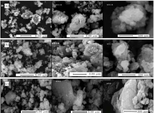
In comparison with the samples before soaking in SBF (Figure 6), the microstructures of the particles soaked for 14 days (Figure 10) show the dierences in their surface morphology. It is obvious from these micrographs that an interaction between the powders and SBF has occurred, a new bone-like apatite layer has been formed, and tiny sediments have precipitated on the surface of the immersed samples.
3.5. Cellular response on apatite samples Figure 11 illustrates the MTT results of 24 h incubation of breast cancer cell in contact with FA and 9FA test concentrations. It is seen that by increasing the FA concentration, the proliferation response reaches a
Figure 11. Viability of BT-20 cell in contact with apatite powders as a result of three individual experiments, while being compared with control wells containing only cell and media.
maximum at 25% FA. However, above 25% concen-tration, the viability decreases, yet it remains above the starting cell quantity. For higher concentrations, cell viability decreases steadily until it reaches the lowest at 100%. The observed cytotoxicity of FA at high concentrations is due to the presence of uoride in the structure of FA [20]; however, it is worth mentioning that uoride has high biological activity and wide biomedical applications to stimulate bone formation. For 9CuFA, for all concentrations, the viability percentage is lower than the control sample, which reveals that the presence of Cu ions in the apatite structure results in an increase of the cytotoxicity eect
Figure 10. FE-SEM photographs of (a) FA, (b) 4CuFA, and (c) 9CuFA powders at dierent magnications after soaking in SBF.
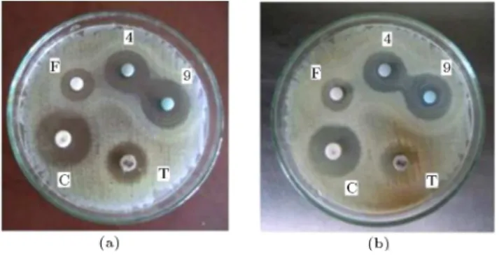
Figure 12. Results of disc diusion test of the synthetized samples after (a) 20 h and (b) 45 h of incubation. Denitions of characters on the plates are: F = pure FA, 4 = 0.4CuFA, 9 = 0.9CuFA, T = Tetracycline antibiogram and C = Cloramphenicolantibiogram.
of uorapatite against BT-20 cell line. However, at 10% CuFA, the cell viability tends to the maximum of around 95% of the control sample, which is the lowest cytotoxicity achievable.
3.6. Antibacterial assessment
Results of antibacterial tests are presented in Figure 12. Both control disks and apatite samples of dierent copper contents clearly show the zones of inhibition. Recent studies have shown that uoride can aect bacterial metabolism (as an enzyme inhibitor) and act as an antibacterial agent [32,33]. This leads to the presence of the zone of inhabitation around the pure FA disk. On the other hand, with an increase of the dose of the copper ions in the apatite structure, a growth in the zone of inhabitation is observed. This shows antibacterial activity improvement. With regard to the antibiotics used and the results derived from the Interpretative Chart of Zone Sizes [21], FA sample shows strong antibacterial property after doping with copper ions, which shows the complete inhibition of the growth of S.aureus bacteria. Indeed, Cu2+ ions react
with bacteria and deactivate the S.aureus metabolism, thus inhibiting its growth even after 45 h of incubation. The mechanism of antibacterial activity of copper ions is as follows: Cu ions eluted from the material's surface are absorbed onto the surface of bacteria cells. These ions damage the cell membrane and penetrate into the bacteria. Cu-ions that have the strong reduction can extract the electrons from the bacteria, causing their cytoplasm to run o and oxidizing their cell nucleus and killing them [34,35].
We should consider that cell responses of bioma-terials are various against dierent cultured cells. For instance, uoride has shown considerably higher cyto-toxicity against tumor cell lines than normal cells [36]. Besides, in this study, just high concentration of the prepared samples is investigated which may result in dierent observation than concentrations lower than 1% of sample test. Considering that biomaterials
may have dierent potential risks to human safety, they can be utilized for various applications with respect to the extent of contact with patient's tissue. Therefore, to determine the exact biological response of copper ions on apatite structure, more exhaustive and comprehensive studies are required.
4. Conclusions
Nanostructured copper doped uorapatites of dierent copper contents were synthesized using CaO, CaF2,
P2O5, and CuO as the starting materials through
mechanochemical route. The results of Williamson-Hall equation and TEM analysis showed that the syn-thetic FA powders were not only nano-structured, but consisted of nano-particles, too. XRD results demon-strated that relatively pure FA and copper doped uorapatite with the substitution ratio of 0.4 could be synthesized after milling for 48 h. Their respective crystallite sizes were 34 and 29 nm. The synthesis time mounted to 72 h, when the substitution ratio of copper doped uorapatite was 0.9 and crystallite size became 19 nm. SEM micrographs along with in-vitro analysis showed the sedimentation of apatite-like tiny particles on the surface of all three samples. In-vitro test revealed that cell viability signicantly increased following the treatment of BT-20 cell with FA from 1% to the high concentration of 50% and reached the max proliferation response at 25%. However, for 9CuFA, the presence of Cu ions showed signs of toxicity, especially at higher concentration than 25%. While high antibacterial activities were discerned for all three samples containing dierent amounts of copper, a positive correlation between copper content and the antibacterial properties was indicated.
Acknowledgment
The authors would like to appreciate the overall sup-port of the research deputy of the Sharif University of Technology and Iran National Science Foundation (INSF).
References
1. Leroy, N., Bres, E., Jones, D. and Downes, S. \Structure and substitutions in uorapatite", Eur. Cell Mater., 2, pp. 36-48 (2001).
2. Wan, Y., Xiong, G., Liang, H., Raman, S., He, F. and Huang, Y. \Modication of medical metals by ion implantation of copper", Appl. Surf. Sci., 253(24), pp. 9426-9429 (2007).
3. Ibrahim, M., Wang, F., Lou, M.M., Xie, G.L., Li, B., Bo, Z., Zhang, G.Q., Liu, H. and Wareth, A. \Copper as an antibacterial agent for human pathogenic multidrug resistant burkholderia cepacia
complex bacteria", Journal of Bioscience and Bioengi-neering, 112(6), pp. 570-576 (2011).
4. Molteni, C., Abicht, H.K. and Solioz, M. \Killing of bacteria by copper surfaces involves dissolved cop-per", Appl. Environ. Microbiol., 76(12), pp. 4099-4101 (2010).
5. Mulla, S.A., Inamdar, S.M., Pathan, M.Y. and Cha-van, S.S. \Ligand free, highly ecient synthesis of diaryl ether over copper uorapatite as heterogeneous reusable catalyst", Tetrahedron Lett., 53(14), pp. 1826-1829 (2012).
6. Kantam, M.L., Venkanna, G., Kumar, K., Balasub-rahmanyam, V., Venkateswarlu, G. and Sreedhar, B. \Mild and ecient alkylation of aldehydes by using copper uorapatite as catalyst", Advanced Synthesis & Catalysis, 350(10), pp. 1497-1502 (2008).
7. Suryanarayana, C. \Mechanical alloying and milling", Progress in Materials Science, 46(1), pp. 1-184 (2001). 8. Aminzare, M., Eskandari, A., Baroonian, M.H., Berenovc, A., Razavi Hesabib, Z., Taheri, M. and Sadrnezhaad, S.K. \Hydroxyapatite nanocomposites: Synthesis, sintering and mechanical properties", Ce-ramics International, 39(3), pp. 2197-2206 (2013). 9. Rhee, S. \Synthesis of hydroxyapatite via
mechanochemical treatment", Biomaterials, 23(4), pp. 1147-1152 (2002).
10. Eskandari, A., Aminzare, M., Hassani, H., Barounian, H., Hesaraki, S. and Sadrnezhaad, S.K. \Densication behavior and mechanical properties of biomimetic apatite nano-crystals", Current Nanoscience, 7(5), pp. 776-780 (2011).
11. Shi, Z., Huang, X., Cai, Y., Tang, R. and Yang, D. \Size eect of hydroxyapatite nanoparticles on proliferation and apoptosis of osteoblast-like cells", Acta Biomaterialia, 5(1), pp. 338-345 (2009).
12. Prabakaran, K. and Rajeswari, S. \Spectroscopic in-vestigations on the synthesis of nano-hydroxyapatite from calcined eggshell by hydrothermal method using cationic surfactant as template", Spectrochimica Acta Part A: Molecular and Biomolecular Spectroscopy, 74(5), pp. 1127-1134 (2009).
13. Torabi, M. and Sadrnezhaad, S.K. \Corrosion be-havior of polypyrrole/hydroxyapatite nanocomposite thin lms electropolymerized on NiTi substrates in simulated body uid", Materials and Corrosion, 62(3), pp. 252-257 (2011).
14. Williamson, G. and Hall, W. \X-ray line broadening from led aluminum and wolfram", Acta Metallurgica, 1(1), pp. 22-31 (1953).
15. Ebrahimi-Kahrizsangi, R., Nasiri-Tabrizi, B. and Chami, A. \Characterization of single-crystal uora-patite nanoparticles synthesized via mechanochemical method", Particuology, 9(5), pp. 537-544 (2011). 16. Kokubo, T. and Takadama, H. \How useful is SBF
in predicting in vivo bone bioactivity?", Biomaterials, 27(15), pp. 2907-2915 (2006).
17. Kokubo, T. \Bioactive glass ceramics: Properties and applications", Biomaterials, 12(2), pp. 155-163 (1991). 18. Zarrabi, A., Shokrgozar, M.A., Vossoughi, M. and Farokhi, M. \In vitro biocompatibility evaluations of hyperbranched polyglycerol hybrid nanostructure as a candidate for nanomedicine applications", J. Mater. Sci. Mater. Med., 25(2), pp. 499-506 (2014).
19. Patel, S., Gheewala, N., Suthar, A. and Shah, A. \In-vitro cytotoxicity activity of solanum nigrum extract against hela cell line and vero cell line", International Journal of Pharmacy and Pharmaceutical Sciences, 1(1), pp. 38-46 (2009).
20. Jantova, S., Theiszova, M., Letasiova, S., Birosova, L. and Palou, T. \In vitro eects of uor-hydroxyapatite, uorapatite and hydroxyapatite on colony forma-tion, DNA damage and mutagenicity", Mutation Re-search/Genetic Toxicology and Environmental Muta-genesis, 652(2), pp. 139-144 (2008).
21. Vandepitte, J., Verhaegen, J., Engbaek, K., Rohner, P., Piot, P. and Heuck, C. \Basic laboratory proce-dures in clinical bacteriology", World Health Organi-zation (2003).
22. Li, M., Xiao, X., Liu, R., Chen, C. and Huang, L. \Structural characterization of zinc-substituted hydroxyapatite prepared by hydrothermal method", Journal of Materials Science: Materials in Medicine, 19(2), pp. 797-803 (2008).
23. Fathi, M. and Mohammadi Zahrani, E. \Mechani-cal alloying synthesis and bioactivity evaluation of nanocrystalline uoridated hydroxyapatite", J. Cryst. Growth, 311(5), pp. 1392-1403 (2009).
24. Nikcevic, I., Jokanovic, V., Mitric, M., Nedic, Z., Makovec, D. and Uskokovic, D. \Mechanochemical synthesis of nanostructured uorapatite/uorhydroxyapatite and carbonated uorapatite/uorhydroxyapatite", Journal of Solid State Chemistry, 177(7), pp. 2565-2574 (2004). 25. Nasiri-Tabrizi, B. and Fahami, A. \Synthesis and
characterization of uorapatite-zirconia composite nanopowders", Ceram. Int., 39(4), pp. 4329-4337 (2013).
26. Albano, M.P. and Garrido, L.B. \Processing of con-centrated aqueous uorapatite suspensions by slip casting", J. Mater. Sci., 46(15), pp. 5117-5128 (2011). 27. Freund, F. and Knobel, R.M. \Distribution of uorine in hydroxyapatite studied by infrared spectroscopy", Journal of the Chemical Society, Dalton Transactions, 11, pp. 1136-1140 (1977).
28. Golden, D. and Ming, D. \Nutrient-substituted hy-droxyapatites: Synthesis and characterization", Soil Sci. Soc. Am. J., 63(3), pp. 657-664 (1999).
29. Lafon, J., Champion, E. and Bernache-Assollant, D. \Processing of AB-type carbonated hydroxya-patite Ca10-x(PO4)6-x(CO3)x(OH)2-x-2y(CO3)y ce-ramics with controlled composition", Journal of the European Ceramic Society, 28(1), pp. 139-147 (2008).
30. Kim, H., Himeno, T., Kokubo, T. and Nakamura, T. \Process and kinetics of bonelike apatite formation on sintered hydroxyapatite in a simulated body uid", Biomaterials, 26(21), pp. 4366-4373 (2005).
31. Cai, Y., Zhang, S., Zeng, X., Wang, Y., Qian, M. and Weng, W. \Improvement of bioactivity with mag-nesium and uorine ions incorporated hydroxyapatite coatings via sol-gel deposition on Ti6Al4V alloys", Thin Solid Films, 517(17), pp. 5347-5351 (2009). 32. Van Loveren, C. \The antimicrobial action of uoride
and its role in caries inhibition", J. Dent. Res., 69, Spec. No: 676-81; discussion 682-3 (1990).
33. Marquis, R.E. \Antimicrobial actions of uoride for oral bacteria", Can. J. Microbiol., 41(11), pp. 955-964 (1995).
34. Hong, I. and Koo, C.H. \Antibacterial properties, corrosion resistance and mechanical properties of cu-modied SUS 304 stainless steel", Materials Science and Engineering: A, 393(1), pp. 213-222 (2005). 35. Dan, Z., Ni, H., Xu, B., Xiong, J. and Xiong, P.
\Microstructure and antibacterial properties of AISI 420 stainless steel implanted by copper ions", Thin Solid Films, 492(1), pp. 93-100 (2005).
36. Otsuki, S., Morshed, S.R., Chowdhury, S.A., Takayama, F., Satoh, T., Hashimoto, K., Sugiyama, K., Amano, O., Yasui, T., Yokote, Y., Akahane, K. and Sakagami, H. \Possible link between glycolysis and apoptosis induced by sodium uoride", J. Dent. Res., 84(10), pp. 919-923 (2005).
Biographies
Raheleh Nikonam Mofrad received her master and bachelor degrees in Materials Science and Engineering from Sharif University of Technology and Ferdowsi University of Mashhad in 2012 and 2010, respectively. In 2014, she joined the Mechanical, Industrial and Aerospace Engineering Department at Concordia Uni-versity in Canada to conduct a research on fabrication and characterization of porous silicon nitride ceramics with ltration applications.
Sayed Khatiboleslam Sadrnezhaad is a distin-guished Professor at the Department of Materials Science and Engineering of Sharif University of Tech-nology. His current interest is in the emerging bio-nano elds of the materials science and engineering discipline. His PhD dates back to 1979 from the Massachusetts Institute of Technology. So far, he has authored and coauthored ve books, more than 600 technical papers and more than 60 patents.
Jalil Vahdati Khaki is a Professor at the Department of Materials Engineering, Faculty of Engineering of Ferdowsi University of Mashhad. His elds of study and research include mechanical alloying, thermody-namics and kinetics of materials, materials processing and extractive metallurgy. He is the editor-in-chief of Journal of Metallurgical and Materials Engineering which is published by Ferdowsi University of Mashhad. Dr. Vahdati has authored 140 journal and conference papers.