Islamic Republic or Iran Fall 1372 November 1993
EVALUATION OF THE ROLE OF CELL-MEDIATED
IMMUNITY IN DERMATOPHYTOSIS
A.R. KHOSRAVI,* M. FRANCO,**
AND
M. MAHMOUD!+
From tIl£' *FclC'ulIy of Velerirwry M('t/icine. and +School of Public Heallh, Tehran Ufliversil)� and thl' **Deparll1l(JnJ of Pathology, SclUJol of Mt!dicint!- amllnUJlIl1l% gy Division. UNESP, BOlIlC�lltll, Brazil.
ABSTRACT
For demonstration of cell-mediated immunity and its role in the process of
dermatophytosis,
98
patients with acute dermatophytosis (group
I)
and
131
chronic
dermatophytosis patients (group
2)
were chosen. In ail patients, lymphocyte
transfonnation and skin test were used.
96
members of group
I (98%)
had positive
delayed-type hypersensitivity (DTH) responses to trichophytin, whereas only
43
subjects
(32.8%)
of group
2
had positive DTH responses.
In
group
I,
positive
lymphocyte blastogenic responses to trichophytin and phytohemagglutinin were
seen in
95 (96.lJ%)
and
lJX (100%)
patients, respectively, but in group
2
the positive
results were observed in4lJ
(34.4%)
and
127 (lJ4.9%)
subjects, respectively. Patients
with chronic dermatophytosis had histories of a number of systemic disorders such
as: atopy
04
patients; diabetes, seven patients; contact dennatitis, four patients; and
tuberculosis, three patients.
M.lIRI, Vol. 7, No.3, 165-169, 1993.
INTRODUCTION
Dennatophytic infections are very superficial in location (i.e .. generally confined to the strntum corneum), and it might seem re.�sonable to assume thai this type of infection would not sensitize the host.' However, the disease process is greatly influenced by the host response to dennatophyte infection. There are a number of studies on the immunology of dennatophytosis.'·l.4 Hosts infected with dermatophytes usually develop cellular hypersensitivity.'
Several studies have demonstmted that animals, experimentally infected with dennatophytes, generally develop delayed hypersensiti vity to antigens of the infecting agent.'·'·' Delayed hypersensitivity responses appear to be imponant in the development and c1eamnce of the lesions of dennatophytosis.' Kerbs et al.' found that in guinea pigs
C"orrespondenceAddress; A.R. Khosravi. Depnnment ofMedic.'l1 Mycology, Facuhy of Veterinary Medicine. Universiry of Tehran. P.O.Box 14155· 6453. Tehran, Iran.
165
with experimental Trichophyton mentagrophytes infections, the degree of maximal erythema occurred when the animals developed cell-mediated immunity.
Lepper' noted that in canle infected with
T.
verr/lCOS/I/Il, the development of inflammation in primary infections coincided with the development of delayed hypersensitivity to fungal antigens.Green el al,' used T. /Ilelltagrophyles infections of guinea pig skin gmfts on nude mice to demonstrate that cell mediated immunity is required to sustain inflammation in the infected skin and to eliminate the infecting organisms. In the bovine model of dermatophytosis, delayed hypersensitivity reactions appeared to promote tile cleanUlce of the infection by increasing the rate of desqumnation of the stratum corneum.'
In man, the importance of cell-mediated immunity, as manifested by delayed-hypersensitivity responses to trichophytin, in the resistance to dennatophytosis has been demonstrated by experimental infections; subjects with positive delayed tests appear to be relatively immune to experimental inoculation with
T.
memagrophyles. whereasCell-Mediated Immunity in Dermatophytosis
chronic'�ly infected subjects with negative delayed skin
tests were susceptible.M-D
PATIENTS AND METHODS
229 patients were selected for this study. The patients,
based on types of ringworm.wert! divided into 2 groups as
follows:
Group
I:In this group 98 subjects with acute
dematophytosis were chosen. The duration of their infection
ranged from
Ito 3 months (mean duration, 2.5 months).
Their ages ranged from
I Ito 36 years (means age, 19
years).
Group 2: In this group, 13
Isubjects with chronic
dematophytosis were selected. The duration of their
infection I1Ulged from 7 to 38 years (meM duration, 21
years). the age I1Ulge being from 41 to 65 years (mean age,
51 years).
Trichophytin (Cruickshank) was used for skin test in
all 229 patients. Each individual was questioned for a
personal history of medical problems such as acne, psoriasis,
contact dennalitis. diabetes, c:md atopy.
Skin Test
Trichophytin was used in s;�ine solution. The patients
were injected intmdenn;�ly on the volar aspect of the
foreann, with trichophytin (10 �g/ 0.1 mL).
TIle sites of injection were observed to delennine the
size 'Uld degree of erythema, and degree of induration at 30
minutes, 24,48, and 72 hours.
At
20 minutes reading, a wheal greater than 10 10m in
diouneler, with or without a
flare.was considered a positive
immediate reaction. The DTH reaction were intCJllfcted as
positive if any degree of erythema, edema, or induration
was present at the 72 hours reading.
Lymphocyte Transformation Test (LTT)
For LIT, 30 mL of blood from each patient was
aspimted into sterile syringes containing I: 500 U heparin
and 2.0 mL of 8% dexuan. The blood was sedimented lor
35 min atlhe room teperature. Lymphocyles were isolaled
and washed Ihree times in HBSS.
Forculture oflymphocyles,RPMlI640medium which
contained 15% heat-inactivated guinea pig serum, 10 10m
hepes huffer, 100 �g/mL streplomycin, 100 U/mL
penicillin, 0.29 mg/mL L-glutmnine, 'Uld 0.15% sodium
hicarhonate was used. Cells were cultured in microtiler
plale al5 x 10" cells per well in 0.2 mL of medium. TI,e
mitogens were added in Ihe following concenualion:
Phylohemagglutinin (PH
A)
4�g/mL and Uichophytin, 5
Ilg/mL. Microcultures were incubated at 37°C in a
humidified aunosphere of 5% CO, in
air
for three days.
TIlen, uitiated thymidine (0.5 cell per well) was added
Fig. 1. Tinea barbae, severe inflammatory reaction (Kerion) produced by infection with M. callis.
Fig. 2. Acute form of tinea corporis cnused by T. Vt!/'TIICDSllfll.
Fig. 3. Tinea unguium. The infection is chronic and of many years' duration.
Fig. 4. Tinea pedis due to T. rubrunl.
36.6
2
No reaction
I
�Acule DChron9
IH= Immediate hypersensitivity30.6
Fig. 5. Comparison of the results of skin test hctwl!cn patients with acute and chronic dermatophytosis.
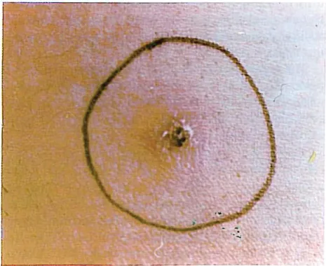
Fig. 6. Delayed�type hypersensitivity reaction, 72 hours after injection of trichophytin in a patient' with acute t.lermalophytosis.
167
and the microtiter plates incubated for an additional
24
hr. and the cultures were harvested.Harvesting was done by depositing the lymphocytes on glass fiber filters. Cells were rinsed with saline, fixed with
lO%
TCA 1md rinsed with water. Portions of filter strips corresponding to indi vidual wells were placed in vials containing10
mL scintillation fluid.Vials were counted in a liquid scintillation spectrophotometer for
2
min.Statistical Analysis
The Chi-Square test was used for tile statistical ;malysis of the results.
RESULTS
Patients with acute dermatophytosis (group
I),
did not have 1my background diseases, but patients with chronic dermatophytosis (group2),
had histories of other disorders: atopy,64
patients; diabetes, seven patients; contact dermatitis, four patients; tuberculosis, three patients.Thirteen patients of group I had tinea capitis,
28
had tinea corporis, and16
had tinea barbae. Most of these patients had severe inflammatory reaction and kerion (Fig.1,2).
34
patients of group2
had tinea pedis and tinea unguium,31
had tinea pedis,29
had tinea unguium,24
had tinea mannum. and13
had tinea mannum ;md tinea pedis (Fig.3,4).
Results of Cell-Mediated Immune Response: In Vivo Responses
Results of skin test on the acute infected patients (group I) and chronically infected volunteers (group
2)
were as follows:Group I: DTH reactions,
96
patients(98%);
no reaction, two patients(2%)
(Fig.5,6).
Fig.7
illustrates the distribution of DTH reactions by size in this group.Group
2:
DTH reactions,43
patients(32.8%);
immediate reaction,40
patients(30.5%);
no reaction,48
patients(36.6%)
(Figs.5,8
and TableI).
Cell-
M
ediated Immune Response: In Vitro Responses GrourJ'I ,�
tive lymphocyte blastogenic to trichophytin and PHA were observed in95 (96.8%)
and98 (lOO%)
patients, respectiv'
ely,Cfig.9).
Group
2:
Positive IYnWh
ocyte blastogenic stimulation to trichophytin and PHA were observed in46 (37.4%)
and127 (94.9%)
patients, respectively (Fig.9).
DISCUSSION
Acquired immunity following dermatophyte infection
Cell-Mediated Immunity in Dermatophytosis
30
�---'
2.
20
15
10
•
O ��==
No reaclion 2-5 6-10 15-20
I
_Negative �DTHI
Fig. 7. Relative frequency (If DTH responses by size (diameter of 72-hour reaclion, mm) in 98 patients with acule dermatophytosis.
Fig. 8. Immediate hypersensitivity reaclion. 20 minutes after injection of trichophytin, in a patient with chronic dermatophytosis.
has been observed in both naturally and experimenk'llly infected man and animrus.4•J8 There is evidence from studies using experimental infections in animals and man that resistance to dennatophyte infection correlates with cell-mediated immunity to fungal antigens.19.11 In man, a number of ill vitro assays of cell-mediated immunity have been carried out.' Lymphocyte transfonnation has been used to demonstrate cell-mediated immunity to dennatophylc antigens in both animals and man.1,7.20 Positi ve lymphocyte transfonnation responses of peripheral blood lymphocytes to trichophytin in patients with dermatophytosis have been found
in
several studies."·" Several studies have demonstrated that animals experimentally infected with dennatophytes generally develop delayed hypersensiti vity to :mtigens of the infecting agent.' Delayed hypersensitivity response appears to be imponant in the developmenl and clearance of the lesionspereent
120 96.9
Trlchophytln PH.
I
� Acule D ChronIcI
Fig. 9. Comparison of the positive results of lymphocyte transformation test
(L 11)
between patients with acute and chronic dennalophylosis.Table I. Analysis of patients with chronic dermatophytosis by type of skin test responses and relation 10 atopy
Type of skin Numhers of patients Numbers of patients
lest responses wilh alopic history
Delayed type hyper- 43(32.8) 7(tO.9%) sensitivity
lrnmediate hypcrsen - 40(30.5%) 36(56.3%) sitivily
Nonrcnctivc 48(36.6%) 21(32.8%)
Total 131(tOO%) 64(tOO%)
of dermatophytosis.'
In our study, we have found that a significantly higher proportion of positive delayed-hypersensitivity responses to trichophytin were seen in acute dennatophytosis than
in
chronic dennatophytosis (P<O.OI). This result provides additiom� support for previous reports which suggested that this occurred.II.D.BA
number of systemic disorders, including connective tissue diseases, systemic corticosteroid thempy, chronicmucocut(meous candidiasis, diabetes mellitus, Cushing's
syndrome and hematologic malignancies have been
associated with chronic dermatophytosis.27.n
Hanfin et
a1."
found that approximately50%
of their chronic dennatophytosis patients were also atopic or had a frunily history of atopy.A
similar history of atopy inpatients with chronic dennatophytosis has been found in other studies.lJ.)S and this combination has been tile "atopic chronic dennatophytosis" syndrome."
Our findings were similar; 64 of the
131
chronicdermatophytosis patients were atopic. whereas no acute dermatophytosis patient had history of atopy or any other disorder.
It seems that most patients wi�t chronicdermatophylOsis do nOt have genemlized deficiency of the cell-mediated immunity, that is, their immunological defects are limited to responses against uichophytin.24•34
ntis report indicates that �tere are differences in the resuhs oflymphocyte transformation test wi�t trichophytin between lymphocytes of patients with chronic and acute dennatophytosis, which are statistically significant (p<
0.05),
whereas with PHA, these differences are not observed.REFERENCES
1. Cox RA: Immunology of the Fungal Diseases. eRe Press Inc.
I-20, 1989.
2.Grappcl SF, BishopCR, BlankF: lmmunology ofdcrmatophytcs and dermatophytosis. Bacteriol Rev 38(2): 222, 1974. 3. Calderon RA. Hay RJ. Shennan GI: Circulating antigens and
antibodies in human and mouse dennalophylosis: use of monoclonal antibody reactive to phosphorylcholinc-likc epitopes. J Gen Microbiol 133 (Pt 9) (12): 2699, 1987.
4. Tndjebakhchc H. Khosravi AR:Theincidencc oftheprccipitating antibodies in human and animals with dermatophytosis in Iran. In press.
5. Kerbs S) Greenberg J, Jesruni K: Temporal correlation of lymphocyte blastogenesis, skin test responses, and erythema during dernlatophyte infections. Clin Exp Immunol 27: 531. 1977.
6. CruickshankCND, WoodSR: Studies on lrichophytinsensitivity.
] Invest Dermatol 35: 219,1960.
7. Green F, Balish E: Trichophyton melllagroplzytes dermatophytosis in gennfree guinea pigs. J Invest Dermatol 75: 476,1980.
8. Lepper AWD: Experimental bovine Trichophyton verrllCOSlIm
infection, the cellular responses in primary lesions of the skin resulting from surface or intradermal inoculation. Res Vet Sci 76: 287, 1974.
9. Green F, Lee KW, Balish E: Chronic T. menlagropltyles
dermatophytosis of guinea pig skin grafts on nude mice. I
Invest Dermatol 79: 125, 1982.
10. Iones HE, Reinhardt JH, Rinliidi MG: Model dermlilophytosis in naturally infected subjects. Arch Dennutol l lO: 36�', 1974.
11. Iones HE, Reinhardt IH, Rinaldi MG: A clinical. mycological. and immunological survey for dermatophytosis. Arch Dermatol 108: 61, 1973.
12. Kaaman T: The clinical significance of cutaneous reactions to trichophytin in dermatophytosis. Acta Dermatovener 58: 139, 1978.
13. Kaaman T: Cell-mediated reactivity in dennatophytosis: differences in skin responses to purified trichophytin in tinea pedis and tinea cruris. Acta Dermatovener 61: 119, 1981. 14. Bloch B: Zur Lehre von den dermatomykosen. Arch Dermatoi
Syph 93: 157, 1908.
15. Greenbaum S5: Immunity in ringworm infections.
1.
Active169
llcquired immunity with a note on complement fixation tests in superficial ringworm infection. Arch Dennatol lO: 279. 1924.
16. Bonk AR, Friedman L, Derbes VJ: Experimental dermatophytosis. J Invest Dermatol 39: 281, 1962.
17. Jones HE, Reinhardt IH, Rinaldi MG: Acquired immunity to dermatophytcs. Arch Dermatol 109: 840, 1974.
18. Kligman AM: The pathogenesis of tinea capitis due to Microsporum adouilli and Microsporum Callis.
1.
Cross obscrvati<Jns following the inoculation of humans. J Invest Dennatol 18: 231. 1952.19. Hunjan BS. Cronholm LS: An animal model for cell-mediated immune responses to dennatophytes. J Allergy Clin Immunol 63: 361. 1979.
20. Kerbs S, Greenberg J, Jesrani K: Temporal correlation of lymphocyte blastogenesis, skin test responses, and erythema during dermatophyte infections. Clin Exp Immunol 27: 526, 1977.
21. Calderon RA, Hay RJ: Cell-mediated immunity in cxperimental murine dermatophytosis. Immunology 53: 465, 1984.
22. Svejgaard E, Thomsen M, Morling N, Christiansen AH: Lymphocyte transformation ill vitro in dermatophytosis. Acta Path Microbiol Scand Sec. C, 84: 511, 1979.
23. Hanifin 1M. Ray LF. Lobitz WC: Immunological reactivity in dermatophytosis. Br J Dermatol 90: 1, 1974.
24. Hay RJ, Shennan G: Chronic dermatophyte infections. II. Antibody and cell-mediated immune responses. Br J Dermatol 106: 191, 1982.
25. Kaaman T, Petrini B, Wasserman S: In vivo and in viero immune responses to trichophytin in dermatophytosis. Acta Dennatovener 59: 229, 1979.
26. Stahl D, Svejgaard E: Lymphocyte transformation in vitro in aCHtedermalophytosis: a folJow-upstudy. AvtaDennatovener 62: 289,1982.
27. Hay RI: Chronic dermatophyte infections. I. Clinical and mycological features. Br J Dermatol 106: I, 1982.
28. Mendel EH: Tinea circinnta and onychomycosis (Trichophyton pllrpllreum) resistance 10 griseofulvin during uncontrolled diabetes. Arch Drm.toI82: 1027,1960.
29. Nelson LM, McNiece KJ: Recurrent Cushing's syndrome with TriciJophylollrubrllm infection. Arch Dennatol80: 700,1959.
30. Canizares D, Shatin H, Kellert AJ: Cushing's syndrome and dermatomycosis. Arch Dennatol 80: 705, 1959.
31. Findling JW, Tyrrell IN, Aron DC, Fitzgerald PA, Young CW, Sohnle PG: Fungal infections in Cushing's syndrome (letter to the editor) Ann Intern Mod 95: 392, 1981.
32. Lewis GM, Hopper ME, Scott MJ: Generalized Tric/zophylolJ rllbrllm infection associated with systemic lymphoblastoma. Arch Dermatol67: 247, 1953.
33. Jones HE, Reinhardt JH, Rinaldi MG: A clinical, mycological, and immunological survey fordermatophytosis. Arch Dermlltol 108:61,1973.
34. Sorensen G, Jones HE: Immediate nod delayed hypersensitivity in chronicdennatophytosis. Arch Dematol l12: 40,1976. 35. Jones HE: The atopic-dermatophytosis syndrome. Acta
Dermatovener Supple. 92: 81. 1980.