Journal website: http://www.jfqhc.com
Distribution Pattern of Cadmium in Liver and Kidney of Broiler
Chicken: An Experimental Study
A. Heshmati
1*, J. Salaramoli
21. Department of Nutrition, Faculty of Medicine, Hamadan University of Medical Sciences and Health Services, Hamadan, Iran 2. Toxicology and Animal Poisoning Research Centre, Faculty of Veterinary Medicine, University of Tehran, Tehran, Iran
Abstract
Background: Cadmium (Cd) is not considered as essential element for living organisms, therefore its presence in tissues is considered as toxic element. From food hazards control point of view, information on the distribution of these toxic elements in the animal tissues is very important. The present study was designed to evaluate Cd distribution pattern in kidney and liver of broiler chicken.
Methods: Forty eight day-old broiler chickens were randomly and equally divided into dif-ferent groups including control, I, II and III. During 42 days Cd was added to their diet in amount of 0, 25, 50 and 100 ppm, respectively. From each group, four chickens were sacri-ficed at 14, 28, 42 days of age, and amount of Cd in their liver and kidney were measured by atomic absorbent spectrophotometer. Analysis of data was performed by two-way ANOVA using SPSS software version 16.0.
Results: Interaction effect of the time duration of Cd diet consumption and the accumula-tion of Cd contents from either liver or kidney in different groups was significant (p<0.05). Increasing dietary Cd levels and exposure days resulted in higher Cd accumulation in kidney and liver. In all groups and in all different ages, kidney Cd levels were higher than those of liver.
Conclusion: Cd content in chicken organs is attributable to its dietary level and the duration of exposure. The mean concentration of Cd accumulated in chicken kidney tissue was higher in comparison to it in liver tissue.
Introduction
Cadmium (Cd) as a toxic heavy metal has been distribut-ed widely and uniformly and with small amounts through-out the earth's crust. Cd is not considered as essential ele-ment for living organisms, therefore its presence in organ-ism tissues is considered as contamination (Kramarova et al., 2005; Rehman et al., 2012). Human activities such as production of dyes, plastics, dry batteries, porcelain and etc. have increased Cd contamination in the environment (Akan et al., 2010).
This element is known as one of the most important envi-ronmental and industrial toxic agents and affects many
*
Corresponding author E-mail: [email protected]
target tissues such as appetite and pain centers, brain, heart and blood vessel, kidney and lungs (Bendeddouche et al., 2014; Hyder et al., 2013). High levels of Cd create acute disorders and long time exposure with small amounts cre-ate chronic adverse effect such as renal disorders and dys-function, enzyme reduction of alanine amino transferase, lactate dehydrogenase, aspartate amino transferase in liver and skeletal changes (Maxwell and Iwegbue, 2008).
Intake of Cd resulted in its accumulation in different organs which its amount depends on the interval of exposure, the quantity ingestion, the production and reproduction phases of the animals, as well as their age and breed (Baykov et al., 1996). This element has a very long
Article type
Original article
Keywords
Chickens Food Safety Cadmium
Received: 20 Sep 2014 Revised: 28 Oct 2014 Accepted: 19 Nov 2014
16 Journal website: http://www.jfqhc.com
biological half-life and may reach up to 20 years, depend-ing on type of animal (De la Fuente et al., 2002).
Distribution of Cd driven from feed among animal target organs is a key for estimating health risk from this exposure and determining the safety of animal origin foods; however, the bioaccumulation of Cd from feed is modified by many dietary components (Akan et al., 2010).
Several researchers studied the distribution of Cd in liver and kidney of animals (Baykov et al., 1996; Doganoc and Gacnik, 1995; Sharma et al., 1979; Swaileh et al., 2009; White and Finley, 1978). The aim of this study was to investigate distribution pat-tern of dietary Cd in liver and kidney tissues of broil-er chicken and its relation with Cd intake dosage and duration.
Materials and methods
Chemicals and experimental animals
All chemicals used for determination of Cd were of analytical grade and purchased from Merck Compa-ny (Darmstadt, GermaCompa-ny). To achieve this study, 48 day-old broiler chickens having a body weight of 40±2 g were purchased from Zarbal Company (Amol, Iran).
Ethical approval
Protocol and experimental design of study was approved by Department of Animal Ethics Committee, Faculty of Veterinary Medicine, Tehran University, Tehran, Iran (Pro-tocol No. 75070096/6/18).
Experimental design
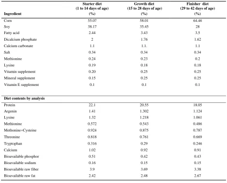
The design of study was based on Bharavi et al. (2011) with a little modification. Forty eight day-old broiler chickens (Ross) were randomly and equally divided into 4 different groups and each group consisted of 12 chickens. Breeding stages were done in metal cages at room tempera-ture and light levels were adjusted based on standard con-ditions at various stages. During the breeding, all the chickens had freely access to food and water. Chicken base diet is given in Table 1. During 42 days, Cd was added to base diet in amount of 0, 25, 50 and 100 ppm. In each stage of sample preparation, added Cd amount accuracy was determined by atomic absorbent spectrophotometer.
Control group chickens were fed from base feed includ-ing starter diet from day 1 to 14, growth diet from day 14 to 28 and finisher diet from day 28 to 42. The formula of these diets in addition to their effective chemical compo-nent has been given in Table 1. The chickens pertaining to group I, II and III were fed with base feed with additional
Cd levels of 25, 50 and 100 ppm, respectively. From each group at 14, 28 and 42 days of age, four chickens were randomly chosen to measure the amount of Cd accumulat-ed in liver and kidney tissue. Chickens were euthanizaccumulat-ed and their liver and kidney removed. The tissue samples were kept at -18 °C until analysis.
Determination of Cd
For measuring Cd amounts, liver and kidney sam-ples were grounded and homogenized. Five ml of percholeric acid and 20 ml of nitric acid were added to 2 g of homogenate sample and heated at 130 °C for 2 h. Choleric acid 10% (10 ml) was added to ash and adjusted by distilled water to 50 ml. The undis-solved particles were filtered off. Final digested sam-ples were analyzed for the presence of Cd using a flame atomic absorption spectrophotometer (Mel-bourne, Australia) under conditions recommended by the instrument manufacturer.
Statistical analysis
Analysis of data was performed using SPSS software version 16.0. To investigate the interaction effect of diet uptake time and diet Cd dose on Cd accumulated in liver and kidney, two-way ANOVA was used. P value less than 0.05 was considered to be statistically significant.
Results
The relations of Cd intake doses and exposure time inter-action with Cd average accumulated in the liver were sig-nificant (p<0.05). Table 2 shows Cd amounts accumulated in the liver at different ages and groups. In groups I, II and III, liver mean concentration of Cd was higher than that in control group. With the increasing levels of diet Cd, con-centrations of Cd in liver increased. In group I, the liver mean Cd amounts in 14, 28 and 42 days of ages showed significant difference compared with control group. There were significant differences between mean of group I (0.64 mg/kg) and group II (5.53 mg/kg) with group III (14.65 mg/kg). The mean accumulated Cd in liver during all days of ages in group II and III are significantly more than group I and control group.
Table 3 shows Cd accumulation in kidney in dif-ferent ages and groups. Diet Cd dosages and expo-sure time interaction effect on the mean of accumu-lated Cd in the kidney was significant (p<0.05). Feeding a diet containing approximately 100 ppm Cd for a period of 42 days was caused 46.65 mg/kg ac-cumulation of this metal in the kidney. There was a significant difference (p<0.05) in the Cd accumulated mean in the kidney of treatment and control groups.
17
Journal website: http://www.jfqhc.com Table 1:Composition of base diets in studied broiler chickens
Starter diet (1 to 14 days of age)
Growth diet (15 to 28 days of age)
Finisher diet (29 to 42 days of age)
Ingredient (%) (%) (%)
Corn 55.07 58.01 64.46
Soy 38.17 35.45 28
Fatty acid 2.44 3.43 3.5
Dicalcium phosphate 2 1.76 1.62
Calcium carbonate 1.1 1.1. 1.1
Salt 0.34 0.34 0.34
Methionine 0.24 0.23 0.2
Lysine 0.19 0.18 0.18
Vitamin supplement 0.20 0.25 0.25
Mineral supplement 0.15 0.25 0.25
Vitamin E supplement 0.1 0.1 0.1
Diet contents by analysis
Protein 22.1 20.55 18.05
Argenin 1.41 1.302 1.124
Lysine 1.32 1.218 1.061
Methionine 0.572 0.543 0.486
Methonine+Cysteine 0.924 0.875 0.787
Threonine 0.818 0.761 0.669
Tryptophan 0.316 0.29 0.246
Calcium 1.02 0.92 0.91
Bioavailable phosphor 0.51 0.42 0.43
Bioavailable sodium 0.16 0.15 0.15
Bioavailable raw fiber 3.9 3.69 3.38
Bioavailable raw fat 2.42 2.48 2.67
Table 2:The mean Cd concentration (mg/kg) accumulated in liver of broiler chicken given different levels of dietary Cd for 42 days
Age (day) Control Group I Group II Group III
14 NDCa* 0.64±0.07Cb** 5.53±1.07Bb 14.65±6.9Ab
28 NDDa 2.43±0.98Cb 13.22±2.99Ba 21.57±9.58Aa
42 0.02±0.003Da 5.36±1.77Ca 15.27±4.9Ba 26.01±3.92Aa
*ND: Not Detected
**Values were presented as mean value±SD (n=4). Different superscript capital letters within a row indicate statistically significant differences (p<0.05) among
values. Different superscript small letters within a column indicate statistically significant differences (p<0.05) among values
Table 3:The mean Cd concentration (mg/kg) accumulated in kidney of broiler chicken given different levels of dietary Cd for 42 days
Age (day) Control Group I Group II Group III
14 NDCa* 1.64±0.07Cb** 17.34±6.06Bb 33.26±3.29Ac
28 NDBa 2.97±1.39Cb 18.76±2.78Bb 38.43±17.82Ab
42 0.03±0.002Da 12.39±4.58Ca 26.33±7.46Ba 46.65±4.03Aa
*
ND: Not Detected
**Values were presented as mean value±SD (n=4). Different superscript capital letters within a row indicate statistically significant differences (p<0.05)
among values. Different superscript small letters within a column indicate statistically significant differences (p<0.05) among values
18 Journal website: http://www.jfqhc.com
Discussion
Cd was absorbed via the gastrointestinal tract and lungs, transported by blood and accumulated in various tissues and organs in the body, mainly kidney and liver because its rate of elimination from these organs is relatively low (Koréneková et al., 2002).
In poultry, feed composition can influence the retention of trace elements. It seems absorption and accumulation of Cd in tissues depends on a wide range of factors including nutritional substance and vitamin status, age and sex of the animal (Massanyi et al., 2003). The ability of liver and kidneys to accumulate high concentration of Cd is a com-mon feature to chickens and many other animals. The ac-cumulation of Cd in liver and kidney tissue is partly at-tributable to the increased levels of metallothionein in these tissues (Sharma et al., 1979).
Our results showed that dietary Cd greatly increased its content in the liver and kidney of broiler chicken; there-fore, Cd accumulation in these organs depended on dietary Cd and exposure time. The rate of accumulation increased along with exposure time and Cd content of diet. We found Cd accumulation in kidney was more than it in liver. These results are in consistence with previous report which showed highest accumulation of Cd occurred in kidney than in liver and the accumulation increase in these organs is a consequence to Cd doses increase in chicken diet (Barregard et al., 2010; Baykov et al., 1996; Sharma et al., 1979; Villar et al., 2005; White and Finley, 1978). Also, the present finding was comparable with those of Kumar et al. (2007) that reported the higher ability of kidneys to accumulate Cd compared liver and muscle in freshwater fish and chicken (Kumar et al., 2007). It is also in con-sistent with the results of Doganoc and Gačnik (1995) that found Cd accumulation is more in kidney than liver when chicken age was increased. They looked for correlation between the animals' age and Cd content in kidney from
Ilirska Bistrica. The positive correlation between the con-centrations of Cd in kidney of game from Ilirska Bistrica
and the age was evident. Age-dependent increase in Cd content is due to long biological half-time of it in the liver and kidney (Doganoc and Gačnik, 1995).
Conclusions
In summary, adding Cd to a chicken diet effects the ac-cumulation of this element in kidney and liver. Cd content in these organs is attributable to its dietary level and the duration of exposure. In this study, it was concluded the mean concentration of Cd accumulated in chicken kidney tissue was higher in comparison to it in liver tissue.
Conflicts of interest
The authors declare that they have no conflict of interest in this study.
Acknowledgements
The authors like to acknowledge authorities of Tehran University, Tehran, Iran for providing fund for this study.
References
Akan J.C., Abdulrahman F.I., Sodipo O.A., Chiroma Y.A. (2010). Distribution of heavy metals in the liver, kidney and meat of beef, mutton, caprine and chicken from Kasuwan Shanu market in Maiduguri Metropolis, Borno state, Nigeria. Research Journal of Applied Sciences, En-gineering and Technology. 2: 743-748.
Barregard L., Fabricius-Lagging E., Lundh T., Molne J., Wallin M., Olausson M., Modigh C., Sallsten G. (2010). Cadmi-um, mercury, and lead in kidney cortex of living kidney donors: impact of different exposure sources. Environ-mental Research. 110: 47-54.
Baykov B.D., Stoyanov M.P., Gugova M.L. (1996). Cadmium and lead bioaccumulation in male chickens for high food concentrations. Environmental Toxicology and Chemistry. 54: 155-159.
Bendeddouche B., Rachid Z., Esma B. (2014). Levels of select-ed heavy metals in fresh meat from cattle, sheep, chicken and camel produced in Algeria. Annual Review and Re-search in Biology. 4: 1260-1267.
Bharavi K., Reddy A.G., Rao G.S., Kumar P.R., Kumar D.S., Prasadini P.P. (2011). Prevention of cadmium bioaccumu-lation by herbal adaptogens. Indian Journal of Pharma-cology. 43: 45-49.
De la Fuente H., Portales-Perez D., Baranda L., Diaz-Barriga F., Saavedra-Alanis E., Gonzalez-Amaro R. (2002). Effect of arsenic, cadmium and lead on the induction of apopto-sis of normal human mononuclear cells. Clinical and Ex-perimental Immunology. 129: 69-77.
Doganoc D.Z., Gačnik K.Ŝ. (1995). Lead and cadmium in meat and organs of game in Slovenia. Bulletin of Environmental Contamination and Toxicology. 54: 166-170.
Hyder O., Chung M., Cosgrove D., Herman J.M., Li Z., Firooz mand A., Gurakar A., Koteish A., Pawliket T.M. (2013). Cadmium exposure and liver disease among US adults.
Journal of Gastrointestinal Surgery. 17: 1265-1273.
Koréneková B., Skalická M., Naï P. (2002). Concentration of some heavy metals in cattle reared in the vicinity of a metallurgic industry. Veterinarski Arhiv. 72: 259-267.
Kramarova M., Massanyi P., Slamecka J., Tataruch F., Jancova A., Gasparik J., Fabis M., Kovacik J., Toman R., Galova J., Jurcik R. (2005). Distribution of cadmium and lead in liver and kidney of some wild animals in Slovakia. Jour-nal of Environmental Science and Health. Part A, Tox-ic/HazardousSubstances and Environmental Engineering. 40: 593-600.
Kumar P., Prasad Y., Patra A.K., Swarup D. (2007). Levels of cadmium and lead in tissues of freshwater fish (Clarias batrachus L.) and chicken in Western UP (India). Bulletin of Environmental Contamination and Toxicology. 79: 396-400.
Massanyi P., Tataruch F., Slameka J., Toman R., Jurik R. (2003). Accumulation of lead, cadmium, and mercury in liver and kidney of the brown hare (Lepus europaeus) in relation to the season, age, and sex in the west Slovakian lowland. Journal of Environmental Science and Health. Part A, Toxic/HazardousSubstances and Environmental Engineering. 38: 1299-1309.
Maxwell C., Iwegbue A. (2008). Heavy metal composition of livers and kidneys of cattle from southern Nigeria.
Veterinarski Arhiv. 78: 401-410.
Rehman K.U., Andalib S., Ansar M., Bukhar S., Naeem N.M., Yousaf K. (2012). Assessment of heavy metal in different tissues of and domestic layers. Global Veterinaria. 9: 32-37.
Sharma R.P., Street J.C., Verma M.P., Shupe J.L. (1979). Cad-mium uptake from feed and its distribution to food
19
Journal website: http://www.jfqhc.com ucts of livestock. Environmental Health Perspectives. 28:
59-66.
Swaileh K.M., Abdulkhaliq A., Hussein R.M., Matani M. (2009). Distribution of toxic metals in organs of local cat-tle, sheep, goat and poultry from the west Bank, Palestinian authority. Bulletin of Environmental Contami-nation and Toxicology. 83: 265–268.
Villar T.D.C., Elaine J.P.K., Flavier M.E. (2005). Lead and cadmium levels in edible internal organs and blood of poultry chicken. Journal of Applied Sciences. 5: 1250– 1253.
White D.H., Finley M.T. (1978). Uptake and retention of die-tary cadmium in mallard ducks. Environmental Research. 17: 53-59.