ABSTRACT
When in a patient more than one tumour in the same or a different organ is diagnosed, multiple primary tumours may be present. For epidemiological studies, different definitions of multiple primaries are used with the two main definitions coming from the project Surveillance Epidemiology and End Results and the International Association of Cancer Registries and International Agency for Research on Cancer. The differences in the two definitions have to be taken into consideration when reports on multiple primaries are analysed. In this review, the literature on multiple primaries is reviewed and summarised. Overall, the frequency of multiple primaries is reported in the range of 2–17%.
Aetiological factors that may predispose patients to multiple primaries can be grouped into host related, lifestyle factors and environmental influences. Some of the most common cancer predisposition syndromes based on a clinical presentation are discussed and the relevant genetic evaluation and testing are characterised. Importantly, from a clinical standpoint, clinical situations when multiple primaries should be suspected and ruled out in a patient are discussed.
Furthermore, general principles and possible
treatment strategies for patients with synchronous and metachronous multiple primary tumours are highlighted.
INTRODUCTION
Significant progress has been made in the prevention, diagnosis and treatment of malignant tumours.1 As a result of screening programmes and improvement in diagnostic procedures, cancer can be detected at an earlier stage. Significant treatment advances have led to increased overall survival in patients with advanced cancer. As of early 2014, nearly 14.5 million people were alive in the USA with a history of cancer. Some of these people are on active anticancer therapy, others have been diagnosed and treated many years ago with no current evidence of tumour. For Switzerland, an estimate reported 2.08% of cancer survivors in 1990 with an expected increase to 3.7% in 2010.2
The fact that patients may have multiple primary tumours is not new and already in 1921 a report found in 3000 cases of malig-nancy ‘4.7% of cases of multiple growth’.3 In epidemiological studies, the frequency of
multiple primaries is reported to be in the range of 2–17%.4–8 Many factors can influence the reported numbers of multiple primaries, namely the definition that was applied (see below), the follow-up time since the longer patients are followed up after a primary cancer diagnosis, the higher the likelihood that they may develop a second malignancy and importantly also the patient population studied.9
Today, the situation of patients with multiple primaries is of increasing relevance and importance. Apart from many reports in the literature on the frequency of multiple primaries, the practical implications of the management of patients with multiple prima-ries are rarely discussed. When in a patient two active malignancies are diagnosed at the same time, the challenge is to find an anticancer therapy strategy that covers both cancer types without increased toxicity or relevant pharmacological interactions and without negative impact on the overall outcome. In a patient with a previous cancer history and potentially prior anticancer therapy, it can be difficult to establish the diagnosis of an addi-tional primary because, for example, newly developed metastases could have developed from the first cancer diagnosis but could also be part of a second malignancy. In daily clin-ical practice, it is important to recognise these situations and to perform the adequate inves-tigations because of relevant implications on subsequent therapeutic management strategies. Multiple primaries also impact enrolment in clinical research protocols because patients with a prior cancer history or with current active secondary malignancy are generally excluded on most clinical trials.
This review will discuss and summarise the topic of multiple primary tumours from an epidemiological viewpoint and will review some of the large cohort studies on multiple primaries. Aetiological factors and genetic syndromes predisposing for multiple prima-ries will be discussed. Furthermore, clinical situations when a treating physician should
Multiple primary tumours: challenges
and approaches, a review
Alexia Vogt,1,2 Sabine Schmid,1 Karl Heinimann,3,4 Harald Frick,5
Christian Herrmann,5 Thomas Cerny,1 Aurelius Omlin1,2
To cite: Vogt A, Schmid S, Heinimann K, et al. Multiple primary tumours: challenges and approaches, a review. ESMO Open
2017;2:e000172. doi:10.1136/ esmoopen-2017-000172
Received 30 January 2017 Revised 6 March 2017 Accepted 17 March 2017
1Department of Oncology and
Haematology, Kantonsspital St. Gallen, St.Gallen, Switzerland
2Oncology, Inselspital and
University of Berne, Berne, Switzerland
3Medizinische Genetik,
Universittsspital Basel, Basel, Switzerland
4Research Group Human
Genomics, Zentrum für Lehre und Forschung, Labor 317/319, Department Biomedizin, University of Basel, Basel, Switzerland
5Cancer Registry St. Gallen
and Appenzell, St. Gallen, Switzerland
Correspondence to
Aurelius Omlin, Department of Oncology and Haematology, Kantonsspital St.Gallen, Rorschacherstrasse 959007 St .Gallen, Switzerland; aurelius. omlin@ kssg. ch
on September 12, 2020 by guest. Protected by copyright.
take the possibility of multiple primaries into consider-ation will be highlighted.
DEFINITION OF MULTIPLE PRIMARIES
Multiple primaries are defined as more than one synchro-nous or metachrosynchro-nous cancer in the same individual. For epidemiological studies, tumours are considered multiple primary malignancies if arising in different sites and/or are of a different histology or morphology group. This avoids misclassification of multifocal/multicentric tumours or metastases as multiple primaries. A cancer is classified as index cancer if there has been no prior record of invasive cancer.10
The definitions and understanding of a multiple primary have changed over the time and may differ from one study to another. The two most common definitions currently used are provided by the Surveillance Epidemiology and End Results (SEER) project and the International Associ-ation of Cancer Registries and InternAssoci-ational Agency for Research on Cancer (IACR/IARC).4 One of the main differences is that according to IACR/IARC, several groups of topography codes of International Classifica-tion of Diseases for Oncology 3rdEdition (ICD-O-3) are
considered one site in the definition of multiple primaries (IARC 2004). For example, the colon is regarded as one site while the SEER considers single tumours of different parts of the colon as single tumours. Further differences are that IACR/IARC apply much broader histological groups and rules not being time dependent. The use of the different guidelines can result in numbers which are different by several percentage points (eg, for breast or colon cancer). European cancer registries generally prefer the use of the IACR/IARC definitions.
The SEER database recommends to use a 2-month period to distinguish between synchronous and meta-chronous multiple primaries.11 Rules according to the IARC suggest the registration of synchronous tumours diagnosed in an interval of less than 6 months (or
metachronous if more than 6 months) if arising in different sites.12
Epidemiology of multiple primaries
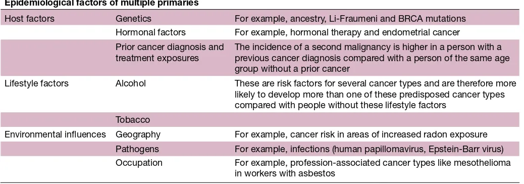
The burden of second primary malignancies in a growing and ageing population of first cancer survivors has increased over the last decades.2 Factors accounting for the increasing frequency of multiple primaries are improved diagnostic tests, increasing and more sophisticated treat-ment, as well as improved screening and surveillance of patients with cancer, for example, screening for colorectal and breast cancers.4 9 13–17 Cancer survivors may be suscep-tible to developing second primary malignancies due to a variety of unique factors, including cancer predisposition syndromes or special tumour characteristics, environ-mental exposures and late effects of therapies (table 1). Caucasian ancestry, index cancer diagnosed at younger age, lower stage and with generally indolent clinical behaviour with longer survival, as well as positive family history are reported to harbour excess risk for multiple primaries.11
The incidence of multiple primaries in a cancer popu-lation varies between 2.4% and 8%, up to 17% within 20 years of follow-up (table 2).
The risk of developing a second primary malignancy is varying in different cancer sites and is reported in a range from 1% (primary liver malignancy) up to 16% (primary bladder cancer).18 Weir et al found an incidence of multiple primaries of 19.7% following the SEER guide-lines (or 16.9% IACR rules) in colon and 21% (SEER; 19.9% IACR) in patients with lung cancer.6 Amer et al found similar incidences of multiple primaries in patients with colon cancer; however, they only reported 5.6% multiple primaries in patients with lung cancer.11
Long-term survival with multiple primaries is variable and is influenced by cancer type and stage at diagnosis. Genetic factors, behavioural influences, lifestyle and comorbidities generally influence patient’s outcomes. In
Table 1 Epidemiological factors of multiple primary tumours
Epidemiological factors of multiple primaries
Host factors Genetics For example, ancestry, Li-Fraumeni and BRCA mutations
Hormonal factors For example, hormonal therapy and endometrial cancer
Prior cancer diagnosis and treatment exposures
The incidence of a second malignancy is higher in a person with a previous cancer diagnosis compared with a person of the same age group without a prior cancer
Lifestyle factors Alcohol These are risk factors for several cancer types and are therefore more
likely to develop more than one of these predisposed cancer types compared with people without these lifestyle factors
Tobacco
Environmental influences Geography For example, cancer risk in areas of increased radon exposure
Pathogens For example, infections (human papillomavirus, Epstein-Barr virus)
Occupation For example, profession-associated cancer types like mesothelioma
in workers with asbestos
on September 12, 2020 by guest. Protected by copyright.
general, black patients have a lower incidence of multiple primaries and also a lower relative survival independent of cancer site or stage at diagnosis (2.37% in black vs 3.41% white women age-adjusted prevalence for cancer survivors in the US population).19 For an overview of epidemiological studies, see table 2.
The global cancer burden according to IARC in 2012 was 14.1 million new cases and 8.2 million cancer deaths and is estimated to increase by 2030 to 21.7 million new cases and 13 million deaths.20 With this increase alone, the number of patients with multiple primaries will grow significantly.
In the following short sections, the occurrence of multiple primaries in several common cancer types will be summarised, not in a systematic approach but to provide some ideas of some of the available data on multiple primaries. For details and further reading, several good reviews are referenced.
Multiple primaries in female patients with breast cancer
Coyte et al assessed the impact of these different defi-nitions for 8386 women diagnosed with breast cancer. Within 5 years after the diagnosis of primary breast cancer, a total of 98 secondary breast cancers were diagnosed.
When applying the SEER guidelines, 79 of the reported secondary breast cancers qualified as multiple primaries, but according to the IARC/IACR guidelines, it was only one case.4
In patients with breast cancer, the incidence of multiple primaries has been reported in the range of 4.1% (Kim et al 108 patients, 45.9 months follow-up, guideline not avail-able) to 16.4% (Weir et al 301 963 patients, max. 10 years follow-up, SEER, in IACR only 10.4%).6 11 21 The median time to a second malignancy was between 5 and 8 years.21 22
Hormonal treatment of a primary breast cancer increases the risk for endometrial, gastric, colon and ovarian cancers with an excess risk for endometrial cancer observed especially after tamoxifen therapy.22
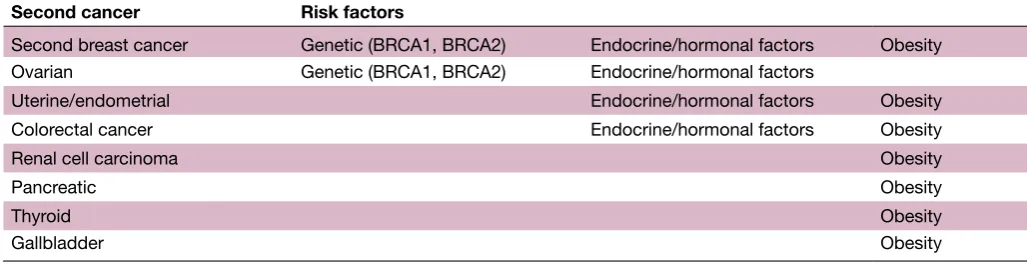
Reproductive/hormonal and genetic factors (eg, BRCA1, BRCA2) as well as obesity are recognised as common risk factors for multiple primaries (table 3).23
For the breast cancer genes BRCA1 and BRCA2, there is a strong association with an increased risk for a second breast or ovarian cancer. Furthermore, there is a strong association between hereditary gastric cancer and lobular breast cancer.9 The risk of a second breast, endometrial or ovarian cancer after breast cancer is inversely associated
Table 2 Incidence of multiple primaries over all cancer sites in the literature
Number of patients
evaluated Geographic region Frequency of multiple primaries Mean follow-up(years)
Definition used for multiple primary
(SEER/IACR/IARC) Reference
19 252 Italy 2.4% 2.5 IACR 5
1 015 564 men USA 15.8% NA SEER 6
951 022 women 14.4% IACR
17.2% SEER
14.55 IACR
334 168 Victoria, Australia 4.3% 5 IACR 8
7.7% 10
12.4% 20
2 919 023 22 European
countries 6.3% NA IACR
7
57 393 West of Scotland 5% 5 IACR 4
938 Netherlands 7% NA NA 17
1873 USA 7.2% 5 SEER 11
11.4% 10
13.3% 15
14.8% 20
17.2% >20
1953 USA 2.15% 20 NA 14
1 635 060 Italy 6.3% 14 IACR 16
82 671 Switzerland 8.17% 6.6 IACR Authors of this
publication
IACR, International Association of Cancer Registries; IARC, International Agency for Research on Cancer; SEER, Surveillance Epidemiology and End Results.
on September 12, 2020 by guest. Protected by copyright.
with postmenopausal status and history of full-term preg-nancy.22
Late toxic effects of radiotherapy and chemotherapy also contribute to the increased risk for a secondary primary tumour after breast cancer. Acute myeloid leukaemia (AML) can be induced during the first 2 years after radio-therapy and is a late effect of adjuvant chemoradio-therapy.22 In addition, thyroid cancer, second malignancies of breast, bone, connective tissue and lung may arise after radio-therapy.
Multiple primaries in patients with prostate cancer
Rates of multiple primaries in patients with prostate cancer range from 1.14% following radiation therapy (Jin et al Meta-analysis from six references, >10 years follow-up) to 8.7% (Weir et al 335 565 patients, max. 10 years follow-up, SEER and IACR).6 24 A systematic review with 21 studies evaluating the risk of secondary malignan-cies in men with prostate cancer who received radiation therapy found an increased risk of bladder cancer (HR 1.67) and colorectal cancer (HR 1.79) with notably an increased risk in patients who were treated with external beam radiation therapy and no increased risk in men who were treated with brachytherapy.25 Radiation therapy has been shown to increase the risk for urothelial and rectal cancers and sarcomas within the radiation area after a disease-free interval of at least 5 years. Increasingly, genetic factors accounting for secondary primaries in prostate cancer are being recognised, most prominently BRCA2 mutations (see also table 5).26
Multiple primaries in patients with Hodgkin’s lymphoma
Multiple primaries rates in patients with Hodgkin’s lymphoma are reported in the range of 7.5% (Weir et al 13 198 patients, max. 10 years follow-up, SEER and IACR) to 7.8% (Bhuller et al 442 patients, up to 39 years follow-up)6 27 and are mainly attributed to late toxic treat-ment effects. Survivors of Hodgkin’s lymphoma with a history of radiation therapy have an increased risk for breast, lung, thyroid and colorectal cancers.27 28 Chemo-therapy is associated with an increased risk for secondary leukaemias and lung, gastrointestinal or bladder cancer, as well as soft tissue and bone sarcomas.9 Secondary malignancies are one of the most relevant causes for
mortality in Hodgkin’s lymphoma survivors.27 Secondary leukaemia and myelodysplastic syndromes are associated with combination chemotherapy, especially etoposide in a dose-dependent manner. Etoposide-related leukaemia is distinguished from other forms of leukaemia that can be induced by alkylating agents by characteristics such as a shorter time from the end of treatment to develop-ment of leukaemia and evidence of translocation 11q23 or 21q22.29
Multiple primaries in patients with lung cancer
In patients with lung cancer, the incidence of multiple primaries ranges from 13.4% (Rosso et al 2 919 023 patients, follow-up not available, IACR) to 22% (Sánchez
et al 1769 patients, 5–23 years follow-up, only patients with survival >3 years (12.3%)).7 Patients diagnosed with early stage lung cancer have an increased risk for second primaries compared with the general population without a prior cancer diagnosis.30 Many second malignancies are related to smoking. The rate of second primaries is slightly lower in first primary adenocarcinoma with a rate of 3.36 per 100 person-years than in squamous cell carcinoma with a rate of 3.77 per 100 person-years. It is highest in small cell lung cancer (SCLC) with 4.46 cases per 100 person-years. There is no significant association with radiotherapy.30
The most common second cancers are in the lung, especially if the first primary was a SCLC. The most common second lung malignancies are adenocarcinomas (29.9%) followed by squamous cell carcinomas (27.1%). SCLCs represent only 7.9% of the second pulmonary malignancies after a first lung cancer.31 Also, colorectal and bladder cancers are often found as second malignan-cies after a primary lung cancer.
Summary on epidemiology of multiple primaries
► Multiple primaries are defined as more than one synchronous or metachronous cancer in the same individual.
► Synchronous and metachronous multiple primaries are distinguished. Definitions depend on whether SEER or the IACR/IARC rules are applied.
► Common risk factors for multiple primaries are as follows: inherited predisposition to cancer;
cancer-Table 3 Common risk factors for multiple primaries in women with breast cancer (adapted from Wood et al9)
Second cancer Risk factors
Second breast cancer Genetic (BRCA1, BRCA2) Endocrine/hormonal factors Obesity
Ovarian Genetic (BRCA1, BRCA2) Endocrine/hormonal factors
Uterine/endometrial Endocrine/hormonal factors Obesity
Colorectal cancer Endocrine/hormonal factors Obesity
Renal cell carcinoma Obesity
Pancreatic Obesity
Thyroid Obesity
Gallbladder Obesity
on September 12, 2020 by guest. Protected by copyright.
promoting aspects of lifestyle, hormonal and environmental factors; treatment of the previous primary cancer and increased surveillance of cancer survivors.32
► The incidence of multiple primaries varies in different primary cancer types and treatment that was applied for these cancers.
Cancer predisposition syndromes and genetic
test-ing
Several clinical scenarios should call a clinician’s atten-tion towards the possibility of an underlying cancer predisposition, in particular the occurrence of multiple primary cancers, diagnosis at young age and in several family members. For example, metachronous breast and ovarian cancers, especially in younger patients, should prompt evaluation of an underlying hereditary breast and ovarian cancer (HBOC) syndrome associated with germ-line mutations in the BRCA1/2 genes, likewise prostate cancer in a young male patient with a family history of breast cancer and melanoma. Similarly, colon cancer in a young woman from a colon and endometrial cancer-prone family points towards Lynch syndrome (LS) (see also table 4 for further clinical scenarios).
Also, in the era of novel targeted agents and check-point inhibitors, germline and somatic mutations are becoming more important with respect to new treatment options and strategies, such as the use of poly-ADP-ribose polymerase (PARP) inhibitors in patients whose tumours display DNA repair defects (BRCA1/2, ATM) or check-point inhibitors in tumours with high mutational load as exemplified by microsatellite instability (MSI). This has opened another field for direct-to-consumer genetic testing, which is increasingly being offered by commer-cial companies through internet portals and has risen widespread concerns about accuracy, usefulness and interpretation of such tests in the absence of medical supervision and genetic counselling.29 33 In the following, we will give a short, non-comprehensive overview of the most relevant cancer predispositions in routine oncology care, namely HBOC, LS (formerly known as hereditary non-polyposis colon cancer, HNPCC), multiple endocrine neoplasia type 1 and type 2 (MEN1, MEN2), von Hippel-Lindau (VHL) disease and Li-Fraumeni syndrome (LFS). For a more detailed overview, we recommend the review by Garber and Offit34 as well as the ESMO guidelines on hereditary cancer syndromes.35 36
Hereditary breast and ovarian cancer syndrome
HBOC is a high-penetrance, autosomal-dominant breast and ovarian cancer predisposition caused by germline mutations in the genes BRCA1 and BRCA2. Its prevalence in an unselected population is estimated to be <10% but is markedly increased (up to 35%) in patients with a positive family history of breast and ovarian cancers, young age at first diagnosis, multiple metachronous breast/ovarian tumours or special ethnic groups (Ashkenazi Jews). The average lifetime risk for breast or ovarian cancer in
BRCA1 and BRCA2 carriers by age 80 is up to ca. 83% and 76%, respectively;37 for men with BRCA2 mutation, life-time risk for breast and prostate cancers is also elevated (up to ca. 8.9%). Criteria for genetic counselling and testing referral vary between countries, but commonly include clinical features (see table 4) as well as predictive models such as BRCAPRO or Manchester Score. However, with higher awareness and apart from surveillance also emerging therapeutic consequences as PARP inhibitors, more widespread testing can be expected.
Lynch syndrome/hereditary non-polyposis colon cancer
In this autosomal-dominantly inherited cancer predis-position, germline loss-of-function mutations in DNA mismatch repair (MMR) genes combined with a second, somatically acquired hit in the tumour lead to MSI and a mutated phenotype resulting in an increased risk for colon and endometrial cancers as well as more rarely to cancers, for example, of the urinary tract, ovary, small intestine, gastric and hepatobiliary tract.38 39 In the overall colon cancer population, the prevalence for LS is approx. 1–3% and lifetime risk in MMR gene mutation carriers for colon and endometrium cancers amounts to approx. 52–82% and 25–60%, respectively.40 41 Referral for genetic counselling and testing is generally based on MSI immu-nohistochemistry results on patient’s tumour tissue in conjunction with family history. As with BRCA1/2-asso-ciated cancers, MSI (sporadic and germline) recently gained more attention due to possible therapeutic conse-quences, as was shown, that checkpoint inhibitors seem to be effective almost exquisitely in colon and gastric cancers with MSI high tumours.42
von Hippel-Lindau disease
VHL disease is an autosomal dominantly inherited tumour predisposition syndrome, which is associated with an increased risk of developing various benign and malig-nant tumours, including retinal capillary and central nervous system haemangioblastomas, phaeochromocy-tomas, renal cysts and clear cell renal cell carcinomas, endolymphatic sac tumours as well as pancreatic cysts, pancreatic neuroendocrine tumours and cystadenomas of the epididymis and the broad ligament.43 The disease penetrance is high and more than 90% of patients harbouring a VHL mutation develop clinical symp-toms before the age of 65 years. Disease expression and severity vary considerably within and between families.44 Preventive screening programmes have improved median overall survival of affected patients.
Li-Fraumeni syndrome
Caused by germline mutations in the ‘guardian of the genome’, the tumour protein p53 (TP53) gene, LFS is characterised by a distinct set of early onset tumours such as sarcoma, breast cancer, brain tumours, leukaemia and adrenocortical carcinoma. Due to the rarity of the syndrome until now, no standard surveillance protocols have been implemented, but recent studies could show
on September 12, 2020 by guest. Protected by copyright.
Table 4 Clinical scenarios and potential genetic cancer syndrome
Clinical picture, patient with a history or diagnosis of
(synchronous or metachronous) Familial cancer syndrome to think of Full picture of familial cancer syndrome
Synchronous bilateral breast cancer or secondary (metachronous) breast cancer
Hereditary breast and ovarian cancer (HBOC) [BRCA1, BRCA2]
•Breast cancer
•Ovarian cancer
Breast and ovarian cancers HBOC [BRCA 1/2] •Male breast cancer
Prostate cancer and pancreatic
cancer or melanoma HBOC [BRCA 2] •Prostate cancer
•Melanoma
(Multiple) renal cell carcinomas VHL •Renal cell carcinoma <47 years
Renal cell carcinoma and
pancreatic cystic lesions VHL •Multiple renal cysts
Haemangioblastomas of central
nervous system (CNS) and retina VHL •Multiple pancreas cysts and pancreatic neuroendocrine tumour
•CNS and retinal haemagioblastomas •Phaeochromocytomas
•Endolymphatic sac tumours
•Cystadenomas of epididymis or broad ligament
Breast cancer and sarcoma Li-Fraumeni syndrome •Soft tissue sarcoma and osteosarcoma
Breast cancer and leukaemia Li-Fraumeni syndrome •Premenopausal breast cancer
•Leukaemia •Brain tumours
•Adrenocortical carcinoma •Lung bronchoalveolar cancer
Colon and endometrial cancers Lynch syndrome •Colon cancer
Colon and ovarian cancers HNPCC •Endometrial cancer
•Ovarian cancer
•Renal pelvis cancers •Ureteral cancers
•Pancreatic and hepatobiliary cancers •Stomach and small bowel cancers Multiple colon polpys and/or colon
cancer Familial adenomatous polyposis •Colon cancer
•Duodenal cancer •Thyroid cancer •Hepatoblastoma Parathyroid adenomas and
pituitary adenomas MEN1 •Pituitary tumours
•Parathyroid tumours
•Endocrine tumours of the gastro–entero– pancreatic tract
•Carcinoid tumours Medullary thyroid cancer and
phaeochromocytoma MEN 2 •Medullary thyroid cancer
•Phaeochromocytoma
Continued
on September 12, 2020 by guest. Protected by copyright.
long-term survival benefit for patients complying with an intensive surveillance protocol for early tumour detec-tion.45
MEN1/MEN2 syndrome
Basis of the autosomal dominantly inherited MEN1 syndrome are germline mutations in the MEN1
gene, predisposing to parathyroid adenomas, pitu-itary adenomas and pancreatic islet cell tumours as gastrinomas, vipomas or insulinomas.46 Surveillance of mutation carriers should start at an early age, usually between 5 and 10 years. MEN2 is associated with medul-lary thyroid cancer and phaeochromocytoma and in nearly 90% of affected patients a germline RET-proto-onco-gene mutation can be found.47
CLINICAL SITUATIONS WHEN TO SUSPECT MULTIPLE PRIMARIES (SYNCHRONOUS OR METACHRONOUS)
As discussed above, multiple primaries can occur in a synchronous or metachronous pattern. In the situation of a new cancer diagnosis, staging examinations are performed to assess the extent of disease but sometimes also to localise a primary tumour in the situation where a metastatic site is the first presentation of malignancy. In patients with a prior cancer diagnosis, often over the period of several years, follow-up tests and examinations are performed in order to rule out relapse.
With the increasing use of more sophisticated imaging methods, namely positron-emission tomography computed tomography (PET-CT) and whole body MRI, it is not uncommon to find suspicious lesions that might have not been detected by standard CT and/or bone scintigraphy. In a large series of 1912 patients who were scanned with PET-CT scans, lesions suspicious of addi-tional malignant tumour were found in 4.1% of which 1.2% were histologically confirmed.48 Not surprisingly, these secondary primaries were in the following localisa-tions: thyroid, colon, breast, oesophagus, bile duct and head and neck regions, where even contrast enhanced CT may easily miss small tumours. In a series of 200 patients with oesophageal cancer who underwent PET-CT for staging, a total of 17% patients had synchronous multiple primary tumours, namely in the stomach, head and neck regions, colon and lung.49
Several clinical features should alert clinicians to the possibility that a patient may have a second primary tumour both synchronous and in the situation of a prior cancer history:
► Atypical metastatic spread of primary tumour (eg, radiologically lytic bone metastases in prostate cancer).
► High tumour burden relative to tumour marker load (eg, extensive liver metastases with low prostate-specific antigen in prostate cancer).
► New metastatic spread (eg, liver, lung) several years after a primary cancer diagnosis.
► Single new metastatic lesion after a primary cancer diagnosis (eg, single pulmonary nodule in a patient with a prior primary head and neck carcinoma).
► Chronological atypical metastatic spread (eg, relapse 5 years after primary small cell carcinoma of the lung).
► Recurrence in patients with exposure to
environmental carcinogens (eg, smoking).
► Suspicion of haematological malignancy after prior chemotherapy (eg, etoposide, anthracyclines).
► Suspicion of secondary malignancy in patients with prior radiation for malignancy and especially if recurrence in prior radiation field.
► Suspicious lesion on imaging (eg, PET-CT) detected at staging or in follow-up.
► Differential standard uptake value (SUV) of suspected lesions on PET-CT (eg, lesions with very high SUV and lesions with low SUV).
In case of a suspected secondary primary, a histological confirmation should be pursued if the patient is consid-ered for active treatment. With the advances in imaging and imaging-guided biopsy techniques, it is in most cases possible to get enough tissue for an adequate diagnosis. Importantly, the primary tissue should always be available for comparison, especially in cases of undifferentiated histologies and clinicians should inform the reporting pathologist about the fact that a primary cancer diagnosis has already been established. In case of a new diagnosis in a patient with multiple metastatic sites, the collection of more than one biopsy has to be considered.
TREATMENT APPROACHES IN PATIENTS WITH MULTIPLE PRIMARIES
Synchronous multiple primaries
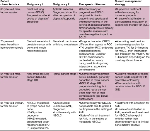
The treatment of patients with synchronous multiple primaries is challenging and often a therapeutic dilemma. In table 5, a few examples of patients with synchronous multiple primaries are summarised to illustrate the diffi-culties that arise in daily clinical practice. Patients with
Clinical picture, patient with a history or diagnosis of
(synchronous or metachronous) Familial cancer syndrome to think of Full picture of familial cancer syndrome
•Parathyroid hyperplasia/adenoma HNPCC, hereditary non-polyposis colon cancer; MEN1, multiple endocrine neoplasia type 1; MEN2, multiple endocrine neoplasia type 2; VHL, von Hippel-Lindau.
Table 4 Continued
on September 12, 2020 by guest. Protected by copyright.
synchronous multiple primaries should be discussed in multidisciplinary team (MDT) meetings and a consensus on a therapeutic strategy can sometimes take more than one MDT to form. Also, the patient should be informed about the situation and therapeutic challenges and often the uncertainty about the prognosis, because the therapy approach needs to be adapted.
In localised disease, the strategy may be surgery or radiation/chemoradiation therapy covering both malig-nancies.50 51
However, in the situation of advanced disease, the antitumour therapy selection is often difficult and mostly not based on evidence from the literature and clinical trials. In patients where both tumours are likely to respond to the same antitumour regimen as may be the case in patients with synchronous squamous cell carcinoma of the head and a second head and neck or
squamous non-small cell lung cancer, the therapeutic decision will involve a systemic therapy, which is likely to be active in for example, a platinum-based chemo-therapy.
In patients with synchronous multiple primaries, the following points should be considered when deciding on an antitumour treatment strategy. Please note that the proposed approaches are individual decisions and not general treatment recommendations. Also, these treatment approaches are not based on prospective trial evidence but rather ‘real-world’ examples of challenging clinical situations.
► What is the most significant tumour in terms of prognosis? Is there any chance of a curative approach or is the situation palliative? If palliative, which tumour is metastasised, what is known about the tumour dynamics (imaging, tumour marker) of
Table 5 Examples of clinical cases of patients with synchronous advanced multiple primary tumours
Patient
characteristics Malignancy 1 Malignancy 2 Therapeutic dilemma Current management strategy
60-year-old man,
former smoker Small cell lung cancer (SCLC)
Progression: after 6 cycles of cisplatin/ etoposide
Aplastic anaemia Diagnosed 4 months after completion of cisplatin/etoposide
•Chemotherapy at progression of SCLC not possible due to grade 4 neutropenia and thrombocytopenia in the setting of aplastic anaemia •Immunosuppressive therapy for aplastic anaemia with possible negative impact on SCLC
•Supportive treatment with eltrombopag for thrombocytopenia •In case of stabilisation of pancytopenia, evaluation of second line therapy for SCLC
71-year-old man, hereditary haemochromatosis
Castration-resistant prostate cancer with bone and lymph node metastases
Renal cell carcinoma
with lung metastases •Drugs active in for CRPC different than agents in RCC •TKI used for RCC endocrine drugs (abiraterone/
enzalutamide) used for CRPC: combinations not tested, no safety data, possible drug–drug interactions, expensive combinations
•Alternating treatment for the two malignancies: for example, TKI for 3–4 months for mRCC, then interruption and treatment for mCRPC for 3–4 months depending on the most significant tumour
64-year-old man,
former smoker Non-small cell lung cancer (NSCLC)
stage IIIB
Rectal cancer stage I •Chemotherapy regimens active in NSCLC generally not active in rectal cancer •NSCLC stage IIIB prognosis-defining, but untreated rectal cancer bears high risk of local complications (eg, bowel obstruction)
•Curative resection of rectal cancer (node-negative) with protective colostomy •Chemoradiation with curative intent for NSCLC (IIIB)
65-year-old woman,
former smoker NSCLC, metastatic to lymph nodes and
bone, KRAS proto-oncogene (KRAS)-mutated, programmed death receptor ligand (PD-L1) expression 0%
Acute myeloid leukaemia (AML) Diagnosed
simultaneously with NSCLC
•Chemotherapy for NSCLC not possible due to grade 4 neutropenia in the setting of AML
•State-of-the art treatment for AML in the setting of metastatic NSCLC
•Treatment with azacitidin for AML
•In case of stabilisation of AML, evaluation of treatment for NSCLC (checkpoint inhibitor rather than
chemotherapy due to limited bone marrow reserve)
on September 12, 2020 by guest. Protected by copyright.
the synchronous multiple primaries? What are the systemic therapy options?
► Should treatment strategy focus on local or systemic therapy? Can one of the synchronous tumours be treated radically and then the second malignancy sequentially?
► Which problems can be anticipated? For example, bowel obstruction in untreated colorectal cancer, liver failure in case of extensive liver metastases.
► If systemic therapy is necessary for, for example, two advanced malignancies, can a regimen be chosen that is active for both diagnoses? If not, what is the potential of interaction between two antitumour regimens (hepatic, eg, cytochrome-P (CYP) or cardiac frequency-corrected QT interval on the ECG)? Is there any literature about the combination (eg, axitinib and enzalutamide (see table 5))? Can systemic chemotherapy for an advanced solid tumour be applied at all (eg, in the case of secondary malignancy in the form of AML)? Can the two malignancies be treated in a cycling manner (eg, systemic treatment for tumour A for 2–3 months, followed by systemic therapy for tumour B)?
► Tumour profiling: Can the tumours be profiled (eg, targeted panel sequencing) and could these tumours have a common genetic background, which allows a common strategy option (eg, synchronous malignancies in BRCA1/2 carriers)?
Importantly, patients with active secondary malignancy are excluded from the vast majority of clinical trials involving novel treatments. For the majority of situations of patients with synchronous multiple primaries, only case reports are available in the literature and these should be applied to an individual clinical situation with caution.
Metachronous multiple primaries
The situation of metachronous multiple primaries can be equally challenging. For the situation when the first malignancy is still active/advanced, the considerations above apply. For the situation where a tumour was treated with curative intent and where a biopsy has confirmed a metachronous second malignancy, several points should be considered:
► Can the second primary cancer be treated with curative intent?
► What prior treatment has the patients had for the prior cancer diagnosis?Prior surgery: For example, in breast cancer? Is a second breast-conserving surgery possible and reasonable? In lung cancer: Is a second lung operation possible?Prior radiation therapy: Is there any overlap with the prior radiation field? Could the second primary be radiation therapy induced? What is the tolerance of the healthy tissue in the previously irradiated area? What is the radiation reserve and does it allow a radiation therapy with curative intent or only a palliative schedule/dose?Prior systemic therapy: What regimen? How long since the last systemic
therapy? What cumulative dose (eg, anthracyclines)? Is there any residual toxicity (eg, neuropathy, bone marrow toxicity)? Is the second primary potentially treatment induced?
► Can any possible complications be anticipated based on the patient’s prior anticancer therapy history?
► Are there carcinogenic factors that can be managed? For example, smoking, alcohol, viral infection.
► Could a cancer predisposition explain the multiple primaries?
As discussed in the sections above, the number of multiple metachronous tumours is likely to increase. Currently, the literature on the management of patients with metachronous multiple primaries is scarce, but it will be important to collect the information possibly through cancer registries in order to learn and identify potential rare complications in these situations.
DISCUSSION AND CONCLUSION
Based on a combination of factors (diagnosis, treatment, demographics), it is expected that in the course of the coming years, the prevalence of patients with multiple primaries will increase. In the literature, depending on definition, the frequency of multiple primaries ranges between 2.4% and 17% and it is important to realise that with longer follow-up the number of multiple primaries increases in all studies significantly and also that most of the epidemiological studies are based on data acquired more than 10 years ago and that therefore with longer follow-up or in subsequent studies the percentage of patients with synchronous but mainly metachronous multiple primaries will increase.
Also, it is important to recognise that certain patient popu-lations are at higher risk of developing multiple primaries, namely male patients and patients with a history of smoking or alcoholism but also patients diagnosed with a primary malignancy at an early stage and lower grade and patients with a hereditary cancer syndrome. Also, the number of octogenarians diagnosed with cancer is increasing and at the same time the frequency of multiple primaries in older patients who are potentially fit enough to receive active antineoplastic therapy.
Treatment-related secondary malignancies as observed in patients with germ cell or Hodgkin’s lymphoma are well characterised but with novel targeted therapies it is currently unclear whether an increased rate of secondary malignancies needs to be taken into consideration. For the B-RAF (rapidly accelerated fibrosarcoma) inhibitor vemurafenib, an increased rate of secondary cutaneous malignancies was demonstrated and requires careful and regular dermatological evaluation for patients on treatment.52 Also, for the PARP inhibitor olaparib, cases of myelodysplastic syndromes and AML have been observed and careful monitoring of patients for haemato-logical toxicity is recommended.53
With the advances and wider availability of genetic testing (eg, gene panels), patients diagnosed with
on September 12, 2020 by guest. Protected by copyright.
multiple primaries will be increasingly investigated for an underlying cancer predisposition. The gain of knowledge on patients with hereditary cancer and cancer survivors will hopefully allow the development of specific manage-ment and surveillance measures.
For clinical trials, generally patients with secondary malignancies are very often excluded unless they have been low grade/stage and were successfully treated at least 3-5 years ago. To reflect more of a real life population and to enable patients with a prior cancer history partic-ipation in clinical trials, the exclusion criteria, especially for early phase clinical trials could be modified to only exclude patients who currently require active anticancer therapy. Admittedly, this may add marked complexity in assessing efficacy and progression and may therefore not be suitable for phase III clinical trials.
Further research is needed, especially with regards to the areas of the treatment of patients with synchronous or metachronous multiple primary cancers. Also, the impact of prior therapies on prognosis, antitumour efficacy and toxicity needs to be better characterised.
Contributors We confirm that all co-authors have contributed to the generation of
the manuscript.
Competing interests AO: advisory role (compensated, institutional): Astellas,
Bayer, Sanofi, Roche, Janssen. Research support (institutional): Teva, Janssen. Travel support: Astellas, Bayer, Sanofi, Roche. SS: advisory role (compensated institutional): Böhringer Ingelheim, BMS. AV, KH, HF, CH and TC report no competing interests.
Provenance and peer review Commissioned; externally peer reviewed.
Open Access This is an Open Access article distributed in accordance with the
Creative Commons Attribution Non Commercial (CC BY-NC 4.0) license, which permits others to distribute, remix, adapt, build upon this work non-commercially, and license their derivative works on different terms, provided the original work is properly cited and the use is non-commercial. See: http:// creativecommons. org/ licenses/ by- nc/ 4. 0/
© European Society for Medical Oncology (unless otherwise stated in the text of the article) 2017. All rights reserved. No commercial use is permitted unless otherwise expressly granted.
REFERENCES
1. American Cancer Society. Cancer facts & figures 2016. 2016. Atlanta: American Cancer Society, 2016.
2. Herrmann C, Cerny T, Savidan A, et al. Cancer survivors in Switzerland: a rapidly growing population to care for. BMC Cancer 2013;13:287.
3. Owen LJ. Multiple malignant neoplasms. JAMA 1921;76:1329–33. 4. Coyte A, Morrison DS, McLoone P. Second primary cancer risk - the
impact of applying different definitions of multiple primaries: results from a retrospective population-based cancer registry study. BMC Cancer 2014;14:272.
5. Buiatti E, Crocetti E, Acciai S, et al. Incidence of second primary cancers in three Italian population-based cancer registries. Eur J Cancer 1997;33:1829–34.
6. Weir HK, Johnson CJ, Thompson TD. The effect of multiple primary rules on population-based cancer survival. Cancer Causes Control 2013;24:1231–42.
7. Rosso S, De Angelis R, Ciccolallo L, et al. Multiple tumours in survival estimates. Eur J Cancer 2009;45:1080–94.
8. Karaholios E, English D, Thursfield V, et al. Second primary cancers in Victoria. 2013. http://www. cancervic. org. au/ downloads/ cec/ Second- Primary- Cancers. pdf
9. Wood ME, Vogel V, Ng A, et al. Second malignant neoplasms: assessment and strategies for risk reduction. J Clin Oncol 2012;30:3734–45.
10. Shah SA, Riaz U, Zahoor I, et al. Carcinoma multiplex. J Coll Physicians Surg Pak 2013;23:290–2.
11. Amer MH. Multiple neoplasms, single primaries, and patient survival. Cancer Manag Res 2014;6:119–34.
12. Ferreti Sea. Airtum cancer registration handbook, 2009.
13. Bajdik CD, Abanto ZU, Spinelli JJ, et al. Identifying related cancer types based on their incidence among people with multiple cancers. Emerg Themes Epidemiol 2006;3:17.
14. Gaskin HS, Hardy RE, Fletcher RL. Multiple primary malignancies in black patients. J Natl Med Assoc 1981;73:1065–8.
15. Donin N, Filson C, Drakaki A, et al. Risk of second primary malignancies among cancer survivors in the United States, 1992 through 2008. Cancer 2016;122:3075–86.
16. AIRTUM Working Group. [Italian Cancer figures, report 2010: cancer prevalence in Italy. patients living with Cancer, long-term survivors and cured patients]. Epidemiol Prev 2010;34:1–188.
17. Hauben EI, Arends J, Vandenbroucke JP, et al. Multiple primary malignancies in osteosarcoma patients. incidence and predictive value of osteosarcoma subtype for cancer syndromes related with osteosarcoma. Eur J Hum Genet 2003;11:611–8.
18. Hayat MJ, Howlader N, Reichman ME, et al. Cancer statistics, trends, and multiple primary cancer analyses from the surveillance, epidemiology, and end results (SEER) program. Oncologist 2007;12:20–37.
19. Mariotto AB, Noone AM, Howlader N, et al. Cancer survival: an overview of measures, uses, and interpretation. J Natl Cancer Inst Monogr 2014;2014:145–86.
20. Ferlay J, Steliarova-Foucher E, Lortet-Tieulent J, et al. Cancer incidence and mortality patterns in Europe: estimates for 40 countries in 2012. Eur J Cancer 2013;49:1374–403.
21. Kim JY, Song HS. Metachronous double primary cancer after treatment of breast cancer. Cancer Res Treat 2015;47:64–71. 22. Ricceri F, Fasanelli F, Giraudo MT, et al. Risk of second primary
malignancies in women with breast cancer: results from the European prospective investigation into cancer and nutrition (EPIC). Int J Cancer 2015;137:940–8.
23. Molina-Montes E, Pérez-Nevot B, Pollán M, et al. Cumulative risk of second primary contralateral breast cancer in BRCA1/BRCA2 mutation carriers with a first breast cancer: a systematic review and meta-analysis. Breast 2014;23:721–42.
24. Jin T, Song T, Deng S, et al. Radiation-induced secondary malignancy in prostate cancer: a systematic review and meta-analysis. Urol Int 2014;93:279–88.
25. Wallis CJ, Mahar AL, Choo R, et al. Second malignancies after radiotherapy for prostate cancer: systematic review and meta-analysis. BMJ 2016;352:i851.
26. Friedenson B. BRCA1 and BRCA2 pathways and the risk of cancers other than breast or ovarian. MedGenMed 2005;7:60.
27. Bhuller KS, Zhang Y, Li D, et al. Late mortality, secondary malignancy and hospitalisation in teenage and young adult survivors of hodgkin lymphoma: report of the childhood/adolescent/young adult cancer survivors research program and the BC cancer agency centre for lymphoid cancer. Br J Haematol 2016;172:757–68.
28. Lisik-Habib M, Czernek U, Dębska-Szmich S, et al. Secondary cancer in a survivor of hodgkin's lymphoma: a case report and review of the literature. Oncol Lett 2015;9:964–6.
29. European Academies Science Advisory Council. Direct-to-consumer genetic testing for health-related purposes in the European Union: the view from EASAC and FEAM EASAC policy report. 2012;18. 30. Sanchez De Cos Escuin J, Rodriguez Lopez DP, Delgado Utrabo I, et
al. Disease recurrence and second tumors in Long-term survivors of lung Cancer. Arch Bronconeumol 2016;52:183–8.
31. Bhaskarla A, Tang PC, Mashtare T, et al. Analysis of second primary lung cancers in the SEER database. J Surg Res 2010;162:1–6. 32. Soerjomataram I, Coebergh JW. Epidemiology of multiple primary
cancers. Methods Mol Biol 2009;471:85–105.
33. Robson ME, Storm CD, Weitzel J, et al. American society of clinical oncology policy statement update: genetic and genomic testing for cancer susceptibility. J Clin Oncol 2010;28:893–901.
34. Garber JE, Offit K. Hereditary cancer predisposition syndromes. J Clin Oncol 2005;23:276–92.
35. Paluch-Shimon S, Cardoso F, Sessa C, et al. Prevention and screening in BRCA mutation carriers and other breast/ovarian hereditary cancer syndromes: ESMO clinical practice guidelines for cancer prevention and screening. Ann Oncol 2016;27:v103–10. 36. Balmaña J, Balaguer F, Cervantes A, et al. Familial
risk-colorectal cancer: ESMO clinical practice guidelines. Ann Oncol 2013;24:vi73–80.
37. Gabai-Kapara E, Lahad A, Kaufman B, et al. Population-based screening for breast and ovarian cancer risk due to BRCA1 and BRCA2. Proc Natl Acad Sci USA 2014;111:14205–10.
38. Lynch HT, de la Chapelle A. Hereditary colorectal cancer. N Engl J Med 2003;348:919–32.
on September 12, 2020 by guest. Protected by copyright.
39. Win AK, Lindor NM, Winship I, et al. Risks of colorectal and other cancers after endometrial cancer for women with lynch syndrome. J Natl Cancer Inst 2013;105:274–9.
40. Aarnio M, Sankila R, Pukkala E, et al. Cancer risk in mutation carriers of DNA-mismatch-repair genes. Int J Cancer 1999;81:214–8. 41. Kohlmann W, Gruber SB. Lynch syndrome. In: Pagon RA, Adam MP,
Ardinger HH, et al. eds: Gene reviews (R). Seattle, WA: 1993. 42. Le DT, Uram JN, Wang H, et al. PD-1 blockade in tumors with
mismatch-repair deficiency. N Engl J Med 2015;372:2509–20. 43. Schmid S, Gillessen S, Binet I, et al. Management of von
hippel-lindau disease: an interdisciplinary review. Oncol Res Treat 2014;37:761–71.
44. Lonser RR, Glenn GM, Walther M, et al. Von Hippel-Lindau disease. Lancet 2003;361:2059–67.
45. Villani A, Shore A, Wasserman JD, et al. Biochemical and imaging surveillance in germline TP53 mutation carriers with Li-Fraumeni syndrome: 11 year follow-up of a prospective observational study. Lancet Oncol 2016;17:1295–305.
46. Pang JT, Thakker RV. Multiple endocrine neoplasia type 1 (MEN1). Eur J Cancer 1994;30A:1961–8.
47. Eng C, Clayton D, Schuffenecker I, et al. The relationship between specific RET proto-oncogene mutations and disease phenotype in multiple endocrine neoplasia type 2. International RET mutation consortium analysis. JAMA 1996;276:1575–9.
48. Ishimori T, Patel PV, Wahl RL. Detection of unexpected additional primary malignancies with PET/CT. J Nucl Med 2005;46:752–7. 49. Miyazaki T, Sohda M, Higuchi T, et al. Effectiveness of FDG-PET in
screening of synchronous cancer of other organs in patients with esophageal cancer. Anticancer Res 2014;34:283–7.
50. Heroiu Cataloiu AD, Danciu CE, Popescu CR. Multiple cancers of the head and neck. Maedica 2013;8:80–5.
51. Zhang Z, Gao S, Mao Y, et al. Surgical outcomes of synchronous multiple primary non-small cell lung cancers. Sci Rep
2016;6:23252.
52. Lacouture ME, O'Reilly K, Rosen N, et al. Induction of cutaneous squamous cell carcinomas by RAF inhibitors: cause for concern? J Clin Oncol 2012;30:329–30.
53. Ricks TK, Chiu HJ, Ison G, et al. Successes and challenges of PARP inhibitors in cancer therapy. Front Oncol 2015;5:222.
on September 12, 2020 by guest. Protected by copyright.