The frequency of autoimmune thyroid disorders in patients with thyroid
dysfunction in Erbil city
Received: 11/10/2016 Accepted: 3/8/2017
Abstract
* Department of Pharmacognosy, College of Pharmacy, Hawler Medical University, Erbil, Iraq. ** Department of Clinical Analysis, College of Pharmacy, Hawler Medical University, Erbil, Iraq.
Introduction
Autoimmune thyroid diseases are resulting from dysregulation of the immune system, which leads to an immune attack to the thyroid gland. These are T cell-mediated organ-specific autoimmune conditions.1
Autoimmune thyroid disorders ascend due to multifaceted interactions between genetic and environmental factors and are described by reactivity to their self-thyroid antigens that are stated as distinctive anti-receptor or inflammatory autoimmune diseases.2 These disorders are categorized
by developing inflammation and producing a wide range of autoantibodies bound for various auto-antigens.3 General and usual screening for thyroid hormone level involve blood tests for Thyroid Stimulating
Hormones TSH, Thyroxin T4 (total and free), and sometimes Triiodothyronine T3
(total and free). The degree of the modification in thyroid function associates
with the severity of the sickness and its outcomes in ill patients. The
mechanisms involved include a reduced conversion of T4 to T3 in the thyroid tissues and modifications in thyroid hormones' binding to serum proteins.5 The anti-thyroperoxidase (TPO), anti-thyroglobulin (anti-Tga), and anti-thyroid stimulating hormone (anti-TSH)
receptor antibodies are the laboratory diagnostic assays for autoimmune thyroid diseases. These group of tests is also known as thyroid microsomal antibodies, one or more of these tests are performed
Background and objective: Thyroid disorders are one of the most frequent pathologies found in the general population, but identifying thyroid disease can be clinically challenging because subclinical thyroid dysfunction and autoimmune thyroiditis are often asymptomatic and usually diagnosed biochemically. This study aimed to distinguish the autoimmune thyroid diseases from other forms of thyroid dysfunctions in patients admitted to PAR hospital in Erbil city.
Methods: blood was withdrawn from healthy subjects, and unhealthy patients suffer from thyroid dysfunction, their age and gender were recorded, and their blood serum were subjected to test the thyroid function antibodies including triiodothyronine T3, thyroxin T4, and thyroid stimulating hormone TSH. Also, autoimmune antibodies were tested including anti-thyroglobulin antibody (anti-TGA) and thyroperoxidase antibody (TPO antibodies).
Results: no significant differences were shown in T3 levels while contrary highly significant differences were shown in T4, TSH anti-TGA and anti-TPO levels between healthy subjects and unhealthy patients groups. The percentages of autoimmune thyroid diseases were (45.2%) as compared to the other forms of thyroid dysfunctions (54.8%). Most of the patients were females in the age group 30-39 years.
Conclusion: In Erbil city population/PAR hospital the prevalence of autoimmune thyroid diseases were more frequent among other thyroid diseases collectively. It is mostly found in females rather than males within the age group 30-39 years.
to determine whether a patient with autoimmune diseases (like systemic lupus
erythematosus, rheumatoid arthritis or pernicious anemia) is at risk of thyroid dysfunction or not.6 This study aimed to predict the autoimmune diseases and distinguish them from the other forms of thyroid disorders in patients admitted to PAR hospital/ Erbil city.
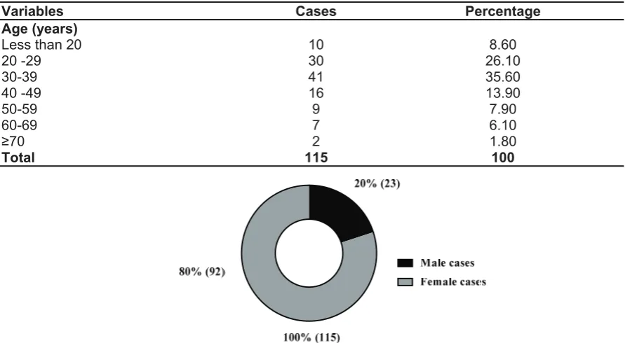
The results of the present study revealed that among the 115 patients admitted to PAR hospital, (80%) were females while only (20%) were males as shown in Figure 1. While there was a clear age-dependent increase in the prevalence
of autoimmune thyroid patients compared with the healthy subjects, the most infected
patients were in age 30-39 years with a high percentage approximately 35.60%
as shown in Table 1.
Methods
Samples Collection
In a cross-sectional study, the data of 115 patients with thyroid dysfunctions including thyrotoxicosis, Graves disease, goiter, nodules, and others were collected from January 2015 until January 2016 from the diagnostic laboratory of PAR hospital. Likewise, data from 50 healthy subjects were used as the comparison group of the study. Information on the whole population was recorded including their gender and age group. Blood samples were withdrawn then serum was separated, and the samples were subjected to test the thyroid
function antibodies including T3, T4,
TSH, anti-TGA, TPO. Enzyme-linked immunoabsorbent assay (ELISA) was used
for determination of antibodies levels in the serum quantitatively.
S t a t i s t i c a l A n a l y s i s a n d D a t a Management
Statistical analysis was evaluated by using the statistical package for the social sciences program (version 18). Independent samples t-test was used for analysis of data. A probability value less 0.05 was considered statistically significant.
Results
Table 1:Distribution of thyroid dysfunction between different gender and age groups.
Figure 1: Percentage of both genders in patients admitted to PAR hospital /Erbil city.
Percentage Cases
Variables
8.60 10
Age (years) Less than 20
26.10 30
20 -29
35.60 41
30-39
13.90 16
40 -49
7.90 9
50-59
6.10 7
60-69
1.80 2
≥70
100 115
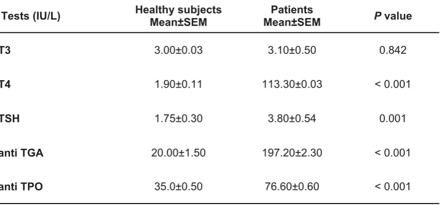
Regarding the thyroid function results from the (Mean ± SD) of T3, T4, and TSH of the patients was significantly higher than the normal ranges of healthy subjects, indicating dysfunction of their thyroid gland. Also, there was a significant increase in serum anti-TGA and TPO levels as compared to the normal ranges of healthy subjects indicating an autoimmune disease as shown in Table 2. The important result of this study was in distinguishing the
percentage of autoimmune thyroid disease patients from the other forms of thyroid dysfunctions, which have been done by comparing the data of patients with
high levels of anti-TGA and TPO antibodies (45.2%) (specific indicators for autoimmune diseases) with those had
only high levels of T3, T4 and TSH antibodies (54.8%) (no specific tests) as
shown in Figure 2.
Table 2: The difference of serum T3, T4, TSH, anti-TGA and anti-TPO levels of patients admitted to PAR hospital as compared to the healthy subject comparison group.
Figure 2: Percentage of autoimmune thyroid diseases from other forms of thyroid dysfunctions in patients admitted to PAR hospital /Erbil city.
Tests (IU/L) Healthy subjectsMean±SEM Mean±SEMPatients P value
T3 3.00±0.03 3.10±0.50 0.842
T4 1.90±0.11 113.30±0.03 < 0.001
TSH 1.75±0.30 3.80±0.54 0.001
anti TGA 20.00±1.50 197.20±2.30 < 0.001
According to the results that been shown above, almost all of the patients who were admitted to PAR hospital were checking their thyroid hormone levels (either normal thyroid tests or thyroid auto-immune test), clearly be seen in Table 2 that the results of (T3) of both healthy subject and unhealthy patients were similar. However, as it is well defined in Table 2 that the results of (T4 and TSH) were exactly the opposite of (T3), in which significant differences were observed in the results of the patients in comparison to the healthy subject data. Moving to (auto-immune tests) as it is shown above in Table 2 the percentage of diseased patients was higher than normal healthy subjects significantly. Accordingly, Figure 2 obviously shows that 45.2% of admitted patients to PAR hospital were suffering from autoimmune thyroid disease, whereas 54.8% of them were suffering from other thyroid dysfunctions.
As mentioned earlier autoimmune disease simply can be differentiating from other thyroid dysfunctions by two major tests which PAR hospital relay
on, anti-thyroglobulin (anti-TGA) and anti-thyroid peroxidase (TPO) tests. Within the 45.2% of thyroid autoimmune disease patients, the highest rate of them were women, since the basic immune response differs between male and female. This is mainly due to a potent immune stimulatory
effect of estrogen and prolactin and a protective role of androgen in this process. Our results are similar to the results of Hollowel et al.8 who reported that
these antibodies were more prevalent in women than men. Also, Perros et al.9
indi-cated that female patients had the highest annual risk of developing thyroid diseases. There is enough evidence to state that genetic factors are important as well. Moreover, the pathogenesis and the origin
of autoimmune thyroid disorders are differing from other thyroid dysfunctions. Since the pathogenesis of autoimmune
disease differs from other thyroid dysfunctions, the treatments are different
from each other. More specifically, autoimmune thyroid disease includes different types each of them with different signs and symptoms and different treatments require. For instance,
Hashimoto's thyroiditis, which is one of the autoimmune disorders causing primary hypothyroidism needs synthetic T4 for treating it. While graves disease result from overactive thyroid gland requires anti-thyroid medications such as propylthiouracil and methimazole.4 In other
words, if there were disorders in thyroid hormone levels, then it is necessary to know whether it is autoimmune or other thyroid dysfunctions since we need to treat them separately according to their different pathogenesis. So this can be differentiating by thyroid antibody tests.7 Infectious
agents have been implicated in the pathogenesis of a variety of autoimmune
diatheses, namely, rheumatic fever,
Reiter's syndrome, systemic lupus erythematosus (SLE), myasthenia gravis,
insulin-dependent diabetes mellitus, Sjogren's syndrome, and the autoimmune thyroid diseases.10 On the other hand, gender has a strong effect on the results of both thyroid tests and autoimmune thyroid test. Through the year 2015, the patients who made thyroid tests in PAR hospital 80% of them were female, on the other hand, only 20% of them were male. Our results are in agreement with the results of
11 who indicated that autoimmune diseases
in a population affect women more than men, Indeed, the age of the patients play a significant role in thyroid tests. According to the tests that have been made at PAR hospital over a year, 35.60 % of the patients were between 31-39 years old. These results are supported by the results of two other studies12,13 who reported that the prevalence of autoimmune disorders is age-related. These age-dependent results are due to many reasons; one of them is that hypothalamus (produce hormone regulate the secretion of thyroid hormone, Thyroid Stimulating hormone Releasing Hormone TSHRH). The amount of these
regulatory hormones stays about the same by increasing the age, but the response of thyroid gland to this hormone will reduce by aging. That is why aging results in hypothyroidism more than hyperthyroidism.11
In Erbil city/PAR hospital the prevalence of autoimmune thyroid diseases was more frequent among other thyroid diseases collectively. It is mostly found in females
rather than males within the age group 30-39 years. Hence, if there is the disturbance in the level of thyroid hormones, it is necessary to identify whether it is the autoimmune disease or other thyroid dysfunction by doing extra blood tests because this will affect the decision of the physician about the treatment and how to monitor and control
the case.
Conclusion
Competing interests
The authors declare that they have no competing interests.
References
1. Antonelli A, Ferrari SM, Corrado A, Di Domenicantonio A, Fallahi P. Autoimmune thyroid disorders. Autoimmun Rev 2015; 14(2):174–80.
2. Tozzoli R, Villalta D, Bizzaro N, Tonutti E, Manoni F. Laboratory diagnosis of autoimmune
thyroid disease. Recenti Prog Med 2001; 92(10):609–17.
3. Szyper-Kravitz M, Marai I, Shoenfeld Y. Coexistence of thyroid autoimmunity with other
autoimmune diseases: friend or foe? Additional
aspects on the mosaic of autoimmunity. Autoimmunity 2009; 38(3):247.
4. Schott M, Scherbaum WA. Autoimmune thyroid disease. Dtsch Arztebl 2006; 103(45):3023–32.
5. Bello G, Ceaichisciuc I, Silva S, Antonelli M. The role of thyroid dysfunction in the critically ill: a review of the literature. Minerva Anestesiol 2010; 76(11):919–28.
6. Iddah M, Macharia B. Autoimmune thyroid disorders. ISRN Endocrinol 2013; 2013.
7. Ladenson PW, Singer PA, Ain KB, Bagchi N, Bigos ST, Levy EG, et al. American Thyroid Association guidelines for detection of thyroid dysfunction. Arch Intern Med 2000; 160(11):1573 –5.
8. Hollowell JG, Staehling NW, Flanders WD, Hannon WH, Gunter EW, Spencer CA, et al. Serum TSH, T4, and thyroid antibodies in the United States population (1988 to 1994): National Health and Nutrition Examination Sur-vey (NHANES III). J Clin Endocrinol Metab 2002; 87(2):489–99.
9. Perros P, McCrimmon R, Shaw G, Frier B. Frequency of thyroid dysfunction in diabetic pa-tients: value of annual screening. Diabet Med 1995; 12(7):622–7.
10. Tomer Y, Davies TF. Infection, Thyroid Disease, and Autoimmunity. Endocr Rev 1993; 14(1):107– 20.
11. Vaidya B, Kendall-Taylor P, Pearce SH. The genetics of autoimmune thyroid disease. J Clin Endocrinol Metab 2002; 87(12):5385–97. 12. Mantovani RM, Mantovani LM, Dias VM. Thyroid
autoimmunity in children and adolescents with type 1 diabetes mellitus: prevalence and risk factors. J Pediatr Endocrinol Metab 2007; 20 (6):669–76.