Open Access
Research article
Brain type carnosinase in dementia: a pilot study
Cynthia M Balion*
1,2, Carolyn Benson
1,2, Parminder S Raina
4,
Alexandra Papaioannou
1,3, Christopher Patterson
1,3and Afisi S Ismaila
4Address: 1Department of Laboratory Medicine, Hamilton Health Sciences, Hamilton, Ontario, Canada, 2Department of Pathology and Molecular
Medicine, McMaster University, Hamilton, Ontario, Canada, 3Department of Medicine, McMaster University, Hamilton, Ontario, Canada and 4Department of Clinical Epidemiology & Biostatistics, McMaster University, Hamilton, Ontario, Canada
Email: Cynthia M Balion* - [email protected]; Carolyn Benson - [email protected]; Parminder S Raina - [email protected]; Alexandra Papaioannou - [email protected]; Christopher Patterson - [email protected]; Afisi S Ismaila - [email protected] * Corresponding author
Abstract
Background: The pathological processes underlying dementia are poorly understood and so are the markers which identify them. Carnosinase is a dipeptidase found almost exclusively in brain and serum. Carnosinase and its substrate carnosine have been linked to neuropathophysiological processes.
Methods: Carnosinase activity was measured by a flourometric method in 37 patients attending a Geriatric Outpatient Clinic. There were 17 patients without dementia, 13 had Alzheimer's disease (AD) and 7 had mixed dementia (MD).
Results: The range of serum carnosinase activity for patients without dementia was 14.5 – 78.5 µmol/ml/h. There was no difference in carnosinase activity between patients without dementia (40.3 ± 15.2 µmol/ml/h) and patients with AD (44.4 ± 12.4 µmol/ml/h) or MD (26.6 ± 15 µmol/ml/ h). However, levels in the MD group were significantly lower than the AD group (p = 0.01). This difference remained significant after adjusting for gender, MMSE score, exercise, but not age, one at a time and all combined. The effect of other medical conditions did not remove the significance between the AD and MD groups. The MD group, but not the AD group, demonstrated a significant trend with carnosinase activity decreasing with duration of disease (from first recorded date of diagnosis to date of blood collection) (r = -0.76, p = 0.049). There was no association with carnosinase activity and MMSE score in the AD or MD group. Both AD and MD patients on any dementia medication (donepezil, galantamine, memantine) had higher carnosinase activity compared to those not taking a dementia medication. Carnosinase activity was higher in patients who regularly exercised (n = 20) compared to those who did not exercise regularly (n = 17)(p = 0.006).
Conclusion: This exploratory study has shown altered activities of the enzyme carnosinase in patients with dementia.
Background
Dementia has emerged as a major clinical, societal and
economic problem, especially in industrialized societies. Alzheimer's disease (AD) is the most common form of
Published: 5 November 2007
BMC Neurology 2007, 7:38 doi:10.1186/1471-2377-7-38
Received: 20 October 2006 Accepted: 5 November 2007
This article is available from: http://www.biomedcentral.com/1471-2377/7/38
© 2007 Balion et al; licensee BioMed Central Ltd.
dementia followed by vascular dementia and mixed dementia (AD with cerebrovascular disease). Early and accurate diagnosis is desirable, as current therapies are most effective in the early stages [1]. Early diagnosis also allows the cognitively aware patient to deal with future issues in medical care, safety, and legal matters [2].
Numerous plasma and serum markers for dementia have been identified, and many show significant differences between patients with dementia and those without dementia, but they lack the sensitivity and specificity needed for diagnosis. Furthermore, no biomarker has yet demonstrated the utility to monitor the progress of the disease, response to therapy or to predict outcome. Most of the proposed biomarkers are not brain specific but reflect other pathologies involving coagulation and inflammation [3], immunological [4], detoxification [5], oxidative stress [6], lipid metabolism and vascular disease [6,7]. Although proteins specific to the neuropathology of amyloid plaques such as amyloid β-protein (Aβ) and amyloid precursor protein (APP) are detectable in periph-eral blood, they have shown little value in diagnosis or monitoring therapy [6].
Successful markers of dementia will most likely be linked to pathological processes within affected brain cells. Car-nosinase (CN1; EC 3.4.13.20) is an enzyme found almost exclusively in brain tissue and serum [8]. It is a member of the M20 metalloproteinase family and specifically degrades dipeptides. Two of these, carnosine (β -alanyl-histidine) and homocarnosine (γ -aminobutyryl-histi-dine), have been linked to aging and dementia.
Carnosine has many reported functions including intrac-ellular buffer, metal- chelator (particularly copper), anti-oxidant, and free radical scavenger [9]. It reacts with pro-tein carbonyl groups serving as an alternative to glucose, reversing the glycation process at the Schiff base stage [10]. Carnosine, through its copper chelating and anti-cating function, inhibits the production of advanced gly-cation endproducts (AGEs) [11]. AGEs result from the reaction of amino groups with reducing sugars resulting in irreversibly cross-link proteins. While AGEs are consid-ered part of the normal aging process [12], accelerated production is implicated in the formation of β-amyloid plaques and neurofibrillary tangles [11]. Carnosine may also serve as an anti-senescence agent since it has been shown to extend the life of human diploid fibroblasts [13] and reduce the rate for telomeric shortening and damage to DNA of human fetal lung fibroblasts [14].
In the olfactory bulb, carnosine prevents the inhibitory effects of copper on synaptic transmission through chela-tion, thus acting as a neuromodulator [15]. Since AD is characterized by early changes in the olfactory system [16]
and high levels of copper are associated with senile plaques and neurofibrillary tangles [17], lower levels of carnosine could decrease copper chelation and impair olfaction through decreased synaptic transmission [15] and neurotoxic effects [18].
Homocarnosine, a specific substrate of carnosinase is degraded into γ-aminobutyric acid (GABA) and L-histi-dine. Post-mortem and imaging studies of AD have shown decreases in GABA in the temporal, frontal, and parietal regions [19]. Decreased histidine levels in the frontal and temporal lobes of AD patients have also been reported [20]. Since histidine is produced when carnosine and homocarnosine is cleaved by carnosinase, this obser-vation may reflect decreased carnosinase activity.
Case studies of carnosinase deficiency have reported symptoms that include progressive mental deficiency, non-progressive mental retardation, developmental delay, spastic paraplegia, seizures, neurosensory hearing loss, retinitis pigmentosa, and progressive childhood dementia [21,22]. Reduced activity levels have been found in other neurological disorders including Parkin-son's disease, multiple sclerosis, and following a cerebrov-ascular event [23,24]. Recently, serum carnosinase levels have been shown to decrease during cardiopulmonary bypass surgery [25]. Ischemia is a major complication of cardiac surgery and a decrease in carnosinase during this procedure may be a neuroprotective mechanism [25] as carnosine may reduce neurotoxicity through its antioxi-dant capacity [18]. Brains afflicted with AD show bio-chemical evidence of ischemia, specifically Aβ deposits and an increase in basic fibroblastic growth factor [26]. Decreased blood flow and oxygenation has been docu-mented in the temporal lobes in AD [27]. These vascular changes may initiate the same decrease in carnosinase activity as seen in patients with cerebrovascular events. Carnosine has also been shown to produce vascular relax-ation whereas its constituent amino acid β-alanine pro-duced vasoconstriction [28].
In light of this growing body of information relating car-nosinase and its substrates to dementia, we undertook an exploratory study to investigate carnosinase activity in patients with dementia.
Methods
Participants
disor-ders, diet, exercise frequency, and recent weight loss. All prescriptions, over-the-counter medications and supple-ments were recorded. Medical history was obtained and confirmed through medical records. The most recent mini-mental state examination (MMSE) score was used as a global measure of cognition [29]. The diagnosis of dementia was based upon the NINCDS-ADRDA criteria [30] incorporating clinical assessment, laboratory and neuroimaging studies where indicated.
Carnosinase activity
Blood samples were collected by venipuncture. Serum was separated after samples were centrifuged at 2,000 × g for 10 min at room temperature, aliquoted and stored at -20°C until analysis. Serum carnosinase activity was assayed by the method described by Lenney et al. [31]. All reagents were purchased from Sigma-Aldrich Canada Ltd. (Oakville, ON). Serum (50 µl) was first incubated at 30°C with 250 µl NH4OH (pH 8.5) and 150 µl CdCl for 10 min. The enzyme reaction was started by adding 100 µl of 100 mM carnosine and allowed to proceed for 30 min at 30°C. At this point the reaction was stopped by adding 500 µl of 0.6 M TCA. The solution was centrifuged 800 × g for 10 min and the supernatant removed. L-Histidine was measured fluorometrically by the method described by Ambrose et al. [32]. Briefly, 50 µl of supernatant was combined with 1.2 ml 0.83 N NaOH and 500 µl 0.2% o -phthalaldehyde (OPA) and incubated at 30°C for 15 min. The reaction was stopped by adding 500 µl 4.0 M H3PO4 and incubated for a further 15 min at 30°C. The solution was cooled at room temperature for 1 hour and read on a SpectraMax M2 spectrophotofluorometer (Molecular Devices, Sunnyvale, CA) at an excitation wavelength of 340 nm and emission wavelength of 450 nm.
Standard curves and detection limits
Standard curves were created using L-histidine and water in place of TCA supernatant in the o-phthalaldehyde assay. Standard curves using combinations of carnosine,
β-alanine, and L-histidine representing different levels of hydrolysis were not significantly different from standard curves with L-histidine and water [33]. Carnosinase activ-ity was calculated using the L-histidine standard curve and was expressed as µmol of L-histidine produced per ml of serum per hour. The lowest limit of detection was deter-mined by adding 3 SD to the mean fluorescence of 20 blank replicates, which corresponded to an enzyme activ-ity of 4 µmol/ml/h.
Statistical analysis
Normal distribution of the data was done using the Kol-mogorov-Smirnov test and Shapiro-Wilk tests. The Pear-son correlation was calculated for relationships among serum carnosinase activity, duration of dementia and MMSE score. The software used for these analyses was
Analyze-it for Microsoft Excel (Analyze-it Software, Ltd., Leeds, England). Group comparisons were performed using the student's t-test, regression analysis and analysis of variance (SPSS 12.0, SPSS Inc., Chicago, IL). Compari-sons were considered to be significant when p < 0.05.
Results
The study population consisted of 17 patients without dementia and 20 patients with dementia. The dementia group included 13 patients with AD and 7 patients with MD. There were more females (n = 29) than males (n = 8) in all groups. There were no significant differences in age between any groups (p > 0.05). Demographic and clinical characteristic data are presented in Table 1.
Carnosinase activity for patients without dementia was 14.5 – 78.5 µmol/ml/h, comparable to the adult range determined by Lenney et al. (18 – 72 µmol/ml/h) using the same method [31]. The results of the analysis of vari-ance showed a significant difference in means across groups (p = 0.04). Further analysis showed there was no difference in carnosinase activity between patients with-out dementia (40.3 ± 15.2 µmol/ml/h) and patients with AD (44.4 ± 12.4 µmol/ml/h) or MD (26.6 ± 15 µmol/ml/ h) (Figure 1). However, the difference between the AD and MD groups was significant (mean difference = 17.8
µmol/ml/h, p = 0.01).
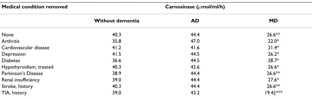
Table 2 shows the pairwise comparisons after adjusting for age, gender, MMSE score and exercise (one variable at a time and all variables combined). The results show there was a significant difference in the means of carnosinase between AD and MD groups after adjusting for each vari-able except for age (p = 0.06). The effect of medical condi-tions, other than dementia on carnosinase activity, was tested by comparison of groups (unpaired t-test) after removal of each medical condition listed in Table 1. Table 3 shows that the AD group remains significantly different (p < 0.05) from the MD group regardless of which medical condition is removed.
However, carnosinase values were higher in both AD and MD for the patients on dementia medications compared to those who were not on dementia medications. Signifi-cance between groups was reached for AD (p = 0.05), but not for MD (p = 0.59). Diet containing meat did not show a difference within the AD or MD groups. Exercise on the day of blood collection showed no difference between patients with dementia and those without dementia, but exercising three or more times a week was associated with an increase in carnosinase activity (n = 37, p = 0.006) (Fig-ure 4).
Discussion
In this study we have shown that levels of carnosinase activity have the potential to differentiate AD from MD with very little overlap between these conditions. As the only known difference between these groups was the pres-ence or abspres-ence of cerebrovascular disease, this suggests that vascular pathology may be the reason for lower car-nosinase activity. Vascular dementia is associated with cer-ebrovascular accident, a condition known to lower carnosinase activity [23,24]. Furthermore, compared to patients without dementia, carnosinase activity remains significantly decreased three months after a cerebrovascu-lar accident [23]. Decreased levels have also been found in
Box-and-whisker plot showing carnosinase activity in without dementia (n = 17), AD (n = 13), and MD (n = 7) groups
Figure 1
Box-and-whisker plot showing carnosinase activity in without dementia (n = 17), AD (n = 13), and MD (n = 7) groups. Comparisons were not significant between with-out dementia and AD or MD groups, however the difference between the AD and MD groups was significant (p = 0.01).
MD AD
Without dementia 80
60
40
20
0
Carn
o
s
in
ase
act
iv
it
y (
µ
mol/ml/h)
Table 1: Demographic and clinical characteristics of the without dementia, AD and MD groups. AD, Alzheimer's disease; MD, mixed dementia
Patient group
Without dementia AD MD
Number of patients 17 13 7
Mean age (range) 80.2 (64–92) 75.5 (58–88) 82.3(72–92)
Female gender 15 8 6
Mean aterial pressure (mmHg) 99 ± 13 103 ± 9 97 ± 7
Duration of dementia (month) - 19 ± 18 11 ± 10
Mini-mental state examination (MMSE) score - 20 ± 7 21 ± 7
Neuroimaging (CT or MRI) 2 5 4
Diet, meat eaten within the last day 13 11 4
Exercise, 3 or more times per week 9 9 2
Medical Conditions:
Arthritis 8 4 4
Cardiovascular disease 5 4 3
Depression 4 2 2
Diabetes 3 1 1
Hypothyroidism, treated 2 1 0
Parkinson's disease 1 0 0
Renal insufficiency 0 0 2
Stroke, history 2 0 0
TIA, history 0 1 2
Dementia medication:
Donepezil 0 6 2
Memantine 0 1 0
Galantamine 0 1 2
Parkinson's disease [24]. In an autopsy series of over 700 Parkinsonism patients, 19% had co-existing cerebrovascu-lar lesions, and 20% of secondary Parkinsonism patients had vascular lesions [34]. Our current findings allow spec-ulation that carnosinase could differentiate "pure" AD from vascular dementia, which cannot be achieved with either CSF Aβ1–42 or CSF total tau [1]. Further evidence supporting a vascular role for carnosinase comes from a recent study showing that its substrate carnosine causes vascular relaxation independent of the endothelium [28].
Several reasons have been suggested for decreased carnos-inase levels in neurological disorders. Wassif et al. [24] suggested that a disruption of the blood brain barrier (BBB) could result in reduced activity of carnosinase seen in several disorders of the CNS. This idea is supported by the ability of cerebral ischemia to alter the BBB, specifi-cally changing endothelial permeability and basal lamina integrity [35]. The association of multiple sclerosis with BBB hyperpermeability further supports this idea [36,37]. Skoog [37] found higher CSF/serum albumin ratios in
several types of dementia including AD and vascular dementia indicating an impairment of the BBB. Recently, aggregation of Aβ has been associated with damage to the structural microvascular and BBB abnormalities in an AD mouse model [38]. Damage to carnosinase-producing cells could be another reason for reduced carnosinase activity. In stroke patients, Butterworth et al. [23] found no relationship between size of the infarct and carnosi-nase activity. If death of carnosicarnosi-nase producing cells were responsible one would expect more cell death to cause a greater decrease in carnosinase activity.
Schoen [25] suggested that the decreased levels of carnos-inase seen during cardiopulmonary bypass surgery were protective as both carnosine and homocarnosine can pro-tect neurons against ischemia and oxidative stress. On the other hand, carnosinase degrades homocarnosine to GABA, an inhibitory neurotransmitter. Lower GABA levels may lead to increased neuronal damage and death. In one study of patients who had suffered a stroke, lower carnos-inase activity was correlated with poorer outcome [23].
Table 3: Mean carnosinase activity in the without dementia, AD and MD groups after removal of medical conditions (Table 1)
Medical condition removed Carnosinase (µmol/ml/h)
Without dementia AD MD
None 40.3 44.4 26.6**
Arthritis 35.8 47.0 22.0*
Cardiovascular disease 41.2 41.6 21.4*
Depression 41.5 44.5 26.2*
Diabetes 36.6 44.5 28.7*
Hypothyroidism, treated 40.3 42.6 26.6*
Parkinson's Disease 38.9 44.4 26.6**
Renal insufficiency 39.0 44.4 27.6*
Stroke, history 40.3 44.4 26.6**
TIA, history 39.0 43.2 19.4‡***
AD, Alzheimer's disease; MD, mixed dementia.
* p-value < 0.05, ** p-value = 0.01, *** p-value = 0.001 between AD and MD ‡ p-value = 0.01 between MD and without dementia
Table 2: Pairwise comparisons between without dementia, AD and MD groups. These comparisons have been adjusted for age, gender, exercise, MMSE score and all four variables combined. Carnosinase activity is expressed as mean difference (SE)
Variable Carnosinase (µmol/ml/h)
Without dementia minus MD Without dementia minus AD AD minus MD
None 12.3 (6.4) -5.5 (5.2) 17.8 (6.7)**
Age 10.8 (6.0) -2.1 (5.1) 12.8 (6.5)‡
Gender 12.2 (6.4) -6.3 (5.5) 18.6 (6.9)**
Exercise 9.8 (6.1) -3.7 (5.0) 13.5 (6.5)*
MMSE score 1.7 (7.6)† -16.4 (7.2) 18.1 (5.9)**
Combined -1.9 (6.9) -14.7 (7.2) 12.8 (6.1)*
AD, Alzheimer's disease; MD, mixed dementia.
A genetic basis for lower carnosinase activity is also possi-ble. The gene for serum carnosinase is located on 18q22.3 [8]. A child with serum carnosinase deficiency was found
to have a deletion distal to 18q21.3 [39] and recently a locus on chromosome 18 was identified for familial AD in Caribbean Hispanics [40]. These two reports, along with a study showing an association between the allelic variation of this gene and carnosinase activity [41], lead to the spec-ulation that lower carnosinase activity could be genetic. In our study, patients with MD may have always had low car-nosinase activity and thus susceptible to dementia. Down-regulation of carnosinase mRNA is an alternative hypoth-esis for lower carnosinase levels. Gene regulation has been shown for the AD marker neural thread protein (AD7C-NTP). It is up regulated in the brains and CSF of patients with AD compared to controls [42].
In our study, carnosinase activity in MD was related to the duration of dementia. This raises the possibility that car-nosinase may have a role in monitoring the progression and therapy of patients with MD. Furthermore, patients treated with dementia medications had somewhat higher carnosinase activities in both AD and MD groups (data not shown). Regular exercise was also associated with increased carnosinase activity and regular physical activity has been associated with a decreased risk of vascular dementia in women [43].
In contrast to other biomarkers for dementia, which are measured in CSF, carnosinase is measured in the serum, which is advantageous as venipuncture is far less invasive than lumbar puncture. Carnosinase activity in CSF is about 30-fold higher than the normal serum/CSF protein ratio indicative of a predominantly brain-derived source
Carnosinase activity in patients who exercised regularly (three or more times per week) compared to those who did not exercise regularly
Figure 4
Carnosinase activity in patients who exercised regu-larly (three or more times per week) compared to those who did not exercise regularly. The mean carnos-inase activity (µmol/ml/h) and 95% confidence intervals for the groups were: No = 31.4 (25.3 to 37.5), Yes = 44.6 (37.5 to 51.7).
0 10 20 30 40 50 60 70 80
Ca
rn
os
in
ase a
ct
ivi
ty (
µm
ol
/m
l/h
)
Exercise - No Exercise - Yes (n = 17) (n = 20)
p = 0.006
Relationship between the duration of dementia and
carnosi-nase activity in patients with MD (䉬, n = 7) and AD (❍, n =
12)
Figure 2
Relationship between the duration of dementia and carnosinase activity in patients with MD (䉬, n = 7) and AD (❍, n = 12). Duration is the time from the first recorded date of diagnosis to the date of blood collection. Duration of dementia was significant for the MD group (--, r = -0.76, p = 0.05) but not for the AD group (----, r = -0.06, p = 0.86).
0 10 20 30 40 50 60 70
0 10 20 30 40 50
Duration of dementia (months)
C
ar
n
o
si
n
ase ac
ti
vi
ty (
µ
mo
l/ml/h
)
Relationship between MMSE score and carnosinase activity
with MD (䉬, n = 7) and AD (❍, n = 13)
Figure 3
Relationship between MMSE score and carnosinase activity with MD (䉬, n = 7) and AD (❍, n = 13). MMSE score were not related to carnosinase activity for the MD (--, r = 0.55(--, p = 0.21) or the AD group (----(--, r = -0.19(--, p = 0.54).
0 10 20 30 40 50 60 70
0 5 10 15 20 25 30
MMSE score
C
ar
no
si
nase a
ct
ivi
ty (
µ
mo
l/ml/h
of carnosinase in CSF [24]. Although studies have not been done to determine the source of serum carnosinase, carnosinase (CN1) is almost exclusively localized to brain tissue.
It is important to comment on the method chosen to measure carnosinase, as there are several methods reported in the literature, making comparisons between studies difficult. Optimization studies for measuring car-nosinase activity were previously performed [33] showing that maximum activity occurs at a pH of 8.5 with Cd2+ as
the metal ion cofactor and confirms those of Lenney et al. [31]. Although Mn2+ is often used as the cofactor
carnosi-nase activity is about 4-fold lower compared to Cd2+.
Car-nosine, although not a specific substrate for carnosinase, is preferred over homocarnosine as it is hydrolyzed more rapidly. These conditions essentially prevent any activity from the related cytosolic nonspecific dipeptidase (previ-ously known as tissue carnosinase) from occurring [44]. Therefore, this assay method achieves high analytical spe-cificity and sensitivity allowing small changes in activity to be detected. These characteristics are beneficial not only for diagnosis but also for monitoring changes in the dis-ease state and treatment.
Since this study was exploratory in nature, designed to see if serum carnosinase activity was altered in dementia, the sample size was small and limited the use of multiple regression analysis to adjust for potential confounding variables. The potential for age, gender, medications and other comorbidities to affect carnosinase activity cannot be ruled out. However, apart from age, univariate analyses for gender, MMSE score and exercise did not change the results. There is limited data in the literature on the effect of age or gender on carnosinase activity [31,33,45,46]. In general, carnosinase activity is absent or low in newborns and gradually increases until adulthood. Only two small studies examined gender and found no difference [31,33]. This study, the first to report carnosinase activity in elderly adults, did not show a difference in activity compared to younger adult populations [31]. However, a larger group of elderly persons is required to show whether there may be an age or gender association with carnosinase activity.
In this study, there were a few patients with medical con-ditions that have been associated with lower values of car-nosinase activity (e.g., stroke, Parkinson's disease). No patients in the AD and MD groups had any of these con-ditions. The number of other medical conditions in any group was too few to adjust for using regression analysis, however, the AD group remained significantly different from the MD group if any one of these exclusions was made.
It is also possible that in the absence of post-mortem con-firmation of diagnosis, some patients may have been mis-classified. Diagnosis was made by mostly clinical assessment and only nine of the twenty patients with dementia had neuroimaging. Neuroimaging does not necessarily improve diagnostic accuracy nor is it required by published criteria for the diagnosis of dementia or AD (though frequently included in the diagnostic evaluation) [47]. For example, in a study of 501 patients evaluated for dementia, 375 had a CT scan. In these 375 patients, 28% of the diagnoses during life were incorrect when com-pared with post mortem pathology. In those who did not undergo CT scanning the diagnosis during life was incor-rect only 18% of the time [48], Furthermore, CT scans may both overestimate the presence of strokes [49] and fail to identify them [50].
Carnosinase activity appears to play a role in dementia. Lower carnosinase activity could be a biochemical response for neuroprotection, a genetic variation in the enzyme, or both. Further research is needed to determine whether carnosinase fulfils the potential for a diagnostic and disease-monitoring measure for dementia.
Abbreviations
Aβ, amyloid β-protein; AD, Alzheimer's disease; AGEs, advanced glycation endproducts; APP, amyloid precursor protein; GABA, γ-aminobutyric acid; MD, mixed demen-tia; MMSE, mini-mental state examination; OPA, o -phtha-laldehyde.
Competing interests
The author(s) declare that they have no competing inter-ests.
Authors' contributions
CMB contributed to the conception, design, laboratory analyses, analysis of data, drafting, writing and revising of the article. CB contributed to laboratory analyses, and writing of the article. PSR contributed to the conception, design, analysis of data, drafting and revising of the arti-cle. AP contributed to patient recruitment and writing and revising of the article. CP contributed to patient recruit-ment and writing and revising of the article. ASI contrib-uted to analysis of data and writing and revising of the article.
Acknowledgements
The investigators gratefully acknowledge Mary Gauld for assisting with patient recruitment. Parminder Raina holds a Canadian Institute of Health Research Investigator Award and Ontario Premier's Research Excellence Award (PREA) in Aging. This study was supported in part by PREA.
References
in clinical neurochemistry [Review]. Brain Res Bull 2003, 61:235-42.
2. Boss MA: Diagnostic approaches to Alzheimer's disease [Review]. Biochim Biophys Acta 2000, 1502:188-200.
3. Gupta A, Watkins A, Thomas P, Majer R, Habubi N, Morris G, Pansari K: Coagulation and inflammatory markers in Alzheimer's and vascular dementia. Int J Clin Pract 2005, 59:52-7.
4. Bièíková M, Řípová D, Hill M, Jirák , Havlikova H, Tallova J, Hampl R: Plasma level of 7-hydroxylated dehydroepiandrosterone (DHEA) metabolites and selected amino-thiols as discrimi-natory tools of Alzheimer's disease and vascular dementia.
Clin Chem Lab Med 2004, 42:518-24.
5. Takahasi M, Stanton E, Moreno JI, Jackowki G: Immunoassay for serum glutamine synthetase in serum: development, refer-ence values, and preliminary study in dementias. Clin Chem
2002, 48:375-8.
6. Irizarry MC: Biomarkers of Alzheimer disease in plasma. Neu-roRx 2004, 1:226-34.
7. Dantoine TF, Debord J, Merle L, Lacroix-Ramiandrisoa H, Bourzeix L, Charmes J-P: Paraoxonase 1 activity: a new vascular marker of dementia. Ann NY Acad Sci 2002, 977:96-101.
8. Teufel M, Saudek V, Ledig J, Bernhardt A, Boularand S, Carreau A, Cairns NJ, Carter C, Cowley DJ, Duverger D, Ganzhorn AJ, Guenet C, Heintzelmann B, Laucher V, Sauvage C, Smirnova T: Sequence identification and characterization of human carnosinase and a closely related non-specific dipeptidase. J Biol Chem
2003, 278:6521-31.
9. Quinn PJ, Boldyrev AA, Formazuyk VE: Carnosine properties, functions and potential therapeutic applications [Review].
Mol Aspects Med 1992, 13:379-44.
10. Szwergold BS: Carnosine and anserine act as effective transg-lycating agents in decomposition of aldose-derived Schiff bases. Biochem Biophys Res Commun 2005, 336:36-41.
11. Munch G, Gerlach M, Sian J, Wong A, Riederer P: Advanced glyca-tion end products in neurodegeneraglyca-tion: more than early markers of oxidative stress? [Review]. Ann Neurol 1998, 44:S85-8.
12. Dukic-Stefanovic S, Schinzel R, Reiderer R, Münch G: AGES in brain ageing: AGE-inhibitors as neuroprotective and anti-demen-tia drugs. Biogerontology 2001, 2:19-34.
13. Holliday R, McFarland GA: A role for carnosine in cellular main-tenance. Biochemistry (Mosc) 2000, 65:873-88.
14. Shao L, Li Q-H, Tan Z: L-Carnosine reduces telomere damage and shortening rate in cultured normal fibroblasts. Biochem Biophys Res Comm 2004, 24:931-6.
15. Trombley PQ, Horning MS, Blakemore LJ: Carnosine modulates zinc and copper effects on amino acid receptors and synaptic transmission. NeuroReport 1998, 9:3503-7.
16. Perry G, Castellani RJ, Smith MA, Harris PL, Kubat Z, Ghanbari K, Jones PK, Cordone G, Tabaton M, Wolozin B, Ghanbari H: Oxida-tive damage in the olfactory system in Alzheimer's disease.
Acta Neuropathol 2003, 106:552-6.
17. Lovell MA, Robertson JD, Teesdale WJ, Campbell JL, Markesbery WR: Copper, iron and zinc in Alzheimer's disease senile plaques. J Neurol Sci 1998, 158:47-52.
18. Trombley PQ, Horning MS, Blakemore LJ: Interactions between carnosine and zinc and copper: implications for neuromodu-lation and neuroprotection [Review]. Biochemistry (Mosc) 2000, 65:807-16.
19. Lanctot KL, Herrmann N, Mazzotta P, Khan LR, Ingber N: GABAer-gic function in Alzheimer's disease: evidence for dysfunction and potential as a therapeutic target for the treatment of behavioural and psychological symptoms of dementia. Can J Psychiatry 2004, 49:439-53.
20. Mazurkiewicz-Kwilecki IM, Nsonwah S: Changes in the regional brain histamine and histidine levels in postmortem brains of Alzheimer patients. Can J Physiol Pharmacol 1989, 67:75-8. 21. Cohen M, Hartlage PL, Krawiecki N, Roesel RA, Carter AL, Hommes
FA: Serum carnosinase deficiency: a non-disabling pheno-type. J Ment Defic Res 1985, 29:383-9.
22. Lunde HA, Gjessing LR, Sjaastad O: Homocarnosinosis: influence of dietary restriction of histidine. Neurochem Res 1986, 11:825-38.
23. Butterworth RJ, Wassif WS, Sherwood RA, Gerges A, Poyser KH, Garthwaite J, Peters TJ, Bath PM: Serum neuron-specific enolase,
carnosinase, and their ratio in acute stroke. An enzymatic test for predicting outcome? Stroke 1996, 27:2064-8.
24. Wassif WS, Sherwood RA, Amir A, Idowu B, Summers B, Leigh N, Peters TJ: Serum carnosinase activities in central nervous sys-tem disorders. Clin Chim Acta 1994, 225:57-64.
25. Schoen P, Everts H, de Boer T, van Oeveren W: Serum carnosi-nase activity in plasma and serum: validation of a method and values in cardiopulmonary bypass surgery [Technical Brief]. Clin Chem 2003, 49:1930-2.
26. Chorsky RL, Yaghmai F, Hill WD, Stopa EG: Alzheimer's disease: a review concerning immune response and microischemia [Review]. Med Hypotheses 2001, 56:124-7.
27. Ishii K, Kitagaki H, Kono M, Mori E: Decreased medial temporal oxygen metabolism in Alzheimer's disease shown by PET. J Nucl Med 1996, 37(7):1159-1165.
28. Ririe DG, Roberts PR, Shouse MN, Zaloga GP: Vasodilatory actions of the dietary peptide carnosine. Nutrition 2000, 16:168-72.
29. McDowell I, Kristjansson B, Hill G, Hébert R: Community screen-ing of dementia: The Mini-Mental State Exam (MMSE) and Modified Mine-Mental State Exam compared. J Clin Epidemiol
1997, 50:377-83.
30. McKhann G, Drachman DA, Folstein M, Katzman R, Price D, Stadlan EM: Clinical diagnosis of Alzheimer's disease-report of the NINCDS-ADRDA Work Group under the auspices of Department of Health and Human Services Task Force on Alzheimer's disease. Neurology 1984, 34:939-44.
31. Lenney JF, George RP, Weiss AM, Kucera CM, Chan PWH, Rinzler GS: Human serum carnosinase: characterization, distinction from cellular carnosinase, and activation by cadmium. Clin Chim Acta 1982, 123:221-31.
32. Ambrose JA, Crimm A, Burton J, Paullin K, Ross C: Fluorometric determination of histidine. Clin Chem 1969, 15:361-6.
33. Balion CM: Regulation of carnosine metabolism: Carnosinase in rat tissues and human sera [MSc thesis]. Saskatoon, Canada: University of Saskatchewan; 1990.
34. Jellinger KA: Alzheimer disease and cerebrovascular pathol-ogy: an update [Review]. J Neural Transm 2002, 109:813-36. 35. Holdenrieder S, Lütjohann D, Geiger S, von Bergmann K, Stieber P,
Hamann GF: Does brain specific 24 S-hydroxycholesterol in plasma indicate the disruption of the blood-brain barrier in patients with ischemic stroke? Neurosci Lett 2005, 368:201-4. 36. Kirk J, Plumb J, Mirakhur M, McQuaid S: Tight junctional
abnor-mality in multiple sclerosis whitematter affects all calibres of vessel and its associated blood-brain barrier leakage and active demyelination. J Pathol 2003, 201:319-27.
37. Skoog I, Wallin A, Fredman P, Hesse C, Aevarsson O, Karlsson I, Gottfries CG, Blennow K: A population study on blood-brain barrier function in 85-year-olds: relation to Alzheimer's dis-ease and vascular dementia. Neurology 1998, 50:966-71. 38. Kumar-Singh S, Pirici D, McGowan E, Serneels S, Ceuterick C, Hardy
J, Duff K, Dickson D, Van Broeckhoven C: Dense-core plaques in Tg2576 and PSAPP mouse models of Alzheimer's disease are centered on vessel walls. Am J Pathol 2005, 167:527-43. 39. Willi SM, Zhang Y, Hill JB, Phelan MC, Michaelis RC, Holden KR: A
deletion in the long arm of chromosome 18 in a child with serum carnosinase deficiency. Pediatr Res 1997, 41:210-3. 40. Lee JH, Mayeux R, Mayo D, Mo J, Santana V, Williamson J, Flaquer A,
Ciappa A, Rondon H, Estevez P, Lantigua R, Kawarai T, Toulina A, Medrano M, Torres M, Stern Y, Tycko B, Rogaeva E, George-Hyslop P, Knowles JA: Fine mapping of 10q and 18q for familial Alzhe-imer's disease in Caribbean Hispanics. Mol Psychiatry 2004, 9:1042-51.
41. Janssen B, Hohenadel D, Brinkkoetter P, Peters V, Rind N, Fischer C, Rychlik I, Cerna M, Romzova M, de Heer E, Baelde H, Bakker SJ, Zirie M, Rondeau E, Mathieson P, Saleem MA, Meyer J, Koppel H, Sauer-hoefer S, Bartram CR, Nawroth P, Hammes HP, Yard BA, Zschocke J, van der Woude FJ: Carnosine as a protective factor in dia-betic nephropathy Association with leucine repeat of the carnosinase gene CNDP1. Diabetes 2005, 54:2320-7.
42. Munzar M, Levy S, Rush R, Averback P: Clinical study of a urinary competitive ELISA for neural thread protein in Alzheimer disease. Neurol Clin Neurophysiol 2002, 2002:2-8.
Publish with BioMed Central and every scientist can read your work free of charge "BioMed Central will be the most significant development for disseminating the results of biomedical researc h in our lifetime."
Sir Paul Nurse, Cancer Research UK
Your research papers will be:
available free of charge to the entire biomedical community
peer reviewed and published immediately upon acceptance
cited in PubMed and archived on PubMed Central
yours — you keep the copyright
Submit your manuscript here:
http://www.biomedcentral.com/info/publishing_adv.asp
BioMedcentral
dementia among persons 65 years of age and older. Ann Intern Med 2006, 144:73-81.
44. Ledig J-P, Vladimir S: Human carnosinase 2. . EP 1122307, Aug 18, 2001
45. van Munster PJJ, Trijbels JMF, van Heeswijk PJ, Schut-Jansen B, Moerk-erk C: A new sensitive method for the determination of serum carnosinase activity using L-carnosine-[I-14C]β-alanyl as substrate. Clin Chim Acta 1970, 29:243-8.
46. Orfanos AP, Guthrie R, Jinks DC: A fluorometric micromethod for estimation of carnosinase in dried blood samples. Clin Chim Acta 1987, 166:219-25.
47. Chertkow H, Bergman H, Schipper HM, Gauthier S, Bouchard R, Fon-taine S, Clarfield AM: Assessment of suspected dementia. Can J Neurol Sci 2001, 28(Suppl 1):S28-S41.
48. Mendez MF, Mastri AR, Zander BA, Frey WH 2nd: A clinicopatho-logical study of CT scans in Alzheimer's disease. J Am Geriatr Soc 1992, 40(2):476-478.
49. Bowen JD, Kamin R, Leverenz JB, Fishel MA, McCormick WC, Kukull WA, Larson EB: Interrater reliability and accuracy in identify-ing ischemic strokes usidentify-ing computed tomography scans in people with dementia. J Am Geriatr Soc 2005, 53(10):1743-1747. 50. Kurita A, Black RS, Blass JP, Deck MD, Nolan KA: Failure of CT scan to detect ischemic lesions in patients with dementia. J Geriatr Psychiatry Neurol 1993, 6(4):245-250.
Pre-publication history
The pre-publication history for this paper can be accessed here: