R E V I E W
Open Access
Long noncoding RNAs in renal diseases
Minghui Liu
1and Jie Ren
2,3,4,5*Abstract
Long noncoding RNAs (lncRNAs) play critical roles in eukaryotic gene regulation and diseases, rather than being merely transcriptional“noise”. Over the past decade, the study of lncRNAs has emerged as a burgeoning field of research and expanded our knowledge of their functions and underlining mechanisms in both normal and malignant cells. However, lncRNAs are still one of the least understood groups of transcripts. Here, we review the classifications and functions of lncRNAs and their roles in renal diseases. This review will provide insights into the roles of lncRNAs in pathogenesis, diagnosis and therapeutics of renal diseases and indications of lncRNAs as potential targets for the treatment of kidney diseases.
Keywords:lncRNAs, Renal diseases
Introduction
Eukaryotic genomes transcribe a broad spectrum of RNA molecules, with extensive diversity in their abun-dance, size and protein-coding capacities. Remarkably, only less than 2% of the human genome is transcribed
into protein-coding RNA [1]. The rest of human genome
is also mostly transcribed, but into a huge array of RNAs without the capability of coding proteins, hence the
name“noncoding RNAs”. They were initially considered
as transcriptional noises or the dark matter of biology
[2]. Till many years after, an increasing number of
non-coding RNAs, especially miRNAs (microRNAs), piRNAs (Piwi-associated RNAs) and lncRNAs, were found to have crucial functions in gene regulation and are heavily involved in multiple physiological and pathological pro-cesses [1].
Unlike well-studied microRNAs, little is known about functions and underlining mechanisms of lncRNAs. In this important emerging field, researches over the past decade have shown that lncRNAs interfere with tissue homeostasis and play a role in renal pathological pro-cesses. However, to date, the study of lncRNAs in renal diseases is still in its infancy. In this review, we summarize available studies indicating that lncRNAs are heavily involved in kidney development and disease, and
propose lncRNAs as novel biomarkers for clinical diag-nosis and potential therapeutic targets in renal diseases.
Identification of lncRNAs
LncRNAs, defined as noncoding RNA molecules longer than 200 nucleotides, were described with an emphasis initially in 2002 by Okazaki et al. in a large-scale
sequen-cing study of full-length cDNA libraries in mice [3].
However, it is very difficult to distinguish lncRNAs from
protein-coding transcripts. Although protein-coding
transcripts are commonly characterized by the presence of an open reading frame (ORF) with more than 100 amino acids, some lncRNAs may also be predicted to
contain such a long ORF [4]. Besides, some transcripts
can be transformed between coding and non-coding iso-forms. For example, SRA (steroid receptor RNA activa-tor), a well-characterized lncRNA, can also encode a protein that functions antagonistically to its alternative
roles as lncRNA [5]. On the other hand, p53 mRNA, the
messenger RNA (mRNA) coding for a tumor suppressor, can also bind the Mdm2 (Mouse double minute 2 homolog) protein and function as a regulator directly at
the RNA level [6]. To date, systematic methods for
lncRNA identification have not been fully established, while a few commonly recognized criteria exists, such as sequence structure, size, presence of ORFs and codon substitution frequency.
© The Author(s). 2019Open AccessThis article is distributed under the terms of the Creative Commons Attribution 4.0 International License (http://creativecommons.org/licenses/by/4.0/), which permits unrestricted use, distribution, and reproduction in any medium, provided you give appropriate credit to the original author(s) and the source, provide a link to the Creative Commons license, and indicate if changes were made. The Creative Commons Public Domain Dedication waiver (http://creativecommons.org/publicdomain/zero/1.0/) applies to the data made available in this article, unless otherwise stated.
* Correspondence:[email protected]
2Beijing Institute of Genomics, Chinese Academy of Sciences, Beijing, China 3Institute for Stem Cell and Regeneration, Chinese Academy of Sciences,
Beijing, China
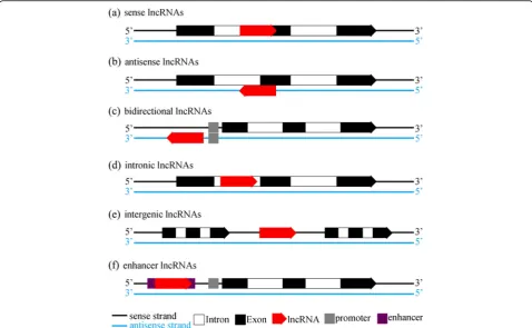
Classifications of lncRNAs
As a broad concept, lncRNAs encompass a few types of RNA transcripts. According to their location in the genome, lncRNAs can be classified into seven broad cat-egories as following: (a) sense lncRNAs, (b) antisense
lncRNAs, (c) bidirectional lncRNAs, (d) intronic
lncRNAs, (e) intergenic lncRNAs and (f ) enhancer
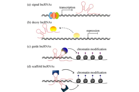
lncRNAs (illustrated in Fig. 1). When it comes to
con-crete functions, lncRNAs can be divided into four groups, namely, (a) signal lncRNAs, (b) decoy lncRNAs, (c) guide lncRNAs and (d) scaffold lncRNAs (described
in Fig. 2). LncRNAs can be found within the nuclear or
cytoplasmic fractions. Cytoplasmic lncRNAs can work as microRNA sponges or miRNA precursors to either re-duce or increase the expression and function of
micro-RNAs [4]. They can also recognize target mRNAs to
interact with the cellular translational machinery [4].
Nuclear lncRNAs exert its effects on chromatin
architec-ture either in acis-acting way (lncRNAs regulate the
ex-pression of neighboring genes) or in a trans-acting way
(lncRNAs regulate the expression of distant genes).
Besides, for some nuclear lncRNAs, it is not clear whether they function incisortrans[7].
Functions of lncRNAs
LncRNAs can regulate gene expression to affect many important physiological processes in multiple roles, to name only a few, as chromatin modifiers, X chromo-some inactivator, enhancers, transcriptional regulators and post-transcriptional regulators.
Chromatin modifiers
LncRNAs have been demonstrated to participate in chromatin modification in a critical way, which subse-quently affects multiple important biological processes
including neurogenesis and stem cell pluripotency [8].
LncRNAs regulate the state of chromatin by recruiting chromatin remodeling proteins to specific genomic loci. For instance, Hox genes are a class of homeotic genes related to the temporal and spatial developmental axes where hundreds of lncRNAs have been shown to be
critical [9]. One of these lncRNAs, HOTAIR (Hox
transcript antisense RNA) originates from HoxC locus and silences HoxD genes spanning over 40 kb by recruiting PRC2 (Polycomb repressive complex-2) in a trans-acting way, finally leading to a repressive
chro-matin state [9]. To be noted, PRC2 is a histone
meth-yltransferase required for epigenetic silencing and thereby an important chromatin modifying factor. Be-sides HOTAIR, thousands of RNAs can bind PRC2 in vivo, although this raises questions about binding spe-cificity and function in different chromatin contexts
[10]. Nevertheless, it has been a prototype for
lncRNAs interacting with PRC2 to alter the
chroma-tin state [11]. Other well-studied lncRNAs known to
bind PRC2 include Xist (X-inactive specific transcript)
[12], Kcnq1ot1 (KCNQ1 overlapping transcript 1)
[13], Braveheart [14], ANRASSF1 [15], etc. For
example, Kcnq1ot1 is a lncRNA acting as an
import-ant mediator for imprinting. The promoter of
Kcnq1ot1 maps to the ICRs (imprinting control re-gions) of Kcnq1 gene, which encodes a protein for a
voltage-gated potassium channel responsible for the
repolarization of the cardiac action potential.
Kcnq1ot1 interacts with Dnmt1 (DNA (cytosine-5)--methyltransferase 1) to establish the placental-specific
imprinting of genes within the Kcnq1 domain [16].
Besides, Kcnq1ot1 induces the methylation of histone H3 on lysine 9 and lysine 27 by recruiting the histone
methyltransferases G9a and PRC2 [13]. Xist is
re-quired for the X-inactivation process during early de-velopment in female mammals, as only one X chromosome will remain active. The other X chromo-somes expressing Xist will be coated with it and packaged into a transcriptionally-inactive
heterochro-matic structure [12]. In this process, Xist will recruit
a series of proteins including PRC2, SPEN, SAF-A (Scaffold Attachment Factor-A) and LBR to initiate
the X chromosome inactivation in cis [17], while
leaving the other X chromosome lacking Xist active
[12]. Another lncRNAs, Firre is also involved in the
X-inactivation process by anchoring the inactive X
chromosome to the position adjacent to the nucleolus
[17]. To sum up, nuclear lncRNAs exerts its effect on
chromatin state mostly through interaction with chro-matin modifying proteins.
Enhancers
A subset of lncRNAs are transcribed from active en-hancers and promote the expression of corresponding protein-coding genes in return, hence the name enhan-cer lncRNAs. In 2010, Kim et al. coined the concept of enhancer RNAs based on the phenomena that RNA polymerase II located to approximately 3000 activated enhancers and that RNAs could be produced from the extragenic enhancer regions of protein-coding genes
[18]. Almost at the same time, Shiekhattar lab reported
lncRNAs with enhancer-like functions. They
character-ized severalcis-acting lncRNAs using GENCODE
anno-tation of the human genome and discovered an RNA-dependent potentiation of gene expression
medi-ated by the ncRNA-a1–7 in particular [19]. Besides, the
Evf2 noncoding RNA, which originates from the Dlx-5/6 ultraconserved region, can interact with Dlx-2 to induce transcriptional enhancement of Dlx-2 in a target and
homeodomain-specific way [20]. The HSR1 (heat shock
RNA-1), which is constitutively expressed in human and rodent cells, works along with eEF1A and actively medi-ates the activation process of the HSF1 (heat-shock
tran-scription factor 1) [21]. Steroid receptor RNA activator
(SRA) also acts as a noncoding transcript to coactivate
steroid receptor [5]. Rosenfeld et al. also reported that
PRNCR1 and PCGEM1 bind to the AR (androgen recep-tor) and potently enhance the AR-mediated gene
activa-tion programs in prostate cancer cells [22]. However, so
far, the functional mechanism of enhancer lncRNAs have not yet been firmly established. More efforts need to be made into revealing the secrets of enhancer lncRNAs in various biological processes.
Transcriptional regulators
Transcriptional regulations of eukaryotic genes are achieved through many ways, including traditional direct interactions of proteins with DNA regulatory elements and, more recently identified, specific interactions be-tween RNAs, DNAs and/or proteins. Thus, lncRNAs are now known as an important facet of such transcriptional
regulations. Forcis-acting lncRNA, its genomic origin is
critical for its function, as it will alter the expression of protein-coding genes nearby. It may function through the transcription activity itself rather than the product: if the promoter of another gene lies in close proximity, it
may cause the collision between transcriptional
machineries on both genes, which is also dubbed “
tran-scriptional interference”. For example, active
transcrip-tion of lncRNA SRG1 will repress the transcriptranscrip-tion of its
downstream SER3 gene in yeast, because the 3′end of
SRG1 overlap with the SER3 promoter. If SRG1 tran-scription is prematurely terminated, the repression of
SER3 will be alleviated [2]. Also, in yeast, transcription
of some lncRNAs facilitates the accessibility of
protein-coding genes to RNA polymerases through alter-ing chromatin structure, such as promotalter-ing transcription initiation at the FBP1 (Fructose-1,6-bisphosphatase 1)
gene [23]. On the other hand, lncRNAs may also act in
trans, affecting transcription via its binding to transcrip-tion factors. For instance, the lncRNA 7SK binds to the elongation factor P-TEFb and downregulates its kinase activity in order to inhibit transcription elongation by Pol II [24].
Post-transcriptional regulators
LncRNAs exert post-transcriptional regulation mainly in two ways, splicing regulation and translational control. Firstly, lncRNAs can either bind to splicing factors in a competition manner or bind to mRNA itself through
base-pairing to block mRNA splicing. MALAT-1
(Metastasis-associated in lung adenocarcinoma
transcript-1) is an abundant ~ 7 kb lncRNA that inter-acts with the serine/arginine-rich (SR) splicing factors. It was suggested to regulate phosphorylation of SR pro-teins to modulate their distribution in nuclear speckles,
thereby affecting alternative splicing of pre-mRNAs [25].
MIAT (Myocardial infarction associated transcript) is another lncRNA containing a highly-conserved tandem repeats of UACUAAC, which has been characterized as a conserved intron branch point that can bind SF1 (spli-cing factor 1) with a higher affinity than the divergent branch point sequence, thus repressing splicing and the formation of spliceosomal complex on other transcripts
[26]. LUST (LUCA-15-specific transcript) is an antisense
transcript of RBM5 (RNA binding motif protein 5) and a sense-strand regulatory sequence of RBM5 in disguise, which is considered to modulate the expression of
RBM5 splicing variants [27]. Secondly, lncRNAs can
bind ribosome or translation factors to control protein translation. For example, snaR (small NF90-associated RNAs) and Gadd7 (growth arrested DNA-damage indu-cible gene 7) are two examples of translational control
by lncRNAs through binding to ribosomes [28]. On the
other hand, BC1 (Brain cytoplasmic RNA 1) and BC200 (200 nt brain cytoplasmic RNA), represent examples of translational repression by lncRNAs through binding to translation factors such as eI4FA (eukaryotic translation initiation factor 4A), PABP (poly (A)-binding protein)
and other factors [29]. Thirdly, some lncRNAs can be
related to both splicing and translation. One example is Zeb2NAT (Zeb2 natural antisense transcript), a ~ 1.2 kb
lncRNA overlapping the 5′ splicing site of an intron,
(zinc finger E-box binding homeobox 2). Zeb2NAT can repress the intron splicing and thereby promote Zeb2
translation [30]. Finally, lncRNAs can also function as
microRNA sponges or microRNA precursors to partici-pate in the post-transcriptional modulation. MicroRNAs are a class of small single-stranded RNAs without protein-coding capacity. MicroRNAs can repress mRNA translation or facilitate mRNA degradation via binding
to the 3′-UTR (3′ untranslated region) of their target
mRNAs. In that case, a few lncRNAs can alter mRNA levels through influencing corresponding microRNA levels. H19, a well-studied lncRNA, functions as a mo-lecular sponge to sequester let-7, while at the same time
serves as a precursor of miR-675-3p [31, 32]. LncRNAs
with similar functions have been reported and summa-rized in reference [33].
Research methods for lncRNAs
Generally, similar experimental procedures have been used for quantification and identification of lncRNA as coding transcripts although with some modification in downstream processing. LncRNA are often quantified together with mRNA using sequencing or microarray techniques, RNA-seq and -chip respectively, in the same biological samples. RNA-seq has the advantage to iden-tify novel RNA transcripts and developed rapidly in the last decades. In addition to the common application of next-generation sequencing (NGS), recent advances in
RNA-seq include single cell sequencing [34], single
mol-ecule sequencing [35], and in situ sequencing of fixed
tissue [36]. On the other hand, transcriptome microarray
is still in use and provides benefits as equally
well-developed data analyses with a lower stochastic vari-ability. Particularly in clinical studies, microarrays even outperform RNA-seq for standard analysis of gene expres-sions when it comes to reproducibility and cost [37].
In terms of functional analysis for lncRNAs, small interfering RNAs or antisense oligonucleotides for knocking down targeted lncRNA, and overexpression constructs to increase certain lncRNA expression levels are traditional methods to reveal their roles in vivo. In recent years, the revolutionizing CRISPR (clustered regularly interspaced short palindromic repeats) systems have been incorporated to maneuver the transcript level by either CRISPR activation or CRISPR inhibin
(CRIS-PRa/i) [38,39] or for genome editing of lncRNA locus of
interest. For nuclear lncRNAs, to study the associations between lncRNAs and chromatin, ChIRP (Chromatin
Isolation by RNA Purification) [40], CHART (capture
hybridization analysis of RNA targets) [41], RAP (RNA
antisense purification) [42] and GRID-seq (capture in
situ global RNA interactions with DNA by deep
sequen-cing) [43] are among the most advanced technologies to
identify the binding sites of lncRNAs genome-wide.
ChIRP, CHART and RAP can study only one known lncRNA, while GRID-seq provides global detection and analysis of RNA-chromatin interactions with high
speci-ficity and sensitivity [43]. To explore the interactions
between lncRNAs and proteins, RIP (RNA
immunopre-cipitation) [44] and CLIP (UV crosslinking and
immuno-precipitation) [45], iCLIP (individual-nucleotide
resolution CLIP) [46] can be utilized to capture the
lncRNA-binding proteins. Similar strategies can be ap-plied to cytoplasmic lncRNAs, which usually function as miRNA sponges or precursors. In addition, with accu-mulating studies of lncRNA, a number of databases have emerged over the past few years with a particular inter-est in curating lncRNAs, to name only a few,
NON-CODE [47], ChipBase [48], lncRNAdb [49], LNCipedia
[50] and LncRNADisease [51].
LncRNAs in renal diseases
There is an increasing number of evidences showing the important roles of lncRNAs in diverse human diseases. However, studies on lncRNAs in renal diseases are still in its infancy and mainly restricted to renal cancer.
There are very few reports on lncRNAs’ function in
other types of renal diseases, such as acute kidney injury, renal fibrosis, polycystic kidney disease, diabetic kidney disease, lupus nephritis and renal transplantation. Here, we discuss major researches published so far on lncRNAs in renal diseases.
Renal cancer
As lncRNAs play important roles in regulating major pathways in cell growth, proliferation, differentiation, apoptosis and survival, dysregulation of lncRNAs can promote tumorigenesis and progression of kidney can-cer, especially in renal cell carcinoma (RCC). For ex-ample, the oncogenic lncRNA HOTAIR promotes RCC tumorigenesis through AXL signaling by acting as a ceRNA (competing endogenous RNA) to sequester miR-217, a tumor suppressor, to facilitate HIF-1 (hypox-ia-inducible factor 1) expression and to upregulate AXL level. The lncRNA-MRCCAT1 (metastatic renal cell car-cinoma-associated transcript 1) is highly expressed in clear cell renal cell carcinoma (ccRCC) and promotes
metastatic properties of ccRCC [52]. Besides, the
upregulations of lncRNA-UCA1 (urothelial
carcin-oma associated 1) [53], lncRNA-ATB [54],
lncRNA-H19 [55] and lncRNA-FTX [56] are also
VHL-mutant RCC cell yet promoting the growth of
VHL-normal RCC cell [57]. Decreases of lncRNAs
such as NBAT-1 (neuroblastoma associated
transcript-1) [58] and CASC2 [59] (cancer
suscepti-bility candidate 2) are associated with poor prognosis in patients with RCC. To date, novel lncRNAs
con-tinue to be identified, such as lnc-BMP2–2,
lnc-CPN2–1, lnc-ACACA-1, lnc-FOXG1–2 and
lnc-TTC34–3, which were predicted by
computa-tional analyses to participate in RNA-protein inter-action networks including spliceosome and other
complexes in RCC [60]. Remarkably, it is reported
that intronic antisense lncRNAs are commonly
expressed in RCC tumors, the majority of which is evolutionarily conserved and possibly modulated by
epigenetic modifications [61]. Besides, a recent study
shows that lncRNA can also be packaged into exo-somes and function critically to promote the
pro-gress of renal carcinoma. Le et al. identified
lncARSR (lncRNA activated in RCC with sunitinib resistance), which acts as competing endogenous RNA for miR-34 and miR-449 to promote c-MET and AXL expression. In addition, lncARSR can be packaged into exosomes and transmitted to sensitive
cells to disseminate sunitinib resistance [62]. Although
studies are accumulating over the past decade, our
know-ledge of the underlying mechanisms of lncRNAs’ role in
renal malignancies is still rudimental, which needs more efforts to be made.
Acute kidney injury
Studies on lncRNAs in acute kidney injury (AKI) can be counted on fingers. One case is the hypoxia-responsive lncRNA GAS5, which is upregulated in renal ischemia/ reperfusion (I/R) injury along with the increased expres-sions of p53 and TSP-1 (thrombospondin 1), promoting
cell apoptosis in kidney [63]. Another case is the
lncRNA MALAT-1, which is highly expressed in ische-mic kidneys and plasma samples of patients with AKI, indicating a potential role of MALAT-1 in the induction
of AKI [64]. However, MALAT-1 was later reported to
be dispensable for renal I/R injury [65]. LncRNA-PRINS
(psoriasis susceptibility-related RNA gene induced by
stress) is induced by HIF-1α under hypoxia and shows
specific interaction with RANTES (regulated on activa-tion, normal T-cell expressed and secreted protein), which recruits circulating leukocytes and aggravates
kid-ney injury [66]. In addition, Lorenzen et al. identified a
novel intronic antisense lncRNA as an independent pre-dictor of mortality in critically Ill patients with AKI, dubbed TapSAKI (transcript predicting survival in AKI), which is enriched in tubular epithelial cells and
in-creased in plasma samples of AKI patients [67].
How-ever, it still remains elusive whether these circulating
lncRNAs are incorporated into exosomes and secreted into the circulating system.
Renal fibrosis / chronic kidney disease
The lncRNA np_5318 is a novel intronic lncRNA, which is located between the first and second exons of Erbb4 in mouse genome and thereby named as Erbb4-IR. Feng et al. reported that TGF-beta 1 can highly increase Erbb4-IR expression via a Smad3-dependent manner in the fibrotic kidney of mouse, suggesting that Erbb4-IR is a specific therapeutic target for chronic kidney disease
[68]. Wang et al. reported the lncRNA ZEB1-AS1 (zinc
finger E-box binding homeobox1-antisense RNA 1) exerted an anti-fibrotic role in diabetic nephropathy
[69]. Xie et al. reported lncRNA H19 overexpression
promoted renal fibrosis [70]. Chen et al. characterized
downregulation of an intergenic lncRNA LINC00963, which suppresses RIF (renal interstitial fibrosis) and OS (oxidative stress) of CRF (chronic renal failure) through activation of the FoxO (forkhead box O) signaling
pathway [71]. Zhou et al. demonstrated that lncRNA
HOTAIR participated in renal interstitial fibrosis through upregulating miR-124 to block Notch1 path-way [72].
Diabetic nephropathy
Hundreds of lncRNAs are deregulated in diabetic
ne-phropathy (DN) [73], which might participate in
patho-genesis of DN, according to a recent study using microarray analysis on lncRNAs in DN. However, only a handful of these deregulated lncRNAs have been dem-onstrated to affect proliferation and fibrosis in DN,
in-cluding CYP4B1-PS1–001 [74], ENSMUST00000147869
[75], NR_033515 [76], Dlx6os1 [77] and LINC00968
[78]. Yet, several lncRNAs can be potential therapeutic
targets for DN, such as MALAT1. Reducing MALAT1 levels can improve renal functions after duodenal-jejunal
bypass in diabetic rats [79]. Increasing expression of
lncRNA TUG1 (taurine upregulated gene 1) can attenu-ate podocyte apoptosis, alleviattenu-ate extracellular matrix
ac-cumulation and protect diabetic rats from DN [80, 81].
Besides, TUG1 was also reported to regulate mitochon-drial bioenergetics in DN [82].
Polycystic kidney disease / lupus nephritis / glomerulonephritis
There are few reports on lncRNAs in polycystic kidney disease, lupus nephritis or glomerulonephritis. One microarray study characterized thousands of lncRNAs in patients with IgA-negative MsPGN (mesangial
prolifera-tive glomerulonephritis) [83]. LncRNA Hoxb3os, which
regulates mTOR signaling, was found to be deregulated
in polycystic kidney disease [84]. The lncRNA NEAT1
characterized as a novel inflammatory mediator in hu-man lupus. Yet it is still unclear whether renal functions
in patients with lupus would be affected by NEAT1 [85].
Kidney transplantation
Even less studies have been carried out when it comes to lncRNAs in kidney transplantation. Chen et al. com-pared differential expressions of lncRNAs by microarray between control samples and samples from patients with acute rejection (AR) after renal transplantation, implying that lncRNAs might take part in the pathogenesis of AR
[86]. Lorenzen et al. detected lncRNAs in urine of
pa-tients with AR and identified urinary lncRNA
RP11-354P17.15–001 as a novel noninvasive biomarker
for renal AR [87]. Ge et al. identified two lncRNAs
(AF264622 and AB209021) from peripheral blood, which
can predict renal AR following transplantation [88].
Shang et al. screened lncRNA expression patterns in re-cipients with urothelial cancer after kidney transplant-ation and suggested that lncRNAs have critical roles in
UC carcinogenesis [89]. However, potential regulatory
mechanisms of lncRNAs in renal transplantation are
sel-domly explored. Only two studies on lncRNA-ATB [90]
and LncRNA-PRINS [91] did some initial research on
the underlying mechanisms involved in the rejection of renal allografts.
Future perspectives for the clinical use of lncRNAs in renal diseases
As a newly developed research field, lncRNAs rapidly
become a“hotspot” and provide new insights for
poten-tial clinical uses. On one hand, numerous lncRNAs have been discovered exhibiting specific expression patterns in various diseases, not only in tissues but also in body fluids including blood and urine. Thereby, lncRNAs could be developed for novel biomarkers to predict and supervise the progression of diseases. However, the quantity and stability of lncRNAs in the circulating and urinary systems make it hardly practical to use lncRNAs as non-invasive biomarkers with current detection limits. On the other hand, many lncRNAs have been demon-strated to directly regulate effector genes critical for the occurrence and development of diseases. From this standpoint, lncRNAs are also attractive therapeutic tar-gets. For example, utilizing antisense oligonucleotides (ASOs) to knockdown MALAT1 can result in slower tumor growth and a reduction in metastasis in mam-mary tumors, indicating MALAT1 ASOs might provide
a potential treatment for breast cancer [92]. In renal
diseases, the potential clinical application of lncRNA studies are similar to other human diseases, focusing on biomarkers and therapeutic targets, and may provide new insights into diagnosis and therapy of renal diseases.
Nevertheless, up to now, no clinical trials of lncRNAs have been documented.
Conclusion
We summarized lncRNAs and their research status in renal diseases in this review. Although lncRNAs studies have increased a lot over the past decade, they are still at the starting stage regarding to kidney diseases for the time being. More efforts need to be made to explore the potential mechanisms and significant roles of lncRNAs during the pathogenesis, diagnosis and treatment of renal diseases, which will expand our understanding of renal disease pathophysiology and drive new strategies for the diagnosis and treatment for renal diseases.
Abbreviations
3′-UTR:3′untranslated region; AKI: Acute kidney injury; AR: Acute rejection; AR: Androgen receptor; BC1: Brain cytoplasmic RNA 1; BC200: 200 nt brain cytoplasmic RNA; CASC2: Cancer susceptibility candidate 2; ccRCC: Clear cell renal cell carcinoma; ceRNA: Competing endogenous RNA; DN: Diabetic nephropathy; Dnmt1: DNA (cytosine-5)-methyltransferase 1; eI4FA: Eukaryotic translation initiation factor 4A; FBP1: Fructose-1,6-bisphosphatase 1; FoxO: Forkhead box O; Gadd7: Growth arrested DNA-damage inducible gene 7; HIF-1: Hypoxia-inducible factor 1; HOTAIR: Hox transcript antisense RNA; HSF1: Heat-shock transcription factor 1; HSR1: Heat shock RNA-1; I/ R: Ischemia/reperfusion; ICRs: Imprinting control regions; Kcnq1ot1: KCNQ1 overlapping transcript 1; LncRNA: Long noncoding RNA; LUST: LUCA-15-specific transcript; MALAT-1: Metastasis-associated in lung adenocarcinoma transcript-1; Mdm2: Mouse double minute 2 homolog; MIAT: Myocardial infarction associated transcript; miRNAs: microRNAs; MRCCAT1: Metastatic renal cell carcinoma-associated transcript 1; mRNA: Messenger RNA; MsPGN: Mesangial proliferative glomerulonephritis; NBAT-1: Neuroblastoma associated transcript-1; NEAT1: Nuclear Enriched Abundant Transcript 1; ORF: Open reading frame; OS: Oxidative stress; PABP: Poly (A)-binding protein; piRNAs: piwi-associated RNAs; PRC2: Polycomb repressive complex-2; PRINS: Psoriasis susceptibility-related RNA gene induced by stress;
RANTES: Regulated on activation, normal T-cell expressed and secreted; RBM5: RNA binding motif protein 5; RCC: Renal cell carcinoma; RIF: Renal interstitial fibrosis; SAF-A: Scaffold Attachment Factor-A; SARCC: Suppressing Androgen Receptor in Renal Cell Carcinoma; SF1: Splicing factor 1; snaR: Small NF90-associated RNAs; SRA: Steroid receptor RNA activator; TapSAKI: Transcript predicting survival in AKI; TSP-1: Thrombospondin 1; TUG1: Taurine upregulated gene 1; UCA1: Urothelial carcinoma associated 1; VHL: Von Hippel-Lindau; Xist: X-inactive specific transcript; ZEB1-AS1: Zinc finger E-box binding homeobox1-antisense RNA 1; Zeb2: Zinc finger E-box binding homeobox 2; Zeb2NAT: Zeb2 natural antisense transcript
Acknowledgements
Not applicable.
Funding
Supported by the Strategic Priority Research Program of the Chinese Academy of Sciences (XDA16010210).
Availability of data and materials
Not applicable.
Authors’contributions
ML designed and drafted the manuscript. JR contributed to revising the manuscript. Both authors read and approved the final manuscript.
Ethics approval and consent to participate
Not applicable.
Consent for publication
Competing interests
The authors declare that they have no competing interests.
Publisher’s Note
Springer Nature remains neutral with regard to jurisdictional claims in published maps and institutional affiliations.
Author details
1
State Key Laboratory of Pharmaceutical Biotechnology, NJU Advanced Institute for Life Sciences, Jiangsu Engineering Research Center for MicroRNA Biology and Biotechnology, Nanjing University, Nanjing, China.2Beijing Institute of Genomics, Chinese Academy of Sciences, Beijing, China.3Institute
for Stem Cell and Regeneration, Chinese Academy of Sciences, Beijing, China.
4University of Chinese Academy of Sciences, Beijing, China.5Cold Spring
Harbor Laboratory, Cold Spring Harbor, NY, USA.
Received: 15 October 2018 Accepted: 15 March 2019
References
1. Ezkurdia I, et al. Multiple evidence strands suggest that there may be as few as 19 000 human protein-coding genes. Hum Mol Genet. 2014; 23(22):5866–78.
2. Ponting CP, Oliver PL, Reik W. Evolution and functions of Long noncoding RNAs. Cell. 2009;136(4):629–41.
3. Okazaki Y, et al. Analysis of the mouse transcriptome based on functional annotation of 60,770 full-length cDNAs. Nature. 2002; 420(6915):563–73.
4. Mercer TR, Dinger ME, Mattick JS. Long non-coding RNAs: insights into functions. Nat Rev Genet. 2009;10(3):155–9.
5. Chooniedass-Kothari S, et al. The steroid receptor RNA activator is the first functional RNA encoding a protein. FEBS Lett. 2004;566(1–3):43–7. 6. Candeias MM, et al. p53 mrNA controls p53 activity by managing Mdm2
functions. Nat Cell Biol. 2008;10(9):1098–105.
7. Ma L, Bajic VB, Zhang Z. On the classification of long non-coding RNAs. RNA Biol. 2013;10(6):925–34.
8. Ng S, Johnson R, Stanton LW. Human long non-coding RNAs promote pluripotency and neuronal differentiation by association with chromatin modifiers and transcription factors. EMBO J. 2012;31(3):522–33. 9. Rinn JL, et al. Functional demarcation of active and silent chromatin
domains in human HOX loci by noncoding RNAs. Cell. 2007;129(7):1311–23. 10. Davidovich C, et al. Toward a consensus on the binding specificity and
promiscuity of PRC2 for RNA. Mol Cell. 2015;57(3):552–8.
11. Davidovich C, Cech TR. The recruitment of chromatin modifiers by long noncoding RNAs: lessons from PRC2. RNA. 2015;21(12):2007–22. 12. Zhao J, et al. Polycomb proteins targeted by a short repeat RNA to the
mouse X chromosome. Science. 2008;322(5902):750–6.
13. Pandey RR, et al. Kcnq1ot1 antisense noncoding RNA mediates lineage-specific transcriptional silencing through chromatin-level regulation. Mol Cell. 2008;32(2):232–46.
14. Klattenhoff CA, et al. Braveheart, a Long noncoding RNA required for cardiovascular lineage commitment. Cell. 2013;152(3):570–83.
15. Beckedorff FC, et al. The Intronic Long noncoding RNA ANRASSF1 recruits PRC2 to the RASSF1A promoter, reducing the expression of RASSF1A and increasing cell proliferation. PLoS Genet. 2013;9:e10037058.
16. Mohammad F, et al. Kcnq1ot1 noncoding RNA mediates transcriptional gene silencing by interacting with Dnmt1. Development. 2010;137(15): 2493–9.
17. Yang F, et al. The lncRNA firre anchors the inactive X chromosome to the nucleolus by binding CTCF and maintains H3K27me3 methylation. Genome Biol. 2015;16:52.
18. Kim T, et al. Widespread transcription at neuronal activity-regulated enhancers. Nature. 2010;465(7295):182–U65.
19. Orom UA, et al. Long noncoding RNAs with enhancer-like function in human cells. Cell. 2010;143(1):46–58.
20. Feng J, et al. The Evf-2 noncoding RNA is transcribed from the Dlx-5/6 ultraconserved region and functions as a Dlx-2 transcriptional coactivator. Genes Dev. 2006;20(11):1470–84.
21. Shamovsky I, et al. RNA-mediated response to heat shock in mammalian cells. Nature. 2006;440(7083):556–60.
22. Yang L, et al. lncRNA-dependent mechanisms of androgen-receptor-regulated gene activation programs. Nature. 2013;500(7464):598. 23. Hirota K, et al. Stepwise chromatin remodelling by a cascade of
transcription initiation of non-coding RNAs. Nature. 2008;456(7218):130–U15. 24. Yik J, et al. Inhibition of P-TEFb (CDK9/Cyclin T) kinase and RNA polymerase
II transcription by the coordinated actions of HEXIM1 and 7SK snRNA. Mol Cell. 2003;12(4):971–82.
25. Tripathi V, et al. The nuclear-retained noncoding RNA MALAT1 regulates alternative splicing by modulating SR splicing factor phosphorylation. Mol Cell. 2010;39(6):925–38.
26. Tsuiji H, et al. Competition between a noncoding exon and introns: Gomafu contains tandem UACUAAC repeats and associates with splicing factor-1. Genes Cells. 2011;16(5):479–90.
27. Rintala-Maki ND, Sutherland LC. Identification and characterisation of a novel antisense non-coding RNA from the RBM5 gene locus. Gene. 2009; 445(1–2):7–16.
28. Parrott AM, et al. The evolution and expression of the snaR family of small non-coding RNAs. Nucleic Acids Res. 2011;39(4):1485–500.
29. Lin D, et al. Translational control by a small RNA: dendritic BC1 RNA targets the eukaryotic initiation factor 4A helicase mechanism. Mol Cell Biol. 2008; 28(9):3008–19.
30. Beltran M, et al. A natural antisense transcript regulates Zeb2/Sip1 gene expression during Snail1-induced epithelial-mesenchymal transition. Genes Dev. 2008;22(6):756–69.
31. Kallen AN, et al. The imprinted H19 LncRNA antagonizes Let-7 MicroRNAs. Mol Cell. 2013;52(1):101–12.
32. Keniry A, et al. The H19 lincRNA is a developmental reservoir of miR-675 that suppresses growth and lgf1r. Nat Cell Biol. 2012;14(7):659–65. 33. Cai X, Cullen BR. The imprinted H19 noncoding RNA is a primary microRNA
precursor. RNA-a Publ RNA Soc. 2007;13(3):313–6.
34. Vitak SA, et al. Sequencing thousands of single-cell genomes with combinatorial indexing. Nat Methods. 2017;14(3):302.
35. Garalde DR, et al. Highly parallel direct RNA sequencing on an array of nanopores. Nat Methods. 2018;15(3):201–+.
36. Lee JH, et al. Highly multiplexed subcellular RNA sequencing in situ. Science. 2014;343(6177):1360–3.
37. Nazarov PV, et al. RNA sequencing and transcriptome arrays analyses show opposing results for alternative splicing in patient derived samples. BMC Genomics. 2017;18:443.
38. Zhu S, et al. Genome-scale deletion screening of human long non-coding RNAs using a paired-guide RNA CRISPR-Cas9 library. Nat Biotechnol. 2016; 34(12):1279–86.
39. Ho T, et al. Targeting non-coding RNAs with the CRISPR/Cas9 system in human cell lines. Nucleic Acids Res. 2015;43:e173.
40. Li W, et al. Functional roles of enhancer RNAs for oestrogen-dependent transcriptional activation. Nature. 2013;498(7455):516.
41. Davis CP, West JA. Purification of specific chromatin regions using oligonucleotides: capture hybridization analysis of RNA targets (CHART). Methods Mol Biol. 2015;1262:167–82.
42. Engreitz J, Lander ES, Guttman M. RNA antisense purification (RAP) for mapping RNA interactions with chromatin. Methods Mol Biol. 2015;1262: 183–97.
43. Li X, et al. GRID-seq reveals the global RNA-chromatin interactome. Nat Biotechnol. 2017;35(10):940–50.
44. Gagliardi M, Matarazzo MR. RIP: RNA Immunoprecipitation. Methods Mol Biol. 2016;1480:73–86.
45. Schaukowitch K, Joo JY, Kim TK. UV-RNA immunoprecipitation (UV-RIP) protocol in neurons. Methods Mol Biol. 2017;1468:33–8.
46. Huppertz I, et al. iCLIP: protein-RNA interactions at nucleotide resolution. Methods. 2014;65(3):274–87.
47. Xie C, et al. NONCODEv4: exploring the world of long non-coding RNA genes. Nucleic Acids Res. 2014;42(D1):D98–D103.
48. Zhou K, et al. ChIPBase v2.0: decoding transcriptional regulatory networks of non-coding RNAs and protein-coding genes from ChIP-seq data. Nucleic Acids Res. 2017;45(D1):D43–50.
49. Amaral PP, et al. lncRNAdb: a reference database for long noncoding RNAs. Nucleic Acids Res. 2011;391:D146–51.
50. Volders P, et al. LNCipedia: a database for annotated human lncRNA transcript sequences and structures. Nucleic Acids Res. 2013;41(D1):D246–51. 51. Chen G, et al. LncRNADisease: a database for long-non-coding
52. Li J, et al. Long noncoding RNA MRCCAT1 promotes metastasis of clear cell renal cell carcinoma via inhibiting NPR3 and activating p38-MAPK signaling. Mol Cancer. 2017;16:111.
53. Li Y, et al. Identification of long-non coding RNA UCA1 as an oncogene in renal cell carcinoma. Mol Med Rep. 2016;13(4):3326–34.
54. Xiong J, et al. High expression of long non-coding RNA lncRNA-ATB is correlated with metastases and promotes cell migration and invasion in renal cell carcinoma. Jpn J Clin Oncol. 2016;46(4):378–84.
55. Wang L, et al. Down-regulated long non-coding RNA H19 inhibits carcinogenesis of renal cell carcinoma. Neoplasma. 2015;62(3):412–8. 56. He X, et al. Knockdown of Long noncoding RNA FTX inhibits proliferation,
migration, and invasion in renal cell carcinoma cells. Oncol Res. 2017;25(2): 157–66.
57. Zhai W, et al. Differential regulation of LncRNA-SARCC suppresses VHL-mutant RCC cell proliferation yet promotes VHL-normal RCC cell proliferation via modulating androgen receptor/HIF-2 alpha/C-MYC axis under hypoxia. Oncogene. 2016;35(37):4866–80.
58. Xue S, et al. Decreased expression of long non-coding RNA NBAT-1 is associated with poor prognosis in patients with clear cell renal cell carcinoma. Int J Clin Exp Pathol. 2015;8(4):3765–74.
59. Cao Y, et al. Downregulation of lncRNA CASC2 by microRNA-21 increases the proliferation and migration of renal cell carcinoma cells. Mol Med Rep. 2016;14(1):1019–25.
60. Blondeau JJC, et al. Identification of novel long non-coding RNAs in clear cell renal cell carcinoma. Clin Epigenetics. 2015;7:10.
61. Fachel AA, et al. Expression analysis and in silico characterization of intronic long noncoding RNAs in renal cell carcinoma: emerging functional associations. Mol Cancer. 2013;12:140.
62. Qu L, et al. Exosome-transmitted lncARSR promotes Sunitinib resistance in renal cancer by acting as a competing endogenous RNA. Cancer Cell. 2016; 29(5):653–68.
63. Geng X, et al. The effect of long noncoding RNA GAS5 on apoptosis in renal ischemia/reperfusion injury. Nephrology (Carlton). 2018.https://doi. org/10.1111/nep.13476.
64. Lelli A, et al. Induction of long noncoding RNA MALAT1 in hypoxic mice. Hypoxia. 2015;3:45–52.
65. Koelling M, et al. Hypoxia-induced long non-coding RNA Malat1 is dispensable for renal ischemia/reperfusion-injury. Sci Rep. 2018;8:3438. 66. Yu T, et al. RANTES mediates kidney ischemia reperfusion injury through a
possible role of HIF-1 alpha and LncRNA PRINS. Sci Rep. 2016;6:18424. 67. Lorenzen JM, et al. Circulating long noncoding RNA TapSAKI is a predictor
of mortality in critically ill patients with acute kidney injury. Clin Chem. 2015;61(1):191–201.
68. Feng M, et al. TGF-beta mediates renal fibrosis via the Smad3-Erbb4-IR Long noncoding RNA Axis. Mol Ther. 2018;26(1):148–61.
69. Wang J, et al. IncRNA ZEB1-AS1 was suppressed by p53 for renal fibrosis in diabetic nephropathy. Mol Therapy-Nucleic Acids. 2018;12:741–50. 70. Xie H, et al. Long non-coding RNA-H19 antagonism protects against renal
fibrosis. Oncotarget. 2016;7(32):51473–81.
71. Chen W, et al. Effects of Long non-coding RNA LINC00963 on renal interstitial fibrosis and oxidative stress of rats with chronic renal failure via the Foxo signaling pathway. Cell Physiol Biochem. 2018;46(2):815–28. 72. Zhou H, et al. LncRNA HOTAIR promotes renal interstitial fibrosis by
regulating Notch1 pathway via the modulation of miR-124. Nephrology (Carlton, Vic). 2018.
73. Chen S, et al. Microarray analysis of long noncoding RNA expression patterns in diabetic nephropathy. J Diabetes Complicat. 2017;31(3):569–76. 74. Wang M, et al. A novel long non-coding RNA CYP4B1-PS1-001 regulates
proliferation and fibrosis in diabetic nephropathy. Mol Cell Endocrinol. 2016; 426(C):136–45.
75. Wang M, et al. Long non-coding RNA ENSMUST00000147869 protects mesangial cells from proliferation and fibrosis induced by diabetic nephropathy. Endocrine. 2016;54(1):81–92.
76. Gao J, et al. LncRNA-NR_033515 promotes proliferation, fibrogenesis and epithelial-to-mesenchymal transition by targeting miR-743b-5p in diabetic nephropathy. Biomed Pharmacother. 2018;106:543–52.
77. Cheng J, et al. Inhibition of lncRNA Dlx6os1 decreases cell proliferation and fibrosis and increases cell apoptosis in diabetic nephropathy. Int J Clin Exp Pathol. 2018;11(7):3302–9.
78. Li Z, et al. LncRNA LINC00968 accelerates the proliferation and fibrosis of diabetic nephropathy by epigenetically repressing p21 via recruiting EZH2. Biochem Biophys Res Commun. 2018;504(2):499–504.
79. Wu D, et al. Downregulation of lncRNA MALAT1 contributes to renal functional improvement after duodenal-jejunal bypass in a diabetic rat model. J Physiol Biochem. 2018;74(3):431–9.
80. Lei X, et al. Astragaloside IV/lncRNA-TUGI/TRAF5 signaling pathway participates in podocyte apoptosis of diabetic nephropathy rats. Drug Design Dev Ther. 2018;12:2785–93.
81. Duan L, et al. Long noncoding RNA TUG1 alleviates extracellular matrix accumulation via mediating microRNA-377 targeting of PPAR gamma in diabetic nephropathy. Biochem Biophys Res Commun. 2017;484(3):598–604. 82. Long J, et al. Long noncoding RNA Tug1 regulates mitochondrial
bioenergetics in diabetic nephropathy. J Clin Investig. 2016;126(11):4205–18. 83. Sui W, et al. Altered long non-coding RNA expression profile in patients
with IgA-negative mesangial proliferative glomerulonephritis. Int J Mol Med. 2012;30(1):173–8.
84. Aboudehen K, et al. Long noncoding RNA Hoxb3os is dysregulated in autosomal dominant polycystic kidney disease and regulates mTOR signaling. J Biol Chem. 2018;293(24):9388–98.
85. Zhang F, et al. Identification of the long noncoding RNA NEAT1 as a novel inflammatory regulator acting through MAPK pathway in human lupus. J Autoimmun. 2016;75:96–104.
86. Chen W, et al. Microarray analysis of long non-coding RNA expression in human acute rejection biopsy samples following renal transplantation. Mol Med Rep. 2014;10(4):2210–6.
87. Lorenzen JM, et al. Long noncoding RNAs in urine are detectable and may enable early detection of acute T cell-mediated rejection of renal allografts. Clin Chem. 2015;61(12):1505–14.
88. Ge Y, et al. A molecular signature of two Long non-coding RNAs in peripheral blood predicts acute renal allograft rejection. Cell Physiol Biochem. 2017;44(3):1213–23.
89. Shang D, et al. Profiling of mRNA and long non-coding RNA of urothelial cancer in recipients after renal transplantation. Tumor Biol. 2016;37(9): 12673–84.
90. Qiu J, et al. Transforming growth factor- activated long non-coding RNA ATB plays an important role in acute rejection of renal allografts and may impacts the postoperative pharmaceutical immunosuppression therapy. Nephrology. 2017;22(10):796–803.