ABSTRACT
TAMBE, NISARG MAHESH. Surface Modification Techniques for Polymeric Biomaterials for use as Tissue Engineering Scaffolds. (Under the direction of Dr. Martin. W. King and Dr. Ahmed El-Shafei)
In the case of biomaterials used as tissue engineering scaffolds, it is the surface that plays an
important role. On exposure to a biological environment, the extra cellular matrix (ECM)
proteins are non-specifically adsorbed onto the surface and the cells interact indirectly with
the surface through the adsorbed proteins. The cell-membrane receptors interact with
intermediary layers of adsorbed proteins. However, most of the polymeric biomaterials lack
the compatibility for cells as well as having poor cellular adhesion due to their hydrophobic
nature. So, it is important for the cells to have biomolecular recognition of the synthetic
polymeric surface in order to eliminate these disadvantages.
The main objective of this study was to harness surface bioactivation technologies to
fabricate porous tissue engineering scaffolds that would be biocompatible and support the
adhesion and proliferation of the cells. Nonwoven polylactic acid (PLA) webs from Ahlstrom
Nonwovens LLC were used as the substrate. PLA web was grafted with maleic acid in order
to functionalize the surface with carboxylic groups which react readily with most other
functional groups. The grafting was done by thermal initiation and plasma initiation methods
of polymerization. The grafting resulted in a strong 1705 cm-1 peak confirming the grafting of carboxylic acid groups without changing the bulk properties of PLA. The plasma initiated
grafting led to a contact angle of 28.2o while the thermal initiated grafting led to a contact
The immobilization of collagen on the functionalized PLA surface via genipin as a spacer
molecule resulted in better cell proliferation of human dermal fibroblast cells. The bioactive
coating was uniform in nature and had excellent coverage over the fibers. This supported the
hypothesis that the attachment of a bioactive coating on the surface improved cell viability of
the scaffold. This is the first report that genipin was used as a spacer molecule between
carboxylic group of maleic acid and amine group of collagen molecule. It is believed that
genipin helps reduce the steric problems between functional groups and large protein
molecules and enables immobilized peptide to move more freely in the biological
Surface Modification Techniques for Polymeric Biomaterials for use as Tissue Engineering Scaffolds
by
Nisarg Mahesh Tambe
A thesis submitted to the Graduate Faculty of North Carolina State University
in partial fulfillment of the requirements for the degree of
Master of Science
Textile Chemistry
Raleigh, North Carolina
2011
APPROVED BY:
_______________________________ ______________________________
Martin W. King Ahmed El-Shafei
Co-chair of Advisory Committee Co-chair of Advisory Committee
________________________________
DEDICATION
BIOGRAPHY
Nisarg Tambe was born on July 16th, 1987 in India. He received his Bachelor of Technology degree in Fiber and Textile Processing technology in May 2009 from Institute of Chemical
Technology, Mumbai, India (formerly known as UDCT). In pursuit of further studies, he
joined North Carolina State University, NC to start his master program in Textile Chemistry
in fall 2009. He expects to graduate in August 2011. Upon completion of his master degree,
he plans to continue expanding his research in biomaterials, polymers and gain a PhD degree
ACKNOWLEDGMENTS
First and foremost, I would like to express my sincere gratitude and appreciation to Dr.
Martin. W. King for giving me the opportunity to work with him and for providing his
continuous guidance and encouragement throughout the study. I also appreciate the support
given by the other member of my advisory committee, Dr. Ahmed El-Shafei. The discussions
with him and Dr. King together were invaluable. This work would not have been possible
without both of their vast knowledge, experience, enthusiasm and support. I would like to
thank Dr. Wendy Krause for agreeing to be on my committee and extending her support for
this project. I also wish to address special thanks to Dr. Susan Bernacki for allowing to use
her research facility and lending her expertise with cell cultures. Many thanks go to Dr.
Marian McCord and Dr. Julie Willoughby for letting me use their plasma lab and contact
angle instrument respectively.
A special thanks to Birgit Anderson, Jeff Krauss, Chuck Mooney, Chuanzhen Zhou (Elaine),
Fred Stevie and Valerie Knowlton for assisting and lending their special expertise in using
various lab equipments for research purposes. I should also thank National Textile Center
(NTC), North Carolina Translational and Clinical Sciences (NC TraCS) at University of
North Carolina, Chapel Hill and College of Textiles for providing financial support during
my graduate program.
I extend great appreciation to my lab mates Jin Di and Ting He for helping me out with the
possible without them. A special thanks to Rupesh Nawalakhe for helping me with the
plasma equipment and Michael Sieber for training me on the contact angle instrument. I also
would like to thank Dr. Sangwon Chung and Leslie Eadie for their initial help in
familiarizing with the biomedical textiles (BMT) laboratory and also all the other members
of the BMT group. A special thanks to Priya Malshe for helping me with statistical
calculations.
Last but not the least, I would like to express my sincere appreciation, love and gratitude to
my parents and grandparents back in India who continue to love, encourage and support me
throughout my life. Without their confidence and support in me over the years, I would not
have made it this far. I hope to succeed in life and make them proud. My gratitude also
extends to my great friends who have been such a big support and a source of encouragement
during the entire study.
TABLE OF CONTENTS
LIST OF TABLES…..………...x
LIST OF FIGURES……..………..xi
CHAPTER 1-INTRODUCTION…...………..……….1
1.1. Goals and Objectives.……..……...…………...…………...…...………...3
1.2. Limitations...….………...…..…...5
CHAPTER 2-REVIEW OF LITERATURE…………...………6
2.1 TISSUE ENGINEERING………...6
2.1.1. Definition………..………...……….6
2.1.2. Regenerative Medicine...10
2.2 TISSUE ENGINEERING SCAFFOLDS...15
2.2.1. Scaffold requirements...15
2.2.1.1. Biodegradability and Biocompatibility...16
2.2.1.2. Surface properties...19
2.2.1.3. Mechanical performance...20
2.2.1.4. Microstructure...22
2.3 SURFACE MODIFICATION TECHNIQUES...24
2.3.1. Non – thermal plasma technology...26
2.3.2. Free radical graft polymerization techniques...30
2.3.3. Living free radical graft polymerization techniques...35
2.4 IMMOBILIZATION OF BIOACTIVE MOLECULES...38
3.1. MATERIALS...42
3.2. METHODS...43
3.2.1. Surface functionalization...43
3.2.1.1. Plasma polymerization...43
3.2.1.2. Thermal initiated graft polymerization...45
3.2.2. Genipin attachment...47
3.2.3. Collagen immobilization...48
3.3 ANALYSIS...48
3.3.1. Surface Chemistry...48
3.3.1.1. Fourier Transform Infrared (FTIR) Spectroscopy...48
3.3.1.2. Contact angle...51
3.3.1.3. Dye Assay...53
3.3.1.4. X-ray Photoelectron Spectroscopy (XPS)...55
3.3.1.5. Time-of-flight Secondary Ion Mass Spectrometry (TOF-SIMS)...57
3.3.2. Mechanical Properties...60
3.3.2.1. Probe bursting strength...60
3.3.3. Surface Morphology...61
3.3.3.1. Scanning Electron Microscopy (SEM)...61
3.3.4. Cell Culture Study...62
3.3.4.1. MTT Assay...63
3.3.4.2. SEM Analysis...64
4.1. Surface Functionalization...66
4.1.1. Plasma initiated polymerization...66
4.1.1.1. Operating variables...68
4.1.1.2. Method variables...75
4.1.2. Thermal initiated polymerization...76
4.1.2.1. Material variables...77
4.1.2.2. Process variables...82
4.1.3. Dye Assay...83
4.2. Genipin attachment and collagen immobilization...89
4.2.1. X-ray Photoelectron Spectroscopy (XPS)...90
4.2.2. Time-of-flight Mass Spectrometry (TOF-SIMS)...95
4.3. Mechanical Properties...107
4.4. Surface Morphology and Fiber Diameter...108
4.5. Cell Culture Study...113
CHAPTER 5-CONCLUSIONS AND RECOMMENDATIONS...124
5.1. Conclusions...124
5.2. Future Work...125
REFERENCES...127
APPENDICES...137
A. Contact angle measurements...138
B. T-test results for bursting strength values...140
LIST OF TABLES
Table 2.1-Key industry parameters...12
Table 2.2-Structures in human use/clinical trials created using TE principle……….14
Table 3.1: Thermal properties of the PLA as provided by the supplier………..42
Table 4.1: Sample description...85
Table 4.2: Elemental composition for each of the treatment………..91
Table 4.3: O/C ratios and C=O % from XPS data for each treatment………....92
Table 4.4: Characteristic peaks of positive and negative spectrum of PLA control………....97
Table 4.5: Sample description and Mean corrected area ratio values………..………..102
Table 4.6: Fiber diameters (mean, standard deviation) measurements……….….110
Table 4.7: Description of four cell-cultured samples……….……....113
LIST OF FIGURES
Figure 2.1: Principle therapeutic strategies for treating tissues in patients………..8
Figure 2.2 – The tissue engineering triad………...9
Figure 2.3: Mesenchymal stem cells phenotype……….10
Figure 2.4: Apligraf® (left) and Vitrix® (right) by Organogenesis………..11
Figure 2.5: Growth of economic value of regenerative medicine………...13
Figure 2.6: Surface modification and immobilization of bioactive compounds……….25
Figure 2.7: Schematic of Apjet atm. pressure plasma reactor using in-situ mode…………..28
Figure 2.8: Pre-activation of substrate with plasma, monomer deposition and post irradiation for monomer fixation………...29
Figure 2.9: Graft polymerization reaction system using a free radical mechanism………….31
Figure 2.10: Flory’s model for a free radical mechanism reaction………..32
Figure 2.11: Kotaka model for graft polymerization………...………32
Figure 2.12: Reaction mechanism of ATRP………36
Figure 2.13: Schematic diagram of Type I collagen fibril structure………40
Figure 2.14: Chemical formula for Genipin………....41
Figure 3.1: PLA nonwoven web (left) and fiber (right) on arrival………..42
Figure 3.2: Contact angle measurements……….52
Figure 3.3 Basic Blue 3...54
Figure 3.4: Principle of XPS………55
Figure 3.5: Photoelectric emission and Auger emission………..56
Figure 4.1: FTIR spectra of grafted vs. untreated PLA……….…………..67
Figure 4.2: He treated vs. He-O2 plasma treated PLA……….69
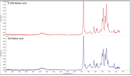
Figure 4.3: 0.25M vs. 1M monomer concentration……….70
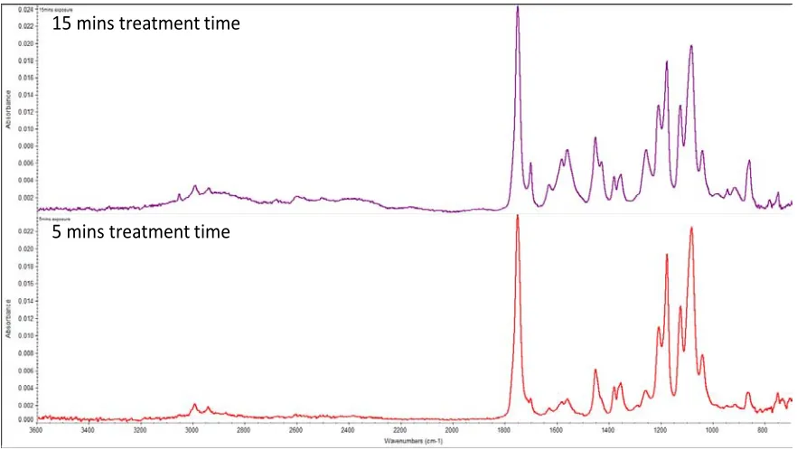
Figure 4.4: 5mins vs. 15mins exposure time………...71
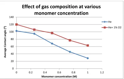
Figure 4.5: Effect of gas composition at various monomer concentrations………72
Figure 4.6: Effect of gas composition at various exposure times………73
Figure 4.7: Comparison of two methods of grafting………...75
Figure 4.8: FTIR spectra of grafted maleic acid vs. untreated PLA………77
Figure 4.9: Effect of initiator concentration on contact angle at 1M Maleic acid…………...78
Figure 4.10: Effect of monomer concentration on contact angle at 0.2M initiator………….79
Figure 4.11: Mohr’s salt vs. no salt addition………...80
Figure 4.12: Effect of Mohr’s salt concentration on the grafting………81
Figure 4.13: Calibration curve for Basic Blue 3...83
Figure 4.14: Absorbance vs. dyebath concentration for 1M MA grafted PLA...84
Figure 4.15: Dye concentration variation for different samples...85
Figure 4.16: Model fit on the dyeing behavior of MA grafted PLA...86
Figure 4.17: log (charge density) plot for various samples...87
Figure 4.18: Genipin attachment on maleic acid grafted PLA………89
Figure 4.19: Full spectra of (a). Untreated PLA (b). Collagen immobilized PLA…………..91
Figure 4.20: Deconvoluted C1s spectra of untreated PLA………..92
Figure 4.21: Deconvoluted N1s spectrum of a collagen immobilized sample……….94
Figure 4.23: (a) Positive mass spectra of maleic acid plasma grafted PLA and (b) genipin
attached on maleic acid grafted PLA………...98
Figure 4.24: Positive (a) and negative (b) spectra of collagen grafted PLA sample……….100
Figure 4.25: Stacked positive mass spectra of sample 2(top), 3(middle), 4 (bottom)……...103
Figure 4.26: Chemical mapping of collagen characteristic peak of the C4H8N+ ion……….104
Figure 4.27: Chemical mapping of the PLA characteristic peak of the C3H7O+ ion……….105
Figure 4.28: Chemical mapping images showing overlay of the collagen C4H8N+ ion image in
green over the PLA C3H7O+ ion image in red for Samples 2, 3, 4………....…106
Figure 4.29: Probe bursting strength values for the treated and untreated samples………..107
Figure 4.30: SEM photo micrographs of the untreated PLA sample……….109
Figure 4.31: SEM photo micrographs of maleic acid plasma grafted PLA sample………..110
Figure 4.32: SEM photo micrographs of the genipin treated PLA sample………111
Figure 4.33: SEM photo micrographs of the collagen immobilized PLA sample………….112
Figure 4.34: MTT assay results for the four samples listed in Table 4.7………..115
Figure 4.35: Untreated PLA sample in the medium (a): with cells (b): without cells……...119
Figure 4.36: Collagen treated PLA sample with a few cells attached………...120
Figure 4.37: Surface activated and collagen immobilized PLA sample………121
Figure 4.38: Surface activated PLA sample with no collagen………...122
Figure 4.39: Close up of fibroblast cell attachment on the immobilized collagen scaffold..123
CHAPTER 1-INTRODUCTION
Organ injury and organ failure constitute one of the major problems faced by patients around
the world. The viable options which are currently available to these patients are organ
transplantation and biotextile implants. Transplantation involves removing a living organ
from a donor’s site and implanting it into the patient’s body used as a replacement for the
injured or diseased organ. The various organs which can be transplanted with a limited
amount of success are the heart, kidney, lungs, liver, etc. The alternative approach is to
implant a permanent biotextile device, such as a heart valve, hernia repair mesh and
endovascular prosthesis. These implants invariably have a limited functional life span, which
leads to device failures, reoperations and the implantation of replacement devices.
However, transplantation is a clinically difficult and complex process with major
immunological challenges. There is always a danger of transplant rejection by the human
body where the body develops an immune response to the “foreign” transplant and causes
transplant failure. In many cases due to the advancements in drugs and medicinal chemistry
the problem can be minimized. However, the problem of dependence on the organ donor is
still prevalent. There is a huge paucity of donor organs required for transplantation leading to
long waiting lists and some malpractices. The World Health Organization (WHO) in its
report entitled “WHO guiding principles on human, cell, tissue and organ transplantation”
dated May 2010 has raised concerns over commercial trafficking in human organs,
particularly donors unrelated to the recipients. Such malpractices are risking human life and
In the USA, there are currently millions of people waiting for organ transplantation with the
annual healthcare costs for the treatment of these patients exceeding $500 billion3, 4. Such high costs and large numbers on the waiting lists have drawn the attention of scientists
throughout the world for an alternative therapeutic approach. Thus, there is a need to cater to
this severe shortage of viable organs for transplantation. This need is now driving growing
interest in the fields of regenerative medicine and tissue engineering for developing viable
clinical treatments for the replacement of diseased and injured tissues and organs. Tissue
engineering (TE) can play a pivotal part in providing an alternative to transplantation. The
development of an innovative, resorbable porous three dimensional (3-D) TE scaffold for use
in organ regeneration looks to be a promising option. Consequently, there is considerable
interest in studies investigating regenerative medicine. However, there are substantial
challenges to be faced by this approach of engineering viable organs. The scaffold should be
biocompatible and should possess sufficient strength and modulus to be able to withstand the
mechanical forces in the bioreactor and in vivo. It should also have sufficient mechanical
integrity and promote cell-cell and cell-matrix interactions. In natural tissues, an important
component is the extracellular matrix (ECM) which surrounds the cells and is essential to
replicate the cell’s native environment5. Since textile structures are porous and have the versatility to be engineered into a wide variety of specific 3D structural geometries, textile or
fibrous structures are ideal candidates for use as thick and multiple layered scaffolds for
regenerative medicine. For creating living tissues, various surface bioactivation technologies
The design of a textile scaffold material for the fabrication of engineered tissues first requires
the selection of an appropriate fiber forming polymer. The polymer should be resorbable,
possess suitable mechanical properties when spun into fibers and its degradation products
should be nontoxic. The hydrolyzable and biocompatible polymers and copolymers of
glycolic acid, lactic acid and ε-caprolactone have been extensively used over the past few years in the medical applications such as surgical sutures6. Polylactic acid (PLA) is a well known polymer which because of its slow rate of resorption has great potential for use as
scaffold material.
1.1. Goals and Objectives
The goal of this study is to harness surface bioactivation technologies to fabricate porous
tissue engineering scaffolds that will be biocompatible and support the adhesion and
proliferation of the cells for use in a wide range of TE and regenerative medicine
applications. The study will also determine the effect of applying different bioactive coatings
to the scaffold surface. The hypothesis behind the study is that incorporation of bioactive
coatings on the scaffold surface will mimic the cell’s native in vivo environment which will
be essential for the formation of natural tissues and an immunologically acceptable construct.
This can be achieved by two steps. Firstly, by chemically surface modifying or activating the
polymer surface and secondly by the application of a bioactive coating on the functionalized
The specific objectives of the study are summarized in the following statements which
includes comprehensive list of steps essential for the promotion of cell growth and
proliferation through the thickness of the porous textile structure.
1) To compare two polymer surface modification techniques, particularly compare
plasma initiated polymerization vs. thermal initiated polymerization to functionalize
the PLA surface to produce carboxylic groups
2) To determine and study the material and process variables for those techniques
a) For plasma initiated, determine optimum material variables(such as monomer
concentration, gas composition, exposure time) and process variables (such as effect
of post-plasma treatment)
b) For thermal initiated, determine optimum material variables(such as monomer
concentration, initiator concentration, effect of homopolymerization) and process
variables (such as curing time and temperature)
3) To identify a spacer molecule to attach any bioactive coating to the surface
4) To attach bioactive coatings onto the surface successfully
a) To study the uniformity of these bioactive coatings
5) To evaluate the relative cell viability of these coatings
6) To evaluate the mechanical properties and morphology of the scaffold
It is anticipated that by achieving these specific objectives, the ultimate goal of mimicking a
native cell environment in the TE scaffold which will be able to serve as a realistic and viable
1.2 Limitations
The use of radio frequency (RF) plasma during the initial stages of the project did not prove
to be a good choice. The system was often not running and offered very limited working time
while in running condition. The vaporizer for attachment of monomer was contaminated with
fluoro compounds which negated the purpose of grafting and led to improper surface
functionalization. Also, due to lack of time and skilled personnel, the cell proliferation and
viability experiments could not be continued. This influenced the results of the MTT assay
which required a substantial number and amount of specimens to minimize the variation
CHAPTER 2 – REVIEW OF LITERATURE 2.1 TISSUE ENGINEERING
2.1.1. Definition
The field of tissue engineering has been defined only since the mid-80’s with emphasis to
provide a firm scientific basis for therapeutic applications7. Langer and Vacanti have defined tissue engineering as “an interdisciplinary field of research that applies the principles of
engineering and life sciences towards the development of biological substitutes that restore,
maintain or enhance tissue and organs”10. The field has emerged from the fact that transplantation though being successful has severe constraints. There is a widespread need of
transplantable tissue and many patients have to face death while waiting for donor organs.
There are complications with transplantation such as a negative immune response which may
result in chronic rejection and loss of organ function over time. Thus, tissue engineering has
the potential for providing a permanent solution to the problem of organ failure which gives
it significant advantages over other prevailing therapies like drugs8. It is expected that this field will have a very broad impact in the future as engineered tissues will reduce the demand
for donor organs and will slowly eliminate the need for organ transplant operations.
Tissue engineering is a new hybridized method developed by combining cells, environmental
growth factors and biomaterial scaffolds to regenerate viable functional tissue for the
replacement or repair of injured or damaged organs9. It involves using the knowledge gained in the fields of embryology, cell biology, biochemistry, molecular biology for application in
used for the application of engineering principles to the living systems. Vacanti J and Vacanti
C stated that “we are in the midst of a biological renaissance and the interactions of the
various scientific disciplines can elucidate not only the potential direction of each field of
study, but also the right questions to address”7. In essence, each field needs to evolve and also continue to grow it’s interactions with other fields so as to address current prevalent
problem.
In one of his papers, Griffith lists the strategies for treating tissues by tissue engineering
principles11. There are three strategies for the treatment of diseased or injured tissue (Figure 2.1):
1. Implantation of freshly cultured or isolated cells as cellular replacements
2. Implanting biomaterials to serve as the extra cellular matrix and be capable of in situ
tissue regeneration
Figure 2.1: Principle therapeutic strategies for treating tissues in patients8
Cellular implantation involves implanting the patient or donor cells into the damaged tissue.
They can also be grown on a resorbable scaffold in vitro and implanted into the patient’s
body. In situ tissue regeneration involves implanting a biomaterial in a patient which as an
extracellular matrix (ECM) stimulates the body cells to proliferate and to repair the tissue.
The combined use of both cells and biomaterials involves growing the tissue on the scaffold
in a bioreactor outside the body and implanting the scaffold when it is developed, viable and
functional.
However, the modern approaches have evolved around the usage of both cells and
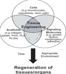
scaffolds do not appear to have the same potential as the combined approach12 and tissue engineering is now defined by the triad components which consist of the following: (Figure
2.2)
1. Cells - They are harvested and isolated from donor tissue
2. Scaffold - Biomaterials serve as the ECM substrates for the cells to attach, grow,
proliferate and differentiate as necessary prior to being implanted at the desired site in
the patient’s body
3. Growth factors – These signaling Biomolecules program the function of the cells and
promote cell adhesion, differentiation, proliferation and migration as required
2.1.2 Regenerative medicine
Within the past decade, many people have worked towards tissue repair or replacement and
demonstrated that the fundamental principles of tissue generation can be successfully applied
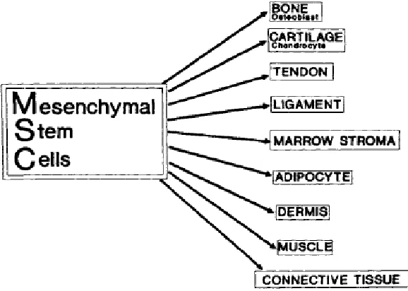
to the actual human therapy. Stem cells and particularly embryonic stem cells have been
successfully demonstrated to be applicable for human therapy. In an embryo, the
mesenchymal stem cell is a pluripotent progenitor cell and can differentiate and eventually
form a wide variety of different tissues. They also have the ability to migrate through the
arterial system and between tissue layers which is the key element for wound repair in
humans13. These mesenchymal stem cells are capable of differentiating through various transitions into different types of phenotypes as shown in Figure 2.3.
Figure 2.3: Mesenchymal stem cells phenotype
Thus by combining the elements of tissue engineering with the science of embryology and
range of scientific and clinical disciplines which are working towards the common goal of
the repair and replacement of cells, tissues and organs for human therapy14. It involves identifying and replicating the actions of cells, scaffolds or extra cellular matrix and growth
factors to stimulate the body’s repair mechanism and then implanting tissues or organs for
implantation in the body. Figure 2.4 shows Apligraf® from Organogenesis which was one of the first regenerative medicine products to be approved in USA for clinical trials. It was an
allogenic bilayered skin substitute for the treatment of venous stasis and diabetic ulcers. The
Vitrix® cellular dermal replacement is comprised of living human dermal cells and the dermal structural protein collagen which can be folded upon itself and inserted into deep
wounds.
Figure 2.4: Apligraf® (left) and Vitrix® (right) by Organogenesis15
It also involves the exploitation of advances in nanotechnology and material science.
William Hastine, CEO of Human Genome Sciences has stated that ““Living things are
engineered to subatomic physical tolerances, and nanotechnology will soon provide that
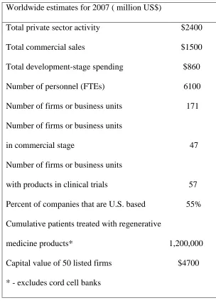
for cells, organs, and tissues that will integrate seamlessly with our natural ones”15. Table 2.1 provides a concise overview of the key parameters for the new global enterprise associated
with the therapeutics of regenerative medicine in 200716.
Table 2.1: Key industry parameters: TE, regenerative medicine and stem cell therapeutics
Worldwide estimates for 2007 ( million US$)
Total private sector activity $2400
Total commercial sales $1500
Total development-stage spending $860
Number of personnel (FTEs) 6100
Number of firms or business units 171
Number of firms or business units
in commercial stage 47
Number of firms or business units
with products in clinical trials 57
Percent of companies that are U.S. based 55%
Cumulative patients treated with regenerative
medicine products* 1,200,000
Capital value of 50 listed firms $4700
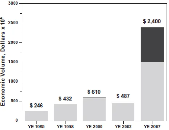
Thus, the concepts in tissue engineering and regenerative medicine increasingly clinically
relevant and both large firms and small new enterprises are committing increasing amounts
of research funding to develop new products17. This field has seen a major growth in economic value as shown in Figure 2.5 with the FierceBiotech daily expecting an explosive
growth to US $ 20 billion in the next 15 years.
Figure 2.5: Growth of economic value of regenerative medicine16 (light gray: spending by company in clinical stages; dark gray: sale by firms in commercial stage)
The commercialization of these concepts has been a major focus of many engineering and
clinical researchers so as to translate the bench-top research to patients as quickly as possible.
Table 2.2 provides a list of tissues which are either already approved for human use or are
currently in clinical trials and so will soon be available on the commercial market as reported
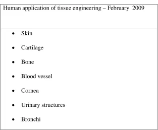
Table 2.2: Structures in human use or clinical trials created using tissue engineering
principles14
Human application of tissue engineering – February 2009
• Skin
• Cartilage
• Bone
• Blood vessel
• Cornea
• Urinary structures
• Bronchi
These achievements point towards an optimistic future for helping many patients with injured
or diseased tissues and/or who are waiting for organ transplantation. The addition of stem
cell technologies into the equation has brought about more options and generates possibilities
to solving the problem of finding viable tissues and organs for these. The field has become
far more diversified in recent years and is also a commercial stage enterprise with more than
60% of economic activity18, 19. While it is fair to say that the concepts have been overpromised and underdelivered, there is still considerable potential for this technology to
2.2 TISSUE ENGINEERING SCAFFOLDS
The outcome for any tissue engineered construct used in regenerative medicine is dictated by
the state of the extra cellular matrix that supports the cell culture, which in turn is controlled
by the various properties of the scaffold. Indeed the phenotype expressed by the cells
depends upon their microenvironment, which includes the biomechanical and biochemical
components of the scaffold, as well as the cell’s extracellular matrix (ECM) 20. The scaffold provides the three dimensional environment which brings the cells into close proximity and
permits them to develop into a thick tissue construct8. So, the principle for the design of the tissue engineering scaffold is to mimic the structure and biological function of the ECM as
closely as possible. The ECM, apart from providing the physical support for cells, provides a
substrate for cell adhesion, attachment and migration and allows for the retention of
differentiated cell functions22. It can also help in regulating cellular proliferation and function by the presence of various growth factors 21. A way to ensure the right three dimensional structure and appropriate matrix in the traditional tissue engineering approach is by seeding
cells onto and into the scaffold20.
2.2.1 Scaffold requirements
There are several design criteria which should be met for the production of tissue engineering
scaffolds23, 24, 25.
1. Biodegradable (or bioresorbable) with controlled rates of loss of strength and mass so
2. Suitable surface chemistry for effective cell adhesion, attachment, differentiation,
growth and proliferation.
3. Possess adequate mechanical properties so as to ensure the integrity of the construct,
to match intended site of application and to be easy to handle, manipulate and secure
at the implantation site
4. Three dimensional and highly porous with a network of open, interconnected pores
for cell-polymer interaction and tissue integration.
5. Good cytocompatibility (or biocompatibility) so as to not induce any adverse
inflammatory or immunological response
6. Ease of processing into various shapes and sizes
7. Ease of sterilization
2.2.1.1 Biodegradability and Biocompatibility
These two requirements are among seven criteria listed above responsible for the successful
design of tissue engineering scaffolds. The way which these requirements are met will vary
depending upon the type of material chosen for the scaffold as well as the particular tissue
engineering application22. The material as well as its degradation products should not provoke inflammation, not cause any cytotoxicity when implanted in vivo. The material
should resorb in a predictable manner because it may lead to chronic inflammation if it
have the potential for fulfilling the above requirements which has lead to their extensive use
for this purpose.
There are two types of biodegradable polymers: natural polymers, synthetic polymers26. Natural polymers such as chitin, chitosan, lignin, elastin, and collagen have been used
previously and have limited applications when used alone due to poor mechanical
performance as well as large batch to batch variation when isolated from biological tissue27. As a result many tissue engineering scaffolds used today are based on synthetic polymer such
as poly hydroxy esters, polyanhydrides, polyorthoesters and polyphosphazenes. These
polymers have a number of advantages over natural materials, such as their ability to tailor
make their mechanical properties and to be engineered to match the degradation rates with
that of regenerated tissue. They can be fabricated into various shapes and their surfaces can
be imparted tailored to give the desired chemical functionality so as to help or induce tissue
attachment and in-growth. The degradation of synthetic polymers is usually via a chemical
hydrolysis mechanism. This means that they are usually resistant to enzymatic activity which
is advantageous in avoiding changes in degradation rates from patient to patient28.
The biodegradability of a polymer depends upon the intrinsic properties of that polymer. The
intrinsic properties include its chemical structure, molecular weight, glass transition
temperature (if any), degree of crystallinity, chain orientation, the nature of hydrolytically
unstable bonds and also level of its hydrophilicity/hydrophobicity. There are other
the amount of stress and strain, the type and concentration of additives and the distribution of
chemical reactive groups within the matrix29, 30.
The most often utilized synthetic polymers are poly α-hydroxy esters such as polylactic acid
(PLA), polyglycolic acid (PGA), polycaprolactone (PCL) and their copolymers31. These polymers undergo hydrolytic degradation through esterification. On degradation, the
components of the monomer are water soluble and so are removed by natural pathways as
part of the human body’s regulatory mechanisms. For example, the degradation product of
PLA is lactic acid which is cleared through the tricarboxylic acid cycle32.
The biocompatibility of the scaffold is related to the inflammatory reaction and the immune
response on implantation. Whenever a patient undergoes a surgical procedure there will
inevitably be some tissue damage that leads to an inflammatory response and a subsequent
natural healing process. With the implantation of a resorbable scaffold, neither the scaffold
nor its degradation products should contribute to more severe, acute or prolonged
inflammatory response. This will depend upon the polymer synthesis, the method of
fabrication and surface modification, cleaning and sterilization techniques. For example, it is
important not to use any additives or solvents which have toxic residuals to ensure that the
2.2.1.2 Surface properties
The surface properties of the scaffold including its chemical nature as well as the topography
have certain specific requirements as it is in direct contact with the cells and the tissue. The
chemical properties of the material surface are responsible for the adsorption of proteins and
other biological molecules which regulate the cells surface activities such as cell adhesion,
migration and material-protein interactions34. The surface morphology affects the structural biocompatibility and the architecture and topography affect the cell activities.
The surface chemistry depends upon the type of material used as a tissue engineering
scaffold. The outermost functional groups are responsible for the binding of cells to the
material. When the material comes into contact with body fluids or the cell culture medium,
the initial response is the adsorption of proteins on the surface. The material then interacts
with the cells through this adsorbed protein layer35. Any changes in the surface chemistry can affect the cell behavior. For example, fluorapatite was incorporated onto the hydroxyapatite
surface which changed the orientation of adsorbed proteins and thus the cell attachment36. There have been various studies which show that the cells discriminate between different
chemistries. The surface energy also plays an important role as it makes the surface either
hydrophilic or hydrophobic. The surface charge provides the local environment with surface
tension as well as the energy of adhesion37. The hydrophilic/hydrophobic character of a surface is one of the initial parameter which affects the protein adsorption. An improved
surface hydrophilicity is important for hydrophobic materials to support cell adherence and
as compared to the hydrophobic surface38. The optimum surface hydrophilicity is a variable depending upon the cell type and the material used. Also, the non-specific relationship
between hydrophilic nature and cell activity found in vitro cannot be directly applied in vivo
because of complicated simultaneous interactions between body fluids, tissues and cells39, 40.
The surface topography or texture can also affect the level of cell activity. There have been
several studies which have shown that the percentage of cell attachment has changed with the
surface roughness. The rugosity of the surface can influence the nature of the host response
as cells are sensitive to specific changes occurring in the surface topography41. The cell response for the roughness depends upon the maturation state of the cells and the type of
morphology the cells assume on attachment. In some of the studies, it was found that the
mesenchymal cells form focal attachments on rougher surfaces while the cells spread on
smoother surfaces37.
2.2.1.3 Mechanical performance
The mechanical performance of a tissue engineering scaffold is important as it will provide
adequate tissue function. The polymeric scaffold should possess similar mechanical
properties to the engineered host tissue at the time of implantation in order to function
structurally42. Depending upon the tissue application, the mechanical performance desired will vary. The strength of biodegradable polymers is low compared to that of metals and
sufficient for most of the applications except possibly bone, cartilage and vascular tissue
engineering involving composite material strategies. These exceptions are generally
load-bearing tissues, and hence the scaffold should provide a high initial modulus, good
mechanical strength as well as resistance to deformation so as to withstand applied pressures.
This is more likely to lead to cell in-growth with the required functionality and support the
formation of an elaborate ECM.
The mechanical performance depends upon the type of polymer, its strength and elasticity, its
rate of degradation or resorption, its fabrication method to form the scaffold, its cellular
application and its resorption mechanism. The engineering construct has to withstand in vivo
stresses and physiological loadings such as compression, tensile loading and pulsatile flow45. Thus, the correct polymer molecular weight, fiber crystallinity and macroscopic scaffold
architecture need to be correct for effective tissue regeneration11. Polymer selection from a mechanical performance point of view depends upon one of two strategies. In the first
strategy one of which applies to load bearing scaffolds, the degradation rate of the material
must be selected so as to retain the scaffold’s strength until the tissue engineered implant is
fully remodeled and it can assume its structural role. In the second strategy, the resorption or
degradation rate of the polymer is adjusted so as to complement the cell proliferation and
extra cellular matrix secretion such that the polymer degrades completely leaving optimum
The method of scaffold fabrication also plays an important role in determining the
mechanical performance as well as porosity of the scaffold. Hutmacher DW mentions a
number of different fabrication technologies each with its own pros and cons24. The conventional techniques, such as solvent casting, leaching, melt molding and gas foaming,
have many limitations which are mentioned by KF Leong et al45. The modern solid freeform fabrication techniques have demonstrated better capabilities and improvements in structural
integrity and mechanical properties.
2.2.1.4 Microstructure
A high internal surface area to volume ratio of the scaffold microstructure is essential to
allow for cell attachment, cell proliferation and tissue in-growth. Creating a more porous
structure helps to increase the surface area as well as the mass transfer properties of the
scaffold47. The mass transfer properties are important for optimal diffusion of nutrients into the structure and waste products out of the construct. The surface area of porous materials
depends upon the pore diameter and its density42. A highly porous scaffold is desired to facilitate cell seeding and migration through the construct48. In one of the study, it showed that a porosity of 90% was an ideal porosity for tissue engineering scaffold. It allowed for
good diffusion and adequate surface area for cell-polymer interactions. However, sometimes
the mechanical properties at this porosity may be insufficient resulting in the mechanical
failure of the scaffold. To avoid this situation, it is necessary to optimize the porosity with
respect to the availability and transport of nutrients, and to match them with biomaterials
fabrication method determines the porosity and pore size distribution. Karande et. al. 49 discuss the various techniques that have been used by different researchers to achieve a range
of levels of porosity. The interconnectivity of the pores also plays an important role to
facilitate nutrient and waste exchange by those cells which are located deep within the
construct47.
An adequate pore size is also very important to provide for cell penetration, extracellular
matrix production and neovascularization of the internal space of the scaffold42. The optimum pore size differs depending upon the application. The macropores (> 50nm
<300µm) are required for cell and tissue penetration, and the micropores (<2nm), mesopores
(>2nm <50nm) permit solute diffusion50. Since the scaffold material is biodegradable, the pore size will increase over time, which is an important consideration when designing the
structure. Also, the shape and tortuosity of the pores can affect the extent and density of
tissue ingrowth.
The permeability and nutrient transport through a scaffold are important functional
characteristics of a scaffold. Permeability is a measure of the ease with which a fluid flows
through the structure. A high permeability means high diffusion which will facilitate good
inflow of nutrients and elimination of waste products. The permeability depends upon the
fluid-material interaction, total porosity, pore size connectivity and distribution. The nutrient
transport is mainly a function of diffusion which in turn is governed by the porosity and
2.3 SURFACE MODIFICATION TECHNIQUES
The most important properties required by biomaterials for tissue engineering and
regenerative medicine are biocompatibility and bioactivity. However, most of the synthetic
biodegradable polymers used for this application do not possess either of these particular
properties. They donot have the ability to support the culture of cells or to promote ECM
secretion. Most of the biochemical reactions in biology occur at surfaces making the study of
surfaces of crucial importance38. The work of Langmuir and Blodgett revolutionized the field of surface science and further research lead to the origin of “biointerface science” and
“surface engineering”. Thus, considerable efforts have been focused on surface engineering
of polymeric surfaces so as to promote their abilities to support cells, promote proliferation
and maintain the cell’s functionality.
The best way to change the surface chemistry of polymeric surfaces is to impart new
functionality so as to optimize cell-polymer interactions52. There are a range of different polar groups which can be grafted onto the polymer surface to improve its wettability and
hydrophilicity without changing its bulk properties. Examples of such polar groups include
carboxyl, carbonyl, hydroxyl, carboxylic acid, amine, aldehyde and thiol. These
functionalized polymer surfaces can then react covalently and immobile bioactive
compounds on their surface. The bioactive compound is generally tethered to the
functionalized surface via an intermediary compound such as a spacer molecule as shown in
Figure 2.6. The spacer molecule reduces the steric constraints of the functional groups and
sometimes to cause denaturation51. The chemical structure and functional groups also play an important role in determining the biocompatibility of the surface, which leads to the reason
why we study the structure-property relationship of biomaterials53.
Figure 2.6: Surface modification and immobilization of bioactive compounds via a spacer
molecule53
There are various methods which have been used successfully for the introduction of
functional groups onto polymer surfaces. Graft polymerization takes place by various
mechanisms depending upon the monomer used and the functionality imparted. There are
primarily four different types of mechanisms for graft polymerization:
1) Ionic mechanism
2) Coupling mechanism
3) Co-ordination mechanism
4) Free radical mechanism
Most of the known methods used for graft polymerization follow one of these mechanisms52. In the past the most commonly used methods have been physical methods like flame and
being replaced by many modern techniques like the use of non thermal plasma, irradiation
for free radical generation and living free radical polymerization methods, all of which are all
based on the free radical mechanism. Hoffman54 divides the methods for polymer surface modification into three types namely: physico-chemical methods, mechanical methods and
biological methods. On the other hand, Oehr55 divides them into physical, chemical and physico-chemical modifications. For surface modification in the medical field, a thin surface
layer with a thickness of about 10-100 nm is required. The layer should have strong
adherence onto the substrate and be readily sterilized. Thus, there is no specific
comprehensive classification for the surface modification of biomaterials, and as such the
different methods belonging to the different classes as outlined above.
2.3.1 Non-thermal Plasma Technology
Plasma is generally known as the distinct fourth state of matter which was first introduced by
Langmuir in 1922. It is a partially ionized gas which consists of highly reactive
multi-component physical and chemical species. It consists of charged particles (like electrons,
positive ions), excited atoms and molecules, radicals, active atoms and UV photons.
However, not all ionized gases are classified as plasmas. A plasma must have a dimension
more than the Debye length, which is the typical length to maintain neutrality over which
significant charge separation can occur56. The plasma produces a very high concentration of energetic and chemically active species even at low temperatures such as room temperature.
In order to reach the plasma state, the input of ionizing energy must be applied continuously
This continuous energy source can easily be provided by electricity which is also convenient
to handle. Radio Frequency (RF) discharges are amongst the most efficient electrical energy
inputs for the generation of plasma57. The use of atmospheric pressure for such applications is fast gaining popularity due to the several advantages it offers. The cost of the equipment is
reduced because no vacuum is required, and thus the material handling is simpler and faster.
The main advantage of atmospheric pressure plasma over other plasma techniques is that it
can provide higher throughput. It can be easily added onto a conventional production line and
become part of a continuous processing line for textiles and other materials58.
With recent advances in this technology, there are multiple processing parameters which can
affect the nature, extent and uniformity of the surface modification treatment. The following
independent parameters of the reactor like the electrode distance, the electrode size, the
monomer flow rate, the discharge power, the exposure time, the type of carrier gas and gas
flow can be varied for atmospheric pressure plasma systems59. Most atmospheric pressure plasmas use mainly noble gases like helium or argon to generate a stable discharge. Then a
small fraction of reactive gases is added to create active species in the plasma. The discharge
power affects the electron density of the reactive species which play an important role in the
surface modification process of the material. The electron density increases with an increase
in the discharge power. Also, the position of the material relative to the discharge is an
important parameter. There are two modes of applying the atmospheric pressure plasma
system; one is in-situ and the other is downstream. In the in-situ mode, the substrate is passed
interactions either on one or both sides with the various dynamic reactive species generated
from the discharge as shown in Figure 2.7. In the downstream mode, the substrate is passed
at a certain distance from the discharge, resulting in higher chemical selectivity and lower ion
bombardment and hence reduced surface damage. The in-situ mode is preferred as it requires
lower power consumption and less gas flow with the reactive species being formed nearer to
the substrate’s surface60.
Figure 2.7: Schematic of Apjet atmospheric pressure plasma reactor using one sided in-situ
mode 61
This non-thermal atmospheric pressure plasma technology can be useful for surface
modification of polymeric surfaces by combining it with conventional wet chemistry
techniques. Thus, plasma induced graft polymerization is a useful approach where the plasma
activates the polymer surface and the monomer in liquid or vapor phase is deposited and
redox or thermal). Alternatively, a post irradiation may be added as an additional step as
shown in Figure 2.852.
Figure 2.8: Pre-activation of substrate with plasma, monomer deposition and post irradiation
for monomer fixation52
In the vapor phase mode, the monomer is introduced into the plasma in the vapor phase and
is converted into reactive fragments which combine to form oligomer and polymer molecules
in the plasma. This type of polymerization occurs with most monomers regardless of their
structures, as there is no need for unsaturated bonds or cyclic structures to generate the free
radical polymerization species. As a result, the polymers formed have a different chemical
structure as well as physical properties from those synthesized by conventional
polymerization techniques. The polymer structures consist of complex units containing
cross-linked, fragmented and re-arranged monomer units unlike those made by conventional
vapor phase grafting tends to result in lower hompolymer formation largely due to the many
surface active sites as compared to solution phase grafting.
There are two commonly used grafting strategies with plasma that influence the structure and
morphology of the grafted polymer. They are referred to as “grafting to” and “grafting from”.
The “grafting to” strategy refers to the binding of the polymer to the surface while the
“grafting from” strategy refers to the initiation of the polymerization at the surface to grow
the polymer. The “grafting to” strategy has lower grafting density due to the short life of the
free radicals and the likelihood of side reactions. The “grafting from” offers a higher chain
density due to no strong hindrance for the monomer addition reaction in good solvent
conditions.
2.3.2 Free radical graft polymerization techniques
Graft polymerization reactions are characterized by the growth of polymer chains from the
surface of the substrate and are one of the universal, accessible and effective methods of
chemical modification. Free radical reactions were popularized in the late 1930’s when
researchers like Kharasch and Mayo were the first to study the kinetics and publish detailed
mechanisms involving free radicals. The free radical mechanism for grafting consumes
monomer units in the process of forming polymer chains from the surface reactive sites. It
generally takes place in aqueous media with a good yield of the final product62. It can be used for a wide range of unsaturated monomers having various physico-chemical properties
which undergo spontaneous free radical graft polymerization are ethylene derivatives
(CH2=CHX) and butadiene derivatives (CH2=CX-CH=CH2).
Figure 2.9: Graft polymerization reaction system using a free radical mechanism62
Many early researchers like Hofmann, Staudinger, Chalmers, and HS Taylor focused their
studies on confirming the reaction mechanism of free radicals. However it was not until 1937
that Flory developed a comprehensive model which describes the free radical mechanism as
shown in Figure 2.10. This model also matched the experimental results of Schulz et. al. who
studied the polymerization of styrene. By the 1940’s, this mechanism was widely accepted
for all free radical polymerization reaction systems and during the mid-40’s, many
companies like American Dow Chemical made extensive investigations to produce synthetic
rubbers using butadiene modified styrene monomer by this grafting process. Various other
Figure 2.10: Flory’s model for a free radical mechanism reaction65
Then in the 1960’s, many researchers started looking into the mechanism for grafting onto
the surface of a substrate via a free radical mechanism based on the Flory model as well as
Mayo’s work. One of the most efficient models was the one proposed by Kotaka in 1976
who studied the kinetics of grafting vinyl monomers onto polydiene64.
Kotaka proposed that decomposition of the initiator only results in the generation of free
radicals and that the termination reaction leads to either graft formation, cross-linking or
homopolymer formation. The live homopolymer chains can chemically bond to the surface
directly via a polymer grafting mechanism, which helps increase the grafting efficiency.
The polymerization reaction is initiated by a reactive species R* produced from an initiator I.
The reactive species can be a free radical, cation or anion which combines with a monomer
molecule by the opening of the π-bond to form a new radical. The radicals can be produced
by a variety of methods, namely by thermal, photochemical, redox, electrolytic, plasma or
sonication techniques. The initiator system should ideally be readily available, stable under
ambient conditions but should decompose and produce radicals at a known and predictable
rate in ambient polymer processing conditions. The thermal, hemolytic dissociation of
initiators is the most widely used mode for initiation65. The compounds having dissociation energies in the range of 100-170 KJ/mol can be used potentially as thermal initiators. For
example, the compounds containing the O-O, S-S, N-O bonds possess the desired range of
dissociation energies. Commonly used initiators include acyl peroxides, hydroperoxides,
persulfates and various other peroxy compounds. The redox initiation method uses
oxidation-reduction reactions to produce radicals that can also be initiated thermally. Commonly,
peroxides together with a reducing agent or a combination of various inorganic reductants
and oxidants are used for radical polymerization. The initiators used at elevated or lower than
ambient temperatures depend upon the rate of decomposition which is influenced by the
is expressed as the initiator half-life (t1/2) which is the time required for the concentration of
the initiator to be one half of its original concentration and is an important indicator of the
activity of the initiator66.
The propagation step has two different possible modes of addition of the primary radical
from the initiator to the double bond of the monomer. It can occur by either head or tail
addition. During the propagation step, many monomer species add onto the active centre
successively causing rapid formation of a polymer molecule. The process continues as a
chain reaction until the active centre is lost by termination or is transferred to another
molecule67. Many propagation reactions follow a single act of initiation. Each addition creates a new radical that has the same identity as the previous one but has an additional
monomer unit attached to it.
Radical 1
Radical 2
There are two possible points of attachment on mono- or disubstituted monomers for a
propagating radical. They can occur either on carbon 1 or carbon 2. The Radical 2 is shown
above, is more stable than Radical 1 due to resonance stabilization effects of the substituents.
Hence, the final polymer product will have an arrangement of monomer units in which
addition is the most predominant propagation method in vinyl monomer polymerizations.
The likelihood of head to head placements is less frequent due to steric and resonance
factors.
Termination is the step where the polymer chain stops growing and annihilation of the
radicals occurs by a bimolecular reaction between them. The two most common ways by
which the radicals can react with each other are by coupling and disproportionation as
represented below. In addition, there are also combinations of these two mechanisms.
The coupling reaction results in the formation of a single polymer molecule, and the
disproportionation reaction results in two polymer molecules, one saturated and the other
unsaturated caused due to the transfer of the hydrogen radical beta from one radical center to
the other68.
2.3.3 Living free radical graft polymerization techniques
The living radical process differs from the conventional chain radical polymerization
technique described above, as it does not involve any chain breaking reactions, which makes
it more desirable. The lifetime of the propagating radicals is very short for conventional
living polymerization, the bimolecular termination is minimized and the lifetime of the living
polymer is prolonged by the introduction of a dormant species during propagation by one of
two different modes of reaction, namely by reversible termination or by reversible transfer68. One of the most commonly used living radical polymerization techniques is atom transfer
radical polymerization, referred to as ATRP, which proceeds with a reversible termination
mode of reaction. An organic halide initiator undergoes a reversible redox process catalyzed
by a transition metal compound such as a cuprous halide. The ATRP mechanism is as
follows:
Figure 2.12: Reaction mechanism of ATRP69
The activation of the initiator which is generally an alkyl halide is an important step in the
reaction. The initiator with the transition metal ligand complex catalyst in the lower oxidation
state forms a dormant species which undergoes an electron transfer reaction with
simultaneous halogen abstraction and expansion of its coordinate sphere. It results in the
formation of the propagating radical R. and the metal complex in the higher oxidation state
complex reduces the steady state concentration of the propagating radical and so minimizes
the termination reaction of the polymers.
A wide variety of monomers can undergo polymerization using this ATRP mechanism which
requires a multi-component system of an initiator, an activator catalyst, a deactivator,
ligands, and solvent. ATRP is a complex reaction system with multiple components which
affect the interactions between the reagents that constitute its equilibrium. It requires both
fast and quantitative initiation, so that the propagating species can begin growing at the same
time, and also it needs rapid reversible deactivation of the propagating radicals. The initiator
reactivity should be matched with the monomer reactivity to achieve a higher concentration
of the dormant species. So, an alkyl halide is generally used as the initiator with the organic
group similar in structure to the propagating radical. The initiation reaction should be fast but
not too rapid as it may result in bimolecular termination. The halide selection is important
and depends on the reactivity of the halide. The fluorides are unreactive. The iodides result in
side reactions and the bromides are more reactive than the chlorides, leading to alkyl
bromides being the preferred initiator. The metal catalyst should possess two oxidation states
that can be achieved by simple electron transfer. It should also have an affinity for halogens,
possess an expandable coordination sphere to accommodate the halogen and should be able
to form a complex with the ligand. Copper has proven to be the most common transition
metal of choice for the catalyst irrespective of the monomer type. The ligand forms a
complex with the cuprous salt and helps to solubilize it in the organic reaction system. For
copper catalysts, multidentate nitrogen ligands are commonly used with bridged and cyclic
the polymerization rate varies with a change in temperature. Hence, there is always an
optimum temperature for any specific ATRP reaction system70.
The use of this ATRP technique for surface modification is fast gaining popularity due to the
progress in understanding this polymerization approach71,72,73. The surface-initiated ATRP allows the functionalization of well defined polymer brushes on the surface of different kinds
of substrates. It is able to provide control over the structural characteristics of the grafted
polymer brush, such as the thickness and density of the brush, its molecular weight and
narrow polydispersity as well as its functionality and mechanism of attachment to the
homopolymer’s surface. These advantages have increased the interest in living radical
polymerization techniques because of the control over these properties which is not possible
by using conventional free radical polymerization methods74. The chain growth behavior of almost all the chains is similar, which leads to a defect-free polymer brush on the surface.
Another unique characteristic of ATRP method is that the polymer chains synthesized by this
method preserve the dormant end groups throughout the polymerization reaction. Thus,
ATRP can be successfully used to covalently tether polymer brushes with predictable and
controlled properties to the surface of polymeric substrate for improved surface
functionalization75.
2.4 IMMOBILIZATION OF BIOACTIVE MOLECULES
In the case of biomaterials used as tissue engineering scaffolds it is the surface that plays an
non-specifically adsorbed onto the surface and the cells interact indirectly with the surface
through the adsorbed proteins. The cell-membrane receptors interact with intermediary layers
of adsorbed proteins. Most synthetic polymeric biomaterials lack the compatibility for cells
as well as having poor cellular adhesion due to their hydrophobic nature. So, it is important
for the cells to have biomolecular recognition of the synthetic polymeric surface in order to
eliminate these disadvantages76. This can be done by incorporating cell binding peptides onto the surface of the material via physical or chemical modification. The immobilization of
signaling peptides renders the surface attractive for cell adhesion so that the surface can
potentially mimic the various roles of the ECM proteins. These bioactive molecules induce
certain specific, predictable and controllable cellular responses from the cells adhering to the
surface.
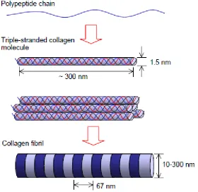
The most useful protein signaling domain commonly used is the Arg-Gly-Asp (RGD)
sequence which is present in most structural proteins such as fibronectin, collagen, and
laminin51. Among these, collagen is the most important biological macromolecule in extracellular matrix. It is the most abundant protein, accounting for about 30% of the total
amount of proteins formed in the human body. In addition, it is biodegradable,
non-immunogenic and specific in its interactions with cells like fibroblasts. Collagen is composed
of a family of nearly 20 related proteins which form triple helices through the three
polypeptide chains winding around each other in a rope like structure. These triple stranded
Figure 2.13: Schematic diagram of Type I collagen fibril structure77
A variety of methods exist to crosslink collagen so as to improve the scaffolds
cytocompatibility, biostability and reduce its rate of in vitro enzymatic degradation. The most
common physical crosslinking methods involve microwave energy, UV irradiation and
thermal drying treatments. However, the degree of crosslinking is difficult to predict and can
lead to denaturation of proteins, although there is no exposure to harmful chemicals. On the
other hand, chemical crosslinking is commonly performed using aldehydes, polyepoxy
compounds and carbodiimides, with glutaraldehyde being the most commonly used