IJOCR
ORIGINAL RESEARCH
Comparative Evaluation of Effect of Pre -Processing Surface
Treatment of Acrylic Teeth on Bonding With Denture Base
Resins”- An
In Vitro
Study
Srinivasa Gowda1, Lalit Kumar2, Komal Sehgal3, Virender Kumar4, G R Ramakrishna Maiya5, K Vidyachandra6,
Shetty Rajeet Sesappa7
ABSTRACT
Aim: To compare the effects of pre-processing mechanical treatment of ridge lap surface of acrylic teeth on bond strength between denture base resin and acrylic teeth.
Materials and Methods: Three groups of specimens with 72 sets of anterior teeth in each group were tested. Seventy two sets of each specimen were divided into three subgroups with 24 sets each for processing with three types of denture cur-ing techniques like heat cure, self cure and microwave cure techniques. The specimen teeth mounted on the metal former were processed as per manufacturer’s recommendations for each curing technique. All specimens were subjected to bond strength and tensile strength testing in Housefield universal testing machine.
Results: The analysis of variance test results indicated that there is a significant difference between the different groups influencing bond strength (P < 0.001) and between the different curing resins (P < 0.001). Multiple comparisons with post hoc test indicated that sand blast group showed higher bond strength. Grinding method showed higher bond strength com-pared to control group in heat cure resin and self-cure resin.
Conclusion: Within the limitation of this study, it is concluded that bond strength of acrylic tooth to denture base resin depends predominantly on surface treatment of ridge lap area of acrylic teeth positively.
Clinical Significance: The selection of compatible combina-tions of acrylic teeth with surface modificacombina-tions of ridge lap area and denture base resins may reduce the incidences of prosthesis failure due to acrylic teeth separation from resin denture base.
1Prosthodontist, 2,3Associate Professor, 4Assistant Professor, 5Senior Lecturer, 6,7Reader
1Department of Prosthodontics, Pune, Maharashtra, India 2-4Department of Prosthodontics, Dr Harvansh Singh Judge Institute of Dental Sciences and Hospital, Punjab University, Punjab, Chandigarh – 160 014, India
5-7Deparment of Oral and Maxillofacial Surgery, Srinivas Institute of Dental Sciences, Mukka, Mangalore, Karnataka, India
Corresponding Author: Srinivasa Gowda, Assistant Professor Department of Prosthodontics, AFMC-Pune, MUHS-Nashik, Pune, Maharashtra, India. Phone: +91-9049866969.
e-mail: [email protected]
Keywords: Bond strength, Heat cure, Microwave cure, Sand blasting, Tensile strength.
How to cite this article: Gowda S, Kumar L, Sehgal K, Kumar V, Maiya GRR, Vidyachandra K, Sesappa SR. Comparative Evaluation of Effect of Pre-Processing Surface Treatment of Acrylic Teeth on Bonding With Denture Base Resins”- An In Vitro Study. Int J Oral Care Res 2018;6(2):13-18.
Source of support: Nil
Conflicts of interest: None
INTRODUCTION
The most common reason for the elderly group of the population to seek dental treatment is for the replace-ment of missing teeth with either partial or complete removable prostheses.[1] Various materials and
tech-niques have been employed for the fabrication of remov-able prostheses. Acrylic denture base resins, introduced in 1937, have enjoyed a continued popularity, which is attributed to its simple processing technique and relative low cost of fabrication process.[1,2] Adequate bonding of
acrylic resin teeth to denture base resin is a critical factor because it enhances the fracture strength and longevity of the prosthesis since teeth are inseparable portion of the prosthesis.[1,2] The 33% of prostheses failure occurs
due to debonding of acrylic teeth from denture base resin. Such incidences are seen in implant - supported removable prostheses also. So such failures are consid-ered a major concern in removable prosthodontics.[1-3]
The acrylic resin tooth is made up of cross linked poly-methyl methacrylate. The monomer cross - linking may not be distributed equally in the denture tooth and the cervical ridge lap area is usually not as cross linked as the incisal portion of acrylic resin tooth.[4] The use of
mechanical retention is the most important means of achieving adequate bond between acrylic resin teeth and denture base resins. The retentive grooves enhances quantity and quality of the surface area in the ridge lap thereby promoting adequate bond strength.[5] Micro
mechanical or mechanical retention achieved by either macroscopic design or microscopic keying, has been shown to significantly affect acrylic tooth retention.[4,5]
oxide particles against the material surface intended for bonding under high pressure. This technique has been used to produce micro roughened surface for orthodon-tic brackets and bands as well as in porcelain-bonded to metal surfaces. Sand blasting of the acrylic resin teeth has been reported to be effective in enhancing the bond-ing between denture tooth and the denture base resin.[6]
However, debonding of acrylic teeth in denture base remains a major problem in removable prosthodontics despite advances in materials science and techniques. It has been estimated that 30% of denture repairs involve tooth debonding in the anterior region of removable prostheses.[1,4,5] This debonding may be attributed to a
lesser ridge lap surface areas available for bonding and the direction of the stresses encountered during func-tion.[6,7] Two factors affect the achievement of adequate
bonding between acrylic teeth and denture base resin. Firstly, the polymerizing denture base resin must come into physical contact with the denture tooth resin, sec-ondly the polymer network of denture base resin must react with the acrylic tooth polymer to form inter woven polymer network.[1,5-7] Debonding may be the result of
incompatible surface conditions during acrylic tooth and denture base resin interaction.[8] The factors that
contribute to this discrepancy are contamination of the joining surfaces and differences in structural compo-nents because of their different processing routes.[7,8]
The maximum stress concentration in dentures occur at the beginning of palatal aspect of tooth- denture interface. Stress concentrations of 74–90 MPa occur at the interface which is in excess of the recommendations by the national standards for adhesive bond strength (American National standard- 31 MPa and Australian National Standard –32MPa).[6] Recently, there has been
an increase in the use of implant supported removable prostheses. This has not only increased biting forces with such removable prostheses but also increased the mechanical failure of the prostheses. Inadequate thick-ness of acrylic resin in the anterior segment of a den-ture as a result of the dimensions of bar and clip attach-ments can also lead to fracture of the denture and teeth debonding from the base.[9] However there are limited
studies or literature which prove the effectiveness of the sandblasting or grinding in ridge lap area of acrylic teeth on the bond strength between the acrylic teeth and denture base resins cured by various methods. With this thing in mind, this study was designed to evaluate the effects of mechanical surface treatments of ridge lap area of acrylic resin teeth on bonding with denture base resins cured by various methods.
Materials used in the present study included solvent resistant cross linked acrylic teeth (Ivostar, Ivoclar Vivadent, Schaan, Liechtenstein), Heat Cure acrylic resin (Trevalon ,(Dentsply India Pvt., Ltd), Acron Acrylic resin for microwave polymerization (Acron MC, GC Corporation Japan), Self Cure acrylic resin (RRTM, Dentsply India Pvt., Ltd.), Dental Plaster (Kalabhai Karson Private Limited Mumbai, Maharashtra, India), Modeling wax (DPI, Mumbai. Maharashtra, India), Cold Mould Seal (DPI, Mumbai, Maharashtra, India), tung-sten steel burs #1508 (Edenta, Schweiz, Switzerland), Sand paper (No. 80, 100 and 120), Rubber wheels (fine and very fine), Universal polishing paste (Ivoclar), Polishing cake (Bego-Germany), Pumice (Micro white, Asian chemicals), Gold Rouge (Bego-Germany) and Distilled water.
Methodology
Solvent resistant cross linked acrylic teeth from canine to canine of maxillary arch were used in this study. Total of 216 acrylic maxillary anterior teeth from canine to canine were used. The metal formers of dimension 70 mm × 25 mm × 7 mm (the design same as used in the American National Standard⁄American Dental Association (ANSI/ADA) Specification No 15) which incorporates a trough of 5 mm width and 1.5 mm depth for mounting the teeth was used in this study. To keep uniform surface for bonding, a silicone (Flexceed, GC Tokyo; Japan) positioning device with an open win-dow (5 mm × 5 mm) was used (Figure 1). Seventy two sets of acrylic teeth were treated by sandblasting of ridge lap surface of 25 mm2 using the silicone
position-ing device (Figure 1). Sandblastposition-ing was done in sand-blaster unit (Type 5417-Kavo EWL, Germany) with alu-minium oxide particles (Bego, Germany) of 250 µ size under 5 kg cm−2 of pressure for 5 s. 72 sets of acrylic
teeth were treated by grinding over the ridge lap surface of 25 mm2 using silicone positioning device. Grinding
Effect of surface treatment of acrylic teeth IJOCR
cure technique. Total 24 sets of teeth were used for each processing technique (Figure 2). Six anterior teeth were mounted and sealed with the metal former using modelling wax (Figure 2). Cervical neck portion of the teeth was completely devoid of wax. The incisal 3rd of
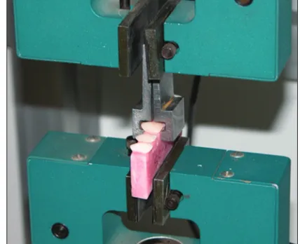
the lingual surface was projected out of metal former completely (Figure 2). The flasking of sealed metal for-mer was completed in split flask (Varsity flask, Jabbar & Co, New Delhi; India) for heat curing and special flask for microwave curing (VIPI-STG-Flask, American Tooth Industries, California, USA). Dewaxing was com-pleted by immersing and washing in boiling water for 10–16 min after the setting of the plaster and the metal former was removed carefully without moving the teeth. The plaster surfaces in the flask coated with sep-arating medium were allowed to dry. The polymethyl methacrylate resins compatible with self curing, heat curing and micro wave curing technique were packed as per manufacturer’s recommendations. Specimens of microwave curing were cured in oven (LG - model Number 1927C, Max 800 W with the frequency of 2450 MHz) for 4 min at 500 W and bench-cooled for 03 h before deflasking. Similarly, heat-polymerising spec-imens were cured in acryliser (Unident Instruments Private Limited, New Delhi; India) for 9 hr at 74°C and bench-cooled for 03 h before deflasking. Auto polymer-ising specimens were cured in room temperature as per manufacturer’s recommendations and bench cooled for 03 h. Deflasking was completed with removal of adher-ing plaster. Specimens were finished and polished. Flat polished specimens were stored in distilled water in 37°C for 09 days before testing. All specimens were sub-jected to tensile force and dislodgement force till failure in universal testing machine (Star Testing System, India) (Figure 3). The testing machine used a direct pull on the incisal portion of the lingual surface in a labial direc-tion at a height above the denture base resin bar with a crosshead speed of 5 mm min−1 (Figure 3). Metal jig
was fabricated to hold the specimen during application of load. The loads at failure and fracture were recorded and calculated to units of stress according to the original bonding area, i.e.,
SW A
S= bond strength in MegaPascals (MPa). W= load in Newton.
A= surface area i.e., (5 mm × 5 mm).
The data obtained were compiled on Microsoft excel sheet. Statistical Package for the Social Sciences ver-sion 17.0 (SPSS Inc., Chicago, Illinois, USA) software was used for statistical analysis. One way analysis of variance (ANOVA) test was used to analyse the tensile
and bond strengths for each of the processing tech-niques. to find out the significant difference with respect to the mean bond strengths and mean tensile strengths among pair of groups and processing techniques, mul-tiple comparisons with post-hoc test using Bonferroni method was carried out.
RESULTS
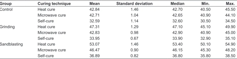
The mean, standard deviation and median of bond strengths recorded during different surface treatments and various curing processes are listed in Table 1. The ANOVA test results indicated that there is a significant difference between the different groups influencing bond Figure 1: Silicone positioning device
Figure 2: Teeth wax-up in the metal former
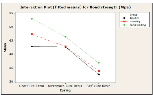
strength (P < 0.001) and between the different curing res-ins (P < 0.001). The interaction (joint effect) of group and curing on bond strength is also found to be significant (P < 0.001). The ANOVA test results indicated that sur-face treatment has a significant difference between the different groups influencing tensile strength (P < 0.001) and also, there is a significant difference between the dif-ferent curings (P < 0.001). The interaction (joint effect) of groups and curing techniques on tensile strength is also found to be significant (P < 0.001). In order to find out among which pair of groups and curing there exists a significant difference with respect to the mean bond strength and mean tensile strength, multiple compar-isons with - post-hoc test using Bonferroni method was done. The results are given as interaction plot for bond strengths in Figure 4. It was noticed that sand blast-ing method group showed higher bond strength in all the types of curing compared to the other two groups. Grinding method group showed higher bond strength compared to control group in heat curing and self curing. But the mean bond strength recorded in microwave cur-ing was almost same for grindcur-ing method group as well as control group. Highest bond strength was recorded in sandblasting method group with heat curing. The sand blasting method group showed lower tensile strength compared to the other two groups with heat curing. Whereas, it showed higher tensile strength with micro-wave curing compared to the other two groups. In heat curing, grinding method group showed higher mean tensile strength followed by control group and sand blasting method group respectively. Grinding method group always showed higher tensile strength compare to control group in different curing techniques. The lowest tensile strength was recorded in control group with self curing whereas the highest tensile strength was recorded in sand blasting method group with microwave curing.
DISCUSSION
The loss of teeth is a matter of great concern to the major-ity of people and their replacement by artificial substi-tutes such as dentures, is vital to lead normal life. One
of the problems encountered in provision of denture is whether the limitations of strength of such prosthesis meets functional demands of oral cavity.[10] Polymethyl
methacrylate despite being most commonly employed in construction of dentures is far from ideal in fulfilling mechanical requirements of such prosthesis.[10,11] There
is a wide variation in materials tested and the method-ology used for constructing and testing the samples for bond strength.[12] Contamination with wax seems to be
the major cause for bond failure between teeth and den-ture base resins.[1,12] Contamination with tin foil
substi-tute reduced the bond strength values in some studies whereas there was no decrease in one study. Application of adhesive bonding agents or chemicals like dichloro-methane to acrylic teeth has demonstrated an improve-ment in the bond strength values.[13,14] The application of
monomer before packing the resin, decreased the bond strength in two studies but there was a definite improve-ment according to other studies.[2,13] Modification of the
ridge lap area of the acrylic resin teeth demonstrated an increase in bond strength, whereas some other studies showed no obvious advantage.[15,16] There was no
differ-ence in the bond strengths in hydrated or unhydrated specimens and thermocycling was found to reduce the bond strength.[14,17,18] The present study was carried out
to compare the effect of pre-processing surface treatment such as sand blasting and grinding of ridge lap area of acrylic teeth on bond strength and tensile strength between acrylic teeth and denture base resin cured with different techniques. This study hypothesized that surface treatments in the ridge lap area of acrylic teeth would provide maximum withholding strength against separation of acrylic teeth from denture foundation. This hypothesis was accepted, as the results demonstrated, that the control group obtained lower bond strength values than the sandblasting and grinding surface treat-ment groups. The sandblasting surface treattreat-ment group with either heat or microwave-polymerization showed significantly higher bond strength and tensile strength values. All bond strength results values were in harmony with the criteria in ISO-3336 of 31 MPa for bond strength
Microwave cure 42.71 1.04 42.65 40.90 44.10
Self-cure 32.59 1.14 32.60 30.50 34.50
Grinding Heat cure 47.31 1.29 47.10 45.10 49.90
Microwave cure 42.83 0.98 42.90 40.90 45.00
Self-cure 33.95 0.67 33.90 32.90 35.10
Sandblasting Heat cure 53.07 1.46 53.40 50.10 54.90
Microwave cure 46.47 0.90 46.15 45.30 48.20
Effect of surface treatment of acrylic teeth IJOCR
test.[4,7,14] In fact, the free surface energy of the resin
sur-face treated by sandblasting with Al2O3 is undoubtedly higher than that of the untreated surface, which may be a reason why sandblasting improves bonding. Cohesive failures that were found may be due to enhanced poly-mer network formed by cross linking agent of the mono-mer between the teeth and resin base.[4,7,14] The type of
failure also needs to be verified because fractures may occur in the denture base or tooth before occurring at the interface between tooth and denture base. This implies that the fracture load has some degrees of relationship to fracture strength of acrylic tooth. Regression analy-sis of relation between bond strength and failure mode indicated that bond strength is directly proportional to cohesive failure of denture base.[4]
Within the limitations of this in vitro study under conditions according to the ANSI/ADA Specification No. 15, the acrylic tooth and denture base resin bond is considered satisfactorily strong if separation does not occur at acrylic tooth-denture base interface and if the tooth remains strongly bonded to the denture base. Using this criteria, the specimens tested in this study cured with either heat, or microwave polymerization bonded satisfactorily. Nevertheless, the microwave based sub-groups exhibited lower bond strengths, indicating that the method of polymerization influenced tooth-to-base bond strength. In fact, microwave curing was reported to have unfavourable intrinsic temperature rise of acrylic monomer while curing resulting in the formation of inter-nal voids.[4,7] Thus, increased number of internal defects
reduce denture base strength in microwave cured resin in comparision to heat cured resin. The thickness of the test specimen bar used in this study revealed that forma-tion of internal voids or bubbles is an explanaforma-tion for the bond failure after microwave polymerization. This find-ing is of clinical importance because the thickness of the denture base material in the tooth-bearing areas usually promotes voids formation.
The limitations of this study are that the effect of quality of acrylic teeth or denture base resins, effect of
chewing or masticatory forces, thermocycling effects were not considered and investigated. Most signifi-cantly, the study has to be verified in clinical scenario wherein the effect of masticatory forces and cyclic loads on bond strength can be analysed. It is suggested that future lab and clinical studies based on relation between shear strength, fatigue strength, impact strength of den-ture base resins and acrylic teeth materials with new technology like interpenetrating polymer network may be carried out to substantiate the data.
CONCLUSION
Within the limitation of this study it is concluded that bond strength of acrylic tooth to denture base depends predominantly on surface treatment of ridge lap area of acrylic teeth positively. The inherent strength of resin denture base material also contributes to bond strength to some extent. But these facts have to be substantiated by evidence based large sample multi centre clinical studies with longer follow up period.
Clinical Significance
Commercially, vast numbers of teeth and denture base resins are available for denture construction. However there is very little or no mention of bond strength or compatibility of acrylic teeth to the denture base resins by the manufacturers. The selection of more compatible combinations of acrylic teeth and denture base resins may reduce the incidences of acrylic teeth separation from resin denture base leading to prosthesis failure.
REFERRENCES
1. Patil SB, Naveen BH, Patil NP. Bonding acrylic teeth to acrylic denture bases. J Gerodontol 2006;23:131-9.
2. Cunningham JL. Shear bond strength of resin teeth to heat-cured and light-heat-cured denture base resin. J Oral Rehab 2000;27:312-6.
3. Rosangela SS, Karin NH, Joao FN. Factors affecting the strength of denture repairs. J Prosthodont 2007;16:302-10. 4. Chung KH, Chung CY, Chung CY, Chan DC. Effect of
pre-processing surface treatments of acrylic teeth on bond-ing to the denture base. J Oral Rehabil 2008;35:268-75. 5. Cardesh HS, Benjamin A, Haim B, Reuven L. Effect of
reten-tion grooves on tooth denture base bond. J Prosthet Dent 1990;64:492-6.
6. Cardesh HS, Liberman R, Helft M. The effect of retention grooves in acrylic resin teeth on tooth denture base bond. J Prosthet Dent 1986;55:526-8.
7. Wala AM. Improving bonding of acrylic teeth to self-polym-erizing denture base resins. Saudi Dent J 2002;14:15-9. 8. Ogawa T, Hasegawa A. Effect of curing environment on
mechanical properties and polymerizing behaviour of methyl methacrylate resin. J Oral Rehab 2005;32:221-6. 9. Hugget R, John G, Jagger GR, Bates JF. Strength of the acrylic
11. Vallittu PK, Docent DT, Ruyter IE, Rer N. The swelling phe-nomenon of acrylic resin polymer teeth at the interface with denture base polymer. J Prosthet Dent 1997;98:194-9. 12. Büyükyilmaz S, Ruyter IE. The effects of polymerization
temperature on the acrylic resin denture base-tooth bond. Int J Prosthodont 1997;10:49-54.
13. Takahashi Y, Chai J, Takahashi T, Habu T. Bond strength of denture teeth to denture base resins. Int J Prosthodont 2000;13:59-65.
14. Chai J, Takahashi Y, Takahashi T, Habu T. Bonding dura-bility of conventional resinous denture teeth and highly
teeth in dentures. Int J Prosthodont 1995;8:69-72.
16. Efstratios P, Athanasios VI. Shear bond strength for compos-ite and autopolymerized acrylic resin bonded to acrylic resin denture teeth. J Prosthet Dent 1999;82:573-8.
17. Minami H, Suzuki S, Kurashige H, Minesaki Y, Tanaka T. Flexural strengths of denture base resin repaired with auto-polymerizing resin and reinforcements after thermocycle stressing. J Prosthodont 2005;14:12-8.