Electronic Thesis and Dissertation Repository
4-18-2011 12:00 AM
From Isolated Tumour Cells to Overt Lymph Node Metastases:
From Isolated Tumour Cells to Overt Lymph Node Metastases:
Biological and Imaging Studies on the Development of
Biological and Imaging Studies on the Development of
Experimental Lymph Node Metastases
Experimental Lymph Node Metastases
Michael M. Lizardo
The University of Western Ontario
Supervisor
Dr. Ann F. Chambers
The University of Western Ontario Joint Supervisor Dr. Ian C. MacDonald
The University of Western Ontario
Graduate Program in Medical Biophysics
A thesis submitted in partial fulfillment of the requirements for the degree in Doctor of Philosophy
© Michael M. Lizardo 2011
Follow this and additional works at: https://ir.lib.uwo.ca/etd
Part of the Medical Biophysics Commons, Neoplasms Commons, and the Oncology Commons
Recommended Citation Recommended Citation
Lizardo, Michael M., "From Isolated Tumour Cells to Overt Lymph Node Metastases: Biological and Imaging Studies on the Development of Experimental Lymph Node Metastases" (2011). Electronic Thesis and Dissertation Repository. 129.
https://ir.lib.uwo.ca/etd/129
This Dissertation/Thesis is brought to you for free and open access by Scholarship@Western. It has been accepted for inclusion in Electronic Thesis and Dissertation Repository by an authorized administrator of
i
(Spine title: Experimental Lymph Node Metastases)
(Thesis format: Integrated-Article)
By
Michael M. Lizardo
Graduate Program In Medical Biophysics
.
A thesis submitted in partial fulfillment of the requirements for the degree of
Doctor of Philosophy
School of Graduate and Postdoctoral Studies The University of Western Ontario
London, Ontario, Canada
CERTIFICATE OF EXAMINATION
Supervisor
______________________________ Dr. Ann Chambers
Co-Supervisor
______________________________ Dr. Ian MacDonald
Examiners
______________________________ Dr. Donna Goldhawk
______________________________ Dr. Tamie Poepping
______________________________ Dr. Peeyush Lala
______________________________ Dr. Bruce Elliott
The thesis by
Michael Lizardo
entitled:
From Isolated Tumour Cells to Overt Lymph Node Metastases:
Biological and Imaging Studies on the Development of Experimental
Lymph Node Metastases
is accepted in partial fulfilment of the requirements for the degree of
Doctor of Philosophy
Date__________________________ _______________________________ Chair of the Thesis Examination Board
iii
tumors indicates poor prognosis for cancer patients. However, the basic biology
that influences the development of LN metastasis is poorly understood. The
following work provides a new lymph node experimental metastasis assay
(LEMA) that permits the assessment of tumor cell fate after they arrest in
draining LNs. Using a mouse melanoma cell model, we discovered that only 8%
of the tumor cells that arrive in the LN are successful in forming overt tumors.
This work also explored the use of imaging approaches to monitor the process
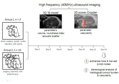
on LN metastasis in mice. To study the dynamic growth of LN metastases in vivo,
we used three-dimentional high frequency ultrasound (HFUS) to non-invasively
and longitudinally monitor the progression of LN metastases in mice. We
observed that growth rates of LN tumors varied from mouse to mouse.
Furthermore, HFUS allowed us to visualize small metastatic deposits and
micrometastases and their growth over time. In order to provide some ground
work for the development of magnetic resonance imaging (MRI) of lymph node
metastases, we developed a gel phantom that simulated the progressive
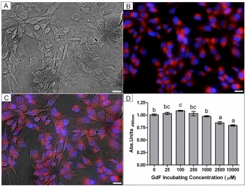
metastatic colonization of LNs. By labeling cells with gadofluorine M (GdF), a
positive contrast agent), we were able to demonstrate that MRI ( at 3T) detected
differences in tumor cell number in our gel phantom that contained a series of
cell pellets with different percentages of labeled tumor cells mixed with unlabeled
tumor cells. We found that the lower limit of detection of GdF-labeled cells was
iv lymph node.
KEYWORDS: lymphatic metastasis, lymph node metastasis, lymph node,
v
Chapter 1 contains concepts and parts of a previously published review
article “A New Breast Cancer Model for Lymphatic Metastasis,” by M.M. Lizardo,
I.C. MacDonald, A.B. Tuck, and A.F. Chambers, in: Cancer Metastasis and the
Lymphovascular System Basis for Rational Therapy, Leong S.P.L. (ed). Springer,
2007. I wrote this review with the assistance from I.C. MacDonald, A.B. Tuck,
and A.F. Chambers.
Chapter 2 contains unpublished material that is currently being prepared
as a manuscript for submission to Clinical and Experimental Metastasis. A.B.
Tuck, I.C. MacDonald, and A.F. Chambers are providing supervision and
assistance in editing the manuscript. I designed and developed the lymph node
experimental metastasis assay (LEMA), and performed the animal surgeries with
the help of N. Hague. I carried out the tissue processing, staining, and
stereological analysis of histology sections. All histological sections were
evaluated by a clinical pathologist A.B. Tuck.
Chapter 3 contains unpublished material that is currently being prepared
as a manuscript for submission to Investigative Radiology. A.B. Tuck, I.C.
MacDonald, J.C. Lacefield, and A.F. Chambers are providing supervision and
assistance in editing the manuscript. I designed the experiments and imaging
protocol. I performed the animal surgeries with the help of N. Hague. I carried out
vi
Chapter 4 contains unpublished materials that are currently being
prepared as a manuscript for submission to Molecular Imaging and Biology. The
manuscript was written with the assistance of J,A. Ronald and Y.Chen. P. Foster,
B. K. Rutt, B. Misselwitz, I.C. MacDonald, and A.F. Chambers are providing
supervision and assistance in editing the manuscript. The design of the
experiments in this chapter was a collaborative effort between J.A. Ronald, and
Y. Chen, and myself. I designed the cell culture experiments, construction of the
MRI phantom, image analysis, and statistical analysis of data. J.A. Ronald
provided the initial idea for the application of gadofluorine in assessing the
detectability of cancer cells; Y. Chen provided his theoretical and practical
knowledge in operating the MRI scanners. B Misselwitz provided positive
vii
guidance and support from my thesis advisors: Dr. Ann Chambers and Dr. Ian
MacDonald. From Ann, I have learned about the importance and urgency of
distilling biological inquiry into translatable knowledge. From Ian, I have gained
invaluable experience in animal surgical techniques and intravital
videomicroscopy. The complementary teachings of these two mentors have
made me a better experimentalist in metastasis research.
I would also like to thank my collaborators at the Robarts Research
Institute: Dr. James Lacefield and Dr. Paula Foster. I have gained a deeper
appreciation about micro-ultrasound and cellular MRI, and I look forward to
incorporating these imaging modalities in my future endeavors in metastasis
research.
To members of the Chambers’ group (past and present): Dr. Kevin
Graham, David Dales, Carl Postenka, Carmen Simedrea, and most importantly
Nicole Hague; I couldn’t have done it on my own. Thank you!
My time in London wouldn’t be as memorable without the friends I have
made throughout the years: Danielle and John Ronald, Jason Townson and
family, Jenn MacLean and family, Amanda Hamilton, Piya Lahiry, and Jen
Mutrie, and Leslie Souter. To the students I have mentored over the years: Baraa
Al-Khazraji, Dean Percy, Lenny Guizzetti, and Matthew Lowerison; it was a
viii
#42511 from the Canadian Institutes of Health Research.
Michael M. Lizardo was funded by studentships from the Translational Breast
Cancer Research Unit of the London Regional Cancer Program, Strategic
Training Fellowship in Vascular Research from the Canadian Institutes of Health
ix
ABSTRACT iii
CO-AUTHORSHIP v
ACKNOWLEDGEMENTS vii
GRANT SUPPORT viii
THESIS TABLE OF CONTENTS ix
LIST OF TABLES xvii
LIST OF FIGURES xviiI LIST OF ABBREVATIONS xxi
CHAPTER 1.0: INTRODUCTION OF LYMPHATIC METASTASIS 1
1.1 NATURAL HISTORY OF LYMPHATIC METASTASIS IN CANCER 1
1.2 LYMPHANGIOGENESIS 2
1.3 LYMPHOVASCULAR INVASION 4
1.4 LYMPH NODE METASTASIS 5
1.4.1 Metastatic progression in the lymph node 6
x
1.6 UNANSWERED QUESTIONS 10
1.7 THESIS HYPOTHESIS 12
1.8 THESIS OBJECTIVE 12
1.8.1 Specific aims 12
1.9 REFERENCES 14
CHAPTER 2.0: ASSESSING THE FATE OF ISOLATED TUMOUR CELLS AND MICROMETASTASES IN THE DEVELOPMENT OF OVERT LYMPH NODE METASTASES 21
2.1 INTRODUCTION 21
2.2 MATERIALS AND METHODS 26
2.2.1 Cell culture and cell labeling 26
2.2.2 Animal model and surgical technique 27
2.2.3 Micro-lymphangiography 28
2.2.4 Lymph node staining and histology bbbbbb 28
2.2.5 Histological and stereological analysis 29
2.2.6 Determining tumour cell survival in lymph nodes DD 30
2.2.7 Statistical analysis 31
2.3 RESULTS 33
xi
tumour burden bnfgnfgnrrb 40
2.3.4 Stereological analysis of histological tumour burden 43
2.3.5 Consistent delivery of reference beads in all time-end point groups wefwefe 46
2.3.6 Assessment of tumour cell survival and fate with respect to formation of micrometastases and overt metastases bbbbbb 49
2.3.7 Sinus histiocytes as a possible source of tumour cell toxicity in the lymph node 53
2.4 DISCUSSION 55
2.5 REFERENCES 60
CHAPTER 3.0: HIGH-FREQUENCY (40 MHz) ULTRASOUND IMAGING OF THE DEVELOPMENT OF EXPERIMENTAL LYMPH NODE METASTASES 66
3.1 INTRODUCTION 66
3.2 MATERIALS AND METHODS 70
3.2.1 Cell culture 70
3.2.2 Animal model and surgical technique 70
3.2.3 Ultrasound B-mode & power Doppler imaging 73
xii
3.2.7 Histological and stereological analysis 79
3.2.8 Statistical analysis 80
3.3 RESULTS 81
3.3.1 Lymph node experimental metastasis assay 81
3.3.2 Changes in lymph node volume during the development of metastases 83
3.3.3 Changes in lymph node roundness index during the development of metastases 86
3.3.4 Changes in lymph node B-mode brightness during the development of metastases 89
3.3.5 Intranodal hyperechoic regions in lymph nodes correspond to small metastatic deposits veverv 92
3.3.6 A positive correlation between ultrasound volumetric and histological area measurements 92
3.3.7 Longitudinal imaging of micrometastatic deposits and small metastases 97
3.3.8 Lymph node percent vascularity of negative lymph nodes and metastatic lymph nodes veverv 99
3.3.9 The sensitivity and specificity of ultrasound parameters in the detection of lymph node metastases 101
3.4 DISCUSSION 104
xiii
4.1 INTRODUCTION 113
4.2 MATERIALS AND METHODS 118
4.2.1 Cell culture and in vitro labeling with Gadofluorine M 118
4.2.2 Visualizing intracellular localization of GdF in
breast cancer cells 119
4.2.3 In vitro MRI of cell pellets labeled at different
GdF-loading concentrations 120
4.2.4 Measurement of T1 relaxation times of cell pellets
labeled at different GdF-loading concentrations 121
4.2.5 Quantification of intracellular GdF by inductively
coupled plasma atomic emission spectroscopy 122
4.2.6 Construction of gel phantom for in vitro MRI
scanning 122
4.2.7 Evaluating 3D SPGR and FIESTA pulse
sequences for imaging an MRI phantom 123
4.2.8 Measurements of T1 relaxation times of cell
pellets containing different ratios of GdF-labeled
tumour cells 124
4.2.9 Statistical analysis 124
4.3 RESULTS 126
4.3.1 Labeling efficiency and cell viability at different
xiv
concentrations of GdF scanned at 1.5T & 3T 130
4.3.4 Quantification of signal enhancement and
T1 relaxation of cell pellets of different GdF-
loading conditions at 1.5T & 3T 130
4.3.5 Intracellular concentration of GdF of cell pellets
over a range of GdF-loading conditions 131
4.3.6 In vitro MRI phantom to assess the detectability
of GdF-labeled tumour cells at varying numbers 136
4.3.7 Comparison of CNR from 3D SPGR and FIESTA
pulse sequences 136
4.3.8 Increased signal enhancement and R1 relaxation
rates of cell pellets with higher numbers of GdF-
labeled tumour cells 140
4.3.9 The change in MR signal enhancement and R1
measurements strongly correlates to the number
of GdF-labeled tumour cells 140
4.4 DISCUSSION 145
4.5 REFERENCES 148
CHAPTER 5.0: GENERAL DISCUSSION 154
xv
DEVELOPMENT OF OVERT LYMPH NODE METASTASES 156
5.2.1 The need for a new model of lymph node
Metastasis 156
5.2.2 LEMA and metastatic inefficiency in the lymph node 156
5.2.3 Clinical relevance 157
5.2.4 Future studies 158
5.3 DISCUSSION – HIGH-FREQUENCY (40 MHz) ULTRASOUND
IMAGING OF THE DEVELOPMENT OF EXPERIMENTAL
LYMPH NODE METASTASES 159
5.3.1 The utility of HFUS in tracking the dynamic
growth of lymph node tumours 159
5.3.2 Non-invasive and longitudinal imaging of
micrometastases 160
5.3.3 Clinical relevance 161
5.3.4 Future studies 161
5.4 DISCUSSION – THE CELLULAR DETECTABILITY OF
POSITIVE CONTRAST-LABELED TUMOUR CELLS AT
CLINICAL FIELD STRENGTH MRI 163
5.4.1 Problems in quantifying iron-oxide labeled
xvi
5.4.3 Clinical relevance 164
5.4.4 Future studies 165
5.5 CONCLUSIONS 167
5.6 REFERENCES 168
APPENDIX 1 – Copyright agreements 170
APPENDIX 2 – Ethics approval for use of animal subjects 171
xvii
CHAPTER 2:
Table 2.1 Metastasis incidence from experimental lymph node
Metastasis assay………….………...39
CHAPTER 3:
Table 3.1 Metastasis incidence from experimental lymph node
Metastasis assay ………...82
Table 3.2 Sensitivity and specificity of ultrasound parameters in
xviii
CHAPTER 1:
Figure 1.1 Schematic diagram of the progression of lymphatic
metastasis ……….….7
CHAPTER 2:
Figure 2.1 Lymph node experimental metastasis assay (LEMA)………….…36
Figure 2.2 A time-dependent experiments to study the progression of
lymph node metastasis progression………....37
Figure 2.3 Visualization and quantification of surface tumour burden
of axillary lymph nodes………..………....42
Figure 2.4 Quantification of time-dependent changes in histological
tumour burden………45
Figure 2.5 Quantification of the average number of reference beads
delivered per lymph node for all time points………..48
Figure 2.6. Quantification of the fate of tumour cells in the lymph node………51
Figure 2.7. A flow chart showing the fate of tumour cells after they
arrest in the axillary lymph node………..52
Figure 2.8 Quantification of sinus histiocytes containing 3 μm beads…...…...54
CHAPTER 3:
xix
development of metastases………..85
Figure 3.4 Ultrasound analysis of lymph node roundness index during
the development of metastases………..88
Figure 3.5 Ultrasound analysis of lymph node B-mode brightness
during the development of metastases………..….91
Figure 3.6 Hyperechoic subregions of the lymph node corresponding
to micro- and macrometastatic deposits……….…95
Figure 3.7 Strong positive correlation between ultrasound volumetric
and histological area measurements………..96
Figure 3.8 Growth of small non-palpable metastatic deposits in lymph
nodes………..98
Figure 3.9 Lymph node percent vascularity of negative lymph nodes
and metastatic lymph nodes………...100
CHAPTER 4:
Figure 4.1. Tumour cell uptake of GdF-cc, labeling efficiency, and
cell viability from GdF-loading………128
Figure 4.2 Visualization of intracellular location of GdF-cc in tumour
cells by immunofluorescent microscopy ………..…129
Figure 4.3 Cellular MRI of cell pellets labeled at different loading
concentrations of GdF……….132
Figure 4.4 Cellular MRI of cell pellets at 1.5T and 3T..……….134
xx
enhancement at 3T in detecting differing amounts of
GdF-labeled tumour cells………....137
Figure 4.7 Comparison of image quality between 3D SPGR and
FIESTA pulse sequences………...139
Figure 4.8 MRI of cell pellets containing different proportions of
GdF-labeled tumour cella………143
Figure 4.9 Changes in MR signal enhancement and R1 values
strongly correlate with the number of tumour cells per
voxel……….………..144
xxi μm
μm2
μM 2D 3D 3D HFUS 3D SPGR Ar ANOVA BSA BW CCR7 cm CNR CO2 COX-2 CXCR3 DAPI dB DTPA EDTA FIESTA FITC FPF micrometer micrometer squared micromolar 2 dimensional 3 dimensional
3 dimensional high frequency ultrasound
3 dimensional spoiled gradient recalled
argon
analysis of variance
bovine serum albumin
bandwidth
chemokine cysteine-cysteine motif receptor type 7
centimeter
contrast-to-noise ratio
carbon dioxide
cyclooxygenase-2
cysteine-x-cysteine chemokine receptor 3
4',6-diamidino-2-phenylindole
decibel
diethylene triamine pentaacetic acid
ethylenediaminetetraacetic acid
fast imaging employing steady state acquisition
fluoroscein isothiocyante
xxii GdF-cc HBSS HFUS HIFU H&E HNO3 ICP-AES Ig ITCs IVFVM IVVM kHz kg Kr LEMA LMVD LN LYVE-1 M mg MHz ms NEX nm
gadofluorine M carbocyanine
Hank’s buffered salt solution
high frequency ultrasound
high intensity focused ultrasound
hematoxylin and eosin
nitric oxide
inductively coupled atomic emission spectroscopy
immunoglobulin
isolated tumour cells
intravital fluorescence videomicroscopy
intravital videomicroscopy
kiloHertz
kilogram
krypton
lymph node experimental metastasis assay
lymphatic microvessel density
lympyh node
lymphatic vessel endothelial receptor 1
molarity
milligrams
megaHertz
milliseconds
number of excitations
xxiii R1 r2 RI ROI SD SE SNR SPIO T1 TE TI TPF TR VEGF-C VEGF-D VEGF-R3
relaxation rate (s-1)
correlation coefficient
roundness index
region of interest
standard deviation
standard error
signal-to-noise ratio
superparamagnetic iron oxide
relaxation time (s)
time to echo
time to inversion
true positive fraction
time to repetition
vascular endothelial growth factor-C
vascular endothelial growth factor-D
Chapter 1.0:Introduction of lymphatic metastasis
1.1 Natural history of lymphatic metastasis in cancer
Lymphatic vessels provide one of the main anatomical routes by which
invasive tumour cells can disseminate from the primary tumour (Pantel and
Brakenhoff, 2004). Certain types of cancer, including breast, prostate, and
melanoma, have a propensity to disseminate via the lymphatics. Yet despite the
prevalence of lymphatic metastasis, experimental work elucidating the underlying
biology, until the last decade, has been relatively limited. In the past decade,
there has been a surge in the number of studies uncovering the molecular
determinants of lymphatic metastasis. The following chapter aims to highlight
pre-clinical experimental work that contributes to our basic understanding of
lymphatic metastasis. Before continuing, however, a brief overview of clinical and
pathological studies that detail the natural history of lymphatic metastasis will
esetablish the disease model which experimental models must mimic.
Physicians have described the spread of cancer to axillary lymph nodes
as early as the 18th century (Weiss, 2000). Since then, a multitude of clinical and
pathological studies have been published, suggesting that the lymphatic spread
of breast cancer cells and formation of axillary lymph node metastases are
common events in the natural history of the disease. The incidence of lymph
node metastases in breast cancer is 30% to 50% (Foster 1996), 10% in prostate
cancer (Swanson et al, 2006), and 50% in malignant melanoma (Buzzell and
least three potentially clinically relevant steps in lymphatic metastasis: 1)
lymphangiogenesis, 2) lymphovascular invasion, and 3) lymph node metastasis.
1.2 Lymphangiogenesis
Neo-vascularization contributes to the dissemination of tumour cells by
increasing the likelihood of tumour cell intravasation into vessels (Folkman,
1992). In the context of lymphatic metastasis, it is reasonable to presume that
increased lymphatic vessel density as a result of lymphangiogenesis would
increase the likelihood of tumour cell invasion into lymphatic vessels. Indeed,
there are clinical studies supporting this notion. Nakamura and colleagues (2005)
examined archived and fresh frozen patient tissue samples, and demonstrated
that elevated expression levels of vascular endothelial growth factor C (VEGF-C)
was associated with increased lymphatic vessel density, lymph node metastases,
and decreased patient survival. Schoppman and colleagues (2006) analyzed
archival tissue from patients with invasive breast cancer and found a significant
association between VEGF-C expression from tumour-associated macrophages
and lymphatic microvessel density (LMVD), as well as LMVD and
lymphovascular invasion. These findings support the idea of the local
peritumoural inflammatory reaction that contains VEGF-C expressing
macrophages, may contribute to lymphangiogenesis and thereby increasing the
To model tumour-associated lymphangiogenesis, Skobe and
associates (2001) engineered MDA-MB-435 human breast cancer cells to
over-express VEGF-C. These cells were able to significantly increase intratumoural
lymphangiogenesis when implanted in nude mice. Moreover over-expression of
VEGF-C was also associated with a 60% increase in the incidence of lymph
node metastasis. Similar findings have been demonstrated in MCF-7 human
breast cancer cells (Karpanen et al, 2001; Mattila et al, 2002). Another growth
factor that was discovered to stimulate lymphangiogenesis is VEGF-D. Stacker
and colleagues (2001) transfected the non-metastatic 239EBNA cells to express
VEGF-D (designated VEGF-D-293 cells). When injected into mice, VEGF-D-293
tumours had a higher amount of lymphatic vessels compared to tumours of the
parental cell line. Furthermore, VEGF-D expression in VEGF-D-293 cells led to a
higher incidence of lymph node metastases compared to parental cells. The
authors demonstrated that the treatment of mice bearing VEGF-D-293 cells with
anti-VEGF-D antibodies reduced the amount of tumour-associated lymphatic
vessels, and inhibited the spread of tumour cells to draining lymph nodes.
Metastasis to draining lymph nodes can also be inhibited by the administration of
antibodies against the cognate receptor VEGF-R3, as demonstrated by Shimizu
and colleagues in 2005.
Another biological factor that has been implicated in inducing tumour
lymphangiogenesis is cyclooxygenase-2 (COX-2; Su et al, 2004; Timoshenko et
cyclooxygenase (COX)-2 in the up-regulation of VEGF-C production is
associated with lymphangiogenesis in patient samples. In an experimental
mouse model, the inhibition of COX-2 by etodolac resulted in both decreased
tumour lymphangiogenesis and lymph node tumour burden (Iwata et al, 2007).
1.3 Lymphovascular invasion
Tumour cell intravasation into lymphatic vessels is requisite for the
initiation of lymphatic metastasis (Nathanson et al, 1997). Lymphatic or vascular
invasion, a sign of poor prognosis, is commonly referred to as “lymphovascular
invasion”, and means any involvement of an endothelial-lined space. Although it
has been said that differentiating between vascular vs. lymphatic space
involvement is not important in terms of prognostic value (both are felt to be
associated with poor outcome), the use of this definition precludes the ability to
tease out the relative significance of a lymphatic vs. a vascular route of
metastasis. The development of new and improved markers of lymphatic
endothelium, however, have allowed the specific evaluation of tumour cells found
within lymphatic vessels, as defined by positive staining for markers such as
lymphatic vessel endothelial hyaluronan receptor-1 (LYVE-1; Banurji et al, 1999),
podoplanin (Breiteneder-Geleff et al, 1999), D2-40 (Kahn et al, 2002), or Prox-1
(Wigle and Oliver, 1999). Using dual-color immunofluorescence staining for
LYVE-1 and podoplanin, Schoppman and colleagues (2004) demonstrated that
lymphatic invasion was associated with an increased risk of developing lymph
found a correlation between high lymphatic microvessel density and lymphatic
invasion. This supports the notion that increased vessel density increases the
likelihood of tumour cell intravasation, in the context of lymphatic vessels
(Folkman, 1992).
1.4 Lymph node metastasis
The prognostic value of axillary lymph node metastasis has been well
known, and provides one of the strongest prognostic factors in breast cancer
(Foster, 1996, Koscielny and Tubiana, 1996; Fisher et al., 1984; Contesso et al,
1977; Nemoto et al, 1980; Peloquin et al, 1991). This is very well established for
metastatic nodal deposits > 2.0 mm (Huvos et al, 1971). With lesser degrees of
nodal involvement, the association with outcome is less clear. The
preponderance of the literature would suggest that micrometastatic nodal
involvement (ie. recognizable on routine H&E stained slides, as > 0.2 mm, but
not > 2.0 mm; ) is also significant, but perhaps with a lesser degree of impact on
prognosis (Maibenco et al, 2006; Park et al, 2009; Weaver et al, 2011). The
clinical significance of nodal deposits no greater than 0.2 mm however (referred
to as isolated tumour cells, pN0i+ by AJCC 6th edition staging criteria) is
1.4.1 Metastatic progression in the lymph node
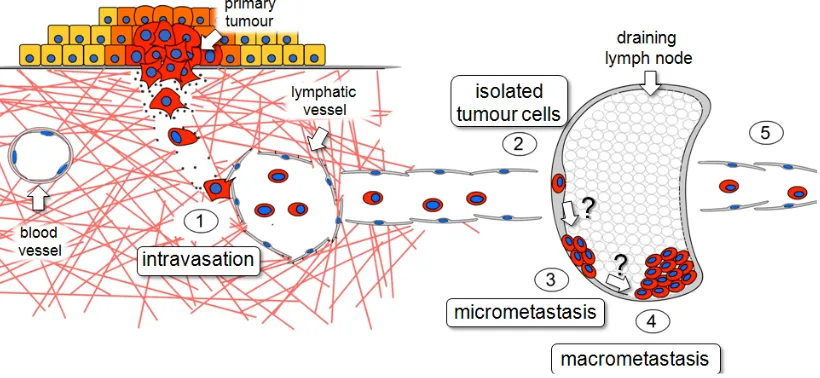
As with step-wise progression of hematogenous (blood-borne) metastasis,
lymphatic metastasis is presumed to be a step-wise phenomenon (Carr and Carr,
1982). From the aforementioned clinical data, we can model that lymphatic
metastasis can occur in five steps, illustrated in the Figure 1.1. The steps
depicted in Figure 1.1 manifest as distinct histopathological entities in the clinic,
some of which have prognostic significance (eg. lymphatic invasion and lymph
node metastasis). However, whether the model represents a true continuous
Figure 1.1Schematic diagram of theprogression of lymphatic metastasis.
(1) Tumour cell entry and transit within lymphatics (lymphatic invasion), (2) arrest
in the draining lymph node as isolated tumour cells (ITCs), (3) formation of
micrometastasis in the lymph node, (4) establishment of macrometastasis in the
lymph node, and (5) further dissemination to downstream lymphatic vessels and
1.4.2 Clues from studies of hematogenous experimental metastasis
Questions regarding the fate of tumour cells once they arrest at the
secondary site can potentially be addressed by using the experimental tools that
were used to ascertain the timing of events and molecular determinants of
hematogenous metastasis, and applying them to the study of the lymphatic
metastasis. For example, Luzzi and colleagues (1998), through the use of
intravital videomicroscopy (IVVM), have demonstrated that tumour cells that are
introduced into systemic circulation arrest and extravasate into secondary organs
with high efficiency. However, only a small proportion of these cells was able to
establish growth at the secondary site. More strikingly, Naumov and colleagues
(1999) employed IVVM to demonstrate the presence of “dormant” single
non-dividing cells that have extravasated into the liver parenchyma and have survived
two weeks after injection. More recently, Heyn et al, (2006) employed magnetic
resonance imaging (MRI) to study the fate of breast cancer cells in a mouse
model of brain metastasis. Their work supports the notion that only a small
proportion of extravasated tumour cells have the capacity to establish
metastases. Using non-invasive imaging techniques to longitudinally monitor the
lymphatic spread of tumour cells in vivo can provide insight into which step(s) of
lymphatic metastasis do tumour cells accomplish with low efficiency, and thereby
1.5 Animal models of lymphatic metastasis
With respect to experimental studies of lymphatic metastasis in breast
cancer, earlier work in the 1970’s by Ian Carr merits discussion. At the University
of Saskatchewan, Carr and colleagues studied a series of rat mammary
carcinomas that reproducibly metastasized to the popliteal lymph node when
injected into the foot pad. In one study using Rd/3 cells, Carr and associates
demonstrated that the primary tumour in the footpad continually seeds the
popliteal lymph node with tumour cells thereby recapitulating the progressive
steps of lymph node metastasis. However, if the footpad was removed before 24
hours have elapsed, progressive lymphatic metastasis did not occur (Carr and
McGinty, 1976). In another study using Walker rat carcinoma cells, cannulation of
the lymphatic trunk efferent to the primary tumour revealed a progressive rise,
with time, in the number of tumour cells leaving the primary tumour (Carr et al,
1980).
To model tumour-associated lymphangiogenesis, Skobe and associates
(2001) engineered MDA-MB-435 human breast cancer cells to over-express
VEGF-C. These cells were able to significantly increase intratumoural
lymphangiogenesis when implanted in nude mice. Moreover over-expression of
VEGF-C was also associated with a 60% increase in the incidence of lymph
node metastasis. Similar findings have been demonstrated in MCF-7 human
There is mounting evidence suggesting tumour cells may actively migrate
towards and intravasate into lymphatic vessels by conscripting the same
mechanisms of adhesion and migration that leukocytes use for entry into the
lymphatic system. In a seminal paper by Muller and associates (2001), the
authors provide clinical and experimental data suggesting that breast cancer
cells express chemokine receptors (CXCR-4) that actively promote tumour cell
migration into lymphatics and draining lymph nodes. In their experimental mouse
studies, the inhibition of CXCR-4 with neutralizing anti-CXCR-4 antibodies
significantly inhibited metastasis to the inguinal and axillary lymph nodes and
lung.
1.6 Unanswered questions
The recent discoveries in lymphatic biology and lymphatic metastasis are
indeed proving to be an exciting time in this field. Such studies are important in
addressing questions regarding the progression of lymphatic metastasis. For
example, during tumour progression, how many cells are shed into the
lymphatics? What proportion of these tumour cells survives to form lymph node
metastases? To what degree do lymph node metastases contribute (if at all) to
systemic dissemination? What biological factors affect the progression of lymph
node metastases? Such questions not only aim to determine the biological
significance of isolated tumour cells (ITCs) and micrometastases, but to also find
These unanswered questions underscore the need for more models of
lymphatic metastasis. Current experimental research is beginning to uncover the
molecular determinants of lymphatic metastasis. However, at the same time, it’s
also important to develop imaging models that permit the in vivo longitudinal
imaging of the progression of lymphatic metastasis. The intersection between
imaging and biological modeling of lymphatic metastasis will be a
forward-thinking approach to investigating the in vivo dynamics of lymphatic metastasis;
and more importantly, such models will permit the non-invasive and longitudinal
1.7 THESIS HYPOTHESIS
Based on previous studies examining tumour cell fate in secondary organs
such as the liver, lung, and brain, it is hypothesized that metastasis
progression in lymph nodes is an inefficient process. Specifically, it is
predicted that cancer cell survival will significantly decrease over
progressive time points.
1.8 THESIS OBJECTIVE
To address the above hypothesis, the primary objective of this thesis is to
develop and characterize a novel lymph node experimental metastasis
assay that recapitulates the progressive steps of metastasis development
in the lymph node. This model will be used to assess tumour cell fate
(cancer cell survival) after their arrest in the lymph node
1.8.1 SPECIFIC AIMS
1) To create a new lymph node experimental metastasis assay (LEMA) that
permits the quantification of tumour cell fate after arrest in draining lymph
nodes
2) To assess the utility of high frequency ultrasound in the non-invasive and
longitudinal characterization of lymph node metastasis development in
3) To develop quantitative methods that utilize positive-contrast MRI to
1.9 References
Banerji S, Ni J, Wang SX, et al. LYVE-1, a new homologue of the CD44
glycoprotein, is a lymph-specific receptor for hyaluronan. Journal of Cell
Biology. 1999; 144: 789-801.
Bhattacharjee RN, Timoshenko AV, Cai J, et al. Relationship between
cyclooxygenase-2 and human epidermal growth factor receptor 2 in vascular
endothelial growth factor –C up-regulation and lymphangiogenesis in human
breast cancer. Cancer Science. 2010; 101: 2026-2032.
Breiteneder-Geleff S, Soleiman A , Kowalski H, et al. Angiosarcomas express
mixed endothelial phenotypes of blood and lymphatic capillaries: podoplanin
as a specific marker for lymphatic endothelium. American Journal of
Pathology. 1999; 154: 385-394.
Buzzell RA, Zitelli JA. Favorable prognostic factors in recruitment and metastatic
melanoma. Journal of the American Academy of Dermatology. 1996; 34:
798-803.
Carr J, Carr I, Dreher B, et al. Lymphatic metastasis: invasion of lymphatic
vessels and efflux of tumour cells in the afferent popliteal lymph as seen in
the Walker rat carcinoma. Journal of Pathology. 1980; 132: 287-305.
Carr I, McGinty F. Neoplastic invasion and metastasis within the lymphoreticular
Carr I, Carr J. Tumor Invasion and Metastasis: Experimental models of lymphatic
metastasis. In: Liotta LA, IR Hart eds. Developments in Oncology 7. Boston,
MA: Martinus Nijhoff Publishers; 1982: 189-206.
Contesso G, Rouesse J, Petit JY, et al. Les facteurs anatomo-pathologiques du
pronostic des cancers du sein. Bulletin du Cancer. 1977; 64: 525-236.
Coussens LM, Werb Z. Inflammation and cancer. Nature. 2002; 420:860-867.
Fisher ER, Sass E, Fisher B. Pathological findings from the NSABP Protocol 4:
discriminants for tenth year treatment failure. Cancer. 1984; 53:712-723.
Folkman J. The role of angiogenesis in tumor growth and metastasis. Seminars
in Oncology. 1992; 6: 15-18.
Foster RS. The Biologic and Clinical Significance of Lymphatic Metastasis in
Breast Cancer. Surgical Oncology Clinics of North America. 1996; 5: 79-104.
Heyn C, Ronald JA, Ramadan SS, et al. In vivo MRI of cancer cell fate at the
single-cell level in a mouse model of breast cancer metastasis to the brain.
Magnetic Resonance Medicine. 2006; 56: 1001-1010.
Huvos AG, Hutter RV, Berg JW. Significance of axillary macrometastases and
Iwata C, Kano MR, Komuro A, et al. Inhibition of cyclooxygenase-2 suppresses
lymph node metastasis via reduction of lymphangiogenesis. Cancer
Research. 2007; 67:10181-10189.
Kahn HJ, Bailey D, Marks A. Monoclonal antibody D2-40, a new marker of
lymphatic endothelium, reacts with Kaposi's sarcoma and a subset of
angiosarcomas. Modern Pathology. 2002; 15:434-40.
Karpanen T, Egeblad M, Karkkainen MJ, et al. Vascular endothelial growth factor
C promotes tumor lymphangiogenesis and intralymphatic tumor growth.
Cancer Research. 2001; 61: 1786-1790.
Koscielny S, Le MG, Tubiana M. The natural history of human breast cancer: The
relationship between involvement of axillary lymph nodes and the initiation of
distant metastases. British Journal of Cancer. 1989; 59: 775-782.
Lauria R, Perrone F, Carlomagno C, et al. The prognostic value of lymphatic and
blood vessel invasion in operable breast cancer. Cancer. 1995; 76:
1772-1778.
Luzzi KJ, MacDonald IC, Schmidt EE, et al. Multistep nature of metastatic
inefficiency: dormancy of solitary cells after successful extravasation and
limited survival of early micrometastases. American Journal of Pathology.
Maibenco DC, Dombi GW, Kau TY, et al. Significance of micrometastases on
the survival of women with T1 breast cancer. Cancer. 2006; 107:1234-1239.
Mattila, M M, Ruohola, J K, Karpanen, T, et al. VEGF-C induced
lymphangiogenesis is associated with lymph node metastasis in orthotopic
MCF-7 tumors. International Journal of Cancer. 2002; 98: 946-951.
Muller A, Homey B, Soto H, et al. Involvement of chemokine receptors in breast
cancer metastasis. Nature. 2001; 410: 24-25.
Nakamura Y, Yasuoka H, Tsujimoto M, et al. Lymph vessel density correlates
with nodal status, VEGF-C expression, and prognosis in breast cancer.
Breast Cancer Research and Treatment. 2005; 91:125-132.
Nathanson SD, Anaya P, Avery M, et al. Sentinal lymph node metastasis in
experimental melanoma: relationships among primary tumor size, lymphativ
vessel diameter, and 99mTc-labeled human albumin clearance. Annals of
Surgical Oncology. 1997; 4: 161-168.
Naumov GN, Wilson SM, MacDonald IC, et al. Cellular expression of green
fluorescent protein, coupled with high-resolution in vivo videomicroscopy, to
monitor steps in tumor metastasis. Journal of Cell Science. 1999;
Nemoto T, Vana J, Bedwani RN, et al. Management and survival of female
breast cancer: results of a national survey by the American College of
Surgeons. Cancer. 1980; 45:2917-2924.
Pantel K, Brakenhoff RH. Dissecting the metastatic cascade. Nature Reviews
Cancer. 2004; 4: 448-456.
Park D, Karesen R, Naume B, et al. The prognostic impact of occult metastasis
in early breast carcinoma. Breast Cancer Research and Treatment. 2009;
118:57-66.
Peloquin A, Poljicak M, Falardeau M, et al. Cancer of the breast: a study of 1520
consecutive patients operated on between 1960 and 1980. Canadian Journal
of Surgery. 1991; 34:151-156.
Querzoli P, Pedriali M, Rinaldi R, et al. Axillary lymph node nanometastases are
prognostic factors for disease-free survival and metastatic relapse in breast
cancer patients. Clinical Cancer Research. 2006; 12:6696-6701.
Schoppmann SF, Bayer G, Aumayr K, et al. Prognostic value of
lymphangiogenesis and lymphovascular invasion in invasive breast cancer.
Annals of Surgery. 2004; 240:306-312.
Schoppmann SF, Fenzl A, Nagy K,et al. VEGF-C expressing tumor-associated
macrophages in lymph node positive breast cancer: impact on
Shimizu K, Kubo H, Yamaguchi K, et al. Suppression of VEGFR-3 signaling
inhibits lymph node metastasis in gastric cancer. Cancer Science. 2004;
95:328-333.
Singletary SE, Greene FL. Revision of breast cancer staging: the 6th edition of
the TNM Classification. Seminars in Surgical Oncology. 2003; 21:53-59.
Skobe M, Hawighorst T, Jackson DG, et al. Induction of tumor
lymphangiogenesis by VEGF-C promotes breast cancer metastasis. Nature
Medicine. 2001; 7:192-198.
Swanson GP, Thompson IM, Basler J. Current status of lymph node-positive
prostate cancer: Incidence and predictors of outcome. Cancer. 2006; 107:
439-450.
Stacker SA, Caesar C, Baldwin ME, et al. VEGF-D promotes the metastatic
spread of tumour cells via the lymphatics. Nature Medicine. 2001; 7: 186-191.
Su JL, Shih JY, Yen ML, et al. Cyclooxygenase-2 induces EP1- and
HER-2/Neu-dependent vascular endothelial growth factor-C up-regulation: a novel
mechanism of lymphangiogenesis in lung adenocarcinoma. Cancer
Research. 2004; 64:554-564.
Timoshenko AV, Chakraborty C, Wagner GF, et al. COX-2 mediated stimulation
of the lymphangiogenic factor VEGF-C in human breast cancer. British
Weaver DL, Ashikaga T, Krag DN, et al. Effect of occult metastases on survivcal
in node-negative breast cancer. New England Journal of Medicine. 2011;
364:412-421.
Weiss L. Metastasis of Cancer: A Conceptual History from Antiquity to the 1990s.
Cancer and Metastasis Reviews. 2000; 19: 219-234.
Wigle JT, Oliver G. Prox1 function is required for the development of the murine
Chapter 2.0: Assessing the fate of isolated tumour cells and
micrometastases in the development of overt lymph node metastases
2.1 Introduction
The dissemination of tumour cells into the lymphatic vasculature is
common in the natural history of many types of cancer. After transit within the
lymphatic vasculature, tumour cells eventually arrest and begin to colonize
draining lymph nodes. The successful establishment of overt lymph node
metastases (> 2 mm in diameter) becomes a clinically relevant event for the
cancer patient since the assessment of the number of lymph node metastases
and extent of nodal involvement is currently standard clinical practice in
evaluating disease aggressiveness and determining patient prognosis (Das and
Skobe, 2008). Although lymph node metastases themselves are not fatal to the
patient, it is hypothesized that lymph node metastases act as reservoirs of
metastatic cells which can disseminate further into systemic circulation
(Sleeman, 2000; Morton et al, 2003). This notion is supported by Rebhun et al
(2008), who demonstrated lymph node tumours can seed tumour cells into the
systemic circulation.
Each step of the (lymphatic) metastatic cascade may be a rate-limiting
step (Chambers et al, 2002; Pantel and Brakenhoff, 2004). From tumour cell
invasion into local stroma, intravasation into nearby vessels, transit within
vessels, arrest at the secondary site, and successful formation of overt
in order to produce efficacious anti-metastatic treatments in the clinic, it is
paramount to study and understand the basic underlying biology that drives each
step of the cascade. Indeed, within the last decade, several groups in lymphatic
metastasis research have studied a particular aspect of the cascade in vivo and
conceived a novel anti-metastatic therapy. For example, Wiley and colleagues
(2001) demonstrated how the lymphatic dissemination of B16 cells
over-expressing chemokine receptor-7 can be inhibited by injecting neutralizing
antibodies against C-C chemokine ligand 21 (cognate ligand of CCR7). In
another study examining tumour-induced lymphangiogenesis, He and colleagues
(2005) studied how blockage of VEGFR3 activation on lymphatic vessels by
soluble VEGF-R3-Ig fusion proteins inhibited lymphangiogenesis and the
incidence of lymph node metastases was reduced.
Despite the plethora of basic research studies examining how tumour cells
gain entry into the lymphatic vasculature, relatively few studies have examined
the events following tumour cell arrest in draining lymph nodes. With the
exceptions of a few studies (Dadiani et al, 2006; Yokoyama et al, 2006), the
majority of studies in lymphatic metastasis have regarded lymph node
metastases as a “black-box” labeled “end-point” rather than being viewed as the
result of a series of discrete steps involving a number of possible pathways
(MacDonald et al, 2002). Clinical data provides evidence that earlier stages of
lymph node metastases can manifest as the following histologically distinct
or clusters of tumour cells less than 0.2 mm in diameter, (2) micrometastases
which are lesions greater than 0.2 mm and no greater than 2 mm, and finally (3)
overt metastases greater than 2 mm (Kahn et al, 2006; Singletary et al, 2002).
From these clinical observations, formation of lymph node metastases is
presumed to begin with isolated tumour cells arresting in the subcapsular sinus
of the lymph node. These tumour cells then proliferate to form a micrometastatic
deposit, which eventually grows into an overt lymph node tumour. Despite the
voluminous amount of clinical observations, exactly how each step progresses to
the next is unknown. What proportion of isolated tumour cells survive to form
micrometastases? What proportion of micrometastases grow into overt lymph
node metastases? What genetic factors positively or negatively regulate the
progression in each of these steps?
Insight into answering these questions comes from animal models of
metastasis where the fate of tumour cells after arrest at the secondary site is
quantified (Luzzi et al, 1998; Cameron et al, 2000, Heyn et al, 2006; Kienast et
al, 2010). To quantify the fate of tumour cells, Luzzi et al (1998) and Cameron et
al (2000) used an experimental metastasis assay in which tumour cells and
similar sized reference beads are directly injected into the vessel upstream of the
target organ. The reference beads permit a “cell accounting” analysis where the
percent survival of the initial injected population of tumour cells that form
metastatic lesions at progressive intervals can be calculated (MacDonald et al,
site, the progression of single tumour cells into overt metastases is an inefficient
process. More specifically, the majority of single tumour cells that arrive in the
secondary site do not survive to form overt tumours, and only 0.02% and 5.8% of
tumour cells survive to form metastases in the liver and lung, respectively (Luzzi
et al, 1998; Cameron et al 2000). Heyn and colleagues (2006) employed
high-resolution cellular MRI to track the fate of magnetically labeled cancer cells
arrested in the brain and determined that 1.5% of the cells were able to form
metastases. Kienast et al (2010) also found similarly low values of efficiency in
the brain; furthermore the authors define several critical steps unique to the brain
microenvironment that contribute to this inefficiency. The studies described
above focused on hematogenous metastasis. However, with respect to
assessing metastatic inefficiency lymph node, there are currently no metastasis
models that permit the quantification of the discrete series of events that occur
after tumour cells arrest in the lymph node.
In the research described herein, the use of the B16F10 melanoma model
is described in a novel metastasis assay, called lymph node experimental
metastasis assay (LEMA), which permits the quantitative assessment of tumour
cell fate after arrest in lymph nodes. We demonstrate how our model
recapitulates the progressive steps of metastasis development: from isolated
tumour cells, micrometastases, and overt lymph node metastases. Furthermore,
using the “cell accounting” technique, we provide quantitative evidence that
found that only a small proportion of cells delivered to the lymph node (0.37%) go
on to form micrometastases, and that even a smaller proportion (~0.08%) that
2.2 Materials and Methods
2.2.1 Cell culture and cell labeling
Briefly, a B16F10 murine melanoma cell line carrying a lacZ expressing
vector (Kirstein et al, 2009) was maintained in -MEM medium containing 10% fetal bovine serum at 37°C and 5% CO2. For labeling cells with 3μm
microspheres, cells were grown in a T75 tissue culture flask using medium with
fetal bovine serum until 80% to 90% confluent.
To determine if macrophages in the lymph node (sinus histiocytes)
phagocytose the tumour cells, tumour cells were labeled with 3μm polystyrene
beads (Polysciences, Warrington, Pennsylvania) in vitro prior to injection into
mice. For cell labeling, 5mL of OptI-MEM medium (Invitrogen, Burlington,
Ontario) containing 1.68 x 10⁸ beads were added to 70-80% confluent
B16F10-LacZ cells (T75 flask) and incubated for 1 h at 37°C. After incubation, the media
was aspirated, and cells were washed thoroughly with Hank’s buffered salt
solution (HBSS) to remove unincorporated 3μm beads. Cells were then
trypsinized, centrifuged, then resuspended in HBSS. The cell pellet was washed
twice in HBSS prior to performing a trypan blue exclusion assay to test cell
viability. Cell viability was between 95-99%. From these cells, a working cell
suspension containing 1.5x10⁷ B16F10-lacZ cells and 7.5x10⁵ 16μm polystyrene
reference beads (Polysciences), at a 20:1 cell:bead ratio, in a volume of 1mL
2.2.2 Animal model and surgical technique
Female C57Bl/6 mice (Harlan Sprague Dawley, Indianapolis, Indiana), 5
weeks of age, were cared for in accordance with standards of the Canadian
Council on Animal Care, under an approved protocol of the University of Western
Ontario Council on Animal Care. During surgery, mice were kept under gas
anesthesia with 1.5% isolflurane in oxygen and restrained on a water-heated
stage. A one-inch incision was made in the lower ventral midline, and skin and
underlying fascia were blunt dissected to expose the 4th mammary fat pad. The
skin flap attached to the mammary gland was reflected back and pinned with
sterile needles for easy access to the mammary fat pad and inguinal lymph node.
Under a dissecting microscope, the surface of the inguinal lymph node was
exposed by removing adjacent mammary fat pad tissue. A suspension of tumour
cells and 16μm reference beads (200μL of working cell suspension described
above) was infused into the lymphatic microvasculature via intranodal injection
with a pulled borosilicate glass needle (inner diameter 200 μm) that was
connected to an 18 gauge needle and 1mL syringe with Tygon tubing. Infusion of
the cell suspension was done over a period of 5 minutes. In order to maintain the
tumour cell to reference bead ratio in the axillary lymph node, further seeding
from tumour cell growth in the inguinal lymph node (site of injection) was
prevented by surgically removing the inguinal immediately after completion of the
injection. Large blood vessels connected to the mammary fat pad were
injected with Metacam (0.1mg/kg; Boehringer Ingelheim Vetmedica Inc, St.
Joseph, Missouri) and saline and allowed to recover.
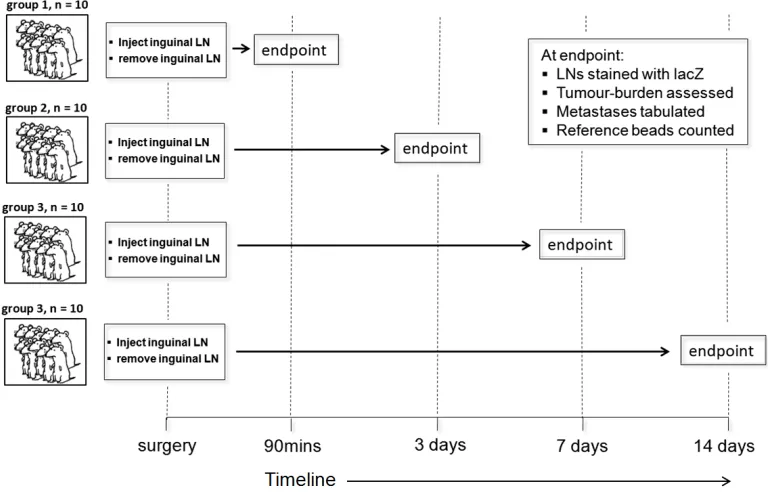
To assess the growth of tumour cells in the axillary lymph node at
progressive time intervals, surgery and tumour cell injection was performed on 40
mice that were divided into 4 time end-point groups: 90 minutes, 3 days, 7 days,
and 14 days post-injection, with 10 mice per group. At the end of each time
interval, axillary lymph nodes were collected for gross morphological
assessment, histological processing and analysis.
2.2.3 Micro-lymphangiography
To highlight the lymphatic drainage pathway from the inguinal lymph node,
a blue tissue marking dye TMDTM (Triangle Biomedical Sciences Inc., Durham,
North Carolina) was injected in the same manner as described in the previous
section.
2.2.4 Lymph node staining and histology
At each time end-point, whole axillary lymph nodes and lungs were
harvested and placed in ice-cold phosphate buffer (0.1 M sodium phosphate
monobasic, 0.1 M sodium phosphate dibasic, pH 7.3 until the organs from all
mice were collected. Organs were subsequently stained with X-gal (Bioshop,
Burlington, Canada) solution as described by Goring et al (1987) to visualize
LacZ-expressing cells. X-gal stained lymph nodes were imaged in whole mount
Canon PowerShot A640 with a vertical and horizontal resolution of 180 dots per
inch..
For histologic examination, fixed organs were paraffin-embedded for
routine histological sectioning (thickness, 5 μm) and staining with hematoxylin
and eosin (H&E). Digital images of histology sections were captured using a
color CCD camera and analyzed using Optimas TM 6.1 image analysis software
(Optimas Corporation, Bothell, Washington).
2.2.5 Histological and stereological analysis
To acquire an estimate of the surface distribution of LacZ-expressing
tumour cells in lymph nodes at each time point, a point count (PP) method
described by Underwood (1970) was used to calculate the fractional surface area
of the lymph node covered with tumour cells. Using ImageJ analysis software
(Abramoff et al, 2004), a point grid consisting of squares with an area of 0.14
mm2 was superimposed onto digital images of whole mount lymph nodes (both
sides of the lymph node were assessed). The point estimate (PP) is calculated by
determining the number of points falling on LacZ/melanin staining divided by the
total number of points falling on the lymph node.
To calculate histological tumour-burden, the same point count method was
applied to digital images of H&E sections of lymph nodes. Using a point grid
consisting of squares with an area of 1000μm2, PP is calculated by determining
of points falling on the lymph node. PP was calculated for three 5 μm-thick
sections (spaced 20 μm apart) per lymph node to obtain an average per mouse.
2.2.6 Determining tumour cell survival in lymph nodes
To determine the percentage of injected melanoma cells that survived as
isolated tumour cells, micrometastases, and/or overt metastases, we based our
calculation on the assumption that metastatic deposits originated from a single
cell (Fidler and Talmadge, 1986). As defined by the American Joint Committee
on Cancer (Green et al, 2002 ; Singletary et al, 2002), isolated tumour cells are
defined as tumour cell clusters that are less than 0.2 mm in diameter;
micrometastases are tumour deposits larger than 0.2 mm but no larger than 2
mm; overt metastases are larger than 2 mm. Therefore, by tabulating the number
of isolated tumour cells, micrometastases, and overt metastases from axillary
lymph node wholemount images (1 wholemount image per mouse, 10 mice per
time point) and the average number of reference beads from corresponding
histology sections (n = 6), the observed tumour cell-to-reference bead ratio for
each lymph node was calculated for each time point. For each mouse in each
time point, the observed ratio was divided by the initially injected tumour
cell-to-reference bead ratio to calculate tumour cell survival. Reference bead counts for
lymph node sections were obtained from the average of 6 histology sections per
lymph node. To estimate the number of the reference beads per lymph node, the
section thickness, 5μm) was multiplied by the number of section volumes that fit
in the total lymph node volume.
2.2.7 Statistical analysis
Statistical analysis was performed using Microsoft Excel 2007 (Microsoft
Corporation, Redmond, Washington) and GraphPad Prism version 4.0
(GraphPad Software Inc., San Diego, California) for Windows XP. When raw
percentage values were binomial in distribution, raw values were transformed
using the arcsine (square root(x)) function in order to normalize the distribution.
The mean and standard deviation of transformed data were analyzed by one-way
ANOVA to compare multiple groups. Upper and lower confidence intervals were
calculated, back-transformed and graphed with the untransformed mean
percentage value for each group. A p value < 0.05 was regarded statistically
significant. To determine which groups were significantly different, Tukey’s
multiple comparisons test was used. When data were not percentage values,
data were tested for normality using Kolmogorov-Smirnov test that is available in
the Graphpad Prism software package.
When data were normal in distribution, parametric one-way ANOVA was
used to compare the means of multiple groups, and post-hoc Tukey’s multiple
comparisons test was performed to determine which groups were signicantly
different. When data were not normally distributed, the means of multiple groups
were compared by non-parametric Kruskal-Wallis ANOVA, and when means
2.3 Results
2.3.1 Experimental lymph node metastasis model
Our first goal was to devise and test a new in vivo model that recapitulated
the progressive stages of metastasis development in the lymph node. This
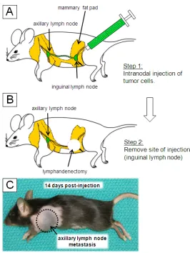
model utilizes two interconnected lymph nodes: the inguinal lymph node
(injection site), and axillary lymph nodes (site of metastasis), as illustrated in
Figure 2.1A. When labeling cells with 3μm beads in vitro, the majority of cells
(~70%) were efficiently labeled with the beads, and labeling did not inhibit cell
growth in vitro or the ability of cells to form metastases in vivo (data not shown).
In the first step illustrated in Figure 2.1, a cell suspension, containing a ~16:1
mixture of B16F10-LacZ cells and 16μm reference beads (see Figure 2.1D), was
injected directly into the inguinal lymph node. By applying slow and steady
pressure to the syringe, the injectate traveled (~40μL per minute) through the
inguinal lymph node, lymphatic duct, and drained into the axillary lymph node.
This is simulated in Figure 1B, where a blue tissue dye was injected into the
inguinal lymph node. In the second step, after injection, the inguinal lymph node
was surgically removed to prevent tumour growth that may further seed the
axillary lymph node, thereby abolishing the injected 16:1 tumour cell-to-bead
ratio. Two weeks post-injection, overt axillary lymph node metastases were
apparent (Figure 2.1C). To assess whether tumour cells were phagocytosed by
immune cells, tumour cells were labeled with 3μm beads prior to injection into the
model to study the development of axillary lymph node metastases, by
performing a time-dependent experiment that permitted the evaluation of
changes in tumour burden at progressive time points as described in the
Figure 2.1Lymph node experimental metastasis assay (LEMA).
A diagram of the experimental lymph node metastasis model is shown (panel A).
A suspension of tumour cells and 16 μm reference beads are intranodally
injected into the inguinal lymph node. Excess fluid pressure cause the
suspension to leave the inguinal lymph node (via efferent lymphatic vessel) into
the lymphatic duct. The cell suspension then arrives in the axillary lymph node
via the afferent lymphatic vessel. Tumour cells and 16 mm reference beads
arrest in the subcapsular region of the afferent side of the axillary lymph node. In
panel B, the drainage route of the cell suspension is simulated where a blue
tissue dye was injected in the same manner to visualize the lymphatic drainage
pathway. After injection with tumour cells, a large axillary lymph node metastasis
became apparent after 14 days (panel C). In panel D, a bright-field micrograph is
shown of the cell suspension containing B16F10-lacZ cells (white *) and 16μm
reference beads (white arrowheads) is shown. In addition, the B16F10-lacZ cells
were pre-labeled with 3μm beads (black arrows) to determine if host cells
phagocytosed tumour cells once they arrive in the lymph node. Scalebar = 20μm
Figure 2.2 A time-dependent study to investigate the progression of lymph
node metastasis progression. Forty mice underwent surgery in which
B16F10-lacZ cells were intranodally injected into the inguinal lymph node of female
C57/Bl6 mice. The inguinal lymph node was then surgically removed, and the
mouse was allowed to recover. Forty mice were divided into 4 groups that were
euthanized at 90 minutes, 3, 7, and 14 days post-injection. Lymph nodes were
then examined for the presence of isolated tumour cells, micrometastases and
2.3.2 Incidence of isolated tumour cells, micrometastases, and overt
metastases
The growth of lymph node metastases over time, including incidences of
each type of metastatic deposit (isolated tumour cells, micrometastases, overt
metastases), is summarized in Table 2.1. The presence or absence of each type
of lesion was tabulated. Shortly after injection into the inguinal lymph node (90
minutes), isolated tumour cells were observed in the axillary lymph node in all 10
mice. After 3 days had elapsed, isolated tumour cells were found in 9/10 mice,
and micrometastases were found in 7/10 mice. At 7 days post-injection, no
isolated tumour cells were found in any of the mice. The incidences of
micrometastases and overt metastases were 1/10 and 2/10, respectively. By 14
days, 1/10 mice had a micrometastasis, and the majority of lymph nodes in mice
(6/10) were overrun by tumour tissue. The assessment of incidence of a
particular metastatic lesion type was binary: only the presence or absence of the
lesion type was denoted. Since this did not provide information about the extent
of nodal involvement, we subsequently assessed metastatic tumour burden by
quantifying the proportion of lymph node surface area occupied by tumour and
2.3.3 Stereological analysis of whole-mount surface tumour burden
To measure the surface area of lymph nodes that was occupied by
tumour, tumour cell expression of -galactosidase (blue in color) on the surface
of the lymph node was assessed. Representative axillary lymph nodes from each
time point are shown in Figure 2.3 A-D. After injection into the inguinal lymph
node, tumour cells drained into the subcapsular sinus of the axillary lymph node,
and can be visualized as blue staining at the lymph node surface (90 minutes;
Figure 2.3A). In Figure 2.3B, the surface distribution of tumour cells was reduced
at 3 days post-injection. The reduction in surface tumour burden continued to
decrease at 7 days. One lymph node harbored a micrometastasis at this
timepoint, as shown in Figure 2.3C. By 14 days, large overt metastases had
formed in the majority of lymph nodes, Figure 2.3D.
The observations above were supported by quantitative measurements of
surface tumour burden as determined by stereological analysis (point count
method) in Figure 2.3E, where the means (± standard deviations) are shown. At
90 minutes post-injection, tumour cells were found on average, to occupy 50.7%
of the lymph node surface area. The surface area occupied by tumour cells
significantly dropped to 10% and then 8%, at 3 and 7 days post-injection,
respectively. By 14 days, the surface area of tumour tissue significantly rose to
54.6%, which reflects the increase in overt metastases at this time point (see
Figure 2.3 Visualization and quantification of surface tumour burden of
axillary lymph nodes. Panels A-D depict the stages of lymph node metastasis
development: initial arrest of tumour cells in the lymph node (A), isolated tumour
cells (B), micrometastasis (C), and an overt metastasis (D), at the time points
indicated. Scalebar = 2 mm. In panel B, inset shows higher magnification
showing clusters of isolated tumour cells (arrowheads). Scalebar = 100μm. The
changes in surface tumour burden are quantified in panel E, where
untransformed means and back transformed standard deviations are shown, n =
10 mice per group. Statistics were performed on transformed data in which the
means were analyzed by one-way ANOVA (p <0.001), and a post-hoc Tukey’s
multiple comparisons test was used to determine which groups were significantly