SYNTHESIS AND CHARACTERIZATION OF MANNICH BASE TRANSITION METAL COMPLEXES:
DNA BINDING AND ANTI-BACTERIAL STUDIES
SIVAKAMI M*, NATARAJAN B, VIJAYACHANDRASEKAR M
Department of Chemistry, SRM University, Kattankulathur, Tamil Nadu, India. Email: [email protected]
Received: 21 June 2014, Revised and Accepted: 15 September 2014
ABSTRACT
Objective: Succinimide (pyrolidine 2,5-dione) is a synthetically versatile substrate used for the synthesis of heterocyclic compounds and as a raw material for drug synthesis. Derivatives of succinimide are of important biological and pharmaceutical interest.
Methods: The novel mannich base 1-((2,5-dioxopyrrolidin-1-yl)(4-methoxyphenyl)methyl)thiourea succinimide, methoxy benzaldehyde and thiourea (SMBTU) has been synthesized in good yield by condensation of equimolar quantities of SMBTU. manganese (II), cobalt (II), nickel (II) and copper (II) complexes of the above ligand have also been synthesized.
Results: Structures of newly synthesized compounds were confirmed by elemental analysis, infrared, ultraviolet-visible and nuclear magnetic resonance. All the complexes adopt octahedral geometry around the metal ions. All the newly synthesized compounds were screened for their anti-microbial activity against Escherichia coli and Bacillus subtilis bacteria by minimal inhibitory concentration technique. The binding of the cobalt chloride complex of the ligand with calf thymus DNA has been investigated using absorption spectroscopy, fluorescence spectroscopy and viscosity measurements.
Conclusion: All the complexes exhibit octahedral geometry. The ligand and its metal complexes have shown significant antibacterial activity. The Co (II) metal complex showed efficient DNA binding ability and the binding constant value is consistent with other typical intercalators.
Keywords: Mannich base, Metal complexes, Anti-bacterial, DNA binding, Fluorescence and Viscosity measurements.
INTRODUCTION
Mannich reaction consists of amino alkylation of an acidic proton placed next to a carbonyl group with formaldehyde and ammonia or any primary or secondary amine. The final product is a β-amino carbonyl compound. Reactions between imides and aromatic aldehydes have also been considered as Mannich reactions. A review of the literature regarding Mannich reactions shows extensive volume on chemical, biological and toxicological feature of Mannich bases [1-6] with vast applications as polymers, dispersants in lubricating oil and pharmaceutical agents. It is well known that compounds containing amide moiety as functional group have been found to possess donor properties and exhibited a wide range of biological activities [7-13]. Transition metals are essential for normal functioning of living organisms and are, therefore, of great interest as potential drugs [14]. The coordination chemistry of nitrogen donor ligands is an interesting area of research. A great deal of attention in this area has been focused on the complexes formed by 3D metals with bidentate ligands using both the nitrogen atoms of the substrates. The study of structural and binding features of various Mannich base complexes can play an important role in better understanding of the complex biological processes. Several drugs showed increased activity as metal chelates rather than as organic compounds [15]. It has been reported in the literature survey that cobalt (II) complexes with octahedral geometry show remarkable intercalative binding affinity as well as DNA cleavage properties [16,17]. Further cobalt is an element of biological interest which is present in the active center of vitamin B12, which regulates the synthesis of DNA indirectly. It is known that there are about eight cobalt dependent proteins [18]. Many cobalt complexes possess antitumor, anti-proliferative, antimicrobial and antifungal activity [19-26]. To the best of our knowledge, no work has been done on this class of metal complexes with the Mannich base ligand succinimide, methoxy benzaldehyde and thiourea (SMBTU). In the continuation of our research work, herein, we report the synthesis of a new Mannich base derived from SMBTUand the metal complexes with Mn (II),
Co (II), Ni (II) and Cu (II). The characterization studies of all the metal complexes have been done with appropriate methods. All the metal complexes were screened for antibacterial activities. The DNA binding studies of the cobalt complex containing the ligand SMBTU is reported.
METHODS
All the reagents and solvents used for the synthesis of ligand and the metal complexes were analar grade of highest available purity and used as such without further purification.
Elemental analysis were performed using Carlo Erba 1108 analyzer and Coleman N analyzer and were found within ±0.5%. The molar conductivities of the metal complexes were measured in approximately 10−3 M ethanol solution using a systronics direct reading digital conductivity meter-304 with dip type conductivity cell. The infrared (IR) spectra was recorded as KBr pellets on Perkin-Elmer 1000 unit instrument. Absorbance in ultraviolet-visible (UV-VIS) region was recorded in dimethylformamide (DMF) solution using UV-VIS spectrometer. The 1H and 13C nuclear magnetic resonance (NMR) of the ligand was recorded on a bruker instrument employing tetramethylsilane (TMS) as internal reference and dimethyl sulfoxide (DMSO) - DMF as solvent. The mass spectral study of the ligand was carried out using liquid chromatography (LC) mass spectrometer. Magnetic susceptibility measurements at room temperature were made by using a gouy magnetic balance. Anti-bacterial activity has been carried out using minimal inhibitory concentration (MIC) technique.
Anti-bacterial activity
colonies (1×10¹⁰ CFU/ml) were in 0.7% of sterile saline, and the final concentrations of metal complexes were varied from 50 µg to 400 µg. 12 hrs incubation was given, and absorbance was taken at 600 nm. 50% of reduction was calculated as MIC. Afterward, the activity of the drug was visualized by well-diffusion assay, and the zone of inhibition was calculated.
Synthesis
Preparation of Mannich base 1-((2,5-dioxopyrrolidin-1-yl) (4-methoxyphenyl)methyl)thiourea
SMBTU were taken in 1:1:1 molar ratio. In aqueous solution of succinimide and thioure, methoxy benzaldehyde was added drop wise, and the mixture was stirred in a magnetic stirrer at room temperature for 8-10 hrs. After a week a solid product formed was filtered, washed with distilled water, dried in an air oven at 60°C and recrystallized using ethanol and chloroform in 1:1 ratio (Fig. 1).
Mechanism
The reaction route for the synthesis of Mannich base (SMBTU) is illustrated in Fig. 2 which involves the condensation reaction of methoxy benzaldehyde with thiourea to form the imine product. This electron deficient imine is then attacked by imide to give the ligand SMBTU.
Synthesis of metal complexes
All the metal complexes of SMBTU were prepared by slow addition of hot methanolic solution of the metal salt with hot ethanolic solution of the ligand in 1:1 molar ratio. The insoluble metal complexes were formed after 2 weeks. It was washed with methanol and ethanol to remove unreacted metal salt and ligand. The products were then dried in an air oven at 60°C. The proposed structures of the metal complexes are shown in Fig 3.
RESULTS AND DISCUSSION
Physical properties and elemental analysis
The physical properties and elemental analysis of the prepared ligand and their metal complexes are described in Table 1. Structures have been
suggested according to these data together with obtained from spectral analysis. The structure of metal complexes was further confirmed by conductivity measurements and magnetic moment determinations. Most of the metal complexes have been found to possess high melting points.
UV-VIS spectroscopic studies
The electronic spectra of the metal complexes were recorded for their solution in DMSO in the range of 180-1800 nm (Table 2).
The electronic spectrum of manganese chloride complex exhibits four absorption bands at 18050/cm, 24985/cm, 29125/cm and 31272/cm for 6A
1g→4T1g, 6A1g→4E2g, 6A1g→4E1g and charge transfer transitions respectively. The μeff value of 4.85 B.M points to a high spin octahedral geometry [27-30].
The cobalt chloride complex shows four absorption bands at 6703/cm, 14,365/cm, 18,742/cm, 29,066/cm assigned for 4T
1g→4T2g, 4T1g→4A2g, 4A
1g→4T1g and charge transfer transition. The μeff value was found to be 4.48 B.M which supports octahedral geometry.
The nickel chloride complex shows absorption bands at 10,525/cm, 15,780/cm and 24,890/cm and 35,235/cm for the transitions 1A
1g→3T1g, 1A
1g→3T2g, 1A1g→3T1g and charge transfer transitions respectively. The μeff value was found to be 3.56 B.M suggestive of octahedral geometry [31].
The copper chloride complex registers absorption bands at 9275/cm, 10374/cm, 12557/cm due to 2B
1g→2A1g, 2B1g→2B2g, 2Eg→2T2g transitions respectively. The charge transfer transition bands occur at 24330 and 28327/cm. The μeff value was found at 2.09 B.M suggesting octahedral geometry.
IR spectra
In order to study the binding mode of the ligand to metal in the complexes, the IR spectrum of the free ligand was compared with the corresponding metal complexes. Selected vibrational bands of the ligand and its metal complexes and their assignments are listed in Table 3. The IR spectrum the free ligand exhibited a strong band at 1690/cm
1+ 2
2
2
2 +1 1+
6
1 2
2
+1 1+ 6
2
3\UUROLGLQHGLRQH 0HWKR[\EHQ]DOGHK\GH 7KLRXUHD GLR[RS\UUROLGLQ\O
PHWKR[\SKHQ\OPHWK\OWKLRXUHD Fig. 1: Synthesis of the ligand SMBTU
Table 1: Analytical data of the ligand SMBTU and its metal complexes
Compound M.Pt°C Color Yield
% Found (Calculated %)C H N O M
SMBTU C13H15N3O3S 123 White 86 53.23 (53.17) 5.15 (5.08) 14.32 (14.26) 16.36 (16.31)
-MnCl2. 2H2O
SMBTU C14H20Cl2MnN3O5S 160 White 90 35.91 (35.86) 4.31 (4.29) 8.97 (8.95) 17.08 (17.01) 11.73 (11.65) CoCl2. 2H2O
SMBTU C14H20Cl2CoN3O5S
164 Light blue 83 35.61 (35.56) 4.27 (4.22) 8.90 (8.88) 16.94 (16.90) 12.48 (12.33)
NiCl2. 2H2O
SMBTU C14H20Cl2N3NiO5S 170 Dull green 94 35.63 (35.58) 4.27 (4.20) 8.90 (8.86) 16.95 (16.89) 12.44 (12.39) CuCl2. 2H2O
DNA binding
One of the most important approaches in the development of drugs and chemotherapy against some cancers, viral and parasitic diseases involve drugs that interact reversibly with DNA. Hence, syntheses of new metal complexes, which can bind with specificity to DNA and bring about its cleavage are of importance in the development of new antitumor agents [35].
Electronic absorption spectra
The binding of calf thymus (CT)-DNA with the synthesized Co (II) complexes was studied using UV absorption spectral method.
The concentration of CT-DNA per nucleotide was measured by using its known extinction coefficient at 260 nm (6600/m/cm). Tris HCl-buffer [5 mM Tris(hydroxymethyl) amino methane, pH 7.2] 50 mM NaCl was used for the absorption, viscosity and thermal denaturation experiments.
Absorption titration experiments were carried out by varying the DNA concentration (0-100 μM) and maintaining the metal complex concentration constant. Absorption spectra were recorded after successive addition of DNA and equilibration (approximately
2
Complex 0Λm (ohm−1
cm2 mol−1)
SMBTU: Succinimide, methoxy benzaldehyde and thiourea
Table 3: Characteristic IR absorption frequencies (cm−1) of SMBTU and its metal complexes
Compound νNH νC=O νC=S νCH (st) νCH (b) N-C-N -OCH3 H2O coordinates M-X M-S
SMBTU 3297 1690 1392 3174 814 1472 1272 -
-MnCl2. 2H2O. SMBTU 3289 1699 1394 3170 811 1462 1271 3746, 1512, 728 422
-CoCl2. 2H2O. SMBTU 3291 1684 1398 3170 808 1467 1271 3778, 1591, 728 425
-NiCl2. 2H2O. SMBTU 3290 1683 1396 3170 808 1463 1271 3747, 1590, 728 423
-CuCl2. 2H2O. SMBTU 3292 1687 1400 3169 808 1466 1268 3747, 1591, 727 424
-IR: Infrared, SMBTU: Succinimide, methoxy benzaldehyde and thiourea which could be assigned to νC=O of the succinimide ring. A band around 3297/cm could be attributed to stretching vibration of νN=H bond [32]. A strong band observed around 1392/cm can be assignable to νC=S vibration mode. In the metal complexes, the band corresponding to νC=O of succinimide ring was shifted to lower frequency range suggesting the coordination of the carbonyl group with a metal ion. There is no shifting of bands at 1400/cm and 750/cm indicating the absence of coordination of C=S group with a metal ion. The N-C-N stretching frequency of the ligand at 1472/cm was shifted toward lower values in all the complexes, indicating the involvement of the nitrogen of thiourea in coordination to the central metal ion. The participation of oxygen and nitrogen in coordination with the metal ion is further supported by the new band appearance of νM-N around 420-425/cm in the far IR region [33,34].
1H NMR Data: (DMSO/TMS, 500.3MHz): δ 2.56 (s, 4H), 3.77 (s, 3H), 3.80 (s, 3H), 6.49 (bs, 2H), 6.93 (d, 1H, J=8.30 Hz), 7.06-7.08 (m, 1H), 7.42 (d, 1H, J=1.80 Hz), 7.75 (s,1H), 10.09 (s, 1H), 11.06 (s, 1H), ppm.
13C NMR Data: (DMSO/TMS, 125.7 MHz): δ 179.4, 156.9, 149.9, 139.5, 127.6, 120.9, 111.3, 108.2, 55.6, 40.0, 29.5.
LC mass data: Calculated for SMBTU C13H15N3O3S m/z=293.08; Found 294.37 (M+1).
Anti-bacterial activity of Mannich base
10 minutes). Absorption titration experiments with CT-DNA had shown intense absorption peaks at 230 and 280 nm in the UV region of the complex due to inter ligand π-π* transition of the coordinated groups in the complex. In addition of increasing amounts of DNA to the complex, both of the two characteristic peaks decreased gradually with the maximum hypochromicity of 15% and 20% respectively, suggesting the strong interaction between the complex and DNA. The observed data were then fitted into Eq. 1 to obtain the intrinsic binding constant Kb [36].
[DNA]/(εa−εf)=[DNA]/(εb−εf)+1/Kb(εa−εf) (1)
where εa, εb, and εf are the apparent, bound, and free metal complex extinction coefficients, respectively, at 263 nm (Fig. 4). A plot of [DNA]/ (εb–εf) versus [DNA] gave a slope of 1/(εb–εf) and intercept y equal to 1/Kb (εb–εf), where Kb is ratio of the slope to the yintercept (Fig. 5).
Fluorescence spectra
Fluorescence quenching experiments were performed with ethidium bromide (EB) bound DNA with increasing concentrations of metal
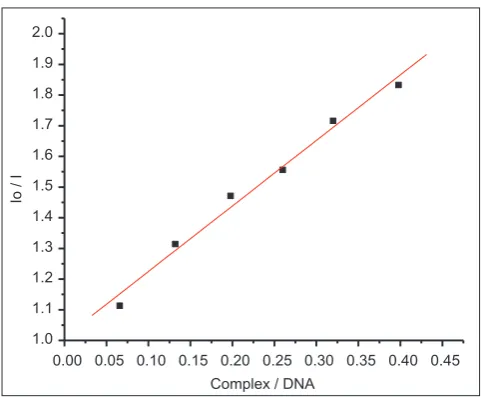
complex to determine the extent of binding between the molecule and DNA. EB is an indicator for fluorescence quenching [37]. The quenching extent of fluorescence EB bound to DNA is used to determine the DNA binding strength of the metal complex. The fluorescence quenching curves of EB bound to DNA in absence and presence of the complex was monitored. The addition of the metal complex to EB bound to DNA has shown a reasonable reduction in emission intensity indicating that the complex is bound to DNA at the sites occupied by EB (Fig. 6). The quenching plots indicate that the quenching of EB bound to DNA by the metal complex is in good agreement with the linear Stern–Volmer equation. In the plot of I0/I versus (complex)/(DNA), K is given by the ratio of the slope to the intercept (Fig. 7).
Viscosity measurements
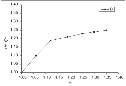
Viscosity measurements are used to explore the binding modes of complex with DNA. Optical photophysical probes provide necessarily, but not sufficient, clues to support a binding model. To clarify further the interactions between the complex and DNA, viscosity measurements were carried out. Viscosity measurements that are sensitive to length change are regarded as the least uncertain and the most critical tests of a binding model in a solution in the absence of crystallographic structural data (Satyanarayana et al., 1992). A classical intercalation model demands that the DNA helix must lengthen as base pairs are separated to hold the binding ligand, leading to the increase of DNA viscosity. In contrast, a partial intercalation ligand could bend the DNA helix, reduce its effective length and, in tandem, its viscosity (Satyanarayana et al., 1993). In Fig. 8, the relative viscosity increased with the addition of metal complex, showing that the cobalt complex bind with DNA in the classical intercalation mode.
Fig. 4: Absorption spectra of Co(II) complex in the absence and presence of the CT-DNA concentration (0-100μΜ)
'1$(
D
(I
[
'1$[0
Fig. 5: Plot of [DNA]/ (εb – εf) versus [DNA] for the complex with DNA
Fig. 6: Emission spectrum of EB bound to DNA in the absence and presence of Co (II) complex
,R,
&RPSOH['1$
Fig. 7: Plot of emission intensity IO/I versus (complex)/[DNA]
Table 4: Diameter of inhibition against bacteria in millimeter (mm) by SMBTU and its metal complexes
Compound E. coli B. subtilis
SMBTU 2.0±0.2 1.9±0.3
MnCl2.2H2O.SMBTU 2.5±0.1 2.3±0.2
CoCl2.2H2O.SMBTU 2.2±0.3 2.1±0.05
NiCl2.2H2O. SMBTU 1.4±0.2 1.3±0.2
CuCl2.2H2O.SMBTU 1.8±0.4 1.7±0.2
In this paper, coordination chemistry of a Mannich base ligand obtained from the reaction of succinimide, methoxy benzadehyde and thiourea is described. Mn (II), Co (II), Ni (II) and Cu (II) complexes have been synthesized using the above Mannich base ligand and characterized on the basis of analytical, magnetic and spectral data. The Mannich base coordinates through its thiourea nitrogen and oxygen of succinimide to the metal ion and acts as a neutral bidentate ligand. All the complexes exhibit octahedral geometry. The ligand and its metal complexes have shown significant antibacterial activity. The Co (II) metal complex showed efficient DNA binding ability and the binding constant value is consistent with other typical intercalators.
REFERENCES
1. Tramontini M. Advances in the chemistry of Mannich bases. Synthesis 1973;1993(12):703-5.
2. Tramontini M, Angiolini L. Further advances in the chemistry of Mannich bases. Tetrahedron 1990;46:1791.
3. Tramontini M, Angiolini L. Mannich Bases: Chemistry and Uses. Boca Raton: RC Press; 1994.
4. Joshi S, Khosla N, Tiwari P. In vitro study of some medicinally important Mannich bases derived from antitubercular agent. Bioorg Med Chem 2004;12(3):571-6.
5. Joshi S, Manikpuri AD, Khare D. Synthetic spectroscopic and antibacterial studies of Mannich bases of 2-chloro 4-nitro benzamide. J Indian Chem Soc 2008;85:1-5.
6. Joshi S, Khosla N, Khare D, Tiwari P. Synthesis and antibacterial screening of novel sulfonamide Mannich bases. Acta Pharm 2002;52(3):197-206.
7. Raman N, Ravichandran S. Synthesis and characterrization of a new schiff base and its metal complexes derived from the Mannich base N-(1-Piperidino benzyl) acetamide. Synth React Inorg Met Org Nano-Metal Chem 2005;35(6):439.
8. Reshetova K, Ustynyuk YA.Binuclear and polynuclear transition metal complexes with macrocyclic ligands.RussChemBull 2004;53:335. 9. Zoupy A, Petit A, Hamelin F, Mathe D. New solvent free organic
synthesis using focused microwaves. Synthesis 1998;1998(9):1213. 10. Gangadasu B, Narender P, Raju BC, Rao VJ. Calcium chloride catalyzed
three component one pot condensation reactions. An efficient synthesis of 3,4-di hydro pyrimidin 2(1H)-ones. IndianJChem 2006;45B:1259. 11. Pelczar MJ, Chan EC, Krieg NR. Microbiology.5th ed. New York:
McGraw-Hill Ltd.; 1998.
12. Scozzafava A, Menabuoni L, Mincione F, Mincione G, Supuran CT. Carbonic anhydrase inhibitors: Synthesis of sulfonamides incorporating dtpa tails and of their zinc complexes with powerful topical antiglaucoma properties. Bioorg Med Chem Lett 2001;11:575-82.
13. Walsh C. Enabling the chemistry of life. Nature 2001;409(6817):226-31. 14. Malhotra E, Kaushik NK, Malhotra HS. Synthesis and studies of ionic
chelates of hafnocene with guanine. IndianJ Chem 2006;45(2):370-6. 15. Chandra S, Shukla D, Gupta LK. Synthesis and spectroscopic studies
of Co(II), Ni(II) and Cu(II) complexes with N donor (N4) macrocyclic ligand (DSLF). J Indian Chem Soc 2008;85:800-6.
16. Pathak P, Jolly VS, Sharma KP. Synthesis and Biological activities of some new substituted aryl azo Schiff bases. Orient J Chem 2000;16(1):161-2.
17. Samadhiya S, Halve A. Synthetic utility of Schiff bases as potential herbicidal agents, Orient J Chem 2001;17(1):119-22.
18. Bernhardt PV, Lawrance GA. Cobalt. In: McCleverty JA, Meyer TJ, editors. Comprehensive Coordination Chemistry. IInd ed., Vol. 6. Oxford: Elsevier; 2003. p. 1-45.
19. Dwyer FP, Gyarfas EC, Rogers WP, Koch JH. Biological activity of complexions. Nature 1952;170:190-1.
20. Hall MD, Failes TW, Yamamoto N, Hambley TW. Bioreductive activation and drug chaperoning in cobalt pharmaceuticals. Dalton Trans 2007;(36):3983-90.
21. Lopez-Sandoval H, Londono-Lemos ME, Garza-Velasco R, Poblano-Melendez I, Granada-Macias P, Gracia-Mora I, et al. Synthesis, structure and biological activities of cobalt(II) and zinc(II) coordination compounds with 2-benzimidazole derivatives. J Inorg Biochem 2008;102:1267-76.
22. Ott I, Abraham A, Schumacher P, Shorafa H, Gastl G, Gust R, et al. Synergistic and additive antiproliferative effects on human leukemia cell lines induced by combining acetylenehexacarbonyldicobalt complexes with the tyrosine kinase inhibitor imatinib. J Inorg Biochem 2006;100:1903-6.
23. Ott I, Schmidt K, Kircher B, Schumacher P, Wiglenda T, Gust R. Antitumour - Active cobalt-alkyne complexes derived from acetylsalicyclic acid; studies on the mode of drug action. J Med Chem 2005;48:622-9.
24. Miodragovic DU, Bogdanovic GA, Miodragovic ZM, Radulovic MD, Novakovic SB, Kaludjerovic GN, et al. Interesting coordination abilities of antiulcer drug famotidine and antimicrobial activity of drug and its cobalt(II) complex. J Inorg Biochem 2006;100:1568-74. 25. Nomiya K, Yoshizawa A, Tsukagoshi K, Kasuga NC, Hirakawa S,
Watanabe JJ. Synthesis and structural characterization of silver(I), aluminium(III) and cobalt(II) complexes with 4-isopropyltropolone (hinokitiol) showing noteworthy biological activities. Action of silver(I)-oxygen bonding complexes on the antimicrobial activities. Inorg Biochem 2004;98:46-60.
26. Lv J, Liu T, Cai S, Wang X, Liu L, Wang Y. Synthesis, structure and biological activity of cobalt(II) and copper(II) complexes of valine-derived Schiff bases. J Inorg Biochem 2006;100:1888-96.
27. Carlin RL. Stereochemistry of Cobalt(II) Complexes in Transition. Metal Chemistry. New York: Marcel Decker; 1965.
28. Lever AB. Inorganic Electronic Spectroscopy. Amsterdam: Elsevier; 1968.
29. Drago RS. Physical Methods in Inorganic Chemistry. New Delhi: Affiliated East West Press; 1978.
30. Khalaji AD. Mononuclear copper(I) complex [Cu (ca2en) (PPh3) (CN)]: Synthesis and characterization. DerChem Sinica 2011;2(6):7-11. 31. Lever AB. InorganicElectronic Spectroscopy. New York: Elsevier;
1968.
32. Karpenko AS, Shibinskaya MO, Zholobak NM, Olevinskaya ZM, Lyakhov SA, Litvinova LA, et al. Synthesis, DNA binding and intferon-inducing properties of isatin and benzo isatin hydrazones. Pharm Chem J 2006;40:595.
33. Reddy V, Patil N, Reddy T, Angadi SD. Synthesis and characterization of Cu (II), Co(II), Ni(II) complexes with Schiff bases derived from 3-(4-chloro phenoxy methyl)-4-amino 5mercapto-1,2,4-triazole. Orient J Chem 2008;24(1):201-6.
34. Agarwal RC, Chandrasekar V. Synthesis and structural studies of some diamine complexes of cobalt (II) and nickel (II) amine and dihydroxy benzoates. J Inorg Nucl Chem 1979;41:1057-61.
35. Kostava I. Lanthanides and anticancer agents. Curr Med Chem Anticancer Agents 2005;5(6):591-602.
36. Wolfe A, Shimer GH, Meehan T. Polycyclic aromatic hydrocarbons physically intercalate into duplex regions of denatured DNA. Biochemistry 1987;26:6392-6.