Serovar Paratyphi A Outbreak Strain and the Human Immune
Response to Infection
Ohad Gal-Mor,aJotham Suez,a,bDana Elhadad,a,bSteffen Porwollik,cEyal Leshem,dLea Valinsky,eMichael McClelland,c Eliezer Schwartz,dand Galia Rahava,b
Infectious Diseases Research Laboratory, Sheba Medical Center, Tel-Hashomer, Israela; Sackler Faculty of Medicine, Tel Aviv University, Tel Aviv, Israelb; The Vaccine Research Institute of San Diego, San Diego, California, USAc; Center for Geographic Medicine and Department of Medicine C, Sheba Medical Center, Tel-Hashomer, Israeld; and Government Central Laboratories, Ministry of Health, Jerusalem, Israele
Enteric fever is an invasive life-threatening systemic disease caused by the
Salmonella enterica
human-adapted serovars Typhi
and Paratyphi. Increasing incidence of infections with
Salmonella enterica
serovar Paratyphi A and the spreading of its
antibiotic-resistant derivates pose a significant health concern in some areas of the world. Herein, we describe a molecular and
phenotypic characterization of an
S
. Paratyphi A strain accounted for a recent paratyphoid outbreak in Nepal that affected at
least 37 travelers. Pulsed-field gel electrophoresis analysis of the outbreak isolates revealed one genetic clone (pulsotype),
con-firming a single infecting source. Genetic profiling of the outbreak strain demonstrated the contribution of specific
bacterio-phages as a prime source of genetic diversity among clinical isolates of
S
. Paratyphi A. Phenotypic characterization in
compari-son with the
S
. Paratyphi A ATCC 9150 reference sequenced strain showed differences in flagellar morphology and increased
abilities of the outbreak strain with respect to its motility, invasion into nonphagocytic cells, intracellular multiplication,
sur-vival within macrophages, and higher induction of interleukin-8 (IL-8) secreted by host cells. Collectively, these differences
sug-gest an enhanced virulence potential of this strain and demonstrate an interesting phenotypic variation among
S
. Paratyphi A
isolates.
In vivo
profiling of 16 inflammatory cytokines in patients infected with the outbreak strain revealed a common profile
of a remarkable gamma interferon (IFN-
␥
) induction together with elevated concentrations of tumor necrosis factor alpha
(TNF-
␣
), IL-6, IL-8, IL-10, and IL-15, but not IL-12, which was previously demonstrated as elevated in nontyphoidal
Salmonella
infections. This apparent profile implies a distinct immune response to paratyphoid infections.
S
almonella enterica
is a Gram-negative, facultative intracellular
pathogen posing a major public health concern worldwide.
The single species
S
.
enterica
consists of over 2,500 closely related
serovars, which share 96 to 99% sequence similarity (14).
Salmo-nella
, like many other bacterial pathogens, harbors clusters of
vir-ulence genes, known as
Salmonella
pathogenicity islands (SPIs),
that have been acquired via horizontal gene transfer. These
genomic clusters are considered to be “quantum leaps” in the
evolution of
Salmonella
(18) and play a fundamental role in its
pathogenesis (20) and host specificity (1).
Several
Salmonella enterica
serovars, such as
S.
Typhimurium
and
S.
Enteritidis, are ubiquitous and “generalist” pathogens that
can infect a broad range of animal hosts, causing different clinical
manifestations, including gastroenteritis or occasionally
septice-mia in humans, lethal diarrhea in calves, or a systemic disease in
genetically susceptible mice. In contrast,
S
. Typhi and
S
. Paratyphi
(collectively referred to as typhoidal serovars) are host specific,
causing an enteric fever disease in humans (38). Enteric fever is an
invasive life-threatening systemic disease with a global annual
es-timation of over 25 million cases, resulting in more than 200,000
deaths (9). In recent years, for unknown reasons, the incidence of
infections with serovar Paratyphi A is increasing, and in some
regions of the globe (particularly in south-east Asia), it is
account-able for up to 50% of all enteric fever cases (33, 35). Enteric fever
is a mostly food- and waterborne disease, and like all
Salmonella
infections is transmitted by the fecal-oral route. Initial
gastroin-testinal infection causes brief, often asymptomatic enteritis
fol-lowed by invasion through the gut mucosa to underlying
macro-phages and lymphoid tissue. Bacteria can survive and multiply
intracellularly within lymphoid follicles, mesenteric lymph nodes,
and the mononuclear phagocyte system. Systemic infection with
bacteremia and fever develops at 8 to 14 days postinfection,
ac-companied by bacterial spreading to systemic sites such as the
liver, spleen, and bone marrow. Secondary infection of the small
bowel can occur via secretion of bacteria through the
enterohe-patic cycle (reviewed in reference 17).
Due to the lack of a suitable animal model, much of our
un-derstanding of typhoidal serovars’ pathogenesis is extrapolated
from the susceptible mouse strains infection with the
nontyphoi-dal serovar (NTS), which normally does not cause a systemic
dis-ease in humans. Although this model has been crucial in
under-standing many aspects of
Salmonella
pathogenicity, conclusions
regarding the virulence of
S
. Paratyphi in humans and host
re-sponse to the infection must be carefully interpreted.
During October 2009, while traveling to Nepal, 38 healthy
young Israeli travelers developed enteric fever caused by an
S
.
Received23 September 2011Returned for modification3 November 2011
Accepted8 December 2011
Published ahead of print21 December 2011
Address correspondence to Ohad Gal-Mor, [email protected].
Supplemental material for this article may be found athttp://cvi.asm.org/.
Copyright © 2012, American Society for Microbiology. All Rights Reserved.
doi:10.1128/CVI.05468-11
on August 17, 2020 by guest
http://cvi.asm.org/
Paratyphi A strain. Herein we describe molecular and cellular
characterization of this outbreak isolate in comparison to the
characterized
S
. Paratyphi A ATCC 9150 strain. In addition, we
studied the human immune response to the
S
. Paratyphi A
infection and determined the inflammatory cytokine profile
during the acute phase of the disease. Our results highlight
patterns of genetic variation among
S
. Paratyphi A strains,
demonstrate differences in virulence capabilities
in vitro
, and
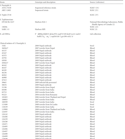
TABLE 1Bacterial strains used in this studyStrain Genotype and description Source (reference)
S. Paratyphi A
ATCC 9150 Sequenced reference strain SGSCa(31)
AKU 12601 Sequenced strain SGSC (21)
S.Typhi CT18 SGSC (37)
S.Typhimurium
DT104 96-5227 Harbors SGI-1 National Microbiology Laboratory, Public
Health Agency of Canada (2)
14028s SGSC (23)
SARC-13 Harbors HPI SGSC (3)
E. coliDH5␣ F⫺80lacZ⌬M15⌬(lacZYA-argF)U169 deoR recA1 endA1 hsdR17(rk⫺mk⫹)supE44 thi-1 gyrA96 relA1⫺
Lab collection
Clinical isolates ofS. Paratyphi A
3182 2009 Nepal outbreak Stool
36056/7 2007 traveler from Nepal Blood
45157 2009 Nepal outbreak Blood
45167 2009 Nepal outbreak Blood
45375 2009 Nepal outbreak Blood
45792 2009 Nepal outbreak Blood
45838 2009 Nepal outbreak Blood
45842/7 2007 traveler from Nepal Blood
46162 2009 Nepal outbreak Blood
46167 2009 Nepal outbreak Blood
46185 2009 Nepal outbreak Blood
46236 2009 Nepal outbreak Blood
46562 2009 Nepal outbreak Blood
46900 2009 Nepal outbreak Blood
47412 2009 Nepal outbreak Blood
50661 2009 infected lab personnel Blood
50676 2009 Nepal outbreak Blood
51190 2009 traveler from Nepal Blood
83698 2003 traveler from India Blood
83753 2003 traveler from India Blood
93223 2004 traveler from Romania Blood
105493 2006 traveler from Thailand and Nepal Blood
108003 2007 traveler from India Blood
108599 2007 traveler from India Stool
113498 2008 traveler from Sri Lanka Stool
118239 2008 traveler from India Stool
119989 2008 traveler from Thailand and India Blood
124597 2009 traveler from India Blood
126142 2009 Nepal outbreak Blood
126206 2009 Nepal outbreak Blood
126239 2009 Nepal outbreak Blood
126240 2009 Nepal outbreak Blood
126471 2009 Nepal outbreak Blood
126495 2009 Nepal outbreak Blood
126496 2009 Nepal outbreak Blood
126497 2009 Nepal outbreak Blood
126498 2009 Nepal outbreak Blood
126499 2009 Nepal outbreak Blood
126640 2009 Nepal outbreak Blood
aSGCS,SalmonellaGenetic Stock Center, University of Calgary, Calgary, Alberta, Canada.
on August 17, 2020 by guest
http://cvi.asm.org/
reveal important aspects of the human immune response to a
paratyphoid infection
in vivo
.
MATERIALS AND METHODS
Bacterial strains and growth conditions.Bacterial strains utilized in this study are listed in Table 1 and include 24S. Paratyphi A blood isolates obtained from the 2009 outbreak and 13 sporadicS. Paratyphi A isolates obtained during 2003 to 2009 (mostly from returning travelers). Bacterial cultures were routinely maintained in Luria-Bertani (LB; BD Difco) liquid medium at 37°C. LB or xylose lysine deoxycholate (XLD; BD Difco) agar plates were used when appropriate.
Genotyping by PFGE.Pulsed-field gel electrophoresis (PFGE) analy-sis was carried out according to the standardizedSalmonellaprotocol defined by CDC PulseNet (43), usingS.Braenderup H9812 as a molecular standard. XbaI-digestedSalmonellaDNA embedded in agarose plugs was subjected to PFGE analysis at 14°C in a CHEF DR III system (Bio-Rad Laboratories) using the following protocol: voltage, 6 V/cm for 19 h; ini-tial pulse, 2.2 s; final pulse, 63.8 s; angle, 120°; buffer, 0.5⫻ Tris-borate-EDTA. PFGE-generated DNA profiles were processed by the Bionumerics software V 5.1 (Applied Maths, Sint-Martens Latem, Belgium) using the Dice coefficients with a 1% position tolerance and optimization values. Cluster analysis was performed by the unweighted-pair group mean anal-ysis (UPGMA) method.
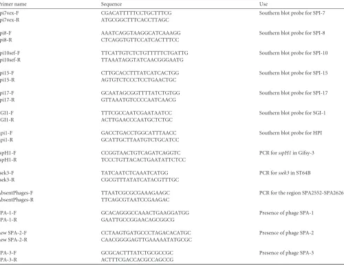
Southern blot hybridization.Primers used in this study are listed in Table 2. DNA primers were purchased from IDT, and PCR was carried out using ReddyMix PCR (Thermo Scientific) or PfuUltra II fusion HS DNA polymerase (Stratagene). For Southern blot analysis, 1g of genomic DNA was digested at 37°C for 16 to 18 h with PstI, subjected to electrophoresis in 1.0% agarose gels before being capillary transferred, and cross-linked onto Hybond-N membranes (Amersham Biosciences). Genomic DNA fromEsch-erichia coliDH5␣was included as a negative control in all hybridizations.S. Typhi CT18 andS.Typhimurium DT104, 14028s, and SARC-13 were used as positive controls. Southern blots were processed using the digoxigenin (DIG) DNA labeling and detection kit (Roche Applied Sciences), followed by an anti-DIG detection according to the manufacturer’s protocol.
CGH.For comparative genomic hybridization (CGH), genomic DNA fromS.Typhimurium LT2 andS. Paratyphi A (clone 45157) was extracted from overnight cultures grown in LB using the GenElute kit (Sigma-Aldrich) according to the manufacturer’s instructions. DNA labeling and hybridization to the STv7E pan-Salmonellamicroarray (http://www.sdibr .org/Faculty/mcclelland/mcclelland-lab/mcclelland-protocols) were performed as previously described (41). An Agilent microarray scanner G2505B was used for image acquisition, and signal intensities were quantified with the Spotreader software (Niles Scientific). Data normalization, analy-sis, and determination of the presence or absence of genes are de-scribed elsewhere (41).
TABLE 2Primers used in this study
Primer name Sequence Use
spi7vex-F CGACATTTTTCCTGCTTTCG Southern blot probe for SPI-7
spi7vex-R ATGCGGCTTTCACCTTAGC
spi8-F AAATCAGGTAAGGCATCAAAGG Southern blot probe for SPI-8
spi8-R CTCAGGTGTTCCATCACTTTCC
spi10sef-F TTCATTGTCTCTGTTTTTCTGATTG Southern blot probe for SPI-10
spi10sef-R TTAAATAGGTATCAACGGGAATG
spi15-F CTTGCACCTTTATCATCACTGG Southern blot probe for SPI-15
spi15-R AGTGTCTCCCTCCTGAACTGC
spi17-F GCAATAGCGGTTTTATCTGTGG Southern blot probe for SPI-17
spi17-R GTTAAATGTCCCCAATCAACG
SGI1-F TTTCGCCAATCGAATAATCC Southern blot probe for SGI-1
SGI1-R ACTTGAACCCAATGCTCTGC
hpi1-F GACCTGACCTGGCATTTAACC Southern blot probe for HPI
hpi1-R GCATTGCTTAATGTCTGCATCC
sspH1-F CCGGTAACTGTCAGATCAGGTC PCR forsspH1in Gifsy-3
sspH1-R TCCCTGTTACACTGAATATTCTCC
ssek3-F TATCAATCTCAAATCATGG PCR forssek3in ST64B
ssek3-R CGCGTTTATATCATACGTTTGC
AbsentPhages-F TTAATCGCGCGAAAGAAGC PCR for the region SPA2552-SPA2626
AbsentPhages-R TTCAGCGTAATCCGAAGAC
SPA-1-F GCACAGGGCCAAACTGAAGGATGG Presence of phage SPA-1
SPA-1-R GAATTGCCGGAACAGCGGCG
new SPA-2-F CCTAAGTGATGCCCTAGACACATGC Presence of phage SPA-2
new SPA-2-R CAACGGGGAGTTGAAAAATATGCGC
SPA-3-F GCGCACTTTATCTGCGCCGC Presence of phage SPA-3
SPA-3-R ACTTTCGACCACGCCAGCCG
on August 17, 2020 by guest
http://cvi.asm.org/
Tissue culture conditions and bacterial infection.The human epi-thelial cell line HeLa and the murine macrophage-like RAW264.7 cell line were cultured in a high-glucose (4.5 g/liter) Dulbecco’s modified Eagle medium (DMEM) supplemented with 10% heat-inactivated fetal bovine serum (FBS), 1 mM pyruvate, and 2 mML-glutamine. The human colonic adenocarcinoma Caco-2 cell line was grown in DMEM–F-12 medium supplemented with 20% FBS and 2 mML-glutamine. All cell lines were
cultured at 37°C in a humidified atmosphere with 5% CO2. Epithelial cells
and RAW264.7 macrophages were seeded at 5⫻104and 2.5⫻105cells/
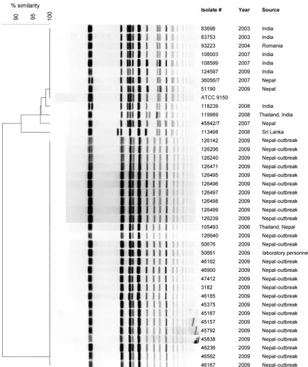
ml, respectively, in a 24-well tissue culture dish 18 to 24 h prior to bacterial infection, and experiments were carried out using the gentamicin protec-tion assay as previously described (15). HeLa and Caco-2 cells were in-fected at a multiplicity of infection (MOI) of ⬃1:50 withSalmonella strains that had been subcultured from an overnight culture and grown FIG 1Molecular fingerprinting ofS. Paratyphi A strains. FortyS. Paratyphi A isolates, including 26 clones isolated from the 2009 outbreak, an isolate from infected laboratory personnel (50661), 12 sporadic strains isolated during 2003 to 2008, and the ATCC 9150 reference strain were subjected to PFGE analysis using macrorestriction fingerprinting with XbaI. The isolate number, year of isolation, and the source are indicated adjacent to each pulsotype. Genetic similarity (%) was based on Dice coefficients and is presented by the phylogenetic tree.
on August 17, 2020 by guest
http://cvi.asm.org/
for 3 h to late logarithmic phase under aerobic conditions or with stationary-phase cultures grown under microaerophilic conditions. RAW264.7 cells were infected at an MOI of 1:10 using overnight stationary-phase-grown cultures. At the desired time points postinfection (p.i.), cells were washed three times with phosphate-buffered saline (PBS) and harvested by addition of lysis buffer (0.1% SDS, 1% Triton X-100 in PBS). Appropriate dilutions were plated on LB plates for bacterial enu-meration by CFU count.Salmonellainvasion was determined by the num-ber of intracellularSalmonellacells at 2 h p.i. divided by the number of infecting bacteria. Survival and intracellular multiplication (in macro-phages and in nonphagocytic cells, respectively) were determined by the number of recovered intracellular salmonellae at 24 h p.i. divided by the number of invading salmonellae at 2 h p.i.
Motility assay. Ten microliters of overnight Salmonella cultures grown in LB broth were placed onto 0.3% agar LB plates. The plates were incubated for 6 h at 37°C without being inverted.
Transmission electron microscopy.Salmonellastrains grown on soft agar plates were suspended in PBS and adsorbed onto 200-mesh Formvar/ carbon-coated copper grids. The grids were negatively stained with 2% aqueous uranyl acetate for 30 s. Images were obtained using a JEOL-1200EX (Jeol, Japan) transmission electron microscope at the Tel-Aviv University Electron Microscopy Facility.
Leukocyte count.White blood cell (WBC) counts were compared between two groups of patients: 24 patients (14 males and 10 females; average age, 27⫾7 years) with positive blood cultures forS. Paratyphi A who were hospitalized in the Sheba Medical Center between 2003 and 2010 and 23 patients (14 females, 9 males; average age, 55⫾18 years) with anE. coli-positive blood culture who were hospitalized between 2010 and 2011. Hematological data were retrieved from the hospital laboratory re-ports documenting the first blood specimens drawn from the patients with their administration. Data retrieval and analysis were done according to the local ethics committee-approved protocol.
Cytokine analysis.In vitroIL-8 analysis was performed following Caco-2 cell infection withS. Paratyphi A strains as described above. Se-creted IL-8 concentrations were determined 18 h p.i. using a Q-Plex array chemiluminescent IL-8 kit (Quansys Biosciences), according to the man-ufacturer’s protocol.In vivocytokine analysis was done using blood sam-ples collected from 10S. Paratyphi A-infected patients. The first blood sample was taken from all patients on admission during bacteremia and prior to antibiotic treatment. A second, matching blood sample was col-lected from the same patients 12 to 14 weeks after recovery. Serum was separated and kept at⫺80°C until final analysis. Cytokines were analyzed using the Q-Plex human cytokine screen system (Quansys Biosciences) based on a multiplex enzyme-linked immunosorbent assay (ELISA) ap-proach according to the manufacturer’s instructions. Chemiluminescent signal acquisition and quantification of spot intensity were done using the Quansys Q-View imager with Q-View software. The concentrations of cytokines were determined against 5-point standard curves using the Q-View software program. The statistical significance was calculated by the one samplettest, against a theoretical mean of 1, with a two-tailedP value.P⬍0.05 was considered to be statistically significant. Informed, written consent was obtained from all subjects. The study was approved by the ethics committee of Sheba Medical Center in accordance with the Helsinki II Declaration.
RESULTS AND DISCUSSION
Molecular fingerprinting of an
S
. Paratyphi A strain accounted
for an outbreak in Nepal.
During October 2009, 38 patients (20
males and 18 females; average age, 24.8
⫾
4.4 years) were
hospi-talized in different medical centers in Israel with acute enteric
fever and positive blood cultures for
S
. Paratyphi A. All of the
patients were returning travelers from Nepal and reported visiting
the city of Pokhara during the same time. To examine a possible
contamination from a single infection source, the genetic
similar-ity between these isolates was determined by PFGE. This analysis
integrated 40
S
. Paratyphi A isolates, including 26 isolates from the
2009 outbreak, 12 sporadic isolates obtained from returning
trav-elers from various places during 2004 to 2008, an isolate from a
laboratory technician who developed a paratyphoid fever in
No-vember 2009, and the
S
. Paratyphi A ATCC 9150 reference strain
(29). Macrorestriction discriminated the examined isolates into 6
distinct profiles (pulsotypes) as shown in Fig. 1. Twenty-five of 26
of the 2009 outbreak isolates had an identical pulsotype,
support-ing the assumption of a common infection source for this
out-break. A single isolate (51190), from a patient who returned from
Nepal later than the others, showed a pulsotype indistinguishable
from ATCC 9150 and a few previous sporadic isolates, suggesting
that isolate 51190 was acquired from a different source than the
rest of the outbreak isolates. Moreover, the PFGE pattern of the
October 2009 outbreak isolates was identical to that of a previous
clone (105493), isolated at 2006 from a patient returning from
Thailand and Nepal, who most likely got infected with the same
strain. The outbreak pulsotype was also identical to the isolate
obtained from the laboratory technician (50661) who got infected
after handling patients’ fecal samples, which might reflect the high
infectivity potential of this outbreak strain.
Nonetheless, despite the different pulsotypes identified, the
overall picture demonstrated a high degree of genetic similarity
between all
S
. Paratyphi A isolates. This observation is in
agree-ment with previous studies that reported low diversity among
worldwide
S
. Paratyphi A isolates and supports the idea of a recent
emergence of
S
. Paratyphi A from a single progenitor (21, 31).
Virulence gene profiling of the outbreak strain.
Many of the
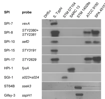
FIG 2Molecular typing of theS. Paratyphi A outbreak strain. Southern blot hybridization and PCR were used to determine the presence of theSalmonella pathogenicity islands 7, 8, 10, 15, and 17, HPI, SGI-1, and the virulence-associated prophages ST64B and Gifsy-3 in the genome of the outbreak strain (SPA 45157).E. coliDH5␣(DH5␣) was used as a negative control for all analyses and S. Typhi CT18 (S. Typhi), S.Typhimurium DT104 (STM DT104),SalmonellaReference Collection C 13 (SARC-13), andS. Typhimu-rium 14028s (STM 14028s) were used as positive controls. The specific genes used as probes to determine the presence of the above loci are listed. The presence of the prophages ST64B and Gifsy-3 was detected by PCR using primers specific tosseK3andsspH1, respectively, while the other images show the results of a Southern blot hybridization using the nonradioactive DIG system.
on August 17, 2020 by guest
http://cvi.asm.org/
Salmonella
virulence factor genes are organized within the
SPIs, genomic islands, and other mobile genetic elements,
in-cluding lysogenic bacteriophages. Thus far, 21 SPIs have been
identified, and in addition to the
Salmonella
genomic island 1
(SGI-1) and the high-pathogenicity island (HPI), they vary in
their distributions among different serovars and even between
isolates of the same serovar (reviewed in reference 45). To
better characterize the outbreak strain with respect to its
viru-lence gene repertoire, we analyzed the presence of multiple
virulence-associated loci in comparison with the ATCC 9150
sequenced strain, using a comparative genome hybridization
approach and a pan-
Salmonella
microarray (41). In addition,
to verify some of the microarray results and to explore the
presence of elements not represented on the array (such as the
Gifsy 3 and ST64B bacteriophages, HPI, and SGI-1), we used
Southern blot hybridizations, PCR, and direct sequencing of
selected targets. Overall, we found that the outbreak strain
con-sists of an SPI inventory similar to that of ATCC 9150, with 14
SPIs, including SPIs 1 to 6, 8 to 11, and 16 to 18, as well as CS54.
Like ATCC 9150, it lacks the SGI-1 and HPI elements, as well as
the Gifsy-3 and ST64B prophages (Fig. 2). More detailed
infor-mation about the presence/absence of more than 150-specific
Salmonella
virulence-associated genes of the outbreak strain is
presented in Table S1 in the supplemental material.
Integrated bacteriophages have been shown to affect the
viru-lence or fitness of
Salmonella
isolates and often encode virulence
factors (4).
S
. Paratyphi A sequenced strains ATCC 9150 and AKU
12601 harbor three particular phages, designated SPA-1 to -3 (31).
Our CGH analysis showed that the 41-kb lambdoid phage SPA-1
(genes SPA2385 to SPA2431) is present in the outbreak strain,
excluding three genes (SPA2409, SPA2412, and SPA2417). In
con-trast, both the 34-kb P2-type phage SPA-2-sopE (SPA2554 to
SPA2600), which carries
sopE
(an invasion-associated gene), and
the 25-kb SPA-3-P2 prophage (SPA2601 to SPA2625) are missing
from the genome of the outbreak strain. In ATCC 9150,
SPA-2-sopE is inserted between two perfect duplicated sequences of
45-bp (ATGTAGGAATTTCGGACGCGGGTTCAACTCCCGCC
AGCTCCACCA) located at positions 2657604 and 2691147 that
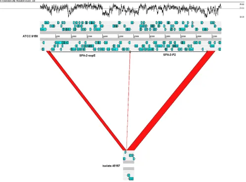
FIG 3The outbreak strain lacks a 65.7-kb region of SPA-2-sopE and SPA-3-P2 prophages. A 75,391-bp section of theS. Paratyphi A ATCC 9150 chromosome (positions 2658172 to 2733562) containing SPA-2-sopE and SPA-3-P2 prophages (top scheme) is compared against the sequence of a 4,591-bp PCR fragment amplified from the genome of the outbreak strain (isolate 45157; bottom scheme). The red regions indicate alignment between the two genomes, and the white regions indicate a missing region of 65,762 bp from the outbreak clone, in relation to the reference strain. Open reading frame (ORF) organization is shown by blue arrows, and the locations of SPA-2-sopE and SPA-3-P2 are indicated by the horizontal bars. The GC content (%) is shown in the upper panel. The figure was created using the Artemis Comparison Tool (6) and modified subsequently.on August 17, 2020 by guest
http://cvi.asm.org/
serves as the integration site for this phage. Similarly, SPA-3-P2 is
inserted between this sequence located at position 2691147 and a
second duplication at position 2723414. PCR analysis using
prim-ers flanking SPA-2-sopE and SPA-3-P2 and sequencing of the
ob-tained PCR product confirmed the absence of 65,762 bp from the
outbreak clone (Fig. 3). To get a broader perspective of the
distri-bution of these phages, we investigated their presence in the
ge-nome of the clinical
S
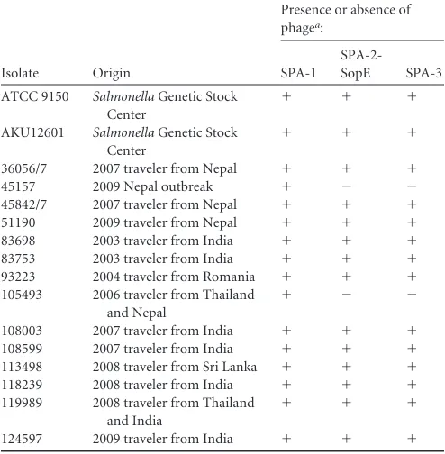
. Paratyphi A isolates using PCR. We found
that only the outbreak strain and isolate 105493, which shared the
same pulsotype, omitted SPA-2-sopE and SPA-3-P2, while the
rest of the isolates harbored all three phages (Table 3). These
re-sults emphasize the contribution of integrated bacteriophages to
the genetic diversity of
S
. Paratyphi A isolates.
Apart from the 75 genes contained within SPA-2-sopE and
SPA-3-P2, at least 31 additional genes (mainly metabolic and
genes with unknown function) were found to have been deleted
from the genome of the outbreak clone (in relation to the ATCC
9150 and/or AKU 12601 genomes; see Table S2 in the
supplemen-tal material). Interestingly, quite a few of these genes are actually
inactivated (pseudogenes) in ATCC 9150 or in other related
ge-nomes and therefore may represent a common pattern of genome
degradation in this group of pathogens.
The outbreak strain presents increased virulence in
compar-ison to the ATCC 9150 strain.
The two hallmarks of
Salmonella
pathogenicity are its ability to invade nonphagocytic cells and to
survive and proliferate within professional phagocytes (reviewed
in reference 19). In order to characterize the pathogenic potential
of the outbreak strain, we studied these abilities in comparison
with the characterized
S
. Paratyphi A ATCC 9150 strain. Previous
studies have shown that
Salmonella
invasiveness is growth phase
as well as oxygen tension dependent (26, 47). Hence,
S
. Paratyphi
A invasion and intracellular replication in a human epithelial cell
line were determined under two sets of conditions, including the
late logarithmic phase under aerobic conditions (LAC) and the
stationary phase under microaerophilic conditions (SMC).
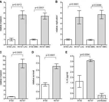
Gen-tamicin protection assays established that the outbreak strain was
able to invade HeLa cells 5-fold better than the reference strain
under both conditions (Fig. 4A). Intracellular replication of the
outbreak strain within nonphagocytic cells was also found to be
⬃
5-fold higher than that of the reference strain (Fig. 4B).
En-hanced invasion of the outbreak strain was also demonstrated in
Caco-2 cells (Fig. 4 C), suggesting that its superior invasion is not
a cell-line-specific characteristic, but represents a common
char-acteristic of this strain.
Infection experiments, using RAW264.7 macrophage-like
cells, were consistent with the results above and showed that the
survival of the outbreak strain was higher than that of the ATCC
9150 strain (Fig. 4 D).
Salmonella
invasion of intestinal epithelial cells leads to
induc-tion and secreinduc-tion of proinflammatory cytokines such as IL-8,
which play an important role in the recruitment of inflammatory
cells to the site of infection and elicitation of the mucosal
inflam-matory response (32). Due to the differences found in the invasion
abilitiy between the outbreak strain and
S
. Paratyphi A 9150, we
also assessed IL-8 secretion following epithelial cell penetration.
Caco-2 cells were infected with
S
. Paratyphi A strains grown under
microaerophilic conditions, and the secreted IL-8 concentration
was measured in the medium 18 h postinfection. Correlated with
the invasion data, epithelial cells that were infected with the
out-break 45157 strain were found to secrete higher levels of the
cyto-kine IL-8 than cells infected with the 9150 reference strain (Fig.
4E), most likely due to the increased invasion ability of the
out-break strain into host cells.
Collectively, these results suggest that the outbreak strain
pres-ents an enhanced virulence potential compared with the ATCC
9150 strain and that different
S
. Paratyphi A isolates vary in their
pathogenicity, at least
in vitro
.
The outbreak strain exhibits enhanced motility and a
differ-ent flagellar morphology.
Motility is an important virulence trait
for
Salmonella
, facilitating invasion into epithelial cells (25, 28).
The observed differences in invasion between the outbreak strain
and the reference strain prompted us to assess their motility using
the soft agar swimming assay. Comparison between
S
. Paratyphi A
strains grown to the stationary phase in rich LB medium
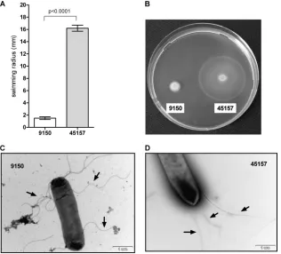
demon-strated significantly higher motility of the outbreak strain (Fig. 5A
and B) and provided a possible mechanistic explanation for its
increased invasion of host cells. To identify potential differences in
the expression or the formation of the flagella between these
strains, we applied transmitted electron microscopy. Although
both strains were found to be flagellated, the flagellar morphology
was found to be different. While ATCC 9150 showed a curly and
thicker flagellar structure, the flagella of the outbreak stain were
straight and thinner (Fig. 5C and D).
Salmonella
mutants that
produce irregular flagella were described more than 40 years ago
and were shown to be impaired in their movement (22). Thus, it is
very likely that the structural differences identified in the flagella
of both strains affect the degree of their motility.
Together, these experiments demonstrated both functional
and phenotypic variations (including in virulence) among
differ-ent isolates of
S
. Paratyphi A.
TABLE 3Distribution of phages among reference and clinical isolates of S. Paratyphi A
Isolate Origin
Presence or absence of phagea:
SPA-1
SPA-2-SopE SPA-3
ATCC 9150 SalmonellaGenetic Stock Center
⫹ ⫹ ⫹
AKU12601 SalmonellaGenetic Stock Center
⫹ ⫹ ⫹
36056/7 2007 traveler from Nepal ⫹ ⫹ ⫹
45157 2009 Nepal outbreak ⫹ ⫺ ⫺
45842/7 2007 traveler from Nepal ⫹ ⫹ ⫹
51190 2009 traveler from Nepal ⫹ ⫹ ⫹
83698 2003 traveler from India ⫹ ⫹ ⫹
83753 2003 traveler from India ⫹ ⫹ ⫹
93223 2004 traveler from Romania ⫹ ⫹ ⫹
105493 2006 traveler from Thailand and Nepal
⫹ ⫺ ⫺
108003 2007 traveler from India ⫹ ⫹ ⫹
108599 2007 traveler from India ⫹ ⫹ ⫹
113498 2008 traveler from Sri Lanka ⫹ ⫹ ⫹
118239 2008 traveler from India ⫹ ⫹ ⫹
119989 2008 traveler from Thailand and India
⫹ ⫹ ⫹
124597 2009 traveler from India ⫹ ⫹ ⫹
aThe presence (⫹) or absence (⫺) of SPA-1, SPA-2-SopE, and SPA-3 was determined by PCR using the primers SPA-1-F and SPA-1-R, new SPA-2-F and new SPA-2-R, and SPA-3-F and SPA-3-R, respectively.
on August 17, 2020 by guest
http://cvi.asm.org/
The immune response to an
S
. Paratyphi A infection
in vivo
.
In humans, an acute inflammatory disease, particularly due to
bacterial infection, is often associated with a high WBC count (39,
44). An accumulation of circulatory leukocytes has been
docu-mented in several
Salmonella
-infected animals, including
mon-keys (11), fowl typhoid in chickens caused by
S
. Gallinarum (16),
and in murine typhoid mediated by
S.
Typhimurium infection
(10). To examine whether paratyphoid patients demonstrate
in-creased WBC levels (leukocytosis) during their acute infection,
peripheral WBC counts of 24 hospitalized paratyphoid patients
were compared to those of a control group of 23 patients with
invasive
E
. coli infections. This comparison showed that while
invasive
E. coli
infections are characterized by increased WBC
numbers ([12.9
⫾
1.4]
⫻
10
3/ml), paratyphoid patients’ counts
([5.4
⫾
0.4]
⫻
10
3/ml) were within the lower end of the accepted
range (4.1
⫻
10
3to 10.9
⫻
10
3/ml) (Fig. 6A). These results
sug-gested that leukocytosis does not characterize the immune
re-sponse to a paratyphoid infection in humans during the acute
phase of the disease, as opposed to some other infectious diseases.
Different cytokines are known to play a pivotal role in
initiat-ing and regulatinitiat-ing the innate and adaptive immune responses
against
Salmonella
. A few clinical studies focusing on NTS
infec-tions in humans have reported the involvement of several
inflam-matory pathways, including gamma interferon (IFN-
␥
) (34, 48),
tumor-necrosis factor alpha (TNF-
␣
) (48), IL-6 (27), IL-8 (27),
IL-10 (34, 48), IL-12 (34, 48), IL-15 (34), and IL-18 (34). Other
studies have examined cytokine concentrations in the serum of
S
.
Typhi-infected patients (5, 24); however, not much is known
about the human immune response to a paratyphoid infection. To
shed some light on this subject, we analyzed the circulating levels
of 16 primary inflammatory cytokines (IL-1
␣
, IL-1

, IL-2, IL-4,
IL-5, IL-6, IL-8, IL-10, IL-12p70, IL-13, IL-15, IL-17, IL-23,
IFN-
␥
, TNF-
␣
, and TNF-

) in the serum of 10 paratyphoid
pa-tients, all of whom were infected with the outbreak strain. Serum
FIG 4The outbreak strain demonstrates an increased virulencein vitro. TheS. Paratyphi A reference strain ATCC 9150 and the outbreak strain (isolate 45157) were grown to either late logarithmic phase under aerobic conditions (LAC) or to a stationary phase under microaerophilic conditions (SMC) and used to infect different host cells. The invasion (A) and intracellular replication (B) of the outbreak strain in relation to the reference strain in HeLa cells are shown, following infection at a multiplicity of infection (MOI) of⬃50:1. (C)S. Paratyphi A strains that were grown to the stationary phase under microaerophilic conditions were used to infect Caco-2 cells at an MOI of⬃50:1. The invasion of the outbreak strain is shown in relation to the invasion of ATCC 9150. (D) RAW264.7 macrophage-like cells were infected with stationary-phaseS. Paratyphi A strains at an MOI of⬃10:1. Survival of the outbreak strain (45157) in the macrophages 24 h postinfection, in relation to the reference strain (9150) is presented. (E) Caco-2 cells were infected withS. Paratyphi A strains grown to the stationary phase under microaerophilic conditions. The secreted IL-8 concentration was measured in the tissue culture medium 18 h postinfection using a quantitative ELISA-based chemiluminescent assay (Q-Plex human IL-8 array). Baseline levels of IL-8 were measured in uninfected cells that were included as a control (uninfected). All panels present the mean and the standard error of the mean (SEM; represented by the error bars) of at least 3 independent infections. An unpairedttest with two tails was used to determine the significance of the differences between the compared data.on August 17, 2020 by guest
http://cvi.asm.org/
that was taken during the acute phase of the disease from each of
these patients was compared to a matching sample obtained from
the same patient 12 to 16 weeks after convalescence.
We found in 10/10 patients a dramatic elevation (
⬃
75-fold) of
IFN-
␥
, in addition to a more moderate induction of IL-6
(18.6-fold), IL-8 (6.2-(18.6-fold), IL-10 (4.4-(18.6-fold), IL-15 (1.6-(18.6-fold), and
TNF-
␣
(3.0-fold) (Fig. 6B). The serum concentrations of the other
10 tested cytokines (IL-1
␣
, IL-1

, 2, 4, 5, 12p70,
IL-FIG 5The outbreak strain demonstrates enhanced motility and different flagellar morphology. TheS. Paratyphi A reference strain (9150) and the outbreak strain (45157) were grown in LB overnight. Ten microliters of each culture was inoculated onto a soft (0.3%) agar plate and incubated at 37°C for 6 h. The ability of the different cultures to move through the soft agar (swim) was measured. The average of five independent experiments with the error bars representing the standard error of the mean is shown (A). One representative experiment was recorded using a Pentax K10D digital camera system (B).S. Paratyphi A strains and surface flagella were negatively stained using 2% uranyl acetate solution and visualized by scanning transmission electron microscopy. Electron micrographs at a⫻20,000 magnification of ATCC 9150 (C) and 45157 (D) are presented. Representative flagellar filaments in each strain are indicated by the black arrows.FIG 6S. Paratyphi A induces a distinct immune responsein vivo. (A) The WBC count is shown for 23 patients with invasive infections ofE. coliand 24S. Paratyphi A-infected patients. Each point represents the WBC count of a single patient, with the mean indicated by the horizontal line. An unpairedttest with two tails was used to determine the significance of the difference between the two groups. (B) Serum samples from 10S. Paratyphi A-infected patients were collected during the acute stage of the disease and 12 to 14 weeks after their convalescence. The serum concentration of 16 inflammatory cytokines was determined by the Q-Plex human cytokine screen kit. The change in the concentrations of 7 cytokines (IL-6, IL-8, IL-10, IL-12, IL-15, TNF-␣, and IFN-␥) during the disease versus convalescence is shown, with the calculatedPvalues below. Points represent the fold change (during disease/convalescence) in the serum concentration of specific cytokines as measured in each patient, with the mean indicated by the horizontal line. n.s., not statistically significant.
on August 17, 2020 by guest
http://cvi.asm.org/
13, IL-17, IL-23, and TNF-

) were not found to fluctuate between
the two time points. To the best of our knowledge, this analysis is
the broadest assessment of the cytokine response to a paratyphoid
infection
in vivo
thus far.
IFN-
␥
, the cytokine which had the most elevated levels among
the paratyphoid patients, is the primary cytokine that drives the
Th1-type immune response and is involved in the clearance of
intracellular pathogens. IFN-
␥
is produced by T-helper cells,
nat-ural killer (NK) cells, and NK-like cells and in synergy with
TNF-
␣
, (which was also found to increase in these patients) is
required for the initiation of antimicrobial functions of the
in-fected macrophages (7, 12, 50). A remarkable induction of IFN-
␥
during paratyphoid fever provided evidence that IFN-
␥
plays a
pivotal role in the human response to an
S.
Paratyphi A infection.
Nonetheless, other type 1 cytokines, including IL-17, IL-23, and
particularly IL-12, were not increased during the acute stage of the
disease. IL-12 is known to activate IFN-
␥
secretion in Th1 and NK
cells and was found to be elevated in NTS infections (34, 48).
While we cannot rule out the possibility of an earlier IL-12
induc-tion, its unchanged level during the acute phase of the paratyphoid
disease may suggest IFN-
␥
induction in an IL-12-independent
manner. A similar mechanism has been reported in the context of
the intracellular parasite
Leishmania major
infection,
demonstrat-ing that the effector function of mature Th1 cells
in vivo
is
inde-pendent of IL-12 (8). The lack of IL-12 induction in the presence
of a dramatic increase in IFN-
␥
may indicate the elicitation of a
different immune response to a paratyphoid disease rather than
the one to an NTS infection. Diverse host-pathogen interplay with
S
. Paratyphi A versus NTS is intriguing and consistent with
accu-mulating clinical evidence. It has been established that invasive
infections caused by NTS, but not by typhoidal serovars, are often
associated with immunocompromised adults, in particular in the
context of HIV (17). This epidemiological observation implies
that certain factors (which are probably malfunctioning in AIDS
patients) are required for the immune defense against systemic
infection of NTS, but not against typhoidal serovars.
Further-more, recent studies have shown that patients with inherited
de-ficiency of the IL-12/IL-23 system (IL-12p40/IL-12R

1) are
highly susceptible to invasive extraintestinal NTS infections, but
not to
S
. Typhi nor
S
. Paratyphi infections, even though some of
these patients live in areas where typhoid is endemic (30, 49).
Collectively, these observations support the possibility that
differ-ent inflammatory pathways may be involved in NTS versus
ty-phoidal infections, including a distinct role for the IL-12 pathway.
Additional cytokines found to be induced during paratyphoid
infection were IL-6, IL-8, and IL-10. IL-6 is produced in the
intes-tinal mucosa and is particularly important due to its pleiotropic
involvement in different pathways (36). Although commonly
considered a proinflammatory cytokine, there is also evidence that
IL-6 has important anti-inflammatory properties and may exert
protective effects in different tissues (51). IL-8 is a potent
neutro-phil chemotactic factor secreted by intestinal inflammatory cells
in response to bacterial invasion (13). IL-8 secretion induced by
some
Salmonella
serovars leads to a massive neutrophil migration
and an elicitation of a mucosal inflammatory response (reviewed
in reference 19). IL-10 is an anti-inflammatory cytokine, which
inhibits antigen presentation to T cells and suppresses
phagocyto-sis and intracellular killing (46). Interestingly, Pie et al. showed
that
S.
Typhimurium induced immunosuppression in mice and
caused the production of large amounts of IL-10 (40). As our
results indicated a moderate increase in IL-6 and IL-10 together
with the lack of leukocytosis, it is tempting to suggest a
pathogen-mediated IL-6/IL-10 induction, as a means to prevent T-cell
pro-liferation and to suppress the cellular immune response of the
host. Nonetheless, further experimental data are certainly
re-quired to support this hypothesis.
The cytokine profile found in the paratyphoid patients is
sim-ilar to previous cytokine analysis of serum from typhoid fever
patients infected with
S
. Typhi that revealed elevated levels of IL-6,
IL-8, TNF-
␣
, and IFN-
␥
(5, 24). In
S
. Typhi, the virulence (Vi)
capsule is presumed to be central to the host-pathogen
interac-tions and is believed to facilitate evasion of the immune system
(42). These results suggest that although
S
. Paratyphi A lacks the
Vi capsule, a significant overlap does exist in the immune response
to
S
. Typhi and
S
. Paratyphi infections.
Conclusions.
Our study describes genetic and phenotypic
characterization of an
S
. Paratyphi A strain that was identified as
the causative agent of a recent paratyphoid outbreak in Nepal.
Comparative PFGE with other
S
. Paratyphi A isolates and
microarray-based CGH analysis showed a high degree of genetic
homogeneity among different isolates of
S
. Paratyphi A. This
ob-servation is consistent with the notion that
S
. Paratyphi A strains
have evolved recently, on an evolutionary time scale, from a single
ancestor (21, 31). Nevertheless, subtle genetic differences between
the outbreak and the reference strains were found, including the
absence of two prophages (SPA-2-sopE and SPA-3-P2) from the
genome of the outbreak strain, as well as sporadically degraded
metabolic and other genes. Differences were also established on
the phenotypic level. The outbreak strain demonstrated enhanced
motility, invasion, and replication in nonphagocytic cells, higher
survival in macrophages, and elevated induction of IL-8 secretion
by host cells. Accumulatively, these results indicate that despite a
very high degree of genetic conservation, different
S
. Paratyphi A
isolates vary in their virulence potential. Cytokine profile analysis
during the acute phase of the disease indicated a remarkable
in-duction of IFN-
␥
in addition to a more moderate increase of IL-6,
IL-8, IL-10, IL-15, and TNF-
␣
, but unchanged levels of IL-12. The
revealed cytokine profile, together with several previously
re-ported clinical observations, suggests a distinct host response to
S
.
Paratyphi A infection in comparison to salmonellosis caused by
NTS strains. Better understanding and a more comprehensive
view of the virulence mechanisms and the immune response to
the
S
. Paratyphi infection might facilitate prevention efforts
and the development of novel therapeutics against this
emerg-ing pathogen.
ACKNOWLEDGMENTS
We thank Nati Keller and the staff of the bacteriology laboratory of the Sheba Medical Center for sharing clinical isolates ofS. Paratyphi A. We also thank the National Microbiology Laboratory Public Health Agency of Canada for theS.Typhimurium DT104 96-5227 strain.
This work was supported by grant 249241 from the European Com-munity’s Seventh Framework Program (PF7/2007-2013).
REFERENCES
1.Baumler AJ, Tsolis RM, Ficht TA, Adams LG.1998. Evolution of host adaptation inSalmonella enterica.Infect. Immun.66:4579 – 4587. 2.Boyd DA, Peters GA, Ng L, Mulvey MR.2000. Partial characterization of a
genomic island associated with the multidrug resistance region ofSalmonella entericaTyphymurium DT104. FEMS Microbiol. Lett.189:285–291. 3.Boyd EF, Wang FS, Whittam TS, Selander RK.1996. Molecular genetic
relationships of the salmonellae. Appl. Environ. Microbiol.62:804 – 808.
on August 17, 2020 by guest
http://cvi.asm.org/
4.Brussow H, Canchaya C, Hardt WD.2004. Phages and the evolution of bacterial pathogens: from genomic rearrangements to lysogenic conver-sion. Microbiol. Mol. Biol. Rev.68:560 – 602.
5.Butler T, Ho M, Acharya G, Tiwari M, Gallati H.1993. Interleukin-6, gamma interferon, and tumor necrosis factor receptors in typhoid fever related to outcome of antimicrobial therapy. Antimicrob. Agents Che-mother.37:2418 –2421.
6.Carver TJ, et al.2005. ACT: the Artemis Comparison Tool. Bioinformat-ics21:3422–3423.
7.Coburn B, Grassl GA, Finlay BB.2007.Salmonella, the host and disease: a brief review. Immunol. Cell Biol.85:112–118.
8.Constantinescu CS, et al.1998. The role of IL-12 in the maintenance of an established Th1 immune response in experimental leishmaniasis. Eur. J. Immunol.28:2227–2233.
9.Crump JA, Luby SP, Mintz ED.2004. The global burden of typhoid fever. Bull. World Health Organ.82:346 –353.
10. Dejager L, Pinheiro I, Bogaert P, Huys L, Libert C. 2010. Role for neutrophils in host immune responses and genetic factors that modulate resistance toSalmonella enterica serovar Typhimurium in the inbred mouse strain SPRET/Ei. Infect. Immun.78:3848 –3860.
11. DeRubertis FR, Woeber KA.1973. Accelerated cellular uptake and me-tabolism of L-thyroxine during acuteSalmonella typhimuriumsepsis. J. Clin. Invest.52:78 – 87.
12. Eckmann L, Kagnoff MF.2001. Cytokines in host defense against Salmo-nella.Microbes Infect.3:1191–1200.
13. Eckmann L, Kagnoff MF, Fierer J. 1993. Epithelial cells secrete the chemokine interleukin-8 in response to bacterial entry. Infect. Immun. 61:4569 – 4574.
14. Edwards RA, Olsen GJ, Maloy SR. 2002. Comparative genomics of closely related salmonellae. Trends Microbiol.10:94 –99.
15. Gal-Mor O, Valdez Y, Finlay BB.2006. The temperature-sensing protein TlpA is repressed by PhoP and dispensable for virulence ofSalmonella entericaserovar Typhimurium in mice. Microbes Infect.8:2154 –2162. 16. Garcia KB-J, Santana A, Freitas-Neto O, Fagliari J.2009. Experimental
infection of commercial layers using aSalmonella entericaserovar Galli-narum strain: leukogram and serum acute-phase protein concentrations. Braz. J. Poult. Sci.11:263–270.
17. Gordon MA. 2008.Salmonella infections in immunocompromised adults. J. Infect.56:413– 422.
18. Groisman EA, Ochman H.1996. Pathogenicity islands: bacterial evolu-tion in quantum leaps. Cell87:791–794.
19. Haraga A, Ohlson MB, Miller SI.2008. Salmonellae interplay with host cells. Nat. Rev. Microbiol.6:53– 66.
20. Hensel M.2004. Evolution of pathogenicity islands ofSalmonella enterica. Int. J. Med. Microbiol.294:95–102.
21. Holt KE, et al.2009. Pseudogene accumulation in the evolutionary his-tories ofSalmonella enterica serovars Paratyphi A and Typhi. BMC Genomics10:36.
22. Iino T, Mitani M. 1967. A mutant of Salmonella possessing straight flagella. J. Gen. Microbiol.49:81– 88.
23. Jarvik T, Smillie C, Groisman EA, Ochman H.2010. Short-term signa-tures of evolutionary change in theSalmonella entericaserovar Typhimu-rium 14028 genome. J. Bacteriol.192:560 –567.
24. Keuter M, et al.1994. Patterns of proinflammatory cytokines and inhib-itors during typhoid fever. J. Infect. Dis.169:1306 –1311.
25. Khoramian-Falsafi T, Harayama S, Kutsukake K, Pechere JC. 1990. Effect of motility and chemotaxis on the invasion ofSalmonella typhimu-riuminto HeLa cells. Microb. Pathog.9:47–53.
26. Lee CA, Falkow S.1990. The ability ofSalmonellato enter mammalian cells is affected by bacterial growth state. Proc. Natl. Acad. Sci. U. S. A. 87:4304 – 4308.
27. Lin CH, et al.2006. The diagnostic value of serum interleukins 6 and 8 in children with acute gastroenteritis. J. Pediatr. Gastroenterol. Nutr.43:25–29. 28. Liu SL, Ezaki T, Miura H, Matsui K, Yabuuchi E.1988. Intact motility
as aSalmonella typhiinvasion-related factor. Infect. Immun.56:1967– 1973.
29. Liu SL, Sanderson KE.1995. The chromosome ofSalmonella paratyphiA is inverted by recombination betweenrrnHandrrnG.J. Bacteriol.177: 6585– 6592.
30. MacLennan C, et al.2004. Interleukin (IL)-12 and IL-23 are key cytokines for immunity againstSalmonellain humans. J. Infect. Dis.190:1755–1757. 31. McClelland M, et al.2004. Comparison of genome degradation in Para-typhi A and Typhi, human-restricted serovars ofSalmonella entericathat cause typhoid. Nat. Genet.36:1268 –1274.
32. McCormick BA, Colgan SP, Delp-Archer C, Miller SI, Madara JL.1993. Salmonella typhimuriumattachment to human intestinal epithelial mono-layers: transcellular signalling to subepithelial neutrophils. J. Cell Biol. 123:895–907.
33. Meltzer E, Schwartz E.2010. Enteric fever: a travel medicine oriented view. Curr. Opin. Infect. Dis.23:432– 437.
34. Mizuno Y, et al.2003. Th1 and Th1-inducing cytokines inSalmonella infection. Clin. Exp. Immunol.131:111–117.
35. Ochiai RL, et al.2005.Salmonellaparatyphi A rates, Asia. Emerg. Infect. Dis.11:1764 –1766.
36. Papanicolaou DA, Wilder RL, Manolagas SC, Chrousos GP.1998. The pathophysiologic roles of interleukin-6 in human disease. Ann. Intern. Med.128:127–137.
37. Parkhill J, et al.2001. Complete genome sequence of a multiple drug resistantSalmonella entericaserovar Typhi CT18. Nature413:848 – 852. 38. Parry CM, Hien TT, Dougan G, White NJ, Farrar JJ.2002. Typhoid
fever. N. Engl. J. Med.347:1770 –1782.
39. Pfafflin A, Schleicher E.2009. Inflammation markers in point-of-care testing (POCT). Anal. Bioanal Chem.393:1473–1480.
40. Pie S, Matsiota-Bernard P, Truffa-Bachi P, Nauciel C.1996. Gamma interferon and interleukin-10 gene expression in innately susceptible and resistant mice during the early phase ofSalmonellaTyphimurium infec-tion. Infect. Immun.64:849 – 854.
41. Porwollik S, Wong RM, McClelland M.2002. Evolutionary genomics of Salmonella: gene acquisitions revealed by microarray analysis. Proc. Natl. Acad. Sci. U. S. A.99:8956 – 8961.
42. Raffatellu M, Wilson RP, Winter SE, Baumler AJ.2008. Clinical patho-genesis of typhoid fever. J. Infect. Dev. Ctries.2:260 –266.
43. Ribot EM, et al.2006. Standardization of pulsed-field gel electrophoresis protocols for the subtyping ofEscherichia coliO157:H7,Salmonella, and Shigellafor PulseNet. Foodborne Pathog. Dis.3:59 – 67.
44. Ryan GB, Majno G.1977. Acute inflammation. A review. Am. J. Pathol. 86:183–276.
45. Sabbagh SC, Forest CG, Lepage C, Leclerc JM, Daigle F. 2010. So similar, yet so different: uncovering distinctive features in the genomes of Salmonella entericaserovars Typhimurium and Typhi. FEMS Microbiol. Lett.305:1–13.
46. Spellberg B, and Edwards JE, Jr. 2001. Type 1/type 2 immunity in infectious diseases. Clin. Infect. Dis.32:76 –102.
47. Steele-Mortimer O.2008. Infection of epithelial cells withSalmonella enterica, p 201–211.InDeLeo F, Otto M (ed), Bacterial pathogenesis: methods and protocols, vol 431. Humana Press, Totowa, NJ.
48. Stoycheva M, Murdjeva M.2005. Serum levels of interferon-gamma, interleukin-12, tumour necrosis factor-alpha, and interleukin-10, and bacterial clearance in patients with gastroentericSalmonellainfection. Scand. J. Infect. Dis.37:11–14.
49. van de Vosse E, Ottenhoff TH.2006. Human host genetic factors in mycobacterial andSalmonellainfection: lessons from single gene disor-ders in IL-12/IL-23-dependent signaling that affect innate and adaptive immunity. Microbes Infect.8:1167–1173.
50. Vazquez-Torres A, Fang FC.2001.Salmonellaevasion of the NADPH phagocyte oxidase. Microbes Infect.3:1313–1320.
51. Xing Z, et al.1998. IL-6 is an antiinflammatory cytokine required for controlling local or systemic acute inflammatory responses. J. Clin. Invest. 101:311–320.