Electronic Thesis and Dissertation Repository
11-13-2018 9:00 AM
Evaluation of the Construct Validity of the Questionnaire to
Evaluation of the Construct Validity of the Questionnaire to
Identify Knee Symptoms Among Individuals Across Canada with
Identify Knee Symptoms Among Individuals Across Canada with
Chronic Knee Pain
Chronic Knee Pain
James Young
The University of Western Ontario
Supervisor
Chesworth, Bert M.
The University of Western Ontario
Graduate Program in Health and Rehabilitation Sciences
A thesis submitted in partial fulfillment of the requirements for the degree in Master of Science © James Young 2018
Follow this and additional works at: https://ir.lib.uwo.ca/etd
Part of the Rehabilitation and Therapy Commons Recommended Citation
Recommended Citation
Young, James, "Evaluation of the Construct Validity of the Questionnaire to Identify Knee Symptoms Among Individuals Across Canada with Chronic Knee Pain" (2018). Electronic Thesis and Dissertation Repository. 5846.
https://ir.lib.uwo.ca/etd/5846
Abstract
The primary purpose of this study was to evaluate the construct validity of the
Questionnaire to Identify Knee Symptoms (QuIKS) measurement scale among individuals with chronic knee symptoms consistent with osteoarthritis. Construct validity was
assessed using tests of convergent and discriminative validity on 15 pre-specified
hypotheses. The secondary objective was to determine the internal consistency reliability of the QuIKS. One hundred and five individuals recruited in select physiotherapy clinics from across Canada were mailed the QuIKS and five other health questionnaires. Fifty-five individuals completed the questionnaire package. While none of the pre-specified construct validity hypotheses were met, nine of the validity analyses supported the construct validity of the QuIKS. The results also supported the internal consistency reliability of the QuIKS, as Cronbach’s alpha for the scale was 0.81. Overall, the QuIKS appears to be a valid tool to quantify illness behaviour in individuals with chronic knee symptoms.
Acknowledgements
I would like to thank my committee for their unwavering support in my pursuit of this Master’s degree and completed thesis.
To Bert, thank you for your never-ending guidance in this project and for always being available to chat whenever I needed. I appreciate your help with all the problems that come with data collection from external sources, a skill that I’m sure will aid me in the future. I have learned a great deal about the value of patience and listening first and foremost, something I will take with me forever.
To Sheilah, thank you for your extreme patience and willingness to help me navigate the world of health measurement scales and statistics. I appreciate the numerous hours you spent explaining (and re-explaining) some of these concepts to me. Your guidance has been truly valuable and has ignited a passion in me for measurement scale development and testing.
Table of Contents
Abstract ... i
Acknowledgements ... ii
List of Appendices ... vi
List of Tables ... vii
List of Figures ... viii
Chapter 1 ... 1
1 Introduction ... 1
1.1 Objectives ... 4
Chapter 2 ... 5
2 Background Literature Review ... 5
2.1 Osteoarthritis ... 5
2.1.1 Definition ... 5
2.1.2 Epidemiology ... 6
2.1.3 Burden ... 7
2.2 Risk Factors for Knee Osteoarthritis ... 9
2.2.1 Non-Modifiable Risk Factors ... 9
2.2.2 Modifiable Risk Factors ... 11
2.3 Management of Knee Osteoarthritis ... 13
2.3.1 Education and Self-management ... 13
2.3.2 Weight Management ... 14
2.3.4 Injections ... 15
2.3.5 Surgery ... 16
2.4 Diagnosing Knee Osteoarthritis ... 17
2.4.1 Clinical Diagnosis for Knee OA ... 17
2.4.2 Diagnostic Imaging for Knee OA ... 19
2.4.3 Plain Film Radiography for Knee OA ... 20
2.4.4 Magnetic Resonance Imaging for Knee OA ... 21
2.4.5 Biomarkers for Knee OA ... 22
2.5 Screening for Knee Osteoarthritis ... 23
2.6 Illness Behaviour ... 27
2.7 QuIKS Questionnaire ... 30
2.8 Construct Validity ... 38
2.9 Internal Consistency Reliability ... 40
Chapter 3 ... 43
3 Methods ... 43
3.1 Design ... 43
3.2 Participants ... 43
3.3 Measures ... 44
3.4 Data Analysis ... 47
3.4.1 Sample Characteristics ... 47
3.4.2 Objective 1: Construct Validation ... 47
3.4.3 Objective 2: Internal Consistency Reliability ... 55
Chapter 4 ... 56
4 Results ... 56
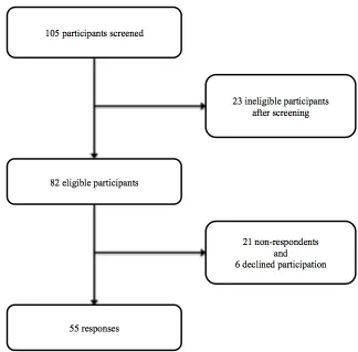
4.1 Response Rate ... 56
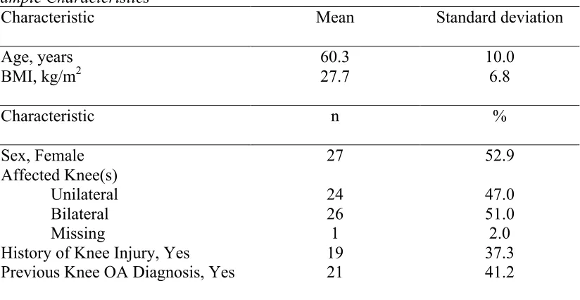
4.2 Sample Characteristics ... 57
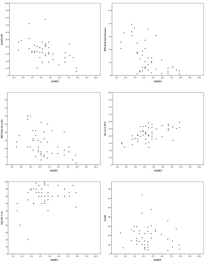
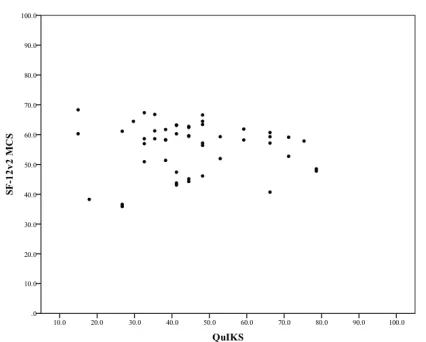
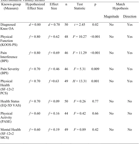
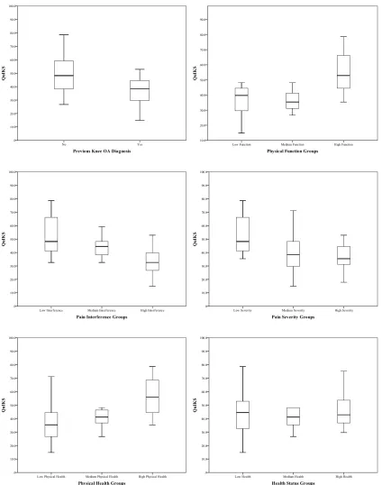
4.3 Objective 1: Construct Validation ... 59
4.4 Objective 2: Internal Consistency Reliability ... 62
Chapter 5 ... 65
5 Discussion ... 65
5.1 Overview of Results ... 65
5.2 Convergent Validity ... 65
5.3 Discriminative Validity ... 66
5.4 Overall Construct Validity ... 67
5.5 Internal Consistency Reliability ... 69
5.6 Limitations ... 70
5.7 Future Research ... 72
Chapter 6 ... 72
6 Conclusion ... 73
References ... 73
Appendices ... 92
List of Appendices
Appendix 1. Copyright agreement for Figure 1 ... 92
Appendix 2. QuIKS and QuIKS-R ... 93
Appendix 3. Letter of Information ... 95
Appendix 4. Consent Form ... 99
List of Tables
Table 1. American College of Rheumatology Diagnostic Criteria for Knee Osteoarthritis
... 18
Table 2. Sample Characteristics ... 58
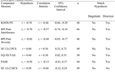
Table 3. Convergent Validity Results ... 59
List of Figures
Figure 1. Model of the process of recognizing the onset of chronic knee problems .. ... 31
Figure 2. Flow diagram of participant enrollment in study ... 57
Figure 3. Scatter plot of Questionnaire to Identify Knee Symptoms total score and other health measures score ... 60
Figure 4. Box plots of mean Questionnaire to Identify Knee Symptoms scores for each known group ... 63
Chapter 1
1 Introduction
Knee osteoarthritis (OA) affects all the tissues of the synovial joint, including the cartilage, bone, meniscus, ligament, tendon, and synovium (Lane et al., 2011). It is estimated that 13% of Canadians and over 300 million people worldwide have OA (Bombardier, Hawker, & Mosher, 2011; Vos et al., 2017). OA is also a major
contributor to disability, as it is ranked as the 10th leading cause of disability in Canada and 12th worldwide (Vos et al., 2017). Additionally, the number of individuals with
OA, and the disability attributed to this condition, is increasing (Hay et al., 2017).
Knee OA has various modifiable and non-modifiable risk factors. Modifiable risk factors, such as obesity and muscle dysfunction, can be influenced through
conservative interventions, and thus are targets for the management of knee OA. While total joint replacement is reserved for individuals with advanced disease or severe pain and functional limitations (Nelson et al., 2014), most knee OA should be managed with education and self-management, weight management, exercise, and corticosteroid injections (McAlindon et al., 2014). However, due to the inability of current diagnostic methods to detect early-stage disease, these treatment strategies have not been comprehensively evaluated in early-stage knee OA, where they are likely to be most effective at preventing disability.
The diagnostic criteria for OA are widely debated, but there has been a recent shift away from imaging-based diagnoses. OA is now considered a clinical diagnosis and imaging is only to be utilized to exclude competing differential diagnoses (Wenham, Grainger & Conaghan, 2014). Major guidelines now support the diagnosis of knee OA based solely on clinical symptoms and examination findings (Altman et al., 1986; National Institute for Health and Care Excellence, 2008; Zhang et al., 2010).
late-stage disease (Jordan, Luta, Renner, Dragomir, Hochberg & Fryer, 1997; O’Reilly, Muir & Doherty, 1996), and thus are not ideal for screening. Patient self-report of symptoms is a cost-efficient method of screening for disease and previous attempts have been made to create screening instruments for knee OA (LaValley et al., 2001; Quintana et al., 2007; Roux et al., 2008; Marra et al., 2007). However, these instruments were developed from current diagnostic criteria and therefore unable to identify the early-onset of disease. An effective screening test should be sensitive to early stages of a disease, when the subsequent course of the disease may still be altered (Fletcher, Fletcher & Fletcher, 2014). Therefore, a new screening instrument that does not rely on late-stage disease criteria would be of benefit.
A new screening tool, the Questionnaire to Identify Knee Symptoms (QuIKS) has been developed to identify individuals with knee OA (Clark, Chesworth, Speechley, Petrella, & Maly, 2014). The QuIKS attempts to quantify illness behaviours of an individual with chronic knee pain (Hamilton, Wong, Gignac, Davis, & Chesworth, 2017). The QuIKS has undergone psychometric evaluation, including reliability and validity testing (Clark, Chesworth, Speechley, Petrella, & Maly, 2014, Hamilton et al., 2015). However, further psychometric testing of the QuIKS has been recommended (Hamilton, Wong, Gignac, Davis, & Chesworth, 2017), including evaluation of construct validity and reliability.
Construct validation determines if the measurement tool is an adequate measure of an unobservable construct (Cronbach & Meehl, 1955). A process of specific, a priori hypothesis testing is recommended to evaluate construct validity (Smith, 2005; Terwee et al., 2007). Convergent and discriminative validity tests are two commonly used construct validation methods (Cronbach & Meehl, 1955; Streiner, Norman, & Cairney, 2015). Convergent validation attempts to hypothesize and quantify how closely a measurement scale of the theoretical construct is related to other constructs and measures, and typically utilizes a correlation analysis undertaken at a single time-point (Streiner, Norman, & Cairney, 2015). Discriminative validation is used to distinguish between predictably different individuals or groups and requires the formulation of a hypothesis about which known-groups of people will have higher or lower amounts of the construct under investigation, observed via scores on the measurement index (Streiner, Norman, & Cairney, 2015). In addition to validity, a measurement scale should demonstrate reliability (Carmines & Zeller, 1979). One type of reliability is evaluated using Cronbach’s alpha, a measure of internal consistency reliability (Cronbach, 1951).
1.1 Objectives
The overall objective of this study was to assess the psychometric properties of a self-administered questionnaire designed to identify the degree of illness behaviour in individuals with knee symptoms consistent with knee OA, and thus, aid in the validation of a tool for use in the clinical and research environment.
Specific objectives to achieve this goal were as follows:
Objective 1:
The primary objective was to evaluate the construct validity of the QuIKS measure in a sample of adults with knee symptoms consistent with knee OA.
Objective 1A: To evaluate the convergent validity of the QuIKS measure.
Objective 1B: To evaluate the discriminative validity of the QuIKS measure.
Objective 2:
Chapter 2
2 Background Literature Review
Osteoarthritis (OA) is a highly prevalent disease and a leading cause of disability in Canada and worldwide (Vos et al., 2017). Early diagnosis of knee OA could result in less disability attributed to this disease (Burstein, 2009), but current diagnostic
methods are only able to identify individuals with late-stage knee OA (Jordan, Luta, Renner, Dragomir, Hochberg & Fryer, 1997; O’Reilly, Muir & Doherty, 1996). This literature review will highlight the epidemiology, diagnosis, and management of knee OA, as well as the potential use of patient self-report questionnaires as screening tools. More specifically, a recap of the development of the Questionnaire to Identify Knee Symptoms will be presented along with a review of psychometric properties, namely construct validity and internal consistency reliability.
2.1 Osteoarthritis
2.1.1 Definition
OA is a complex disease that is difficult to define (Symmons, Mathers & Pfleger, 2006). OA was originally defined as a collection of disorders affecting the articular cartilage and subchondral bone of synovial joints (Altman et al., 1986). Newer
synovial joint, including the cartilage, bone, menisci, ligament, tendon, and synovium (Lane et al., 2011).
2.1.2 Epidemiology
It is estimated that over 300 million individuals worldwide have OA (Vos et al., 2017). OA has been shown to have a high prevalence wherever such statistics are available (Reginster, 2002), but estimates will vary depending on the case definition of OA utilized (Zhang & Jordan, 2010; Felson, Naimark, Anderson, Kazis, Castelli, & Mennan, 1987; Jordan et al., 2007; Lawrence et al., 2008; Quintana, Arostegui, Escobar, Azkarate, Goenaga, & Lafuente, 2008). Canadian prevalence estimates also differ based on the choice of OA definition (Kopec et al., 2007). The prevalence of OA was 10.8% in 2001 in the British Columbian population (Kopec et al., 2007), while another study found roughly 1 in 8 Canadians, or 13% of the population, had osteoarthritis in 2010 (Bombardier, Hawker, & Mosher, 2011).
OA is the most common joint disorder in the United States (Felson et al., 2000) with the knee being the most commonly affected (Newman et al., 2003). American prevalence estimates range from 10-19%, depending on the study sample and case definition used (Felson et al., 198; Jordan, Linder, Renner, & Fryer, 1995; Jordan et al., 2007; Jordan, Linder, Renner, & Fryer, 1995; Jordan et al., 2007). These
American figures are comparable to European and Asian estimates (Arden & Nevitt, 2006; Tang et al., 2016; Park et al., 2017). One global review found the knee OA prevalence to be 24% worldwide, but also provided individual estimates of self-reported, radiographic, and symptomatic knee OA (Pereira, Peleteiro, Araujo, Branco, Santos, & Ramos, 2011).
The prevalence of OA is rising worldwide, as a 30% increase in the number of prevalent cases was observed between 2006 and 2016 (Vos et al., 2017). This trend has also been observed in Canada. One study found that prevalence of all forms of arthritis has increased from 13.4% to 17.6% between 1994 and 2002, an increase of roughly 50% of the percentage of Canadians reporting arthritis (Perruccio, Power, & Badley, 2006). It is expected the prevalence of arthritis in Canada will continue to rise with estimates between 21% and 26% by 2021 (Perruccio, Power, & Badley, 2006). Another projection found a 47% increase in the prevalence of self-reported arthritis is likely by 2031 (Badley, Rothman, & Wang, 1998). Based on a simulation model used in the 2011 report from the Arthritis Alliance of Canada, the projected prevalence of specifically OA is 25% of the population by 2040, or over 10 million Canadians (Bombardier, Hawker, & Mosher, 2011).
Kopec et al., (2008) found that incidence rates of OA increased in both British
Columbian men and women between 1996-97 and 2003-04. The Arthritis Alliance of Canada used these findings in a 2011 report to predict future incidence rates of OA in the entire Canadian population (Bombardier, Hawker, & Mosher, 2011). Compared to 37,342 new diagnoses of OA in 2010, 469,467 new diagnoses of OA are predicted for 2040, which equates to a new OA diagnosis every 60 seconds. All available
prevalence and incidence estimates suggest that OA will affect a large number of Canadians in the near future.
2.1.3 Burden
Knee OA has placed a large burden on health care systems worldwide. In the 2016 Global Burden of Disease Study, OA was the 12th leading cause of years lived with disability in the world (Vos et al., 2017). OA is one of the most common
2009). Over 350,000 total joint replacements are performed annually in the United States for knee and hip OA (Arden & Nevitt, 2006). This problem is compounded by population projections that show a drastic increase in the future population of persons over the age of 60 years (United Nations Department of Economic and Social Affairs, Population Division, 2017). This is likely to further increase the number of
individuals with knee OA and overall economic burden (Woolf & Pfleger, 2003; Sun, Wu, & Kalunian, 2007). One study projected that by 2030, 25% of the total US population and 33% of the working adult population will have arthritis (Hootman & Helmick, 2006), of which a large proportion will have OA, causing significant impact throughout the entire economy.
In Canada, arthritis of all forms is the leading cause of disability and health care utilization (Health Canada, 2003), with annual cost estimates of $4.4 billion (Health Canada, 2002). OA in particular is the 10th leading cause of years lived with disability in Canada (Vos et al., 2017). An individual with OA typically costs the Canadian health care system around $5700 USD per annum (Maetzel, Li, Pencharz, Tomlinson, & Bombardier, 2004), which would rise by roughly $8600 if undergoing joint
replacement (Hawker et al., 2009).
2.2 Risk Factors for Knee Osteoarthritis
Knee OA is a complex condition that has many differing and potentially interacting risk factors. Several risk factors for the development and progression of this multi-factorial condition have been identified in the literature. Some risk factors, such as sex, age, previous trauma, and knee alignment, cannot be altered through conservative interventions, and are thus considered non-modifiable risk factors. Modifiable risk factors, such as obesity and muscle dysfunction, can be influenced through
conservative interventions. Non-modifiable risk factors may provide an opportunity to identify individuals at increased risk of developing knee OA, while modifiable factors may provide unique intervention targets to limit onset and progression of disease and provide symptomatic management for patients.
2.2.1 Non-Modifiable Risk Factors
Sex
The prevalence and incidence of knee OA is significantly greater in women than men (Oliveria, Felson, Reed, Cirillo, & Walker, 1995). Females are two times more likely than men to have OA and typically exhibit higher levels of disability (Statistics Canada, 2007). Females are also more likely to have more severe knee OA (Srikanth, Fryer, Zhai, Winzenberg, Hosmer, & Jones, 2005). However, sex does not seem to affect the progression of knee OA (Felson et al., 1995; Schouten, van den Ouweland, & Valkenburg, 1992). Higher disease rates and severity in females may be caused by the role of estrogen (Zhang & Jordan, 2010) or due to sex differences in bone strength, alignment, neuromuscular strength, pregnancy, and ligament laxity (Johnson &
Hunter, 2014).
Age
increase in the aging population (Arden & Nevitt, 2006). As individuals age, it is likely joints become more vulnerable to biomechanical insult and have a decreased reparative capacity (Arden & Nevitt, 2006). Moreover, age-related reductions in physical activity, muscle weakness, and knee joint laxity may also contribute to increased rates of knee OA in the elderly (Loeser & Shakoor, 2003; Rudloph, Schmitt, & Lewek, 2007). However, the exact mechanism(s) by which increasing age affects knee OA is not currently understood (Johnson & Hunter, 2014).
Trauma
Knee injury is one of the strongest risk factors for OA (Zhang & Jordan, 2010) and individuals who suffer a knee injury are at an increased risk of early-onset disease (Johnson & Hunter, 2014). Ruptures of the anterior cruciate ligament, meniscal tears and articular cartilage injuries are strongly linked to the development of knee OA (Roos, Ostenberg, Roos, Ekdahl, & Lohmander, 2001; Lohmander, Ostenberg, Englund, & Roos, 2004; Lohmander, Englund, Dahl, & Roos, 2007). Anterior
cruciate ligament tears appear to be the most likely to result in knee OA, but the exact mechanism leading to this increased risk is poorly understood (Slauterbeck, Kousa, & Clifton, 2009). It is possible that OA develops as a result of the tissue damage during the initial injury (Buckwalter, 2002; Friel & Chu, 2013) or as a result of altered loading patterns that develop secondary to the knee injury (Chaudari, Briant, Bevill, Koo, & Andriacchi, 2008). Knee OA prevalence rates of up to 70% post-injury are commonly reported in the literature (Johnson & Hunter, 2014), but recent evidence suggests these figures may be overestimated. One study found a 13% prevalence of knee OA after an isolated anterior cruciate ligament rupture, but a higher prevalence of 21-40% if a meniscal injury occurred in combination (Oiestad, Engebretsen, &
Storheim, 2009).
Alignment
bow-legged, is typical in patients with medial compartment knee OA (Hunter, Sharma, & Skaife, 2009). Abnormal alignment has been shown to be strongly associated with increased structural damage in the compartment under greatest mechanical stress (Sharma, Song, Felson, Cahue, Shamiyeh, & Dunlop, 2001). For example, individuals with varus malalignment were four times more likely to experience progression of medial compartment knee OA, while individuals with valgus malalignment were five times more likely to undergo lateral compartment progression (Cerejo, Dunlop, Cahue, Channin, Song, & Sharma, 2002).
The role of knee alignment in the development of knee OA is less understood than its role in disease progression (Johnson & Hunter, 2014; Zhang & Jordan, 2010). There is biomechanical rationale that suggests knee alignment deformities could contribute to the development of knee OA, but this has not been well studied (Arden & Nevitt, 2006). The Rotterdam study found an increased risk of incident knee OA in varus and valgus aligned knees (Brouwer et al., 2007), while no increased risk of knee OA was found with knee alignment in the Framingham cohort (Hunter et al., 2007). This may indicate that malalignment is only a risk factor for disease progression and not for incident knee OA (Hunter et al., 2007). It is also possible the degree of malalignment may affect the incidence and/or progression of knee OA by influencing the impact of other risk factors, such as muscle dysfunction or obesity, thereby altering the load profile placed on the knee joint (Hunter, Sharma, & Skaife, 2009).
2.2.2 Modifiable Risk Factors
Obesity
Leyland, Peat, Cooper, Arden, & Prieto Alhambra, 2016). It has been shown that for every 5-unit increase in BMI, there is a 35% increased risk of developing knee OA (Jiang et al., 2012), while a 5 kg reduction in body weight decreases the risk by 50% (Felson, Zhang, Anthony, Naimark, & Anderson, 1992; Christensen, Bartels, Astrup, & Bliddal, 2007).
The progression of knee OA is also affected by obesity (Felson et al, 2000). Increased weight has been shown to accelerate the structural deterioration of knee OA (Cooper et al., 2000). The risk of total knee arthroplasty is 41% greater in overweight BMI individuals and at least 97% greater in obese BMI individuals compared to normal weight individuals (Leyland et al, 2016). As obesity is an established risk factor for knee OA development and is increasing in prevalence, it is likely that a greater number of individuals will develop knee OA in the future (Johnson & Hunter, 2014).
Muscle Weakness
2.3 Management of Knee Osteoarthritis
The management of knee OA is targeted at the modifiable risk factors of the disease. Many guidelines for the management of knee OA have been published and are generally consistent in their recommendations (McAlindon et al., 2014; Fernandes et al., 2013; Hochberg et al., 2012; Nelson, Allen, Golightly, Goode, & Jordan, 2014). Patient management should be individualized based on patient expectations, risk factors, and pain and disability levels (Fernandes et al., 2013). One internationally developed guideline recommends that education and self-management, weight
management, exercise, and corticosteroid injections are appropriate for all individuals with knee OA (McAlindon et al., 2014). Individuals with knee OA may also undergo surgical intervention if non-surgical interventions fail to adequately manage the disease. While arthroscopic procedures are no longer supported, high tibial osteotomies may be of value, and total joint replacement is recommended for
individuals with end-stage disease or severe pain and functional limitations (Nelson et al., 2014; Richmond et al., 2010).
2.3.1 Education and Self-management
Education and self-management strategies are recommended as core treatments for all individuals with knee OA (McAlindon et al., 2014; Fernandes et al., 2013). Self-management strategies are defined as complex behavioural interventions targeted at patient education and behaviour modification, which encourage people with chronic disease to take an active role in the management of their own condition (Kroon, van der Burg, Buchbinder, Osborne, Johnston, & Pitt, 2014). In 2014, the Cochrane Library published a review of 29 randomized controlled trials assessing the
effectiveness of self-management education programs (Kroon et al., 2014). Compared to usual care for individuals with OA, these interventions may improve
2.3.2 Weight Management
Weight management, defined as addressing weight loss if overweight or obese, is considered standard management for knee OA (McAlindon et al., 2014; Fernandes et al., 2013). Weight management encompasses a range of strategies, including dietary interventions, increasing physical activity, and eating behaviour education (Fernandes et al., 2013). While weight loss shows only small and likely insignificant
improvements in pain levels, it has been shown to have a greater impact on disability (Christensen, Bartels, Astrup, & Bliddal, 2007). A 5% reduction in total body weight over a 20-week period is likely the cut-point to provide significant improvements in disability (Christensen et al., 2007). This improvement appears to be accomplished through dietary modification, exercise intervention, or both, which allows patient preference to be incorporated into the shared decision-making process (Christensen et al., 2007).
2.3.3 Exercise
Therapeutic exercise has been widely studied in the management of knee OA. All patients with knee OA should receive an individualized exercise program, which may involve resistance exercise, aerobic exercise, and range of motion or stretching
exercises (McAlindon et al., 2014; Fernandes et al., 2013). Patients may also perform these exercises in aquatic settings, when appropriate. A 2015 Cochrane Review concluded there is high-quality evidence that land-based exercises have short-term benefits on knee pain and physical function (Fransen, McConnell, Harmer, Van der Esch, Simic, & Bennell, 2015). While the treatment effects of exercise are considered small, they are comparable to the reported effects of non-steroidal anti-inflammatory drugs (Fransen et al., 2015). A more recent Cochrane Review found that exercise may help improve pain, function and depression, while also providing a wide range of health benefits to people (Hurley et al., 2018).
proprioception and balance (Juhl, Christensen, Roos, Zhang, & Lund, 2014). A stratified meta-analysis based on type of exercise intervention found that quadriceps-specific exercise, when performed three times a week under supervision, provides the most pain reduction (Juhl et al., 2014). More recently, combination exercise and education interventions have been introduced. One such example is the Good Life with osteoArthritis in Denmark (GLA:D) program. The GLA:D program has been administered to over 9800 participants with OA in Denmark and has been found to have a significant impact on pain, quality of life, physical function and physical activity levels (Skou & Roos, 2017). This program is currently being implemented globally, and preliminary evidence shows meaningful results for people living in Canada with OA (Davis et al., 2017).
2.3.4 Injections
Patients who do not achieve adequate management of their knee OA through education, weight management, and exercise may elect to receive intra-articular injections. Intra-articular injections may include corticosteroid, hyaluronic acid, platelet-rich plasma, or stem cells. Corticosteroid injections are supported for the management of knee OA (McAlindon et al., 2014; Hochberg et al., 2012), although a recent high quality randomized controlled trial suggests these injections are no better than placebo and may have damaging long-term effects to cartilage health (McAlindon et al., 2017). A 2015 Cochrane Review found that corticosteroid injections may provide clinically important benefits, but are generally short-lived (Jüni et al., 2015). Viscosupplementation with hyaluronic acid injections is not currently recommended for the management of knee OA (McAlindon et al., 2014; Hochberg et al., 2012), as insufficient evidence exists to support their use (Bellamy, Campbell, Robinson, Gee, Bourne, & Wells, 2006).
had a high risk of bias (Laudy, Bakker, Rekers, & Moen, 2015). While some
randomized controlled trials have shown stem cell injections to be effective, a second systematic review reported all trials were at a high risk of bias, and as such, stem cell injections could not be recommended for the treatment of knee OA (Pas, Winters, Haisma, Koenis, Tol, & Moen, 2017).
2.3.5 Surgery
If patients do not achieve adequate symptomatic control through non-surgical management, surgical interventions are available for knee OA. Three main types of surgery are used for knee OA: arthroscopic lavage and/or debridement, high tibial osteotomy, and total joint replacement. Arthroscopic surgeries are no longer recommended in surgical guidelines (Nelson et al., 2014; Richmond et al., 2013; Zhang et al., 2010) as many high quality randomized controlled trials have shown them to be ineffective (Moseley et al., 2002; Kirkly et al., 2008; Sihvonen et al., 2013). A review of all available trials on arthroscopic procedures for knee OA found inconsequential benefits and an increased risk of harm (Thorlund, Juhl, Roos, & Lohmander, 2015). As a result, arthroscopic procedures should not be used in the management of knee OA.
2.4 Diagnosing Knee Osteoarthritis
The diagnosis of knee OA is a much-debated topic and numerous case definitions have been used throughout the literature. OA is currently considered a clinical diagnosis with imaging reserved for excluding differential diagnoses (Wenham, Grainger & Conaghan, 2014). The American College of Rheumatology, National Institute for Health and Care Excellence and European League Against Rheumatism all support the diagnosis of knee OA based on clinical symptoms and examination findings (Altman, et al., 1986; National Institute for Health and Care Excellence, 2008; Zhang, et al., 2010). The only imaging guidelines for OA recommend that imaging is not required to make the diagnosis in patients with a typical presentation of OA (Sakellariou et al., 2017). These guidelines also note there is a lack of literature suggesting any
additional value of imaging over clinical findings in making a diagnosis of OA (Sakellariou et al., 2017). It has also been acknowledged that the diagnosis of knee OA can be made in the presence of specific clinical and examination features, even if radiographs appear normal (Zhang et al., 2010).
2.4.1 Clinical Diagnosis for Knee OA
Table 1
American College of Rheumatology Diagnostic Criteria for Knee Osteoarthritis
Category of Diagnostic
Criteria Criteria
Clinical Knee pain
+ at least 3 of 6:
- Age >50 years
- Stiffness <30 minutes - Crepitus
- Bony tenderness - Bony enlargement - No palpable warmth
Clinical and
Radiographic Knee pain + Osteophytes + at least 1 of 3: - Age >50 years
- Stiffness <30 minutes - Crepitus
Clinical and
Laboratory Knee pain + at least 5 of 9: - Age >50 years
- Stiffness <30 minutes - Crepitus
- Bony tenderness - Bony enlargement - No palpable warmth
- Erythrocyte sedimentation rate <40mm per hour
- Rheumatoid factor <1:40 - Synovial fluid signs of
osteoarthritis
Note. mm = millimeters, Adapted from Altman et al. (1986).
examination signs (Zhang et al., 2010). This diagnosis can be made without
radiographs and applies even when radiographs appear normal (Zhang et al., 2010). Further analysis found that adults aged 45 years and older have a 99% probability of having radiographic knee OA when all six symptoms and signs are present (Zhang et al., 2010). These recommendations are echoed in the National Institute for Health and Care Excellence guidelines (National Institute for Health and Care Excellence, 2014). These guidelines state that OA should be diagnosed clinically without investigations when a person is over 45 years of age and has activity-related joint pain with either no morning stiffness or morning stiffness lasting less than 30 minutes. Currently, knee OA should be diagnosed clinically without need for further investigation or imaging in most cases. However, the diagnostic accuracy of any such clinical examination is hindered by the lack of a consensus reference standard.
2.4.2 Diagnostic Imaging for Knee OA
Imaging examinations are commonly used to aid in OA diagnosis (Demehri,
2.4.3 Plain Film Radiography for Knee OA
Plain film radiography is the imaging modality of choice in clinical practice (Demehri, Guermazi & Kwoh, 2016) and has historically been used as the primary technique to obtain a diagnosis of OA (Cibere, 2006; Peterfy, 2002). A number of radiographic grading systems have been developed to diagnose OA. The most commonly used is the Kellgren-Lawrence grading, which uses a global score of 0-4 to grade the joint (Kellgren & Lawrence, 1957). The Kellgren-Lawrence score is primarily based on the presence of osteophytes and joint space narrowing, which are associated with
moderate to severe disease (Bedson & Croft, 2008). A score of 2 or greater is the traditional cut-off point for a definitive diagnosis of OA.
The American College of Rheumatology has also developed diagnostic criteria for knee OA that utilize plain film radiographs (Altman, et al., 1986). Table 1 includes the radiographic criteria for knee OA. However, these diagnostic criteria may not be applicable to general clinical practice, as they were developed using cases with more advanced disease than is likely to be encountered in the general population (Cibere, 2006). Moreover, the comparator group was made up of individuals with rheumatoid arthritis, and as a result, these criteria are better suited to differentiate OA from inflammatory joint conditions (Cibere, 2006).
The use of radiography in the diagnosis of OA has a number of limitations. X-rays are insensitive to the earliest pathological changes seen in OA (Hunter & Guermazi, 2012) and can appear normal in symptomatic joints (Guermazi, et al., 2012). This is thought to be due to the fact that articular cartilage changes, which are not visualized on radiographs, are likely altered before bony changes are evident (Hunter et al., 2007; Peterfy, 2002). This is compounded by the limitations of assessing a
The discordance of clinical symptoms and structural changes visualized on plain film radiographs is well documented (Bedson & Croft, 2008; Chan et al., 1991; Hannan, Felson, & Pincus, 2000; Szebenyi et al., 2006; Toivanen et al., 2007). As such, a review of the literature on radiographic imaging for OA concluded X-ray images have a limited role in the clinical assessment of OA (Peat, Croft & Hay, 2001). Overall, the limited ability of radiography to detect OA features at an early stage of disease
questions the utility of plain film radiographs as a diagnostic tool (Cibere, 2006). As such, a reliance on plain film radiographs to diagnose OA is not recommended.
2.4.4 Magnetic Resonance Imaging for Knee OA
While only bone can be visualized on plain film radiographs, advanced imaging techniques, such as MRI have the ability to image all the tissues involved in an osteoarthritic joint (Conaghan, 2006). While MRI has an unparalleled ability to evaluate articular cartilage (Eckstein, Cicuttini, Raynauld, Waterton & Peterfy, 2006), it can also visualize ligaments, synovium, menisci and subchondral bone (Conaghan, 2006). As such, MRI is uniquely suited to assess the joint as a whole organ and may be a more sensitive tool for the diagnosis of OA (Peterfy, 2002).
It has been shown that MRI is better able to detect osteophyte formation than plain films (Chan et al., 1991). However, the potential utility of MRI in the diagnosis of OA may stem from whole-organ evaluation. MRI has the ability to visualize bone marrow edema, which has been shown to be associated with painful knees (Conaghan, 2006). One study found in a sample of 400 individuals with radiographic knee OA that bone marrow edema was present in 78% of the painful knees but only in 30% of knees in the non-painful group (Felson et al., 2001). It is also possible that OA symptomology may be attributed to synovitis, joint effusions, and meniscal and ligamentous
pathology, but considerably less research exists evaluating these structures (Conaghan, Felson, Gold, Lohmander, Totterman & Altman, 2006).
evaluating structural morphology and damage (Hunter et al., 2011). However, MRI does not currently have a place in routine clinical practice for diagnosing OA
(Conaghan, 2006). This is reflected in international imaging guidelines for OA, which state that should imaging be needed to exclude differential diagnoses, conventional radiography should be used before other modalities (Sakellariou et al., 2017). This may be in part due to the fact that MRI abnormalities can be frequent even in those with normal knee radiographs (Taouli et al., 2002) and those without knee pain (Guermazi et al., 2012). Additionally, MRI was shown to be of no benefit in
diagnosing OA if clinical features were present prior to imaging (Petron et al., 2010). While MRI technologies have great potential to play a role in the diagnosis of OA, the current body of literature suggesting MRI may be useful for routine practice is limited (Conaghan, 2006). This relative lack of literature is likely related to the limited ability to modify the OA disease process, and thus limited application of advanced imaging techniques in the diagnosis of OA (Peterfy, 2002). However, more studies are currently underway and further developments in MRI technology and image analysis tools hold promise for future disease evaluation (Conaghan, 2006; Conaghan, Felson, Gold, Lohmander, Totterman & Altman, 2006; Eckstein, Cicuttini, Raynauld,
Waterton & Peterfy, 2006).
2.4.5 Biomarkers for Knee OA
structural changes could allow for earlier detection of knee OA (Brandt, Doherty & Lohmander, 1998).
Increased levels of various biomarkers have been found in individuals with OA (Cibere et al., 2009, Hunter et al., 2007). In 2012, the OA Biomarkers Consortium selected twelve serum and/or urine biochemical markers to investigate (Hunter et al., 2014). Since that time, several novel biomarkers have been discovered (Watt, 2018). However, many of these biomarkers have not undergone the rigorous testing required to be considered diagnostic markers. This is complicated by the lack of a gold
standard definition for early OA that limits the ability of biomarkers to be evaluated in diagnostic studies (Watt, 2018). Moreover, recent high quality studies have shown that biomarkers may lack the specificity needed to be of clinical utility (Watt, 2018). OA biomarkers are not localized to a specific joint and concentration levels can be influenced by age, sex, body composition, diet, various comorbidities, and drug interactions (Kraus, 2006). While biomarkers are likely to play a role in the diagnosis of knee OA in the future, too little is currently understood to provide any meaningful contribution to the clinical or research setting at this time.
2.5 Screening for Knee Osteoarthritis
patients to potentially harmful radiation (LaValley, McAlindon, Evans, Chaisson, & Felson, 2001). Staged-screening approaches, such as patient self-report of symptoms followed by radiography or physical examination, have been suggested (Cooper, McAlindon, Coggon, Egger & Dieppe, 1994; O’Reilly, Muir & Doherty, 1998; Hopman-Rock, Odding, Hofman, Kraaimaat & Bijlsma, 1997; March, Schwarz, Carfrae & Bagge, 1998; Oliveria, Felson, Klein, Reed & Walker, 1996), but still require costly radiographs and/or clinical evaluation.
Patient self-report of symptoms may be a cheaper alternative to more costly screening strategies. Various symptom questions to predict the presence of knee OA have been studied, but these focus on individual questions rather than on development of a screening instrument (March, Schwarz, Carfrae & Bagge, 1998; O’Reilly, Muir & Doherty, 1996). However, it has been argued that no single symptom can identify patients with knee OA (Corti & Rigon, 2003). Moreover, it remains unknown if multi-item questionnaires can predict symptomatic knee OA without need for additional, more costly evaluations (LaValley et al., 2001).
The goal of a screening questionnaire is to select a group of people in whom further investigations will yield a high rate of OA diagnoses, known as high specificity, without missing a substantial number of patients with OA, known as high sensitivity (Roux, et al. 2008). Previous attempts have been made to create screening instruments for knee OA (LaValley et al., 2001; Quintana et al., 2007; Roux et al., 2008; Marra et al., 2007). However, these instruments were largely based on current diagnostic criteria and their diagnostic testing is limited.
displayed adequate diagnostic test performance. As such, they concluded that all three instruments resulted in too much misclassification to be used as a single-step screening process (LaValley et al., 2001).
The next attempt at developing a screening tool for knee OA was undertaken in Spain in 2007 (Quintana et al., 2007). Quintana and colleagues developed the Knee and Hip Osteoarthritis Screening Questionnaire that used questions specific to knee OA, questions specific to hip OA, and shared questions for both hip and knee OA
(Quintana et al., 2007). When compared to the reference standard of examination by an orthopedic surgeon, including knee radiographs, the knee questionnaire was found to have a sensitivity of 94.5%, but a poor specificity of only 43.2%. Moreover, only 44% of individuals identified by the questionnaire underwent the reference standard examination, raising questions about the validity of the results. In addition, only individuals over the age of 60 were sampled, which may limit the ability of the knee questionnaire to detect disease in younger individuals at an earlier stage. As a result, the authors suggest that screening for knee OA in the general population still requires individuals to self-report knee symptoms and then be followed up with a clinical examination (Quintana et al., 2007).
In 2008, a questionnaire to identify knee OA was developed in France to be
administered over the telephone (Roux et al., 2008). When tested against the reference standard of clinical evaluation and radiographic evidence, the instrument displayed high sensitivity and specificity. The validation sample also included individuals as young as 45 years, which means this instrument may have utility in populations of individuals with early disease. However, less than 23% of the 479 participants who were identified by the questionnaire underwent reference standard testing, questioning the internal validity of the study results. Therefore, the authors concluded that
A Canadian study evaluated the effectiveness of a screening questionnaire for knee OA administered by pharmacists (Marra et al., 2007). Pharmacists administered the questionnaire to 411 participants, of which 274 screened positively. Only 70% of these individuals received the reference standard of clinical examination and
radiographs and it was found that the questionnaire could correctly identify over 80% of individuals with undiagnosed knee OA, but considerable bias may exist. The authors concluded that pharmacists could play an important role in the identification of undiagnosed knee OA and that much of this OA is in the early stages and likely most amenable to intervention (Marra et al., 2007). However, only participants who screened positively on the questionnaire received the reference testing, and thus, no calculation of sensitivity or specificity was possible. This is problematic, as it is not known how many subjects excluded by the questionnaire truly had knee OA, known as false negatives. At minimum, this questionnaire requires further testing in a more rigorous diagnostic study before it can be used as a screening instrument for knee OA.
These previous attempts at developing a multi-item questionnaire to screen for knee OA (LaValley et al., 2001; Quintana et al., 2007; Roux et al., 2008; Marra et al., 2007) illustrate the potential of this relatively inexpensive strategy. Unfortunately, the previously developed tools may not be sensitive to early stage knee OA, as they were developed using current diagnostic criteria and were not adequately tested in patient samples likely to suffer from early disease. Furthermore, several methodological flaws, such as lack of patient perspective in questionnaire item generation, low
participation rates in validation procedures, and weak diagnostic study design limit our understanding of the utility of these screening instruments. Most notably, not one study included a “period of time for follow-up” in which the disease may be allowed to develop in individuals who initially tested negative (Fletcher, Fletcher & Fletcher, 2014).
However, the diagnostic performance of the previously created instruments suggests there is potential that a screening questionnaire could be developed that will
tested in rigorous diagnostic studies. With proper identification of early-stage knee OA, treatments that can alter the disease course, such as exercise and weight-loss programs can be tested and implemented earlier in the disease process. There is a clear need to develop a single-step screening instrument that is able to detect early-stage knee OA.
2.6 Illness Behaviour
Knee OA is a chronic disease where individuals often adapt their behaviours to
manage symptoms and disability (Gignac, Cott, & Badley, 2002). Through interviews with 248 individuals with OA, it was found that almost all people make at least one adaptation and that most make multiple adaptations (Gignac, Cott, & Badley, 2002). Analysis revealed that these adaptations were largely motivated by real and perceived changes or losses in function, providing evidence that patient perceptions may play a more significant role in mediating behavioural changes than presumed (Gignac, Cott, & Badley, 2002). There is a psychosocial context in which individuals with chronic knee pain/OA make decisions about their health and need for care. Further knowledge of this process may result in earlier identification of knee OA and better care for these individuals.
Many theoretical models have been developed in the behavioural and social sciences of how individuals appraise and respond to health conditions (Baltes & Baltes, 1990; Diefenbach & Leventhal, 1996; Mechanic, 1986). Three main models of illness contribute to our understanding of illness perception and behaviour in individuals with chronic knee pain/OA. The first is the model of selective optimization with
compensation, which describes general adaptation processes across a spectrum of illnesses (Baltes & Baltes, 1990). It has three components describing adaptation: selection or restriction of certain activities, optimization of functional capacity, and compensation or modifying behaviours to perform certain activities (Baltes & Baltes, 1990).
participants interviewed, most reported using all three types of adaptation from the model. However, it is difficult to differentiate between selection, optimization, and compensation, as these components were found to be interrelated (Gignac, Cott, & Badley, 2002). The findings of this work provide evidence that an individual’s perceptions play a role in facilitating the adaptation behaviours to disability from OA, but this model does not explain the cognitive processes that drive behaviour (Gignac, Cott, & Badley, 2002).
Second is the common-sense model of illness representation, or Leventhal’s self-regulatory model of illness behaviour (Diefenbach & Leventhal, 1996; Leventhal, Meyer, & Nerenz, 1980). This theoretical framework attends to both internal and external sources of information to create illness representations (Diefenbach & Leventhal, 1996). This model positions the individual as an active problem solver based on both cognitive and emotional psychological processes that are ignored by the biomedical model (Diefenbach & Leventhal, 1996; Leventhal, 1970).
Meta-analysis of studies using the common-sense model of illness representation has shown that an individual’s thoughts about their illness are moderate-to-strongly related to coping behaviours (Orbell & Hagger, 2003). These findings illustrate a consistency in the way in which individuals cognitively represent illness in a range of health conditions (Orbell & Hagger, 2003). There is however, no empirical evidence supporting the validity or utility of the common-sense model in individuals with chronic knee pain/OA.
Third is Mechanic’s model of illness behaviour, which posits that symptoms may be perceived, evaluated, and acted upon differently by individuals (Mechanic, 1986; Mechanic & Volkart, 1960). This model predicts large variability in reactions to symptoms and illness, as it is the necessary link between biomedicine and behavioural science (Mechanic & Volkart, 1960; Mechanic, 1995). The model of illness behaviour represents the interaction of bodily dysfunction with both a psychological and
defining and interpreting symptoms. Third is remedial actions taken by the individual, and lastly is utilization of help from various sources (Mechanic, 1986; Mechanic, 1995). These four phases make up the socially defined state, which informs the decision-making process and need not be associated with an altered biological state (Febrega, 1973).
Mechanic argued that this illness behaviour model may be more efficient than the diagnostic disease model in addressing the burden of illness and disability (Mechanic, 1995). The disease model does not take into account variability in behavioural responses among individuals with the same disease (Sirri, Fava, & Sonino, 2013). Sirri et al., (2013) argue that illness behaviour can explain the “major prognostic differences among deceptively similar patients” and thus, may provide an improved alternative to the biomedical disease model for illness recognition and medical care for patients, including those with chronic knee pain/OA.
There have been many other attempts at the operationalization of components of the illness behaviour construct (Prior & Bond 2013; Hamilton et al., 2017). For example, the Fear Avoidance Beliefs Questionnaire (Waddell, Newton, Henderson, Somerville, & Main, 1993), Coping Strategies Inventory (Carver, Scheier, & Weintraub, 1989), Knee Osteoarthritis Fears and Beliefs Questionnaire (Benhamou et al., 2013), and the Arthritis Self-Efficacy Scale (Lorig, Chastain, Ung, Shoor, & Holman, 1989), among others, have all been created to measure components of illness behaviour. However, all of these measures lack items that tap at least one domain from Mechanic’s model of illness behaviour and/or have not been validated in a population with chronic knee pain/OA (Hamilton et al., 2017). The only measure with some evidence of validity in this population that specifically addresses all domains of Mechanic’s model of illness behaviour is the Questionnaire to Identify Knee Symptoms (Hamilton et al., 2017).
2.7 QuIKS Questionnaire
Stemming from the potential utility of a measure able to quantify illness behaviour in individuals with chronic knee pain consistent with OA, a series of studies were undertaken to develop such a tool. The first study by Maly and Cott (2009) aimed to identify the process individuals use to recognize and address chronic knee problems that could be precursors to knee OA, as they hypothesized that patient experience could provide an ideal method to screen for individuals with pre-diagnostic knee OA (Maly & Cott, 2009). A grounded theory methodology was utilized to identify a theoretical model of this process. Twenty-six individuals at varying stages of
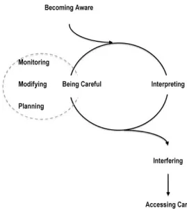
Figure 1. Model of the process of recognizing the onset of chronic knee problems. Reprinted from “Being careful: a grounded theory of emergent chronic knee problems”, by M. Maly and C. Cott, 2009,
Arthritis Care and Research, 61(7), p.939. Copyright 2009 by John Wiley and Sons (Appendix 1).
The process of recognizing and addressing begins with a stage of becoming aware of chronic knee symptoms. This stage is characterized by the admittance and acceptance of a chronic knee problem by the individual, typically associated with increased
frequency and consistency of the knee symptoms. A major determinant of this stage is the acknowledgment of difficulty participating in physical activities due to the impact of the knee symptoms (Maly & Cott, 2009).
cognitive contextual factors, as many participants reported that interactions with others, such as friends, co-workers, and health care practitioners, along with personal experiences and memories from family members with chronic musculoskeletal symptoms, informed their decision-making process (Maly & Cott, 2009).
The second part of the cyclical process, being careful, was defined by the investigators as the perceptions, intentions, and behaviours of the individual used to avoid potential harms while engaging in physical activities. At this stage, individuals typically have the cognitive perception of diminished physical ability, leading to intentions and behaviours to perform physical activities with greater care. Three main actions were identified that compose being careful: 1) monitoring, where attention is given to factors that trigger knee symptoms; 2) modifying, where numerous strategies are employed to adjust physical activities in response to symptoms and to protect the knee; and 3) planning, where intentions to maintain physical activity despite knee symptoms are formed, through the use of anticipatory strategies to prevent harm, such as exercise. The authors describe the process of being careful during daily activities as a cognitive and behavioural approach to self-management of chronic knee symptoms, even before a formal diagnosis of knee OA is made (Maly & Cott, 2009).
The investigators found that the cyclical process of interpreting and being careful continues until the occurrence of an event that signals the inadequacies of this self-management strategy, termed as the interfering stage in the model. The interviews revealed that this interfering event always involved the inability to carry out a task of significant personal value, such as sport or a leisure activity, which caused the individual to seek more formal care for their knee complaint, termed accessing care. The authors posited that the individual’s interpretation of important daily activities is the key criterion for the signal to access care for chronic knee problems in this study population (Maly & Cott, 2009).
model, particularly being careful, could be used as a way of identifying pre-diagnostic knee OA prior to these individuals seeking care and formal diagnosis. While no tool existed to quantify the being careful construct, the Arthritis Self-Efficacy Scale (Lorig, Chastain, Ung, Shoor, & Holman, 1989) may capture elements of being careful, but a specifically designed tool is likely required to detect pre-diagnostic knee OA
experiences (Maly & Cott, 2009).
Using the process model identified by Maly and Cott (2009), a multi-phase research project was designed to develop an instrument capable of identifying emerging knee problems, ultimately named the Questionnaire to Identify Knee Symptoms (QuIKS) (Appendix 2) (Clark, Chesworth, Speechley, Petrella, & Maly, 2014). The aim was to develop a self-administered questionnaire for clinical and research settings, as all diagnostic procedures for knee OA available at that time identified advanced disease only.
In the second phase of tool development, expert review, a 16-member panel of Canadian OA experts reviewed the 33 generated items and response scales. The review panel included rheumatologists, orthopedic surgeons, family practitioners, physical therapists and health care researchers with expertise in OA and measurement scale development and validation. Recommendations on clinical utility, scaling, ambiguous and inappropriate items, and additional constructs to consider were developed by this panel. The expert recommendations were used to inform the drafting of a 35-item questionnaire, organized into three sections: medications and treatments, activities, and living with knee problems (Clark, Chesworth, Speechley, Petrella, & Maly, 2014). Although not specifically stated by the developers of the measure, this phase provides evidence of content validity of the QuIKS (Streiner, Norman, & Cairney, 2015).
In the third phase of the QuIKS development, item reduction, principal components analysis informed the selection of only items that explain a high proportion of the test score variance for the final questionnaire. Participants were recruited from an Ontario family medicine clinic through medical chart review. Patients with evidence of knee pain lasting longer than two weeks, with no diagnosis of knee OA or any other condition that explained their symptoms were invited to complete the preliminary 35-item QuIKS questionnaire. Data from 105 completed questionnaires was used in the principle component analysis. The final solution resulted in a 13-item questionnaire, composed of four subscales, named: medications, monitoring, interpreting, and modifying (Clark, Chesworth, Speechley, Petrella, & Maly, 2014). These subscales were scored individually, as this best captured each individual respondent (Clark, Chesworth, Speechley, Petrella, & Maly, 2014).
Through this three-phase development process, a short, self-administered instrument, named the QuIKS, was created with the aim of identifying individuals at increased risk of developing or in the early stages of knee OA (Clark, Chesworth, Speechley,
Petrella, & Maly, 2014). This instrument, while primarily designed as a research tool, may also have utility in the clinical setting with limited burden to patients, an
important property emphasized during expert review. Furthermore, evidence was provided through the QuIKS development process of content validity (from the expert panel) and internal consistency reliability from a sample of individuals with early knee symptoms. However, the developers recommend that these results be confirmed in an independent sample, as estimates of these properties tend to be overly optimistic when they are derived from development samples (MacCallum, Wideman, Zhang, & Hong, 1999). Moreover, the authors recommended that future research assess the predictive validity of the QuIKS, but note that this may prove difficult as the lack of criterion standard may limit the design (Clark, Chesworth, Speechley, Petrella, & Maly, 2014).
Hamilton et al., (2017) performed a systematic review of measures capturing
components of Mechanic’s model of illness behaviour (Mechanic, 1986). Utilizing a systematic methodological framework for scoping reviews, they found sixteen different measures used in 71 studies demonstrating at least content validity in a sample of individuals with knee pain or OA. Each measure captured at least one component of Mechanic’s definition of illness behaviour, but only one measure, the QuIKS, captured all four components of illness behaviour (Hamilton, Wong, Gignac, Davis, & Chesworth, 2017).
Most of the measures identified captured only one or two of the components of illness behaviour, as they were originally conceptualized to quantify constructs such as coping strategies, self-efficacy, and fear avoidance behaviours (Hamilton, Wong, Gignac, Davis, & Chesworth, 2017). Therefore, the QuIKS is the most comprehensive measure providing complete coverage of illness behaviours experienced by an
Chesworth, 2017; Streiner, Norman, & Cairney, 2015). Hamilton and colleagues recommended that the QuIKS undergo further studies of its validity to determine whether its psychometric properties are sound (Hamilton, Wong, Gignac, Davis, & Chesworth, 2017).
Hamilton and colleagues also performed a study to investigate the validity of the QuIKS in individuals with chronic knee problems, such as OA (Hamilton et al., 2015). With no criterion measure available for illness perception and behaviour, criterion validity could not be tested (Clark, Chesworth, Speechley, Petrella, & Maly, 2014; Streiner, Norman, & Cairney, 2015). Therefore, investigations of the validity of the QuIKS need to focus on construct validity (Streiner, Norman, & Cairney, 2015).
Hamilton et al., (2015) used two objectives to evaluate the construct validity of the QuIKS measure. The primary objective was to use Rasch analysis to determine if the QuIKS captures a unidimensional construct, as combining the subscales of the QuIKS into a single measure may reflect the higher-order construct of illness behaviour (Hamilton et al., 2015). Rasch validation has been argued as a form of construct validity in itself (Velozo, Seel, Magasi, Heinemann, & Romero, 2012). The secondary objective was to test the known-groups validity and convergent validity of the Rasch-validated QuIKS (QuIKS-R) (Hamilton et al., 2015).
As per the secondary objective, mean QuIKS-R scores from each of the three enrolled groups were used to compare the three known-groups to determine whether there was evidence of construct validity. The a priori hypothesis was the distribution of QuIKS-R scores should vary by group with the highest scores in the pre-surgical group and lowest scores in the subjects with healthy knees. Total scores on the QuIKS-R were found to be significantly different between all three known-groups in the hypothesized directions, with moderate effect sizes. As hypothesized a priori, the healthy knee group exhibited significantly less illness perceptions and behaviours than the chronic knee pain group, who in turn exhibited significantly less illness perceptions and behaviours than the pre-surgical group. These findings support the construct validity of the QuIKS-R in a sample of individuals with chronic knee symptoms.
To complete the secondary objective, the total QuIKS-R scores were correlated with the five subscales (pain, symptoms, function in daily living, function in sport and recreation, and knee-related quality of life) of the Knee injury and Osteoarthritis Outcome Score (KOOS) to evaluate the convergent validity. As predicted, total QuIKS-R scores had significant moderate correlations with scores on the KOOS subscales. Spearman correlation coefficients ranged from 0.45 to 0.77, supporting the construct validity of the QuIKS-R in a population of individuals with chronic knee symptoms.
While evidence of construct validity for the QuIKS-R was shown through its
2.8 Construct Validity
Construct validity, originally described by Cronbach and Meehl (1955), is a
framework used to interpret a measure of a latent trait that is not directly observable or measurable. This framework is utilized when no criterion standard is accepted as entirely adequate to measure the quality (Cronbach & Meehl, 1955). Measures of unobservable psychological phenomena, which can be valuable to explaining human behaviour, can be validated using these principles (Smith, 2005). The validation of these phenomena, or constructs, is demonstrated through predictable relationships derived from theoretical hypotheses between a measure of a particular construct and other measures of similar constructs (Kirshner & Guyatt, 1985; Cronbach & Meehl, 1955; Nunnally, 1978; Carmines & Zeller, 1979).
Smith (2005) has proposed a model for construct validation. The first requirement for construct validation is the specification of the construct in question. This requires explicit definition of the construct under investigation. Once defined, the construct must undergo translation into informative hypotheses that can be tested. Based on the hypothesis generated, specification of the appropriate research design can be made. Following data collection, an explanation of how the observations pertain to
predictions must be formulated. Based on the relationship between the observations and predicted outcomes, revision of theory and construct can be performed. It is through this framework that a construct can be evaluated and shown to have validity.
The key feature of the model prosed by Smith (2005) is the focus on testing of
Two commonly utilized construct validation methods are convergent validation and discriminative validation (Cronbach & Meehl, 1955; Campbell & Fiske, 1959; Streiner, Norman, & Cairney, 2015). Convergent validation attempts to hypothesize and quantify how closely a measurement scale of the theoretical construct is related to other constructs and measures (Cronbach & Meehl, 1955; Kirshner & Guyatt, 1985; Streiner, Norman, & Cairney, 2015). Simply stated, two measures of similar
constructs should be hypothesized to show similar scores. Conversely, two measures of unrelated constructs should show less similar scores. As such, convergent
validation typically utilizes a correlation analysis of data collected at a single time-point (Kirshner & Guyatt, 1985; Streiner, Norman, & Cairney, 2015). The correlation statistic selected depends on the nature and properties of the measurement scales under investigation (Bonett & Wright, 2000; Streiner, Norman, & Cairney, 2015).
While there is no specific correlation value at which a measure can be said to exhibit convergent validity, it has been suggested that a moderate Pearson coefficient value of 0.5 or greater is supportive (Guyatt, Norman, Juniper, & Griffith, 2002). However, a correlation value between two measurement scales that is too high (greater than 0.9) suggests that both scales are measuring the same construct, and thus, are redundant and unneeded (Streiner, Norman, & Cairney, 2015). Therefore, moderate to strong correlational values observed in convergent validation designs can be considered to support construct validity.
The second commonly utilized test of construct validity is known as discriminative validation. Discriminative validation is used to distinguish between predictably different individuals or groups (Streiner, Norman, & Cairney, 2015). This
methodology requires the formulation of a hypothesis about which known-groups of people will have higher or lower amounts of the construct under investigation,
observed via scores on the measurement index (Cronbach & Meehl, 1955; Kirshner & Guyatt, 1985; Streiner, Norman, & Cairney, 2015). For example, a group of
As discriminative validation involves testing the predicted differences between known-groups of individuals, statistical analysis typically involves tests of the mean difference. A between groups t-test or ANOVA can be considered for discriminative validation analyses. While no specific magnitude of mean difference, or effect size, is required to show discriminative validity, one would expect differences of at least moderate effect size between observably-distinct groups (Cohen, 1988).
The validation procedures of a construct and measurement scale must be designed and conducted using rigorous scientific methods. Quality criteria have been proposed to ensure the internal validity of the inferences made from these study designs (Terwee et al., 2007, Mokkink et al., 2010). A convergent validation design requires that
expected correlational magnitude and direction be hypothesized prior to data collection and analysis (Terwee et al., 2007). Similarly, a discriminative validation design requires that the predicted magnitude and direction of the differences between known-groups be stated a priori (Terwee et al., 2007). Tools such as the Consensus-based Standards for the selection of health status Measurement Instruments
(COSMIN) Checklist can be used to ensure the methodological quality when designing a construct validity study (Mokkink et al., 2010). However, these quality criteria were derived from expert opinion, as there is no empirical evidence to suggest that certain correlational or mean difference values must be attained to support
construct validity (Terwee et al., 2007). Even Cronbach and Meehl (1955), predicted that a single “construct validity coefficient” was unlikely to be developed, due to the approximate nature of the validation process.