DEPENDENCE OF HALF-TRANSLOCATION FREQUENCY UPON X-RAY DELIVERY RATE’
IRWIN H. HERSKOWITZ AND S. ABRAHAMSON2
Department of Zoology,3 Indiana University, Bloomington, Indiana
Received December 12, 1955
T HAS BEEN definitely established for X-ray-induced mutations in plants that
I
lowering the intensity of treatment causes a decrease in the number of chromo- soma1 rearrangements obtained (SAX 1939a,b, 1940, 1941; SAX and BRUMFIELD 1943). This observation could result from either or both a necessity for several “units” of X-rays to accumulate in order to produce a break (and as the rate of delivery decreases some units are lost before others are produced), and a decrease in rejoinability of breaks with time (by the restitution of some breaks before others are produced, and/or by the movement of broken ends so far apart from others that their chance of ever combining is reduced). Study of Drosophila sperm for gross rearrangement frequency after X-ray treatment of inseminated females revealed no effect of extremely daerent rates of treatment (MULLER 1938, 1939a,b, 1940; RAY-CHAUDHURI 1939, 1944; MAKHIJANI 1945), supporting none of the hypotheses mentioned for sperm known to be mature. Recently, HAAS, DUDGEON, CLAYTON and STONE (1954), using delivery rates higher than those used earlier, have reported that when sperm were treated in males the higher rate of delivery gave more translo- cations than the lower rate. One would want this intensity effect repeated with samples of sperm of greater uniformity, for example with sperm treated in insemi- nated females, however, before accepting it without reservation, since it has been shown that the translocation frequency from sperm delivered by treated males on one day can be twice what it is on a successive day (and possibly in successive copu- lations on the same day) (ABRAHAMSON and TELFER 1954).While a study of breakage dependence upon X-ray intensity can decide whether break production requires the accumulation of “units”, and the best evidence is that no such accumulation is required for breakage itself, a study of restitution and rearrangement can tell whether breaks are temporarily unjoinable and accumulate until a specific time or whether broken ends can join to other broken ends a t any time. The infrequency of gross deficiencies and inversions and of reciprocal transloca- tions in the germ cells of female Drosophila has prevented the latter type of study in this organism until recently, although MULLER, VALENCIA and VALENCIA (1949) inferred union between fragments to occur fairly promptly, i.e. prior to the move- ments of the meiotic divisions. Such a study became possible once it was shown (HERSKOWITZ and MULLER 1953; MULLER and HERSKOWITZ 1954; HERSKOWITZ
‘This work was supported by a grant to DR. H. J. MULLER and associates from the Atomic 2 Eigenmann Fellow in Zoology.
X-RAY INDUCED HALF-TRANSLOCATIONS I N DROSOPHILA 42 1
1954a,b; ABRAHAMSON, HERSKOWITZ and MULLER 1954, 1956; PARKER 1954a,b) that a certain type of multi-break rearrangement does occur frequently in oocytes after X-ray treatment. This type of mutation was termed a “half-translocation” because it involved, of the four pieces produced by breaking two non-homologous chromosomes, only two pieces which joined eucentrically (making up one half of a reciprocal translocation) and were retained in the fertilized egg, the other centric piece having been cast into a polar body joined or unjoined (what the situation usually is has not yet been proven) to the other acentric fragment.
The type of half-translocation studied here was one in which an attached-X chromosome was broken into two arms, only one of which was retained in the egg after joining eucentrically to a piece of another broken chromosome, the other, reciprocal, pieces going into a polar body. All the present experiments were designed to detect whether there was any difference in percentage of offspring with detached- X’s (detected whenever these became viable half-translocations) following different X-ray delivery rates. Part of the results presented here were reported by HERSKO- WITZ and ABRAHAMSON (1955).
MATERIAL A N D METHODS
Using Drosophila melanogasler, virgin females were obtained, carrying the i‘snoc” attached-X chromosome (sc ct” OG Ptg car.snz2 In49 y ) and either no Y chromosome or a modified Y (y+ scvl.Ys), and were aged for a t least 3 days under excellent nu- tritional conditions. The virgins were then irradiated and mated in one half pint bottles to males carrying an X (y scsl B In49 v) and a normal Y chromosome. When 1000r were delivered the number of parents per bottle was 30 9
0
and 6 0 38
; for treatments of (or about) 2000r, the number of parents per bottle was approximately doubled. From eggs oviposited within the first 4 days after the completion of irradia- tion (the parents having been transferred to fresh food bottles once or twice within this period, except in Experiment I where eggs deposited only during the first two days after treatment were used), the F1 flies were scored for detached-X half-translo- cations. Whether or not the irradiated mothers carried the modified Y chromosome, daughters carrying detached-X half-translocations were phenotypically non-ver- milion and “heterozygous” Bar-eyed while triplo-X daughters also would be non-er and (wide) Bar, but sterile. The unexceptional daughters were round-eyed. Occa- sionally, by paternal nondisjunction, vermilion “homozygous” Bar-eyed females were produced. F1 females due to paternal nondisjunction are not included in the tables. Sons carrying a detached-X were round-eyed, whereas unexceptional sons were vermilion and Bar-eyed (and, incidentally, sterile). The irradiations were ad- ministered to non-narcotized, well-aerated flies, a t a peak kilovoltage of 200 and a t 20 ma through a 1 mm thick A1 filter; a Victoreen dosimeter was used to check the machine’s output, just before the first and sometimes also between and after treat- ments in each experiment. The delivery rate to the flies was varied either by changing the target distance or by stopping the X-ray machine for different periods of time before the total dose was delivered, or both. The details of these procedures are given with the results of the specific experiments.1
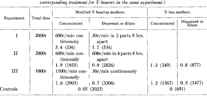
0 . 7 (3306) (2025)TABLE 1
Percentages of half-translocations in the F I f r o m diferent experiments. ( T h e totals of FI scored are given in parentheses. The exposure factors .for Y-less mothers were identical to those f o r the
corresponding treetment for Y-bearers in the same experiment.)
1.2 (1567)
1
0 . 5 (1477) 0 (691)Experiment Total dose
I
I1
I11
Controls
2000r
2000r
l00Or
Modified Y-bearing mothers
I
Y-less mothersConcentrated
600r/min con- tinuously 5 . 4 (336) W r / m i n con-
tinuously 1.9 (1935) 1100r/min con- 1 . 6 (2905)
tinuously 0.05
1
Dispersed or dilute Concentrated1
D i sOr ~ ~ ~ ~ ~I
30r/min in 2 parts 8 hrs.I
I
apart1 . 7 (534)
W r / m i n in 4 parts 8 hrs. apart
0 . 9 (2836)
30r/min continuously
1 . 3 (549) 0.8 (877)
liver, all the treated flies for that experiment were handled alike, in special plastic containers, one side of which was covered with dacron netting, which could be placed on food to permit the virgins to feed between the parts of the irradiation procedure.
All experiments were carried out a t room temperature.
RESULTS AND DISCUSSION O F INDIVIDUAL EXPERIMENTS
In the first type of experiment (Exp. I, table 1) a total dose of 2000r was given either continuously a t 600r/min. (taking only 3/20’’ for delivery) or a t 30r/min. in
2 parts 8 hours apart (requiring 33/20’’ to deliver each of the two parts). Thus the females subjected to the dilute treatment had X-rays delivered a t a rate 20X slower than females given the intense treatment and had in addition an intermission of 8 hours midway in the irradiation. The half-translocation rate obtained was 5.4 % for the intense and 1.5 % for the dilute treatments ( P
<
.005) in oocytes of the modi- fied Y-bearing females. This result proves that the breaks produced in oocytes which enter into gross rearrangements do not always remain unjoined, as they do in the mature sperm (until after fertilization), for 8 hours after being produced. The amount of decrease in half-translocation frequency with protracted treatment permits the estimation that about two thirds of the breaks which, in the intense treatment, were present in cells having a second break with which they half-translocated, were, in the protracted treatment, in cells where this second break had already either resti- tuted or become “unjoinable”.X-RAY INDUCED HALF-TRANSLOCATIONS I N DROSOPHILA 423
tions. If in the dilute treatment all breaks produced in the first one quarter of the irradiation which were ever to restitute or rearrange did so before any breaks in the second one quarter of the treatment were produced, etc., then the total half-translo- cation frequency for the dilute would equal 4 X the rate induced by one quarter of the total dose. Since it was found (HERSKOWITZ 1954b) that half-translocations in- crease, in this dose range, as the
%
power of the dose (however, see General Dis- cussion also), then for “total dose/4” one would expectW
the half-translocations pro- duced by the total dose, and since there were 4 such one quarter doses given in the dilute one would expect from the dilute 4X
%
or46
the number of half-transloca- tions obtained from the intense treatment. However, the rate for the dilute would be more than that for the intense if there were an appreciable number of breaks open and rejoinable 8 hours after treatment. The mutation percentage obtained for the dilute being just about one half that for the intense strongly supports the view that almost all if not all breakages which are ever going to join do so within 8 hours following treatment.Because the percentage for the dilute treatment in Experiment I is less than
56
the rate of the intense (actually 1/3.6, with treatment having been given in only two parts 8 hours apart), there is good reason to believe that breaks were becoming un- available for interchange during the extra 63’20” the X-ray machine was operating in the dilute treatment. Thus the first two experiments strongly suggest that within an hour after breaks are produced an appreciable number of broken pieces can resti- tute or interchange and that if they are to do either they must do so within 8 hours of their production.The third experiment (Exp. 111, table 1) required giving lOOOr in continuous treat- ments either at a rate of 1100r/min. (54” delivery time) or a t 30r/min. (33’20” de- livery time). The incidences of half-translocations for intense and dilute treatments were, respectively, 1.6 % and 0.7 % for modified Y-bearing mothers ( P
< .005).
Since an average of about 16 minutes elapsed between the production of successive breaks in the dilute treatments but averaged less than one half minute in the intense, the reduction in half-translocation percentage for the dilute to about one half the value for the intense is strong evidence that under these experimental conditions a t least one half the breaks that are to undergo restitution or interchange do so within about 16 minutes of their production.Whenever, as in Experiments I1 and 111, Y-less females were also treated a t the times modified Y bearers were, a drop in half-translocation frequency with dosage dilution was obtained which was similar in magnitude to that for modified Y bearers. Although this drop was not statistically significant, its indecisiveness was probably caused only by its small sample size.
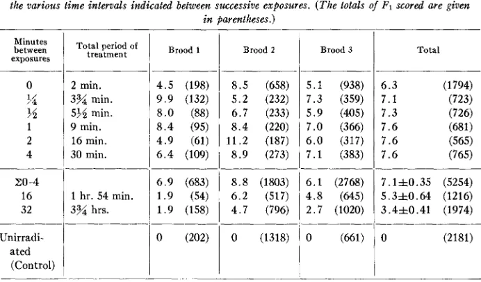
TABLE 2
Percentages of half-translocations produced by 2ZOOr delivered at lla)r/min. i n 8 equal periods with the various time intervals indicated between successive exposures. (The totals of Ft scored are given
i n parentheses.)
Minutes between exposures 0
?4
55
1 2 4 20-4 16 32 Unirradi- ated (Control)Total period of
treatment
2 min.
3% min.
555 min.
9 min.
16 min.
30 min.
1 hr. 54 min.
3% hrs.
Brood 1
4 . 5 (198) 9 . 9 (132) 8 . 0 (88) 8 . 4 (95) 4 . 9 (61) 6 . 4 (109)
6 . 9 (683)
1 . 9 (158) 1 . 9 (54)
0 (202)
Brood 2
8 . 5 (658) 5 . 2 (232) 6 . 7 (233) 8 . 4 (220) 11.2 (187) 8 . 9 (273)
8 . 8 (1803) 6 . 2 (517) 4.7 (796)
0 (1318)
Brood 3
~
5 . 1 (938) 7 . 3 (359) 5 . 9 (405)
7.0 (366) 6 . 0 (317) 7 . 1 (383)
6 . 1 (2768) 4 . 8 (645) 2 . 7 (1020)
0 (661)
Total
7 . 1 f 0 . 3 5 (5254) 5 . 3 f 0 . 6 4 (1216) 3 . 4 f 0 . 4 1 (1974)
0 (2181)
X-RAY INDUCED HALF-TRANSLOCATIONS I N DROSOPHILA 425 vals (where the treatment took almost 2 hours) and to 3.4 f 0.14%
(P
<
.OOOl) with the 32-min. intervals (which required nearly 4 hours to deliver). The frequency for the 32-min. intervals is only that for intervals of 0-4 minutes. However, since it appears significantly higher than 1/3.5 of the latter value, it is probable that, under these latter experimental conditions, some breaks that are produced 32 or more minutes apart cooperate to produce half-translocations, although most of the break- ages which join do so within 4 hours of their production.GENERAL DISCUSSION
The present establishment that the number of detached-X half-translocations depends upon the amount of dispersal of the X-ray dose, is a t the same time an in- dependent demonstration that half-translocations have an origin that is multi- event and not single (such as the production of a break which is a t the same time “healed” to form a new telomere), supporting the other evidences for multi-event origin obtained from X-ray dosage studies (HERSKOWITZ and MULLER 1953; HERS- KOWITZ 1954a,b; and, independently, PARKER 1954a) and from genetic studies for identification of half-translocations (ABRAHAMSON, HERSKOWITZ and MULLER 1954, 1956; PARKER 195413).
The higher rate of half-translocation in oocytes containing the modified Y chromo- some (y+ scvl.Ys) than in Y-less oocytes, discovered by PARKER (1954b), has been confirmed in all 4 possible comparisons in the present experiments (table 1). At least a part of the reason for this difference, it may be proposed here, lies in the fact that, in an oocyte, where the chromosomes are assumed to be in tetrad condition, when a half-translocation between the X and an autosome enters the mature egg there is about one chance in four that the reciprocal centromere-bearing piece will also enter, thereby preventing the detection of a viable half-translocation either because it pro- duces a full translocation or (when this centromere-bearing piece has not joined to an acentric fragment) death due to dicentric formation. However, when a half-transloca- tion between the X and Y enters the egg the reciprocal, homologous, centromere- bearing piece will almost always enter a polar body since homologous centromeres disjoin during chromosome reduction.
It should be noted that among the possible sources of heterogeneity in different experiments, other than the irradiation itself, are variations in daily temperature, humidity, nutrition, and age of the females used, requiring one to proceed with cau- tion when comparing results of different experiments. All of these factors were care- fully randomized, however, within each experiment.
spring of all irradiated series, in no case did the direction of change, in one treatment as compared with another, differ from what is given in the tables, or produce a change which would require modification of the conclusions drawn from the results as pre- sented. For these reasons it is concluded that the number of triplo-X flies scored er- roneously as half-translocations is negligible. Accordingly we have not listed sepa- rately in the tables the phenotypes, as regards y, sc or sex, of the exceptional flies.
Despite the fact that only the eggs oviposited within the first 4 days after the com- pletion of treatment were used in these experiments, and all were “oocytes” when treated, there was a significant decrease in mutability in successive eggs oviposited. Analysis of the data shows that in every case in Experiments I1 and 111, as was found also in earlier work (see table 1 in HERSKOWITZ 1954b), the eggs laid the first days after treatment have more mutations in them than those oviposited soon after. This is undoubtedly partially responsible for the mutation rate with the continuous treatment in Experiment 11, where eggs from all 4 days were used, being lower than the rate with the same treatment in Experiment I, where only eggs from the first 2 days were used. In Experiment IV, while the mutation rate in brood 2 is lower than the rate in brood 1 in only 2 of
7
possible comparisons (probably due to chance variation in the percentages of the smaller sample sizes in brood l ) , there was a lower mutation rate in brood 4 in7
of 8 cases both when compared to the rates for brood 2 alone and for broods 1 and 2 combined.The dilute treatment in Experiment I11 (table 1) and the treatment having in- tervals of 4 minutes in Experiment IV (table 2) gave contradictory results; whereas both were delivered in about the same period of time (33’20” and 30’, respectively) the former showed a significant decrease in mutation rate from the corresponding concentrated treatment but the latter showed no decrease. I n other words, the former case had a considerable amount of rejoining during the treatment period, while the latter had little or none. There are, however, two differences between the conditions of irradiation in these experiments, either or both of which may be responsible for the results. One difference is in the dose administered (one being about twice the other), the other is in the rate a t which the X-rays were applied to the cells (slow and continuous in the former experiment and quick and discontinuous in the latter). WOLFF and ATWOOD (1954) obtained similar results in experiments with Vicia, where a lower dose produced fewer rearrangements when given a t a lower rate than when given a t a higher rate, but a higher dose gave the same number of rearrangements for both intense and dilute treatments. The basis for such results is still undetermined.
X-RAY INDUCED HALF-TRANSLOCATIONS I N DROSOPHILA 427
earlier dosage study the intensity was about 250r/min. If the higher dose used (4000r), delivered in 16 minutes, did not prevent rejoining before the irradiation was completed then the exponent (observed to be the 3/2 power of the dose) was cor- rectly estimated, but if the higher dose prevented rejoining while the lower dose (1000r) did not, or did so to a lesser degree, the exponent was an overestimation if taken as representing the relation of rearrangement frequency to breakage frequency. Which of these situations obtained is unknown a t present.
A significant observation is that while the numbers of parent females irradiated a t the different intensities in Experiment IV were very nearly equal (except that for the 0 min. intervals twice this number was used) the total number of offspring ob- tained increased in the 16 min. as compared with the 0-4 min., and in the 32 min. a s compared with the 16 min. intervals. I t is also noteworthy that the dispersed or dilute has more offspring than its corresponding concentrated treatment in every case in table 1 where the numbers of parents used were the same for both treatments within an experiment (Experiment I11 is to be excluded, however, since more parents were used in the dilute). This statistically significant increase in fecundity n ith dilution of the dose has been a subject for further investigation and will be reported in the next paper of this series (ABRAHAMSON and HERSKOWITZ, in manuscript).
SUMMARY
The frequency of half-translocations obtained following detachment of attached-X chromosomes was employed as the measure of multi-break chromosomal rearrange- ments induced by X-rays administered a t various concentrations to Drosophila melanogaster oocytes. I t was found that higher concentrations produced more half- translocations than lower ones, proving that breaks in oocytes can join in new ar- rangements before fertilization, unlike what is the case for breaks in fully mature spermatozoa (those in inseminated females).
The results show that of the joinings which will take place the great majority do so within 8 hours, and that about one half do so within 4 hours. Under certain experi- mental conditions (as in Exp. 111, see p. 423) a t least one half the breaks that join can do so within hour, but under other conditions (Exp. IV, see pp. 423-425) joinings in this period were not detected.
The relation these results have to earlier work on the dosage-frequency relationship for half-translocations and in confirming the influence of a modified
Y
chromosome on increasing their frequency is discussed.It was found again that among oocytes oviposited during the first four days fol- lowing treatment there were significantly more mutations in the eggs laid in the first days than in those oviposited in later ones. There was also a significant decrease in fecundity of the parent females in this 4-day period when the treatments were more concentrated.
LITERATURE CITED
ABRAHAMSON, S., I. H. HERSKOWITZ, and H. J. MULLER, 1954 Genetic proof for half-transloca- tions derived from irradiated oocytes of Drosophila melanogasler. Genetics 39: 955. (Abstract) Identification of the half-translocations produced by X-raying attached-X chromosomes in Drosophila melanogaster. Genetics 41: 410419.
ABRAHAMSON, S., and J. D. TELFER, 1954 Sex chromosome loss and translocation frequencies in
Drosophila melanogaster after X-raying sperm in males or in females. Genetics 39: 955-956. (Abstract)
HAAS, F. L., E. DUDGEON, F. E. CLAYTON, and W. S. STONE, 1954 Measurement and control of some direct and indirect effects of X-radiation. Genetics 39: 453471.
HERSKOWITZ, I. H., 1954a The joinability of chromosome breakage points produced by X-rays in Drosophila oocytes. Science 119: 581. (Abstract)
1954b The relation between X-ray dosage and the frequency of simulated healing of chromosome breakages in Drosophila melanogaster females. Proc. Nat. Acad. Sci. U. S. 40: 576-585.
Evidence against the healing of X-ray breakages in chromosomes of female Drosophila melanogaster. Genetics 38: 669. (Abstract)
Mutation frequency as conditioned by the manner of application of radiation. Indian J. Genet. Plant Breeding, 3: 15-31.
HERSKOWITZ, 1. H., and H. J. MULLER, 1953 MAKHIJANI, J. K., 1945
MULLER, H. J., 1938 The remaking of chromosomes. Collecting Net 13: 181, 183-195, 198. 1939a The mechanism of structural change in chromosomes of Drosophila. Proc. 7th Inter. 1939b Report of investigations with radium. Med. Res. Council Spec. Rep. Serv. No. 236:
1940 An analysis of the process of structural change in chromosomes of Drosophila. J. Genet. MULLER, H . J., and I. H. HERSKOWITZ, 1954 Concerning the healing of chromosome ends pro- MULLER, H. J., R. M. VALENCIA, and J. I. VALENCIA, 1950 The production of mutations at indi- PARKER, D. R., 1954a Role of the Y chromosome in induced detachment of attached-X chromo- Radiation-induced exchanges in Drosophila females. Proc. Nat. Acad. Sci. U. S. 40:
The validity of the Bunsen-Roscoe law in the production of muta- tions by radiation of extremely low intensity. Proc. 7th Inter. Congr. Genet. J. Genet. Suppl. p. 246; and Proc. Roy. Soc. Edinburgh B 62: 66-72.
The time factor in X-ray irradiation. Science 89: 400-401. (Abstract) Congr. Genet. J. Genet. Suppl. pp. 221-222.
14-15.
40: 1-66.
duced by breakage in Drosophila melanogaster. Am. Naturalist 88: 177-208.
vidual loci in Drosophila by irradiation of oocytes and oogonia. Genetics 36: 126. (Abstract) somes in Drosophila melanogasler. Genetics 39: 986. (Abstract)
1954b 795-800.
RAY-CHAUDHURI, S. P., 1939
SAX, K., 1939a
19391, The time factor in X-ray production of chromosome aberrations. Proc. Nat. Acad. Sci. 1940 An analysis of X-ray induced chromosomal aberrations inTradescantia. Genetics 26 : 41-68. 1941 Types and frequencies of chromosomal aberrations induced by X-rays. Cold Spring Harbor SAX, K., and R. T. BRUMFIELD, 1943 The relation between X-ray dosage and the frequency of WOLFF, S., and K. C. ATWOOD, 1954 Independent X-ray effects on chromosome breakage and
U. S. 26: 225-233.
Symposia Quant. Biol. 9: 93-103.