Article
1
A tunable nanoplatform of nanogold functionalised
2
with angiogenin peptides for anti-angiogenic therapy
3
of brain tumours
4
5
Irina Naletova1, Lorena Maria Cucci2, Floriana D’Angeli3, Carmelina Daniela Anfuso3, Antonio
6
Magrì4, Diego La Mendola1,5,*, Gabriella Lupo3,* and Cristina Satriano1,2,*
7
1 Consorzio Interuniversitario di Ricerca in Chimica dei Metalli nei Sistemi Biologici (CIRCMSB), Via Celso
8
Ulpiani 27, I-70126 Bari, Italy; [email protected]
9
2 Hybrid NanobioInterfaces Lab (NHIL), Department of Chemical Sciences, University of Catania, Viale
10
Andrea Doria 6, I-95125 Catania, Italy; [email protected]
11
3 Department of Biomedical and Biotechnological Sciences, University of Catania, Via Santa Sofia 89, I-95123
12
Catania, Italy; [email protected]; [email protected]
13
4 Institute of Crystallography Catania, National Council of Research (IC-CNR), Via Paolo Gaifami 18, I-95126
14
Catania, Italy; [email protected]
15
5 Department of Pharmacy, University of Pisa, via Bonanno Pisano 6, I-56126 Pisa, Italy
16
17
* Correspondence: [email protected], Tel.: +39- 050-2219533 (D.L.M.); [email protected], Tel.:
18
+39-095-4781158 (G.L.); [email protected], Tel.: +39-095-7385136 (C.S.)
19
20
21
Abstract: Angiogenin (ANG), an endogenous protein that plays a key role in cell growth and
22
survival, has been scrutinised here as promising nanomedicine tool for the modulation of pro-/
anti-23
angiogenic processes in brain cancer therapy. Specifically, peptide fragments from the putative cell
24
membrane binding domain (residues 60-68) of the protein were used in this study to obtain
peptide-25
functionalised spherical gold nanoparticles (AuNPs) of about 10 nm and 30 nm in optical and
26
hydrodynamic size, respectively. Different hybrid biointerfaces were fabricated by peptide physical
27
adsorption (Ang60-68) or chemisorption (the cysteine analogous Ang60-68Cys) at the metal nanoparticle
28
surface, and the cellular assays were performed in the comparison with ANG-functionalised AuNPs.
29
Cellular treatments were performed both in basal and in copper-supplemented cell culture medium,
30
to scrutinise the synergic effect of the metal, which is another known angiogenic factor. Two brain
31
cell lines were investigated in parallel, namely tumour glioblastoma (A172) and neuron-like
32
differentiated neuroblastoma (d-SH-SY5Y). Results on cell viability/proliferation, cytoskeleton actin,
33
angiogenin translocation and VEGF release pointed to the promising potentialities of the developed
34
systems as anti-angiogenic tunable nanoplaftforms in cancer cells treatment.
35
Keywords: plasmonics; nanomedicine; theranostics; copper; VEGF; glioblastoma; differentiated
36
neuroblastoma; peptidomimetics; qPCR; actin.
37
38
39
2 of 30
Table of Contents
40
41
Gold nanoparticles (AuNP) functionalised with peptides mimicking the cell membrane binding site
42
of angiogenin protein (ANG) can perturb the VEGF-regulated nuclear transcription process, which
43
results in an enhanced anti-angiogenic response in tumour cells.
1. Introduction
46
In recent decades, protein-nanoparticle and peptide-nanoparticle conjugates have emerged as
47
powerful nanomedicine tools, enabling for biomedical applications in prevention, diagnosis and
48
treatment of disease [1,2]. Unfunctionalised, bare nanoparticles (NPs), are often able to match several
49
desired function of theranostic platforms, including peculiar optical, electrical, magnetic properties
50
of nanometer-sized materials [3], tunable geometries and tailored size and surface chemistry [4],
51
intrinsic biological properties such as anti-angiogenic nanogold [5,6] or antibacterial nanosilver [7,8].
52
On the other hand, hybrid peptide- or protein-NP conjugates enable addressing many of the
53
difficulties that arise as results of in vivo applications. Specifically, both naturally derived and
54
synthetic polypeptides may offer improved biocompatibility [9], targeted delivery [10] and
55
prolonged lifetime before clearance, to ensure an efficient therapeutic action [1,11].
56
Angiogenin (ANG) is a secreted ribonuclease (also known as RNase 5), identified in media from
57
cancer cells, but also present in normal tissues, such as plasma and amniotic fluid, and secreted from
58
vascular endothelial cells, aortic smooth muscle cells, fibroblasts [12]. Angiogenin induces
59
neovascularization by triggering cell migration, invasion, proliferation, and formation of tubular
60
structures [12-15].
61
Physiologically, ANG is overexpressed during inflammation, exhibiting wound healing
62
properties as well as microbicide activity and conferring host immunity [16]. However, uncontrolled
63
activity of angiogenin is implicated in pathological processes.
64
The protein was isolated for the first time from medium conditioned by a human
65
adenocarcinoma cell line (HT-29) [17]. A high expression of angiogenin has been described in
66
different types of cancers and to their malignant transformation [12], including gliomas that are brain
67
tumours fast-growing, aggressive and with a poor prognosis [18].
68
ANG expression has been identified also in neurons and acts as a part of the secretome of
69
endothelial progenitor cells (EPCs) [19,20]; the modulation of ANG and EPCs as repair-associated
70
factors has been found in stroke patients and mouse models of rehabilitation after cerebral ischemia
71
[21]. Mutations in the ANG gene have been characterized in amyotrophic lateral sclerosis (ALS) [22]
72
and Parkinson's disease (PD) [23]. Moreover, endogenous angiogenin levels are dramatically reduced
73
in an alpha-synuclein mouse model of PD and exogenous angiogenin protects against cell loss in
74
neurotoxin-based cellular models of PD [23]. Genetic studies revealed that angiogenin treatment
75
delays motor dysfunction and motor neuron loss, also prolonging the survival in superoxide
76
dismutase 1 (SOD1) mouse model of ALS [14].
77
In motor neurons, ANG can be upregulated by hypoxia thought the stimulation of rRNA
78
transcription of endothelial cells [24]. Such a process is critical for the cellular proliferation induced
79
by other angiogenic proteins, including vascular endothelial growth factor (VEGF) [25]. While the
80
predominant role of VEGF in the formation of new blood vessels is unquestioned, several recent
81
studies demonstrate that VEGF also has trophic effects on neurons and glia in the Central- (CNS) and
82
Peripheral- (PNS) Nervous System, promoting neurogenesis, neuronal patterning, neuroprotection
83
and glial growth [26]. Therefore, VEGF modulates neuronal health and nerve repair; and exogenous
84
angiogenin delivery can be considered a promising tool of anti-angiogenic therapy for treating
85
gliomas, where malignancy is highly related to angiogenesis.
86
Copper is an essential metal that plays a key role in the CNS development and function, and its
87
dyshomeostasis is involved in many neurodegenerative diseases as Alzheimer’s disease (AD), PD
88
and ALS [27,28]. Furthermore, copper is known to be a strong angiogenic factor, with metal serum
89
levels raising in a wide variety of human cancers [29,30]. Noteworthy, copper increases the
90
expression of ANG and regulates its intracellular localization [31]. Moreover, ANG binds copper ions
91
and the metal interaction largely influences its interaction with endothelial cells as well as its
92
angiogenic activity [32-34]. Taking into account the correlations between angiogenin protein and
93
copper in physiological and pathological conditions of the brain, a promising pharmacological
94
approach in brain tumours therapy is the use of ANG as molecular target, whose activity may be
95
modulated by the presence of copper ions.
4 of 30
As an alternative to protein-based drugs, peptides mimicking functional domains of the whole
97
protein are becoming more relevant as drug candidates, to address problems exhibited by the protein
98
in in vivo applications such as the additional effect or functions or binding sites for other ligands, the
99
immunological clearance before reaching their target site [35,36].
100
Three domains of angiogenin have been demonstrated essential to the protein to explicate its
101
biological activity, i.e.,: the catalytic site (involving His-13, Lys-40, and His-114 residues), the nuclear
102
translocation sequence (encompassing residues 31-35, RRRGL); the putative cellular binding site
103
(residues 60-68, KNGNPHREN) [37,38]. In previous works, peptide fragments encompassing such
104
different domains of the protein have been synthesized and used as mimicking model of the whole
105
protein [6,39,40]. In particular, hybrid nano-assemblies of gold nanoparticles (AuNPs) functionalized
106
with different peptides encompassing the putative cellular binding site of the protein (Ang60-68) have
107
been demonstrated able to maintain their activity on cytoskeleton actin reorganization in a tumour
108
cell line of human neuroblastoma [40].
109
Here, we report on the investigation of AuNPs functionalised with the peptide Ang60-68 or its
110
analogous having a cysteine residue in the C-terminus (Ang60-68Cys), in the comparison with the
111
whole ANG protein. Such systems have been scrutinised as potential nanomedicine platforms
112
towards a brain cancer of human glioblastoma (A172 cell line). To compare the response of tumour
113
and non-tumour brain cells, differentiated neuroblastoma (d-SH-SY5Y line) have been included in
114
the study as model neuron-like cells.
115
Effects on cell proliferation, cytoskeleton actin changes, angiogenin translocation and VEGF
116
expression upon the cell treatments with peptides- or protein-functionalized nanoparticles, in the
117
absence or presence of copper ions, shed new light in the link between different factors involved in
118
angiogenesis processes of non-tumour and tumour model brain cell cultures. Indeed, since ANG
119
plays a key role in cell growth and survival, and the role of VEGF in brain tumour angiogenesis has
120
been demonstrated [41], new perspectives in the therapeutic approaches may rely on the tiny
121
modulation of the pro-/ anti-angiogenic processes [42] for brain cancer treatment.
122
123
2. Results
124
2.1. Physicochemical characterisation of hybrid peptides- and protein-NP conjugates
125
2.1.1. Optical (plasmonic) properties changes of AuNP upon interaction with peptides/protein.
126
Peptide-functionalised NPs were fabricated by two different approaches, namely a purely
127
physical adsorption and a prevalent covalent grafting, with the peptide sequences Ang60-68 and Ang
60-128
68Cys, respectively. As positive control, samples of ANG-functionalised AuNPs were prepared by
129
using the whole protein.
130
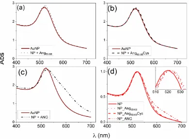
Figure 1 shows the UV−visible spectra of gold nanoparticles, before and after the addition of
131
Ang60-68n, Ang60-68Cys or ANG, respectively. The plasmon peak parameters, i.e., the wavelength at the
132
maximum absorbance (max = 519 nm) and the full width at half maximum (FWHM = 54 nm) point to
133
the formation of a monodisperse gold colloidal solution of spherical nanoparticles with an optical
134
diameter of 11 nm [43].
135
The addition of Ang60-68 (Fig. 1a) or Ang60-68Cys (Fig. 1b), at the concentration of 3·10-5 M for both
136
peptides, induces comparable red-shifts (Δλmax = 3 nm) and increase of absorbance at the maximum
137
(Abs ~ 0.07), according to previous findings [40].
138
The addition of the whole protein ANG (Fig. 1c), at the concentration of 1·10-7 M, leads to a
139
significantly larger red-shift in the plasmon peak (Δλmax = 4 nm) compared to those found upon the
140
addition of the peptides. Moreover, an opposite trend in the absorbance at the maximum, with a
141
decrease (Abs = -0.09) in comparison to the bare AuNPs and a broadening of the plasmon band
142
(FHWM = 11 nm, corresponding to the appearance of a shoulder at around 600 nm) are visible, most
143
likely due to a partial nanoparticle aggregation.
145
146
Figure 1. (a-c) UV-visible spectra of AuNPs in 1 mM MOPS-TCEP buffer (1:1 mol ratio) before and
147
after the addition of: (a) 30 M Ang60-68, (b) 30 M Ang60-68Cys; (c) 100 nM ANG. (d) UV-visible spectra
148
of the pellets collected after two rinsing steps by centrifugation (8,000 r.p.m, for 15 min) and
re-149
suspension in 1 mM MOPS-TCEP buffer.
150
As to the hybrid systems used for the cellular experiments, Fig. 1d shows the UV-visible spectra
151
of the protein/peptide-NP pellets samples after two washing steps. The red-shift in the plasmon peak
152
with respect to the bare AuNPs is still visible (Δλmax ~ 3 nm) for Ang60-68 and Ang60-68Cys peptides as
153
well as for ANG protein). This finding confirms the irreversible adsorption of the peptides and
154
protein molecules and hence the successful surface functionalisation of the gold nanoparticles by the
155
used biomolecules.
156
157
2.1.2. Hydrodynamic size and conformational features of peptides- and protein-functionalised NPs
158
in the absence or presence of copper ions.
159
The hydrodynamic size of ~ 30 nm for the aqueous dispersion of gold nanoparticles (1.7·108
160
NP/mL) did not change significantly upon addition of the peptide solution (3·10-5 M), for both Ang
60-161
68 and Ang60-68Cys fragments. On the other hand, by addition of ANG (1·10-7 M), the nanoparticle size
162
was largely increased in comparison to the bare AuNP, thus suggesting nanoparticle aggregation
163
prompted by the presence of the protein at the nanoparticle surface.
164
The Ang60-68_NP and Ang60-68Cys_NP pellets maintained a size range comparable to that of
non-165
rinsed nanoparticles (both bare and functionalized), while a slight decrease in the size was found for
166
ANG_NP, where a fraction of loosely bound proteins molecules was therefore likely rinsed off by the
167
washing steps of the protein-nanoparticle hybrids.
168
Noteworthy, after the addition of copper ions, the average dimension of nanoparticles was still
169
unchanged for bare AuNP, instead a dramatic increase in the hydrodynamic diameter was found for
170
Ang60-68_NP (to ~ 180 nm) and Ang60-68Cys_NP (to ~300 nm), respectively.
6 of 30
Table 1. Hydrodynamic size of the different NPs before and after the functionalisation with the
172
peptides or the protein, and after the addition of before and after addition of 20 M CuSO4.
173
Hydrodynamic size (nm)
peptide/protein NP +
peptide/protein(1) peptide/protein_NP
(2) peptide/protein_NP + Cu(II)(3)
- 29 ± 3 30 ± 2 31 ± 5
Ang60-68 28 ± 4 37 ± 4 175 ± 10
Ang60-68Cys 30 ± 3 29 ± 5 281 ± 42
ANG 53 ± 4 41 ± 6 47 ± 5
1 peptide/protein-added NP samples; 2 peptide/protein_NP samples after the centrifugation and rinsing steps; 3
174
same pellets as 2 added with copper ions.
175
The Ang60-68 peptide is able to bind copper at physiological pH by the involvement of one
176
imidazole, two deprotonated amide nitrogen and one carboxyl oxygen atoms, respectively [44]. The
177
UV-vis parameters measured for the equimolar solutions at pH= 7.4 of copper(II) and Ang60-68Cys
178
(max = 624 nm; = 100 M-1cm-1) were very similar to those of analogous complex formed with
Ang60-179
68 (max = 630 nm; = 120 M-1cm-1).
180
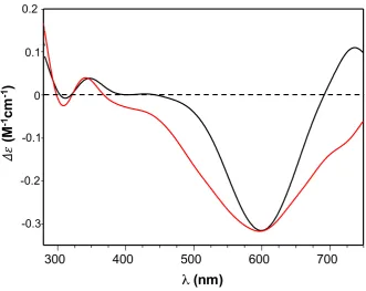
Accordingly, the CD spectra of both Ang60-68+Cu(II) and Ang60-68Cys+Cu(II) (Figure 2) showed a
181
minimum around 600 nm, assigned to copper d-d transition, and a broad band with a maximum
182
approximately at 350 nm, assigned to charge transfer to the metal ion by the imidazole nitrogen
183
(Nim→Cu(II)) and the deprotonated amide nitrogen (Namide→ Cu(II)).
184
185
Figure 2. CD spectra of Ang60-68 + CuSO4 (black line) and Ang60-68Cys + CuSO4 (red line) at pH = 7.4.
186
Equimolar concentration of peptide and copper were used: [peptide] = [Cu(II)]= 1x10⁻³ M.
187
188
189
2.2. Biological characterisation of the interaction between peptides- or protein-NP conjugates and brain
191
tumour (A172 line) or non-tumour (d-SH-SY5Y) cells
192
2.2.1. Determination of angiogenin expression in glioblastoma (A172), undifferentiated and
193
differentiated neuroblastoma (SH-SY5Y) cell lines.
194
To analyse the endogenous levels of ANG expression in the tested cancer cells (glioblastoma
195
A172 and neuroblastoma SH-SY5Y) and neuronal-like cells (differentiated neuroblastoma,
d-SH-196
SY5Y), we performed Western blot analyses of protein extracts from crude cell lysates (Figure S1 in
197
Supplementary Material). Results confirmed that in tumour cells the expressed level of protein were
198
significantly higher than in differentiated neuroblastoma (Fig. S1a,b). Moreover, to control the
199
specific interaction of anti-angiogenin antibody with Ang60-68 or Ang60-68Cys in the comparison with
200
ANG, the peptides and protein samples were analysed by Western and dot blotting assays. The used
201
anti-angiogenin antibody detected only the whole protein but did not interact with the two peptide
202
fragments (Fig. S1c,d).
203
2.2.2. Cell viability
204
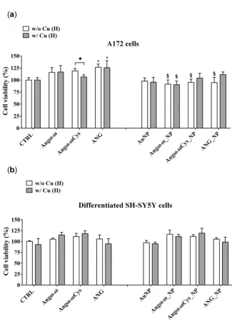
MTT assays were carried out to assess the effect on cell viability (Figure 3) of peptides- or
205
protein-functionalised NPs, in the absence or presence of copper ions, for brain glioblastoma (A172
206
line) and differentiated neuroblastoma (d-SH-SY5Y), respectively.
207
In the tumour A172 cell line (Fig. 3a), a significant increase on viability (+25%; p ≤ 0.05 vs. control
208
untreated cells) was found after the treatment with ANG, both in absence and in the presence of
209
added Cu(II). The cells incubation either with free peptides of Ang60-68Cys or Ang60-68 did not induce
210
any significant change on cell viability; similar results were found for cells treated with peptides in
211
the presence of copper. As to nanoparticle-treated cells, the incubation with bare AuNP, both in the
212
absence and with Cu(II), did not modify the cell viability in comparison with control cells. The
213
incubation with Ang60-68_NP reduced the cell viability by about 20-25% (p ≤ 0.05 vs. the respective
214
peptide and peptide + Cu(II) controls), both in the absence and in the presence of copper. As to Ang
60-215
68Cys_NP, a significant cell viability decrease in the absence of Cu(II) (-25%; p ≤ 0.05 vs. the respective
216
free peptide) was nullified by the incubation in presence of copper. A similar trend was found for the
217
cells incubated with ANG_NP, where a reduced cell viability (- 20%; p ≤ 0.05 vs. the respective free
218
protein) in the absence of copper but no significant difference in presence of copper were found.
219
In the non-tumour d-SH-SY5Y cell line (Fig. 3b), none of the treatments used resulted in a
220
statistically significant decrease of cell viability in comparison to untreated control cells. Trypan blue
221
staining confirmed the above reported results (data not shown).
8 of 30
225
Figure 3. Cell viability determined by MTT assay of A172 (a) and d-SH-SY5Y (b) cell lines. Cells were
226
grown in basal culture medium (control: CTRL) and in culture medium supplemented with: Ang60-68
227
(30 M), Ang60-68Cys (30 M); ANG (100 nM), AuNP (9.4 nM = 1.4108 NP/mL), Ang60-68_NP (1.4 nM =
228
4.0106 NP/mL, [Ang60-68] = 2.8·10-12 M), Ang60-68Cys_NP (1.4 nM = 4.0106 NP/mL, [Ang60-68Cys] = 2.6·10
-229
12 M), ANG_NP (1.2 nM = 3.4106 NP/mL, [ANG]= 0.2·10-12 M). All conditions were evaluated in
230
presence or absence of metal ions (copper sulphate: Cu(II), 20 M). The bars represent means ± SD of
231
three independent experiments performed in triplicate (S.D. = standard deviation). Statistically
232
significant differences, determined by one-way ANOVA are indicated: *p ≤ 0.05 versus CTRL; § p ≤
233
0.05 versus the respective treatment with free peptides/protein.
234
235
2.2.3. Cytoskeleton actin reorganisation and intranuclear angiogenin
236
Cell migration is a critical step in tumour invasion and metastasis; the regulation of this process
237
is often monitored in therapies for treating cancer. Reorganization of the actin cytoskeleton is the
238
primary mechanism of cell motility and is essential for most types of cell migration [45].
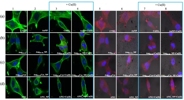
Confocal laser scanning microscopy (LSM) demonstrated substantial differences between the
240
tumour A172 (Figure 4) and non-tumour d-SH-SY5Y (Figure 5) cell lines in the organization of the
241
actin cytoskeleton, both before and after the treatments with the peptides/protein-conjugated
242
nanoparticles, as well as the incubation in the copper-supplemented medium.
243
Combined staining for F-actin (green) and nuclei (blue) for untreated glioblastoma (Fig. 4, panel
244
a1, CTRL) clearly shows their polygonal shape along with different types of actin dorsal fibres and
245
transverse arcs, typical for the lamellipodial actin meshwork [46]. The cell treatment with bare AuNP
246
and/or the addition of Cu(II) (Fig. 4, panels a2-4) increased actin stress fibres. In contrast to A172 cells,
247
F-actin staining of untreated d-SH-SY5Y cells (Fig. 5, panel a1, CTRL) analysed by LSM showed
248
several distinct types of actin structures with broad leading edges, including a lamellipodium with a
249
loose meshwork of actin filaments, an actin rich lamella, dorsal ruffles, transverse arcs and stress
250
fibers, as expected for differentiated neuroblastoma [47].
251
A172 cells treated with the free peptides or protein showed a diffuse actin staining for several
252
lamellipodia protruding from the cell body in all directions. The addition of copper did not change
253
significantly the actin staining for Ang60-68 (Fig. 4, see panels b1 and b3), instead visibly decreased the
254
lamellipodia structures for Ang60-68Cys (Fig. 4, panels c1 and c3) and ANG (Fig. 4, panels d1 and d3),
255
respectively. Both in absence and in the presence of copper, A172 cells treated with Ang60-68_NP (Fig
256
4, panels b2 and b4) and ANG_NP (Fig 4, panels d2 and d4) still displayed a similar actin staining
257
than those incubated with the free peptide or protein, respectively. On the contrary, after incubation
258
with Ang60-68Cys_NP, both in the absence and presence of copper (Fig 4, panels c2 and c4), cells
259
contained very few, if any, actin stress fibres in the central regions and lamellipodia structures.
260
For d-SH-SY5Y cells treated with the two peptides or the protein or their nanoparticle conjugates,
261
irrespective of the incubation in copper-supplemented medium or not, the most notable change was
262
a generally less dense meshwork of actin filaments after the treatment with Ang60-68 (Fig. 5b, panels
263
1-4) or Ang60-68Cys (Fig. 5c, panels 1-4). The central region of these cells contained neither ventral
264
stress fibres nor dorsal ruffles or transverse arcs detectable by LSM. On the contrary, numerous and
265
diffuse actin structures, as well as prominent actin stress fibres along the entire cell border were found
266
for cells treated with ANG samples (Fig. 5d, panels 1-4).
267
LSM imaging of intracellular angiogenin in glioblastoma and differentiated neuroblastoma cells
268
confirmed the western blot results (see Fig. S1) that untreated non-tumour d-SHSY5Y cells (Fig. 5a,
269
panel 5) showed lower levels of endogenous ANG in the cytoplasm and in the nucleus than untreated
270
tumour A172 cells (Fig. 4a, panel 5).
271
Cells incubated with ANG for 2 hr exhibited a strong increase of the red staining, confirming the
272
cellular uptake of exogenous angiogenin. In A172 cells the staining was especially enhanced in the
273
presence of copper ions for the nuclear and perinuclear regions (Fig. 4d, panels 7-8). In d-SH-SY5Y
274
cells, an increased red staining was visible in vesicles in the cytoplasm and in the neurites (Fig. 5d,
275
panels 5-8), according to intracellular angiogenin localisation reported by Thiyagarajan et al. in
276
similar neuronal cell lines [48]. We also observed the presence of intense punctuate structure of
277
angiogenin in perinuclear and neurite regions, suggesting formation of resembling secretory granules.
278
Noteworthy, A172 cells treated with the peptide fragments Ang60-68 (Fig. 4b, panels 5-8) or Ang
60-279
68Cys (Fig. 4c, panels 5-8) showed a diffuse cytoplasmic staining and a weak staining in the nucleus,
280
neurites and cell membrane. A negligible staining of nuclear angiogenin was found after cell
281
incubation with peptide-conjugated nanoparticles in the presence of copper ions.
283
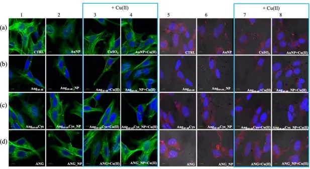
284
Figure 4. Confocal micrographs of A172 cells. Actin Green® 488 (in green, ex/em= 488/500-530 nm) and Hoechst33342 (in blue, ex/em=405/425–475 nm) were used as F-actin
285
and nuclear markers, respectively. Antibody against angiogenin shows angiogenin localisation in red (ex/em=543/560–700 nm) and micrographs are merged with optical
286
bright field images (in grey). Before treatments, cell were rinsed with fresh culture medium and incubated for 2 hr with basal culture medium (control: CTRL) and in
287
culture medium supplemented with: Ang60-68 (30 M), Ang60-68Cys (30 M); ANG (100 nM), AuNP (9.4 nM = 1.4108 NP/mL), Ang60-68_NP (1.4 nM = 4.0106 NP/mL, [Ang
60-288
68] = 2.8·10-12 M), Ang60-68Cys_NP (1.4 nM = 4.0106 NP/mL, [Ang60-68Cys] = 2.6·10-12 M), ANG_NP (1.2 nM = 3.4106 NP/mL, [ANG]= 0.2·10-12 M). Scale bar = 10 m.
290
Figure 5. Confocal micrographs of d-SH-SY5Y cells Actin Green® 488 (in green, ex/em = 488/500-530 nm) and Hoechst33342 (in blue, ex/em = 405/425–475 nm) were used
291
as F-actin and nuclear markers, respectively. Antibody against angiogenin shows angiogenin localisation in red (ex/em=543/560–700 nm) and micrographs are merged
292
with optical bright field images (in grey). Before treatments, cell were rinsed with fresh culture medium and incubated for 2 hr with basal culture medium (control: CTRL)
293
and in culture medium supplemented with: Ang60-68 (30 M), Ang60-68Cys (30 M); ANG (100 nM), AuNP (9.4 nM = 1.4108 NP/mL), Ang60-68_NP (1.4 nM = 4.0106 NP/mL,
294
[Ang60-68] = 2.8·10-12 M), Ang60-68Cys_NP (1.4 nM = 4.0106 NP/mL, [Ang60-68Cys] = 2.6·10-12 M), ANG_NP (1.2 nM = 3.4106 NP/mL, [ANG]= 0.2·10-12 M). Scale bar = 10 m.
2.2.4. VEGF release and synthesis.
296
VEGF has been identified as the most important pro-angiogenic factor released by cancer cells
297
and it its concentration in the tissue of glioblastomas has been demonstrated significantly higher than
298
that in normal brain [49]. Moreover, VEGF has a crucial role in neurogenesis, neuronal patterning,
299
neuroprotection and glial growth [26,50]. Figure 6 shows the VEGF release after incubation for 24 hr
300
of tumour A172 cells and d-SH-SY5Y with peptides- or protein-conjugated NPs, in the absence or
301
presence of copper ions.
302
303
304
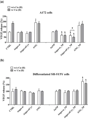
Figure 6. VEGF release in the medium of confluent cultures of A172 (a) and differentiated d-SH-SY5Y
305
cells (b). Cells were grown in basal culture medium (control: CTRL) and in culture medium
306
supplemented with: Ang60-68 (30 M), Ang60-68Cys (30 M); ANG (100 nM), AuNP (9.4 nM = 1.4108
307
NP/mL), Ang60-68_NP (1.4 nM = 4.0106 NP/mL, [Ang60-68] = 2.8·10-12 M), Ang60-68Cys_NP (1.4 nM =
308
4.0106 NP/mL, [Ang60-68Cys] = 2.6·10-12 M), ANG_NP (1.2 nM = 3.4106 NP/mL, [ANG]= 0.2·10-12 M).
309
All conditions were evaluated in presence or absence of metal ions (copper sulphate: Cu(II), 20 M).
310
Sandwich ELISA with monoclonal anti-VEGF antibody was used. The bars represent means ± SD of
311
three independent experiments performed in triplicate (S.D. = standard deviation). Statistically
312
significant differences, determined by one-way ANOVA are indicated: *p ≤ 0.05 versus CTRL; § p ≤
313
0.05 versus the respective treatment with free peptides/protein; ♦p ≤ 0.05 versus the same treatment
314
w/o Cu (II).
In A172 cells (Fig. 6a) the treatments with copper alone increased the VEGF release by about 2.0
316
folds (p ≤ 0.05 vs. control untreated cells), confirming the relevant role of this cation in cancer
317
progression [51].
318
The incubation of the cells with Ang60-68Cysor with Ang60-68 did not modify the VEGF release in
319
comparison to control cells, both in the absence and in the presence of Cu(II) whereas the treatments
320
with ANG or ANG + Cu(II) increased the VEGF release by about 2.3 folds (p ≤ 0.05 vs. control
321
untreated cells).
322
The treatments with bare AuNP, both in the absence and in the presence of copper ions, did not
323
modify significantly the VEGF release in comparison to control cells. Surprisingly, the incubation
324
with AuNPs functionalized with peptide fragment Ang60-68Cys and Ang60-68, induced a significant
325
reduction of VEGF release respectively by 27% and by 30%, in comparison to the corresponding
326
control (free Ang60-68Cys and free Ang60-68). Moreover, further reduction of the release was found
327
when the incubation with peptide fragment Ang60-68Cys was performed in presence of copper.
328
No difference was found in VEGF release after treatment of A172 cells with ANG_NP in
329
comparison to cells treated with free ANG.
330
The incubation of d-SH-SY5Y cells with ANG did not modulate the VEGF release, as well as
331
with Ang60-68 and Ang60-68Cys, both in absence and in presence of copper, in comparison to the
332
respective controls.
333
Differently by A172 cells, in non-tumour d-SH-SY5Y cells (Fig. 6b), only the treatment with
334
AuNP functionalized with ANG, both in the absence and in presence of copper, induced an increase
335
of the VEGF release in comparison to untreated control cells. The incubation of control cells with
336
copper did not modulate the VEGF release.
337
The concentration of VEGF released by A172 and d-SH-SY5Y was 133 pg/ml ± 10.1 and 58 pg/ml
338
± 4.3, respectively.
339
These results were confirmed by determination of VEGF mRNA levels (Figure 7). In A172 cells
340
(Fig. 7a) the treatments with ANG significantly increased mRNA transcription, whereas ANG_NP
341
induced a significant reduction of transcription in comparison with free ANG but with values higher
342
than control cells. No differences were found after treatment with free Ang60-68, free Ang60-68Cys as
343
well as bare NPs. On the other hand, the incubation with Ang60-68_NP and Ang60-68Cys_NP induced a
344
significant reduction of mRNA transcription by about 2.2 and 2.7 folds, respectively, in comparison
345
to the respective control (free Ang60-68 and free Ang60-68 Cys). Moreover, further reduction of the
346
transcription was found after incubation in the presence of copper.
347
In non-tumour d-SH-SY5Y cells (Fig. 7b), only the treatment with AuNP functionalized with
348
ANG, both in the absence and in presence of copper, induced an increase of the VEGF mRNA
349
transcription in comparison to the respective control (free ANG).
14 of 30
352
Figure 7. VEGF mRNA levels determination by qPCR in A172 (a) and differentiated d-SH-SY5Y cells (b).
353
Cells were grown in basal culture medium (control: CTRL) and in culture medium supplemented with:
354
Ang60-68 (30 M), Ang60-68Cys (30 M); ANG (100 nM), AuNP (9.4 nM = 1.4108 NP/mL), Ang60-68_NP (1.4 nM
355
= 4.0106 NP/mL, [Ang60-68] = 2.8·10-12 M), Ang60-68Cys_NP (1.4 nM = 4.0106 NP/mL, [Ang60-68Cys] = 2.6·10-12
356
M), ANG_NP (1.2 nM = 3.4106 NP/mL, [ANG]= 0.2·10-12 M). All conditions were evaluated in presence or
357
absence of metal ions (copper sulphate: Cu(II), 20 M). Relative quantification is referred to untreated cells
358
(CTRL). Data normalized with respect to the expression level of S18 mRNA. The bars represent means ±
359
SD of three independent experiments performed in triplicate (S.D. = standard deviation). Statistically
360
significant differences, determined by one-way ANOVA are indicated: *p ≤ 0.05 versus control; § p ≤ 0.05
361
versus the respective treatment with free peptides/protein.
362
3. Discussion
368
In this study, two brain cell lines, namely tumour glioblastoma (A172) and differentiated
369
neuroblastoma (d- SH-SY5Y) neuron-like cells, were scrutinised after incubation with hybrid
370
nanoassemblies made of gold nanoparticles functionalised with angiogenin protein or with two
371
different angiogenin-mimicking peptides (Ang60–68 and its cysteine derivative at the C-terminus,
372
Ang60-68Cys) containing the ANG residues from 60 to 68, which is the the exposed protein loop region
373
that is part of a cell-surface receptor binding site [52].
374
The Ang60-68 peptide has been demonstrated to specifically interact with cytoskeleton actin [6],
375
whereas the Ang60-68Cys peptide has been successfully used to tailor gold nanoparticles by chemical
376
grafting [40]. Gold, indeed, being a soft acid, binds to soft bases like thiols, to form stable Au-S bonds
377
(40-50 kcal/mol) that are able to replace the citrate shell on the nanoparticle surface due to the strong
378
affinity binding of the thiol groups with the metal [53].
379
To functionalize the gold nanoparticles with Ang60–68, Ang60-68Cys or ANG, the biomolecules
380
were added to the colloidal dispersion (1.7·108 AuNP/mL) at the concentration respectively of 3·10-5
381
M for the peptides and 1·10-7 M for the protein, and the shifts in the plasmon band were monitored
382
(Figure 1).
383
The optical interface established between the biomolecules and the metal nanoparticle surface,
384
as investigated by UV–visible spectroscopy, clearly evidenced an irreversible immobilisation of the
385
peptides and protein molecules onto AuNPs (Fig. 1d).
386
Noteworthy, a red-shift in the wavelength of maximum absorption (𝜆𝑚𝑎𝑥) as well as a
387
broadening in the FWHM of the plasmon peak were found for both peptides- and protein-added
388
nanoparticles in comparison to bare AuNPs. These spectral changes point to an increase in the
389
nanoparticle optical size, which is dependent on the following two concomitant processes: i)
390
nanoparticle surface decoration by biomolecules adsorption; ii) nanoparticles aggregation. The latter
391
contribution was most evident for the protein, as displayed by the plasmon peak broadening and the
392
appearance of a shoulder approximately at 600 nm of wavelength. Hence, the NP functionalisation
393
by the biomolecules immobilisation resulted in peptide-conjugated NPs with lower tendency to
394
aggregation than the protein-conjugated NPs. To better understand these findings, the
395
nanoparticle coverage (𝛤, in molecule/NP) was calculated from the changes in 𝜆𝑚𝑎𝑥 by using
396
equations (1), (2) and (3) (see Materials and Methods). The estimated values from the experimental
397
spectroscopic data as well as the theoretical coverage calculated by considering an ideal monolayer
398
in the two limit configurations respectively of end-on or side-on, are given in Table 2.
399
Table 2. Protein fraction shell value (𝑔) and peptide/protein coverage (Γ) calculated from the
400
changes in the wavelength of maximum absorption (𝜆𝑚𝑎𝑥) of AuNP plasmon peak for
401
peptide/protein added nanoparticles. The ideal monolayer coverage of the peptide/protein in the
402
end-on and side-on limit configuration are given for comparison.
403
sample 𝒈(1) 𝜞 (ng/cm2) 𝜞
(2)
(molecules/NP)
Ideal monolayer coverage (3) (molecules/NP)
End-on Side-on
Ang60-68_NP 0.74 79 194 177 83
Ang60-68Cys_NP 0.74 79 178 202 149
ANG_NP 0.90 165 32 23 10
1Values calculated from eq. (1) in Materials and Methods by considering the refractive index values (𝑛) at 550
404
nm of 1.335 for water [54] and 1.38 for a pure protein [55], respectively. 2Values calculated from eq. (3) in
405
Materials and Methods, given the molecular weights (MW) of 1105.5 g/mol for Ang60-68, 1209.3 g/mol for Ang
60-406
68Cys and 14,200 g/mol for ANG, respectively. 3Calculated by using the average molecular dimensions (in nm3)
407
respectively of (1.7 x1.5 x 3.2) for Ang60-68, (1.6 x1.4 x1.9) for CysAng60-68[40], and (7 x 6.2 x 3.2) for ANG [56].
408
From Table 2 is evident that a multilayer coverage can be figured out for ANG_NP, while most
409
likely a monolayer in end-on configuration and a sub-monolayer coverage can be assumed for Ang
16 of 30
68_NP and Ang60-68Cys_NP, respectively. Hence, in the case of ANG, many protein molecules
411
adsorbed at the nanoparticle surface and formed a ‘thick’ shell that could perturb the mechanism of
412
electrostatic stabilisation for colloidal gold [3], thus explaining the partial aggregation measured in
413
UV-visible spectra. As to the two peptide fragments, their smaller size lead to the formation of a
414
thinner and stiffer shell around the nanoparticles then that formed by the protein molecules. This
415
picture is further supported by the coverage calculated for the pellets recovered after the washing
416
steps. Indeed, for ANG_Au pellet, a loss of unbound and/or weakly bound proteins of about 78% can
417
be estimated by the protein fraction shell decrease to 𝑔 = 0.5, which corresponds to coating thickness
418
and absorbed protein mass of 𝑠 = 1.54 nm and 𝛤 = 36 ng/cm2, respectively. On the other hand, for
419
both Ang60-68_NP and Ang60-68 Cys_NP pellets, the calculated values for the peptide shell are still 𝑔 =
420
0.74, 𝑠 = 3.36 nm and 𝛤 = 79 ng/cm2. To note, even if the measured plasmon peak changes were
421
comparable upon their approaching at the interface with the gold nanoparticles, the cysteine residue
422
in Ang60-68Cys is expected to drive, through the thiol-gold bonding [40], a more ordered and compact
423
biomolecule gathering at the nanoparticle surface in comparison to Ang60-68, which is in agreement
424
with the estimation of a sub-monolayer coverage in Ang60-68Cys_NP.
425
The same trend of optical size change was observed in the hydrodynamic size, determined by DLS
426
(Table 1), which also evidenced different nanoparticles aggregation induced by the addition of
427
copper ions to the peptides- or protein-functionalised NPs (i.e., hydrodynamic size increase
428
approximately of 373%, 866% and 15% for Ang60-68_NP, Ang60-68Cys_NP and ANG_NP, respectively)
429
but no size change for the bare AuNPs.
430
Transition metals, such as copper, can prompt the aggregation of proteins and peptides through
431
the formation of metal complexes [57]. It is known that ANG is able to bind to copper ions [39] and
432
bridged copper complexes can lead to the formation nanoparticles clusters. This effect was more
433
evident for the hybrids CysAng60-68_NP,where the prevalent chemisorption process leads to a more
434
ordered arrangement of the biomolecules around the nanoparticles.
435
The differences found in the presence of copper ions could also be due to different binding
436
modes between the copper and the peptides- or the protein-functionalised nanoparticles. The
UV-437
visible parameters of copper complexes formed by Ang60-68 and Ang60-68Cys were similar, suggesting
438
that the metal ion experiences the same coordination environment with both peptides. However, the
439
observed blue-shift (= 6 nm) and the parallel decrease of molar absorbance coefficient ( = 10) for
440
Ang60-68Cys + Cu(II) compared to Ang60-68 + Cu(II), suggest a slight increase of ligand field strength
441
and a more planar disposition of donor atoms bound to the metal ion [58,59].
442
The CD spectra (Figure 2) confirmed the involvement of imidazole and deprotonated amide
443
nitrogen as donor atoms in metal binding for copper complexes formed by the two peptides [44]. The
444
sharper peaks around 300 nm evidenced the slight increase of metal binding affinity of Ang60-68Cys.
445
Furthermore, the CD broad band in the d-d transition region suggested that the extra cysteine residue
446
at C-terminus may affect peptide backbone conformation of Ang60-68Cys more than it happens for
447
Ang60-68+Cu(II) system [60]. As for the protein, the main copper anchoring sites are the RNase catalytic
448
sites His-13 and His-114 [34]; therefore ANG displays a different coordination mode compared to
449
copper complexes formed by Ang60-68 and Ang60-68Cys. However, it has been hypothesized that in the
450
presence of excess copper a second metal ion can bind to the 60-68 region of ANG affecting protein
451
binding with cell membrane [33]. The different metal coordination modes may potentially tune the
452
biological response of functionalized nanoparticles.
453
The tests of cell viability/proliferation, cytoskeleton actin, angiogenin translocation and VEGF
454
release were scrutinised both in basal and in copper-conditioned medium. Noteworthy, copper is
455
another co-player of the angiogenesis process [29,30].
456
The cell response to nanoparticles is strongly dependent on the cell line, since, for instance,
457
different cell models can overexpress different receptors at the membrane that may trigger the
458
nanoparticle internalisation. ANG stimulates the expression of ANG receptors which mediate its
459
nuclear translocation [24,61]; when nuclear translocation of ANG is inhibited, its angiogenic activity
460
is abolished [62].
Neuroblastoma SH-SY5Y cells are used as a model of dopaminergic neurons as the cells possess
462
similar biochemical functionalities of neurons. They are able to synthesize dopamine and also express
463
dopamine transporter on the cell membrane [63]. On the other hand, SH-SY5Y cells have very low
464
levels of the redox protein thioredoxin that together with glutathione redox cycle represents the major
465
cellular redox buffer [64], acts as a growth factor and is found to be overexpressed in many human
466
primary cancers including glioblastoma cells [65].
467
As to the cell viability effects measured on tumour glioblastoma (A172) and non-tumour
468
differentiated neuroblastoma (d- SH-SY5Y) cell lines, our results (Figure 3) pointed to the very
469
promising potentialities of peptide- and protein-functionalised gold nanoparticles to decrease the
470
proliferation of tumour cells.
471
Indeed, after 24 hr of A172 cells incubation with Ang60-68_NP, Ang60-68Cys_NP and ANG_NP a
472
significantly decreased viability was found compared the cells treated with the free peptides or
473
protein molecules as well as to the untreated control. Noteworthy, at the used experimental
474
conditions, the bare AuNPs as well as the free peptides were found to not affect the cell viability,
475
whereas the free protein increased the viability in comparison to untreated cells, both in the absence
476
and in the presence of copper ions. Another interesting cues was found for the experiments
477
performed in copper-supplemented medium, where cell treatments with Ang60-68Cys_NP+Cu(II) and
478
ANG_NP + Cu(II) nullified the above mentioned decrease of cell viability, whereas for cells
479
treatments with Ang60-68_NP+ Cu(II) no significant difference were found with respect to Ang60-68_NP.
480
These findings confirmed the higher capability in the copper binding for the Ang60-68Cys- conjugated
481
nanoparticles with respect to Ang60-68-NP, as discussed above from CD results.
482
In contrast to A172 cells, no toxicity nor increase in viability was observed for all the incubation
483
conditions of differentiated neuroblastoma cells, as expected for not proliferating non-tumour cells.
484
These findings further support the good potentialities of our peptides- and protein-conjugated NPs
485
as cell specific, anti-angiogenic nanomedine tools.
486
Angiogenin is a protein with an extreme positive charge (pI >10.5), thus generally can avidly
487
bind the cellular membrane [66]. Indeed, ANG binds to the membrane surface actin of vessel
488
endothelial cells and activate the matrix protease cascades.
489
In the cytosol, angiogenin encounters an endogenous inhibitor protein, known as ribonuclease
490
inhibitor (RI), which binds to angiogenin to form a complex with a dissociation constant value in the
491
low femtomolar range, stabilized largely by favourable Coulombic interactions, as RI is highly
492
anionic [67]. It has been demonstrated that upregulating RI suppresses tumour growth and tumour
493
microvessel density through suppression of ANG function [68].
494
In order to explain the different response of two neural lines, we analysed via confocal
495
microscopy the remodelling of actin filaments as well as the ANG translocation induced by the
496
peptides- or protein-conjugated NPs, both in the absence and in the presence of Cu(II) (Figures 4-5).
497
Our results in tumour A172 cell line showed that the cell treatment with bare AuNPs and/or the
498
addition of Cu(II) significantly increased actin stress fibres, while after incubation either with the free
499
Ang60-68 peptide as well as its NP-conjugated derivative, an enhanced actin staining for several
500
lamellipodia protruding from the cell body in all directions were found, with no significant changes
501
observed in the presence of copper. A similar strong actin staining for lamellipodia in Ang60-68Cys
502
peptide-and ANG protein-treated cells was instead decreased by the presence of copper. Finally, after
503
incubation with Ang60-68Cys_NP, both in the absence and presence of copper, cells contained very
504
few, if any, actin stress fibres in the central regions and lamellipodia structures.
505
As to the non-tumour d-SHSY5Y cell line, no significant changes in the cytoskeleton actin were
506
found for cells incubation in the presence or not of copper ions, but only a generally less dense actin
507
meshwork after the treatment with Ang60-68 or Ang60-68Cys samples. Numerous and prominent actin
508
stress fibres along the entire cell border were found for cells treated with ANG samples.
509
As migration and, thus, infiltration of glioma cells is largely governed by reshaping the
510
cytoskeleton, it is no surprise that the composition and organization of the cytoskeleton in glioma
511
cells differs strongly from that of healthy brain cells, such as the neuron-like differentiated
18 of 30
neuroblastoma cells. In a study on glioblastoma multiforme (GBM), the most lethal brain tumour,
513
Memmel et al. found that inhibition of cell migration was associated with massive morphological
514
changes and reorganization of the actin cytoskeleton [50].
515
ANG can interact with the actin, a protein able to form different polymeric structures inside the
516
cells, which is essential to maintain the cell structure and motility [69]. The result of the binding to
517
the actin is the inhibition of the polymerization with consequent changing of the cell cytoskeleton.
518
These modifications play a fundamental role during proliferation of both endothelial and tumour
519
cells [70]. The role of ANG in cell migration, necessary for tumour invasion and metastasis, has been
520
confirmed by an important study which detected elevated levels of secreted and cell surface-bound
521
ANG in highly invasive metastatic breast cancer cells. It has been indeed demonstrated that ANG
522
interacts with the plasminogen activation system, thus increasing plasmin formation and cell
523
migration of tumour cells [71].
524
Under physiologic conditions, ANG is present in the nucleus and in the cytoplasm, where is
525
held in an inactive state through interaction with its known inhibitor RNH1, which prevents random
526
cleavage of cellular RNA. A minor pool of ANG is secreted and is internalized by surrounding cells
527
with a mechanism of endocytosis receptor mediated (reviewed in Shawn M. [66]. In stressed cells
528
ANG dissociates from his inhibitor and becomes active. In this condition, nuclear ANG translocate
529
from nuclear to the cytoplasmic compartment where cleavage mature tRNA, releasing two smaller
530
RNA fragments, termed 5’- and 3’ tiRNAs. The post-transcriptional tRNA processing is necessary to
531
allow the tRNA to regulate in specific manner the transcription [72]. Moreover, tRNA fragments can
532
bind to cytochrome c and block the apoptosoma assembling, thereby inhibiting caspase-3, with
533
consequent increasing of cell viability and proliferation [73]. Noteworthy, the nuclear concentration
534
of ANG increase in the endothelial cells under the stimulation with bFGF, VEGF, aFGF, EGF, and
535
FBS. Authors hypothesized that the endogenous angiogenin participate in endothelial cell
536
proliferation induced by other angiogenic factors [25]. Recombinant ANG play important role in
537
neuroprotection against excitotoxic and endoplasmic reticulum (ER) stress in primary motor neuron
538
cultures, and in SOD1G93A mice [14].
539
By LSM we were able to visualise the different intracellular localisation of endogenous
540
angiogenin in A172 and d-SH-SY5Y cells, but similar effects on angiogenin translocation or uptake
541
by the treatment with the peptides- or protein-conjugated nanoparticles, respectively.
542
For tumour glioblastoma, we found the endogenous angiogenin localised in the nucleus and in the
543
cytosol, while neuron-like differentiated neuroblastoma displayed a weaker angiogenin staining
544
(according to western blotting analysis, Fig. S1), mainly localised in cytoplasm. Indeed, A172 cells
545
treated with the free protein or the protein-conjugated NPs exhibited a strong angiogenin staining of
546
the nuclear and perinuclear regions, especially for incubation in copper-supplemented medium. In
547
d-SH-SY5Y cells, most of the protein was visible in the cytoplasm as large speckles but is also present
548
in the nucleus, in the neurites and the membrane. In both cell lines, upon the treatment with bare
549
AuNPs and/or the incubation in copper-supplemented medium, structural perturbation of
550
intracellular angiogenin was observed, with an intense punctuate structure in perinuclear and neurite
551
regions that suggested the formation of resembling secretory granules.
552
The treatment with peptides or peptide-conjugated nanoparticles was able to translocate angiogenin,
553
with a diffuse cytoplasmic staining and a weak staining in the nucleus, neurites and cell membrane
554
after the incubation with Ang60-68 or Ang60-68Cys; in the presence of copper-supplemented medium,
555
most of the protein remains in the cytoplasm and is absent from the neurites and membrane.
556
Nuclear angiogenin plays different roles. It is localised inside the nucleolus, centre of synthesis
557
and assembly of the ribosomes, where stimulates rRNA production, required for cellular
558
proliferation [74]. Moreover, ANG bind the histone protein and cause a modification which regulates
559
mRNA transcription. The ability to bind the DNA allows to ANG to function as a adaptor protein
560
which recruit other modifying enzymes with methyltransferase or acetyltransferase activity [12].
561
Moreover, ANG has a nuclear localization sequence (NLS), containing Arg 33, which equips the
562
protein for nuclear import [75]. After entering the nucleus, ANG accumulates in the nucleolus, which
is the site of ribosome biogenesis. Within the nucleolus, ANG stimulates ribosomal DNA (rDNA)
564
transcription [76].
565
Our results demonstrate that ANG was able to enter glioma cells and to induce their
566
proliferation. In A172 cancer cells, the MAPK/ERK signalling pathway could be responsible of the
567
ANG phosphorylation which prevent the binding with the RI. Consequently, free ANG could
568
exercise his effect in the nucleus, promoting DNA transcription and cell proliferation [77]. The effect
569
of ANG on A172 cells, but not of ANG_NP, was highlighted by the increase of VEGF transcription
570
and release, after the ribonuclease activity of the protein in the nucleus.
571
The lower VEGF release after incubation of the cells with ANG_NP could be determined by the
572
presence of NPs, which could prevent the phosphorylation of the protein, essential step for its nuclear
573
translocation. A low concentration of phosphorylated protein could explain the reduction of VEGF
574
release after incubation with ANG_NPs in comparison to free ANG.
575
The peptide fragments Ang60-68Cys and Ang60-68 were able to enter the cells but only few of them
576
could cross nuclear membranes, because they are missing of the nucleolar targeting, specifically of
577
Arg 33; the consequent effect is a level of release of VEGF very similar to control cells. Instead,
Ang60-578
68Cys_NP and Ang60-68_NP entered the nucleus and successively they could bind rDNA.Probably,
579
the fragments were not able to catalyse the digestion of the RNA on the promotor site, due to the lack
580
of the catalytic sequence. Consequently, the dissociation of the transcription termination factor
I-581
interacting protein (TIP5) from the rDNA promoter did not occur. The ensuing steric obstruction by
582
the binding of Ang60-68Cys_NP and Ang60-68_NP to rDNA could block the binding of other native
583
angiogenin molecules thereby significantly reducing rDNA transcription and VEGF production.
584
The shield-effect of the NPs towards peptide fragments Ang60-68Cys and Ang60-68 could represent an
585
interesting strategy to modulate VEGF release by glioma cells.
586
The different response of the SH-SY5Y cells to ANG could depend by the interaction of the peptide
587
with his inhibitor, as the signalling pathway determining the phosphorylation are switch off in not
588
tumour cells. In this condition, the protein could remain inactive in the cytosol, bound to its inhibitor.
589
In differentiated SH-SY5Y cells, the hybrid ANG_NP increased VEGF release in comparison to
590
free ANG probably because in absence of NPs, after the adhesion to- and crossing- through plasma
591
membrane, it binds its inhibitor inside the cells. The hybrid ANG_NP protects the protein by the bind
592
with the inhibitor, thereby increasing VEGF mRNA transcription and VEGF release in comparison to
593
free ANG probably because the presence of the NPs could protect the protein by the binding with the
594
inhibitor, thereby increasing VEGF mRNA transcription and VEGF release.
595
4. Materials and Methods
596
4.1 Chemicals
597
Gold(III) chloride trihydrate and trisodium citrate dihydrate were purchased from Sigma-Aldrich.
598
Ultrapure MilliQ water was used (18.2 mΩ·cm at 25 °C, Millipore). 3-(N-morpholino)propanesulfonic
599
acid) MOPS buffer solution (added with 0.27 mM KCl and 13.7 mM NaCl) was prepared at a
600
concentration of 1 mM in the presence of tris(2-carboxyethyl)phosphine (TCEP) at 1:1 molar ratio, and
601
pH was corrected to 7.4 (25 °C). Glassware was cleaned with aqua-regia rinsing (HCl:HNO3, 1:3 volume
602
ratio) and then washed with MilliQ water before starting the experiments.
2-(1-H-benzotriazole-1-yl)-603
1,1,3,3-tetramethyluronium tetrafluoroborate (TBTU) was purchased from Novabiochem (Switzerland);
604
N,N-diisopropyl-ethylamine (DIEA), N,N-dimethylformamide (DMF), 20% (v/v) piperidine in DMF
605
solution, N-hydroxybenzotriazole (HOBT), triisopropylsilane (TIS), trifluoroacetic acid (TFA), were
606
purchased from Sigma Aldrich. The designed primers for angiogenin (ANG) protein expression were
607
purchased from Eurofins GWM. The over-expression plasmid (pET22b(+)-ANG), including a
codon-608
optimized gene for ANG, was obtained from Sloning BioTechnology. Dulbecco’s modified eagle
609
medium (DMEM), Ham's F-12 medium (F12), streptomycin, L-glutamine, fetal bovine serum (FBS)
610
were provided by Lonza (Verviers, Belgium). DMEM high glucose 30-2002 was provided by ATCC
611
(LGC Standards S.r.l., Italy). All other reagents were obtained from Sigma Aldrich at the highest
612
commercially available grade.