Article
1
Synthesis, X-ray Crystal Structures, Computational
2
Studies and Catechol Oxidase Activity of New
3
Acylhydrazone Derivatives
4
Khalid Karrouchi 1,2,3, Smaail Radi 2,*, El bekkaye Yousfi 4, Nada Kheira Sebbar 5, Jamal Taoufik 1,
5
Younes Ouzidan 6, Hazem A. Ghabbour 7,8, Yahia N. Mabkhot 9,*, Salim S. Al‑Showiman9 , Seham
6
Alterary9, M’hammed Ansar 1
7
1 Laboratoire de Chimie Thérapeutique, Faculté de Médecine et de Pharmacie, Université Mohammed V,
8
Rabat, Morocco; [email protected] (K.K) ; [email protected] (J.T) ;
9
[email protected] (M.A)
10
2 Laboratoire de Chimie Appliquée et Environnement (LCAE), Faculté des Sciences, Université Mohamed I,
11
60000 Oujda, Morocco; [email protected] ; [email protected] (S.R)
12
3 Laboratoire National de Contrôle des Médicaments, Direction du Médicament et de la Pharmacie, Ministère
13
de la Santé, Rabat, Morocco;
14
4 Institution Supérieure des Professions Infirmières et Techniques de Santé, Oujda, Morocco;
15
[email protected] (E.Y)
16
5 Laboratoire de Chimie Organique Hétérocyclique, Pharmacochimie, Faculté des sciences, Université
17
Mohammed V, Rabat, Morocco; [email protected] (N.K.S)
18
6 Laboratoire de Chimie Organique Appliquée, Faculté des Sciences et Techniques, Université Sidi Mohamed
19
Ben Abdellah, Fès, Morocco; [email protected] (Y.O)
20
7 Department of Pharmaceutical Chemistry, College of Pharmacy, King Saud University, P. O. Box 2457,
21
Riyadh 11451, Saudi Arabia; [email protected] (H.A.G)
22
8 Department of Medicinal Chemistry, Faculty of Pharmacy, University of Mansoura, Mansoura 35516,
23
Egypt. [email protected] (H.A.G)
24
9 Department of Chemistry, Faculty of Science, King Saud University, P.O. Box 2455, Riyadh 11451, Saudi
25
Arabia; [email protected] (Y.N.M), [email protected] (S.S.A), [email protected](S.A)
26
27
* Correspondence: [email protected]; [email protected] ; [email protected]; Tel.: +212-536-500-601.
28
Abstract: To make low-cost catalytic materials that mimic the activity of tyrosinase enzymes
29
(Catechol oxidase) is an exciting challenge of biochemical technology. Herein, we report the
30
synthesis of a series of acylhydrazone-pyrazoles based biomolecule materials (L1-L7) with superior
31
catecholase activity. These biomolecules were synthesized by a one pot chemical condensation
32
between 5-methyl-1H-pyrazole-3-carbohydrazide and benzaldehyde derivatives. The X-ray single
33
crystal diffraction (XRD) for two ligands L1 and L2 have been studied and the molecular structures
34
were optimized and confirmed using the density functional theory (DFT/B3LYP) method. Copper
35
(II) complexes of the biomolecules (L1-L7), generated in-situ, and were studied for their catalytic
36
activities towards the oxidation reaction of catechol to ortho-quinone according to two parameters:
37
the nature of the ligand and the nature of counter anion. The L7-CuSO4 was found to have an
38
excellent catalytic activity (105.42 μmol·L−1·min−1) among the catalysts recently reported in the
39
existing literature.
40
Keywords: Acylhydrazone; Pyrazole; X-ray crystallography; DFT/B3LYP; Catecholase activity.
41
42
1. Introduction
43
Copper has been known as an essential bioelement; its biological role(s) has been renowned
44
only in the last decades due to: (i) the rapid evolution of bioinorganic chemistry, (ii) a succeed
45
interaction between model complexes and (iii) protein biochemistry [1-4]. Especially, copper
46
containing metalloproteins have attracted a lot of attention in bioinorganic chemistry for their
47
biological catalytic activity and their property of reversibly binding and activating dioxygen [5-8].
48
The oxidation of organic substrates with molecular oxygen under sweet conditions is of wide
49
interest for industrial and synthetic processes both from a biochemical, economical and
50
environmental point of view. The synthesis and investigation of functional model complexes for
51
metalloenzymes with catechol oxygenase or catechol oxidase activity is therefore of great promise
52
for the development of new and efficient catalysts for oxidation reactions [9]. Two major enzymes
53
play the key role in these reactions, catechol dioxygenases and catechol oxidases. Most functional
54
mimics of catechol oxidase are mono- or dinuclear Cu(II) complexes [10-15].
55
Accordingly, aroyl hydrazones are rather interesting as they present a combination of donor
56
sites, such as a protonated/deprotonated amide oxygen atom, imine nitrogen atom of the hydrazone
57
moiety and an additional donor site (usually N or O) provided from the aldehyde or ketone forming
58
the Schiff base [16-19]. Nowadays, hydrazones form large variety of complexes with chemical,
59
structural, biological and industrial importance [20-24].
60
On the other hand, pyrazole derived metal ion complexes have been widely studied in recent
61
years owing to their high diversity of biological activity, ranging from antioxidant, antibacterial,
62
antitumoral, and antiamoebic activities [25-30]. Great interest is also shown in catechol oxidase
63
model complexes containing pyrazole ligands [31-35].
64
In continuation of our recent work in this field [36], we report herein the synthesis and the
65
characterization of more active biomolecules based on new acylhydrazone-pyrazole ligands. DRX
66
analysis, DFT calculations and catechol oxidase activity of the synthesized compounds were studied.
67
All parameters that can affect the catalytic activity were investigated.
68
2. Results and discussion
69
2.1. Synthesis and characterization
70
The hydrazones described here were facilely synthesized according to the procedures outlined
71
in Scheme 1. Commercially disposable aldehydes were refluxed in absolute ethanol with the
72
5-methyl-1H-pyrazole-3-carbohydrazide (2) during 2-5 h. The reaction was followed by TLC until
73
completion. The solution was then cooled to room temperature and the resulting precipitate was
74
collected by filtration to provide the corresponding hydrazones in excellent yield (Table 1).
75
The structures of all compounds were confirmed based on their spectroscopic data (1H-NMR,
76
13C-NMR, FT-IR, and ESI-MS).
77
78
Scheme 1. The synthetic routes of compounds L1-L7, Reagents and conditions: (a) hydrazine
79
hydrate (80%), ethanol, reflux 5h; (b) ethanol, acetic acid, reflux, 2-5 h.
80
Table 1. Synthesis and characterization data of the compounds L1-L7.
81
Compounds R1 R2 R3 Molecular formula Mr (g/mol) Yield (%) M.p (°C)
L1 H NO2 H C12H11N5O3 273.25 84 283-285
L2 Cl H H C12H10ClN4O 297.14 95 258-260
L3 H Br H C12H10BrN4O 307.15 80 300-302
L4 H Cl H C12H10ClN4O 262.69 64 301-303
L6 OH H OCH3 C12H11BrN4O2 323.15 63 254-256
L7 H CH3 H C13H14N4O 242.28 59 292-294
2.2. X-Ray Crystal Structure Description
82
The compounds L1 and L2 were analyzed by X-ray diffraction. Refinement parameters and
83
crystal data are listed in Table 2. Supplementary data are deposited at CCDC under deposition
84
numbers 1583324 and 1583326.
85
Figures 1 and 2 show the symmetric unit of L1 and L2, respectively. The selected bond lengths
86
and bond angles and hydrogen bonds are listed in Table S1 and S3 in the supplementary materials.
87
All the bind lengths and angles are in normal ranges. In the crystal structures, the pyrazole ring
88
(N1/N2/C2-C4) makes diheadral angle with the phenyl ring (C7-C12) 28.45° and 6.69° in L1 and L2,
89
respectively. The presence of the para nitro group in L1 makes the phenyl ring more out of the plane
90
of the pyrazole ring, in respect to the ortho chloro substitution in L2. In the crystal packing, L1
91
molecules are linked via two intermolecular hydrogen bonds between N1—H1N1···O1i and
92
C11—H11A···N2ii, Symmetry codes: (i) x, y−1, z; (ii) x−1, y+1, z (Table S2). On the other hand, the L2
93
molecules are arranged together by three hydrogen bonds between N3—H1N3···N2i,
94
N1—H1N1···O1ii and C6—H6A···N2i, Symmetry codes: (i) −x+1, y, −z+3/2; (ii) x+1/2, −y+3/2, z+1/2
95
(Table S4).
96
97
Figure 1. Asymmetric unit of L1 (CCDC 1583324).
98
99
Figure 2. Asymmetric unit of L2 (CCDC 1583326).
100
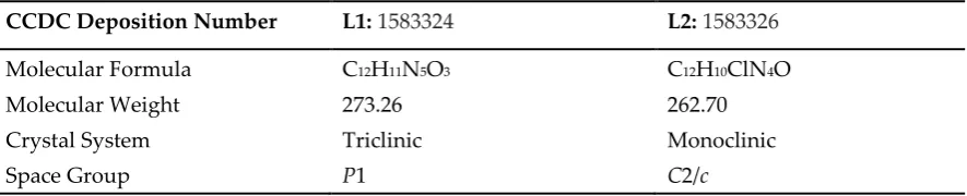
Table 2. Refinement parameters and crystal data for L1 and L2.
101
CCDC Deposition Number L1: 1583324 L2: 1583326
Molecular Formula C12H11N5O3 C12H10ClN4O
Molecular Weight 273.26 262.70
Crystal System Triclinic Monoclinic
2.3. Computational studies
102
The optimized geometry of compounds L1 and L2 were been obtained at B3LYP/6-31G* level.
103
Some optimized geometric parameters are also listed in Figure 3 and Tables 3, 4 and 5.
104
L1 L2
Figure 3. Optimized geometry of L1 and L2.
105
The actual and optimized bond lengths and bond angles obtained by X-ray crystallographic
106
study as well as by geometry optimization at B3LYP/6-31G level of theory of structure L1 and L2 are
107
reported in Table 4 and table 5 succefly.
108
In case of X-ray structure of compound L1, the observed bond lengths of N1-N2, N1-C2 and
109
N2-C4 bonds in five membered pyrazole ring are 1.340 (3) Å, 1.338 (4) Å and 1.327 (4) Å respectively.
110
The calculated bond lengths, through DFT method, of same pyrazole ring, are 1.342Å, 1.364Å and
111
1.335Å respectively, which are very close to the actual values. From Table 2, it is clear that actual C-C
112
and C-H bond lengths are also in close agreement with calculated values. The calculated bond
113
angles for N2N1C2, N4N3C5 and O1C5N3 bond angles of L1 are 114.34, 120.66and 123.16°
114
respectively, which are close to corresponding actual angles obtained from X-ray. The actual values
115
of above bond angles are 113.7 (2), 119.4 (2) and 123.3 (2) ° respectively.
116
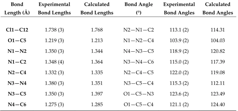
In case of X-ray structure of compound L2, the observed bond lengths of Cl1-C12, O1-C5 and
117
N2-C4 in the pyrazole derivative are 1.738 (3) Å, 1.219 (3) Å and 1.332 (3) Å respectively. The
118
a (Å) 4.6957 (3) 12.6881 (14)
b (Å) 7.2287 (4) 16.905 (2)
c (Å) 9.8261 (7) 13.073 (2)
α (°) 104.084 (3) 90.0
β (°) 90.848 (3) 115.330 (4)
γ (°) 107.980 (3) 90.0
V (Å3) 306.25 (3) 2534.6 (6)
Z 1 8
Dcalc (Mg·m−3) 1.482 1.377
Crystal Dimension (mm) 0.31 × 0.29 × 0.14 0.55 × 0.14 × 0.13
μ (mm−1) 0.11 0.30
Tmin/Tmax 0.966, 0.984 0.855, 0.963
Measured Reflections 10086 27408
Indices Range (h, k, l) −6/6, −9/9, −12/12 −16/16, −21/21, −16/16
θ Limit (°) 2.2-27.5 2.2-27.5
Unique Reflections 2795 2912
Parameters 190 172
Goodness of Fit on F2 1.04 1.02
calculated bond lengths, through DFT method, of same bond lengths, are 1.768Å, 1.213Å and 1.335Å
119
respectively, which are very close to the actual values. The calculated bond angles for N2N1C2,
120
N1N2C4 and O1C5N3 bond angles of L2 are 114.31, 104.03 and 123.49° respectively, which are close
121
to corresponding actual angles obtained from X-ray. The actual values of above bond angles are
122
113.1 (2), 103.9 (2) and 123.6 (2)° respectively (Table 4).
123
Table 3. Selected structural parameters by X-ray and theoretical calculations of L1.
124
Bond Length (Å)
Experimental Bond Lengths
Calculated Bond Lengths
Bond Angle (°)
Experimental Bond Angles
Calculated Bond Angles
O1—C5 1.213 (3) 1.212 N2—N1—C2 113.7 (2) 114.34
O2—N5 1.213 (4) 1.231 N1—N2—C4 103.5 (2) 104.01
O3—N5 1.224 (4) 1.232 N4—N3—C5 119.4 (2) 120.66
N1—N2 1.340 (3) 1.342 N3—N4—C6 116.8 (2) 118.00
N1—C2 1.338 (4) 1.364 O2—N5—O3 123.9 (3) 124.56
N2—C4 1.327 (4) 1.335 N2—C4—C3 112.2 (3) 111.35
N3—N4 1.360 (3) 1.346 N2—C4—C5 120.8 (2) 119.04
N3—C5 1.360 (3) 1.401 N3—C5—C4 115.2 (2) 112.12
N4—C6 1.275 (3) 1.285 O1—C5—N3 123.3 (2) 123.16
N5—C10 1.471 (4) 1.468 O1—C5—C4 121.6 (2) 124.71
Table 4. Selected structural parameters by X-ray and theoretical calculations of L2.
125
Bond
Length (Å)
Experimental
Bond Lengths
Calculated
Bond Lengths
Bond Angle
(°)
Experimental
Bond Angles
Calculated
Bond Angles
Cl1—C12 1.738 (3) 1.768 N2—N1—C2 113.1 (2) 114.31
O1—C5 1.219 (3) 1.213 N1—N2—C4 103.9 (2) 104.03
N1—N2 1.350 (3) 1.344 N4—N3—C5 118.9 (2) 120.82
N1—C2 1.348 (4) 1.364 N3—N4—C6 115.0 (2) 117.39
N2—C4 1.332 (3) 1.335 N2—C4—C5 122.0 (2) 119.08
N3—N4 1.380 (3) 1.351 N3—C5—C4 115.3 (2) 112.11
N3—C5 1.350 (3) 1.397 O1—C5—N3 123.6 (2) 123.49
N4—C6 1.275 (3) 1.285 O1—C5—C4 121.1 (2) 124.40
126
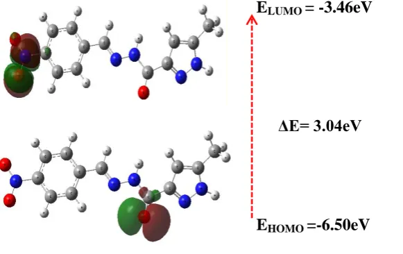
The total energy, energy of HOMO and energy of LUMO, as well as other parameters for
127
structures L1 and L2 are obtained theoretically and listed in Table 5. The HOMO and LUMO
128
electrons density distributions of L1 and L2 are given in (Figure 4 and 5). After the analysis of the
129
theoretical results obtained, we can say that the molecules L1 and L2 have a non-planar structure.
130
The analysis of the wave function indicates that the energy space between the molecular orbit
131
HOMO and LUMO determines the chemical stability and the electrical transport properties of the
132
molecule. The red and green colors of the molecular orbital ridge respectively represent the positive
133
and negative phases. The HOMO of L1 shows the charge density localized on the
134
4-nitrobenzaldehyde ring, but LUMO is characterized by a charge distribution on the hydrazone
135
function, indicating that this moiety can influence the electron transition. The HOMO of L2 has a
localized charge density on the pyrazole and hydrazone function, but LUMO is characterized by a
137
charge distribution on the 2-chlorobenzaldehyde ring and the hydrazone function. The energy
138
difference between HOMO and LUMO of L1 and L2 is about 3.04 and 6.01 eV respectively. The
139
energy of the smaller band space increases the stability of the molecule. The molecular boundary
140
orbitals of L1 and L2 (HOMO-LUMO) are shown in Figures 4 and 5.
141
Table 5. Calculated energies of L1 and L2.
142
Molecular Energy(a.u) L1 L2
TE (eV) -26209.0 -33150.1
EHOMO(eV) -6.5021 -6.2047
ELUMO(eV) -3.4611 -0.1918
Gap ΔE(eV) 3.0410 6.0129
µ(D) 11.2421 5.3366
143
We also studied the HOMO-LUMO gap of L1 and L2 to discover their reactivity towards the
144
catechol oxidation reaction. We found that the complex formed in-situ with L1 (3.04 eV) has a lower
145
HOMO-LUMO difference compared to the L2 complex (6.01 eV). It proves that the ligand L2 is more
146
reactive with respect to the oxidation of catechol with respect to the ligand L1. On the other hand,
147
the calculated HOMO energies are respectively -6.50, -6.20 eV for the ligand L1 and L2. It shows that
148
the HOMO orbital is strongly stabilized in L2 and ready to accept an electron, which facilitates the
149
catechol oxidation reaction. Thus, the oxidation tendency of catechol (L2>L1) is very consistent with
150
our experimental observation.
151
E
LUMO= -3.46eV
E
HOMO=-6.50eV
Figure 4. HOMO-LUMO energy diagram of L1.
152
153
154
155
156
157
E
LUMO= -0.19e.V
E
HOMO=-6.20e.V
Figure 5. HOMO-LUMO energy diagram of L2.
158
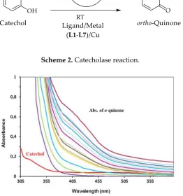
2.4. Catecholase activity: spectrophotometric study
159
The catechol oxidation reaction catalysed by the different copper complexes is studied by
160
following the concentration of O-quinone produced using a UV-Vis spectrometer. This study is
161
performed by monitoring the high absorption peak of o-quinone resulting from the catalyzed
162
reaction (Scheme 2). The copper complex is obtained in situ [31] by mixing the copper salt and the
163
ligand just before the introduction of the catechol solution. All of these solutions are placed together
164
in the spectrophotometer cell at 25 ° C. Thus, the formation of o-quinone is followed by the increase
165
of the absorbance at 390 nm as a function of time (Figure 6).
166
167
Scheme 2. Catecholase reaction.
168
169
Figure 6. Increase of o-quinone band at 390 nm after addition of L3 and CuSO4.
170
The variation of the absorbance as a function of time for each ligand and with different copper
171
salts is presented in the curves of Figure 7.
172
Figure 7. Plot of absorbance vs time for the oxidation of catechol catalyzed by copper complexes
173
formed with different ligands (L1-L7) and copper salts.
174
All cures exhibit an increasing absorbance in the time indicating that all of ligands are
175
catecholase activity. On the other hand, we can note that the curves are not exactly identical which
176
shows a difference in the catalytic activity.
177
The calculated catecholase activity of differentes ligands with different copper salts were
178
presented in Table 6.
179
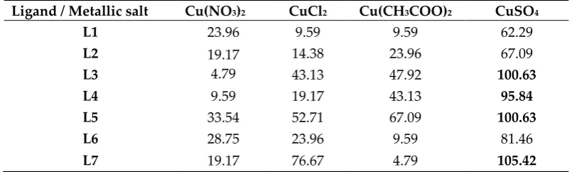
Table 6. Catecholase activity of complexes in methanol (µmol.L-1.min-1) with different copper salts.
180
Ligand / Metallic salt Cu(NO3)2 CuCl2 Cu(CH3COO)2 CuSO4
L1 23.96 9.59 9.59 62.29
L2 19.17
4.79
14.38 23.96 67.09
L3 43.13 47.92 100.63
L4 9.59 19.17 43.13 95.84
L5 33.54 52.71 67.09 100.63
L6 28.75 23.96 9.59 81.46
L7 19.17 76.67 4.79 105.42
181
From the results of the Table 6, we can note that all the ligands have a considerable catalytic
182
activity. Indeed, a maximum activity of about 100 ± 5 µmol.L-1.min-1 is obtained for the L3[CuSO4],
183
L4[CuSO4], L5[CuSO4] and L7[CuSO4] complexes. Low activity values of 4.79 μmol.L-1.min-1 are
184
obtained for the L3[Cu(NO3)2] and L7[Cu(CH3COO)2] complexes. The rate of oxidation appears to be
185
influenced by the ligand and copper salt parameters. The effect of ligand nature on oxidation rate is
186
less important for Cu(NO3)2 salt because mean absolute deviation is lower than 7.6. However, in the
187
cases of CuCl2 and Cu(CH3COO)2, the effect is more important because mean absolute deviation is
188
quite high than 19.9.
189
The effect of changing the copper salts on the oxidation rate is not the same for different
190
ligands. The salt which gives the highest reaction rate is CuSO4 salt and the one which gives the
191
lowest rate is Cu(NO3)2. The effect of the copper salt can be explained by a difficulty in the formation
192
of the L[Cu] complex. Indeed, ions such as NO3- and CH3OO- can form stronger bonds with Cu2+
193
which can prevent the formation of complexes.
194
The ligand L2 with a radical (Cl) in position (R1) ortho of the phenyl gives an activity lower
195
than the ligand L4 which has a radical (Cl) in position (R2) para of the phenyl. This result can be
196
explained by a steric hindrance effect of the radical in the position (R1) ortho and which can prevent
197
the formation of the copper complex. It is also possible that the formed complex has a geometry that
198
is not adapted to the substrate. We can notice the same observation for the ligand L6 which gives a
199
low rate reaction since it also has an (OH) radical in the ortho position of the phenyl.
The L3, L4 and L5 ligands differ by the R2 radical, such as Cl, Br and F respectively. It is noted
201
that, with the exception of L5, the L3 and L4 ligands give similar activities. These activities remain
202
lower than those obtained by L5. This difference may be due to the electronegativity values of these
203
halogens. On the one hand for fluoride, the high electronegativity gives a better activity of the
204
complex. On the other hand, Cl and Br have neighboring electronegativities that give similar
205
activities. This is explained by the stability of the ligands by mesomeric effect in the case of
206
electronegative radicals.
207
2.5. Comparison with Alternative Catalysts
208
Table 7 attests the catalytic activity by recent catalysts reported in the literature. It is clear that
209
the acylhydrazone-pyrazole derivatives, in particular ligand L7, described in this work present
210
better values and higher activity for the effective aerobic oxidation of the catechol into o-quinone. To
211
our knowledge, the catalytic activity observed for ligand L7 (105.42 µmol.L–1.min–1) is the most
212
important among the catalysts described in the literature. This best catalytic activity is probably due
213
to the stability of corresponding copper complex (catalyst) favored by the intense coordination
214
bonds of the Schiff base.
215
Table 7. Comparison of the catalytic activity of various catalysts toward oxidation of the
216
catechol into o-quinone, established in the same conditions, as given in previous literature.
217
Cu(II)-Ligands Cu(II) Salt
Used
Oxidation Rate
(µmol·L−1·min−1) Ref.
Ligand L7 CuSO4 105.42 -
bipyrazolic tripode-3-hydroxypropyl CuCl2 4.378 [31]
bipyrazolic tripode-4-hydroxyphenyl CuCl2 1.458 [32]
C,N-bipyrazole Cu(CH3COO)2 4.440 [34]
(3,5-dimethyl-pyrazol-1-ylmethyl)-amino]-p
ropionitrile CuSO4 8.710 [35]
N’-(diphenylmethylene)-5-phenyl-1H-pyraz
ole-3-carbohydrazide CuSO4 72.920 [36]
bipyrazolic tripode-prop-2-ylacetate Cu(CH3COO)2 11.825 [37]
bipyrazolic tripode-3-hydroxypropyl CuSO4 28.990 [38]
indole-3-chalcone Cu(CH3COO)2 31.780 [39]
3. Conclusions
218
In this work, we report the synthesis of seven new acylhydrazone-pyrazoles based biomolecule
219
materials (L1-L7), with superior catecholase activity, in excellent yields. L1 and L2 structures were
220
investigated by X-ray single crystal diffraction (XRD). The theoretical calculations of L1 and L2
221
through the density functional theory (DFT/B3LYP) method well supported the experimental
222
findings. Ligands (L1–L7) and different Cu(II) salts demonstrate an efficient activity to catalyze the
223
aerobic oxidation of the catechol into o-quinone compared to others recent catalysts described in the
224
literatures. Interestingly, ligand L7 exhibits an extremely high rate of oxidation, attaining 105.42
225
μmol.L−1.min−1, which is, to our knowledge, the best catalytic activity among the reported catalysts.
226
Cu(II) ligand complexes were generated in-situ and the results obtained show that the oxidation
227
depend highly on two parameters: the nature of the ligand and the nature of salts. The results
228
suggest that these new materials have potential for the oxidation of the catechol into o-quinone, thus
229
opening important perspectives.
230
4. Experimental Section
231
Melting points were measured using a Büchi B-545 digital capillary melting point apparatus
233
and used without correction. Reactions were checked with TLC using aluminum sheets with silica
234
gel 60 F254 from Merck. UV-visible (UV-vis) absorption spectra were recorded using a Perkin Elmer
235
Lambda 35 ES UV/VIS spectrophotometer using quartz cuvettes of 1 cm pathlength. Spectra IR were
236
recorded on a Perkin-Elmer VERTEX 70 FT-IR spectrometer covering field 400-4,000 cm-1. The
237
spectra of 1H NMR and 13C NMR were recorded in solution in DMSO-d6 on a Bruker spectrometer
238
(300 MHz). The chemical shifts are expressed in parts per million (ppm) by using tetramethylsilane
239
(TMS) as internal reference. The multiplicities of the signals are indicated by the following
240
abbreviations: s, singlet; d, doublet; t, triplet; q, quadruplet; and m, multiplet, and coupling
241
constants are expressed in Hertz. Mass spectra were collected using an AB Sciex API 3200
242
LC/MS/MS system, equipped with an ESI source. The chemical reagents used in synthesis were
243
purchased from Fluka, Sigma and Aldrich.
244
4.2. Synthesis
245
The synthesis of the intermediate and target compounds was performed according to the
246
reactions outlined in Scheme 1. The new ligands (L1-L7) were synthesized and characterized
247
according to the reported literature method [36, 40, 41].
248
General procedure for the preparation of N′-[(aryl)methylene]-5-methyl-1H-pyrazole-4-carbohydrazides
249
(L1-L7): To a solution of 5-methyl-1H-pyrazole-3-carbohydrazide (2) (1 mmol) in 10 mL of ethanol
250
was added an equimolar amount of the appropriate benzaldehyde derivative in the presence of
251
acetic acid. The mixture was maintained under reflux for 2 h, until TLC indicated the end of reaction.
252
Then, the reaction mixture was poured in cold water, and the precipitate formed was filtered out
253
washed with ethanol and recrystallized from ethanol.
254
N’-(4-nitrobenzylidene)-5-methyl-1H-pyrazole-3-carbohydrazide (L1): Yield 84% (solid), M.p. 283-285°C;
255
IR (ATR, ν(cm-1)) : 3320 (NH), 1681 (C=O), 1512 (N=CH) ; 1H-NMR (300 MHz, DMSO-d6, δ(ppm)): δ =
256
2.28 (s,3H, CH3), 6.44 (s, 1H, H-pyrazole), 7.90 (d, J = 8.7 Hz, 2H, H-Ar), 8.27 (d, , J = 8.7 Hz, 2H,
257
H-Ar), 8.58 (s, 1H, -CONH),11. 92 (s, 1H, N=CH), 13.13 (s, 1H, NH-pyrazole); 13C NMR: (300MHz,
258
DMSO-d6, δ (ppm)): 10.77 (CH3), 105.52 (CH, C4-pyrazole), 124.25 (CH, C3-Ar), 128.33 (CH, C2-Ar),
259
140.71 (C, C1-Ar), 141.40 (C, C3-pyrazole), 145.11 (CH, N=CH), 146.01 (C, C5-pyrazole), 148.14 (C,
260
C-NO2).; 159.10 (C, C=O); MS: m/z = 274.1 (M+H)+.
261
N’-(2-chlorobenzylidene)-5-methyl-1H-pyrazole-3-carbohydrazide (L2): Yield 78%, M.p. 258-260°C; IR
262
(ATR, ν(cm-1)) : 3182 (NH), 1665 (C=O), 1552 (N=CH) ; 1HNMR (300 MHz, DMSO-d6, δ(ppm)): δ =
263
2.27 (s, 3H, CH3), 6.49 (s, 1H, H-pyrazole), 7.39 - 7.99 (m, 4H, H-pyrazole), 8.90 (s, 1H, -N=CH), 11. 92
264
(s, 1H, -CONH), 13.09 (s, 1H, NH-pyrazole) ; 13C NMR: (300MHz, DMSO-d6, δ (ppm)): 10.77 (CH3),
265
105.40 (CH, C4-pyrazole), 127.31 (CH, C5-Ar), 128.01 (CH, C6-Ar), 130.34 (C, C3-Ar), 131.70 (C,
266
C4-Ar), 132.41 (C, C1-Ar), 133.57 (C, C2-Ar), 140.56 (C, C3-pyrazole), 143.65 (CH, N=CH), 146.16 (C,
267
C5-pyrazole), 159.05 (C, C=O); MS: m/z = 263.1 (M+H)+.
268
N'-(4-bromobenzylidene)-5-methyl-1H-pyrazole-3-carbohydrazide (L3): Yield 80 %, M.p. 300-302 °C; IR
269
(ATR, ν(cm-1)) : 3296 (NH), 1677 (C=O), 1617 (N=CH); 1H-NMR (300 MHz, DMSO-d6, δ(ppm)): δ =
270
2.26 (s,3H, CH3), 6.49 (s, 1H, H-pyrazole), 7.42 (d, J = 8.7 Hz, 2H, H-Ar), 7.61 (d, J = 8.7 Hz, 2H,
271
H-Ar), 8.45 (s, 1H, CONH), 11.67 (s, 1H, N=CH) 13.09 (s, 1H, NH-pyrazole) ; 13C NMR: (300MHz,
272
132.28 (C, C1-Ar), 134.32 (C, C4-Ar), 140.57 (C, C3-pyrazole), 146.27 (CH, N=CH), 146.36 (C,
274
C5-pyrazole), 158.91 (C, C=O). MS: m/z = 308.1 (M+H)+.
275
N'-(4-chlorobenzylidene)-5-methyl-1H-pyrazole-3-carbohydrazide (L4): Yield 64 %, M.p. 301-303 °C; IR
276
(ATR, ν(cm-1)) : 3397 (NH), 1677 (C=O), 1618 (N=CH); 1H-NMR (300 MHz, DMSO-d6, δ(ppm)): δ =
277
2.27 (s,3H, CH3), 6.49 (s, 1H, H-pyrazole), 7.47 (d, J = 8.7 Hz, 2H, H-Ar), 7.68 (d, J = 8.7 Hz, 2H,
278
H-Ar), 8.45 (s, 1H, CONH), 11.67 (s, 1H, N=CH) 13.09 (s, 1H, NH-pyrazole) ; 13C NMR: (300MHz,
279
DMSO-d6, δ (ppm)): 10.77 (CH3), 105.35 (CH, C4-pyrazole), 129.04 (CH, C3-Ar), 129.37 (CH, C2-Ar),
280
133.98 (C, C1-Ar), 134.70 (C, C4-Ar), 140.57 (C, C3-pyrazole), 146.27 (CH, N=CH), 147.23 (C,
281
C5-pyrazole), 158.91 (C, C=O). MS: m/z = 263.2 (M+H)+.
282
N'-(4-fluorobenzylidene)-5-methyl-1H-pyrazole-3-carbohydrazide (L5): Yield 75 %, M.p. 310-312 °C; IR
283
(ATR, ν(cm-1)) : 3336 (NH), 1677 (C=O), 1619 (N=CH); 1H-NMR (300 MHz, DMSO-d6, δ(ppm)): δ =
284
2.27 (s,3H, CH3), 6.48 (s, 1H, H-pyrazole), 7.47 (d, J = 8.7 Hz, 2H, H-Ar), 7.68 (d, J = 8.7 Hz, 2H,
285
H-Ar), 8.46 (s, 1H, CONH), 11.59 (s, 1H, N=CH) 13.07 (s, 1H, NH-pyrazole) ; 13C NMR: (300MHz,
286
DMSO-d6, δ (ppm)): 10.77 (CH3), 105.31 (CH, C4-pyrazole), 116.19 (CH, C3-Ar), 129.50 (CH, C2-Ar),
287
131.63 (C, C1-Ar), 140.55 (C, C3-pyrazole), 146.28 (CH, N=CH), 158.89 (C, C5-pyrazole), 161.80 (C,
288
C=O), 165.08 (C, C4-Ar). MS: m/z = 247.1 (M+H)+.
289
N'-(4-hydroxy-3-methoxybenzylidene)-5-methyl-1H-pyrazole-3-carbohydrazide (L6): Yield 65 %, M.p.
290
226-228 °C; IR (ATR, ν(cm-1)) : 3483 (OH), 3258 (NH), 1648 (C=O), 1590 (N=CH); 1H-NMR (300 MHz,
291
DMSO-d6, δ(ppm)): δ = 2.26 (s,3H, CH3), 3.80 (s,3H, OCH3), 6.46 (s, 1H, H-pyrazole), 6.80 (d, J =
292
8.1 Hz, 1H, H-Ar), 7.00 (d, J = 8.1 Hz, 1H, H-Ar), 7.26 sd, 1H, H-Ar), 8.33 (s, 1H, CONH), 9.51 (s, 1H,
293
OH), 11.38 (s, 1H, N=CH) 13.03 (s, 1H, NH-pyrazole) ; 13C NMR: (300MHz, DMSO-d6, δ (ppm)): 10.77
294
(CH3), 56.00 (OCH3), 105.20 (CH, C4-pyrazole), 109.20 (CH, C6-Ar), 115.86 (CH, C3-Ar), 122.43 (CH,
295
C2-Ar), 126.42 (C, C1-Ar), 148.20 (C, C3-pyrazole), 148.47 (CH, N=CH), 149.23 (C, C5-pyrazole),
296
149.85 (C, OCH3), 158.91 (C, OH), 158.91 (C, C=O). MS: m/z = 257.1 (M+H)+.
297
5-methyl-N'-(4-methylbenzylidene)-1H-pyrazole-3-carbohydrazide (L7): Yield 59 %, M.p. 292-294 °C; IR
298
(ATR, ν(cm-1)) : 3224 (NH), 1654 (C=O), 1606 (N=CH); 1H-NMR (300 MHz, DMSO-d6, δ(ppm)): δ =
299
2.26 (s,3H, CH3), 2.31 (s, 3H, CH3), 6.50 (s, 1H, H-pyrazole), 7.23 (d, J = 8.1 Hz, 2H, H-Ar), 7.55 (d, J =
300
8.1 Hz, 2H, H-Ar), 8.42 (s, 1H, -NH), 11.98 (s, 1H, N=CH) 13.10 (s, 1H, NH-pyrazole) ; 13C NMR:
301
(300MHz, DMSO-d6, δ (ppm)): 11.06 (CH3), 21.48 (CH3), 105.27 (CH, C4-pyrazole), 127.42 (CH,
302
C2-Ar), 129.88 (CH, C3-Ar), 132.28 (C, C1-Ar), 140.13 (C, C1-Ar), 141.73 (C, C3-pyrazole), 146.95 (CH,
303
N=CH), 147.13 (C, C5-pyrazole), 158.80 (C, C=O). MS: m/z = 243.1 (M+H)+.
304
4.3. X-ray Crystallographic Analysis
305
The compounds of L1 and L2 were obtained as single crystals by slow evaporation from ethanol
306
solution of the pure compound at room temperature. Data were collected on a Bruker APEX-II D8
307
Venture area diffractometer, equipped with graphite monochromatic Mo Kα radiation, λ = 0.71073 Å
308
at 296 (2) K, respectively. Cell refinement and data reduction were carried out by Bruker SAINT.
309
SHELXT was used to solve structure [42, 43]. The final refinement was carried out by full-matrix
310
least-squares techniques with anisotropic thermal data for no hydrogen atoms on 𝐹. CCDC 1583324
311
and 1583326 for L1 and L2, respectively. The supplementary crystallographic data for these
312
compounds can be obtained free of charge from the Cambridge Crystallographic Data Centre via
313
www.ccdc.cam.ac.uk/data_request/cif.
314
4.4. DFT computational method
The computational studies of compounds L1 and L2 were performed at the B3LYP/6-31G level
316
of theory using Gaussian 09 package programs [44, 45]. The optimizations geometries of L1 and L2
317
were performed using the Berny analytical gradient optimization method [46].
318
4.5. Catecholase activity measurement
319
Kinetic measurements were made spectrophotometrically on UV-Vis spectrometer, following
320
the appearance of o-quinone over time at 25 °C (390 nm absorbance maximum Ɛ = 1600 L.mol-1.cm-1
321
in methanol [33]. The complexes were prepared in-situ by successively mixing 0.15 mL of a solution
322
(2 × 10⁻³ M) of CuX2, nH2O (X = Cl-, NO3-, CH3COO- or SO4-), with 0.15 mL of a solution (2 × 10⁻³ M) of
323
ligand, then adding 2 mL of a solution of catechol at a concentration of 10-1 M.
324
Supplementary Materials: The following are available online at www.mdpi.com/link, Table S1: Selected
325
geometric parameters (Å, °) for L1, Table S2: Hydrogen-bond geometry (Å, °) for L1, Table S3: Selected
326
geometric parameters (Å, °) for L2, Table S4: Hydrogen-bond geometry (Å, °) For L2.
327
Acknowledgments: The authors extend their appreciation to the Deanship of Scientific Research at King Saud
328
University for funding this work through research group No. (RGP-007). Sincere appreciation is also extended
329
to the PPR2-MESRSFC-CNRST-P10 project (Morocco).
330
Author Contributions: K. K., S. R. and M. A. carried out of the experimental work, performed the structural
331
analysis and cooperated in the preparation of the manuscript. N. K. S. and Y. O. performed the density
332
functional theory calculations. E. Y. carried out the catalytic activity. J. T. cooperated in the preparation of the
333
manuscript and interpretation of the results. Y. N. M. and H. A. G. determined the X-ray crystal structure and
334
Y. N. M also paid the publication fees.
335
Conflicts of Interest: The authors declare no conflict of interest.
336
References
337
1. Binolfi, A.; Quintanar, L.; Bertoncini, C. W.; Griesinger, C.; Fernández, C. O. Bioinorganic chemistry
338
of copper coordination to alpha-synuclein: Relevance to Parkinson's disease. Coord. Chem. Rev. 2012,
339
256, 2188-2201, https://doi.org/10.1016/j.ccr.2012.05.004.
340
2. Chanu, O. B.; Kumar, A.; Lemtur, A.; Lal, R. A. Synthesis and characterization of homotrimetallic
341
copper complexes derived from bis(2-hydroxy-1-naphthaldehyde)oxaloyldihydrazone. Spectroc.
342
Acta A. 2012, 96, 854-861, https://doi.org/10.1016/j.saa.2012.07.113.
343
3. Kern, T.; Monkowius, U.; Zabel, M.; Knör, G. Synthesis, crystal structure and charge transfer spectra
344
of dinuclear copper(I) complexes bearing 1,2-bis(arylimino)acenaphthene acceptor ligands. Inorg.
345
Chim. Acta. 2011, 374, 632-636, https://doi.org/10.1016/j.ica.2011.02.042.
346
4. Shokohi-pour, Z.; Chiniforoshan, H.; Momtazi-borojeni, A. A.; Notash, B. A novel Schiff base derived
347
from the gabapentin drug and copper (II) complex: Synthesis, characterization, interaction with
348
DNA/protein and cytotoxic activity. J. Photochem. Photobiol. B. 2016, 162, 34-44,
349
https://doi.org/10.1016/j.jphotobiol.2016.06.022.
350
5. Holland, P. L.; Tolman, W. B. A structural model of the type 1 copper protein active site: N2S (thiolate)
351
S (thioether) ligation in a Cu (II) complex. J. Am. Chem. Soc. 2000, 122, 6331-6332, DOI:
352
10.1021/ja001328v.
353
6. Karlin, K. D.; Tyeklár, Z. Bioinorganic chemistry of copper; Springer Science & Business Media, 2012.
354
7. Blackman, A. G.; Tolman, W. B. Copper-dioxygen and copper-oxo species relevant to copper
355
oxygenases and oxidases. Struct. Bond. 2000, 97, 179-212, https://doi.org/10.1007/3-540-46592-8.
356
8. Gerdemann, C.; Eicken, C.; Krebs, B. The crystal structure of catechol oxidase: new insight into the
357
function of type-3 copper proteins. Acc. Chem. Res. 2002, 35, 183-191. DOI: 10.1021/ar990019a.
358
9. Gentschev, P.; Möller, N.; Krebs, B.: New functional models for catechol oxidases. Inorg. Chim. Acta.
359
2000, 300, 442-452. https://doi.org/10.1016/S0020-1693(99)00553-8.
360
10. Neves, A.; Rossi, L. M.; Bortoluzzi, A. J.; Mangrich, A. S.; Haase, W.; Werner, R. Synthesis, structure,
361
physicochemical properties and catecholase-like activity of a new dicopper (II) complex. J. Braz.
362
11. Banu, K. S.; Chattopadhyay, T.; Banerjee, A.; Bhattacharya, S.; Suresh, E.; Nethaji, M.; Zangrando, E.;
364
Das, D. Catechol oxidase activity of a series of new dinuclear copper (II) complexes with 3, 5-DTBC
365
and TCC as substrates: syntheses, X-ray crystal structures, spectroscopic characterization of the
366
adducts and kinetic studies. Inorg. Chem. 2008, 47, 7083-7093. DOI: 10.1021/ic701332w.
367
12. Os rio, R. E.; Peralta, R. A.; ortoluzzi, A. J.; de Almeida, V. R.; Szpoganicz, B.; Fischer, F. L.; Terenzi,
368
H. n.; Mangrich, A. S.; Mantovani, K. M.; Ferreira, D. E. Synthesis, Magnetostructural Correlation, and
369
Catalytic Promiscuity of Unsymmetric Dinuclear Copper (II) Complexes: Models for Catechol
370
Oxidases and Hydrolases. Inorg. Chem. 2012, 51, 1569-1589. DOI: 10.1021/ic201876k.
371
13. eyazit, N.; Çatıkkaş, .; ayraktar, Ş.; Demetgül, C. Synthesis, characterization and catecholase-like
372
activity of new Schiff base metal complexes derived from visnagin: Theoretical and experimental
373
study. J. Mol. Struct. 2016, 1119, 124-132. https://doi.org/10.1016/j.molstruc.2016.04.047.
374
14. Mistri, S.; Paul, A.; Bhunia, A.; Manne, R. K.; Santra, M. K.; Puschmann, H.; Manna, S. C. A combined
375
experimental and theoretical investigation on the Cu (II) sensing behavior of a piperazinyl moiety
376
based ligand, and catecholase and biological activities of its Cu (II) complex in combination with
377
pyridine 2, 5-dicarboxylate. Polyhedron 2016, 104, 63-72. https://doi.org/10.1016/j.poly.2015.11.030.
378
15. Camargo, T. P.; Peralta, R. A.; Moreira, R.; Castellano, E. E.; Bortoluzzi, A. J.; Neves, A. New
379
mononuclear copper (II) complex based on a salen derivative ligand with an unusual coordination
380
and its catecholase activity. Inorg. Chem. Commun. 2013, 37, 34-38.
381
https://doi.org/10.1016/j.inoche.2013.09.039.
382
16. Stadler, A.-M.; Harrowfield, J.: Bis-acyl-/aroyl-hydrazones as multidentate ligands. Inorganica
383
Chimica Acta 2009, 362, 4298-4314.
384
17. Konar, S.; Jana, A.; Das, K.; Ray, S.; Golen, J. A.; Rheingold, A. L.; Kar, S. K. A rare pentanuclear
385
cadmium(II) complex and two new mononuclear zinc(II) complexes of pyrazole derived ditopic
386
ligands – Synthesis, crystal structures and spectral studies. Inorg. Chim. Acta. 2013, 397, 144-151.
387
https://doi.org/10.1016/j.ica.2012.12.003.
388
18. Gokce, C.; Gup, R. Synthesis and characterisation of Cu (II), Ni (II), and Zn (II) complexes of furfural
389
derived from aroylhydrazones bearing aliphatic groups and their interactions with DNA. Chem. Pap.
390
2013, 67, 1293-1303. https://doi.org/10.2478/s11696-013-0379-8.
391
19. Salem, N. M. H.; El-Sayed, L.; Foro, S.; Haase, W.; Iskander, M. F. Metal complexes derived from
392
hydrazoneoxime ligands: III – Synthesis, characterization and electrospray ionization mass spectra of
393
some nickel(II) complexes with aroylhydrazoneoximes. Polyhedron 2007, 26, 4161-4172.
394
https://doi.org/10.1016/j.poly.2007.05.010.
395
20. Naskar, S.; Mishra, D.; Chattopadhyay, S. K.; Corbella, M.; Blake, A. J.: Versatility of 2,
396
6-diacetylpyridine (dap) hydrazones in stabilizing uncommon coordination geometries of Mn (II):
397
synthesis, spectroscopic, magnetic and structural characterization. Dalton Trans. 2005, 2428-2435.
398
DOI:10.1039/B503891J.
399
21. Uppadine, L. H.; Lehn, J. M.: Three‐Level Synthetic Strategy Towards Mixed‐Valence and
400
Heterometallic [2× 2] Gridlike Arrays. Angew. Chem. Int. Ed. 2004, 43, 240-243,
401
doi:10.1002/anie.200352937.
402
22. Ruben, M.; Lehn, J.-M.; Vaughan, G. Synthesis of ionisable [2× 2] grid-type metallo-arrays and
403
reversible protonic modulation of the optical properties of the [Co II4 L 4] 8+ species. Chem.
404
Commun. 2003, 1338-1339, DOI:10.1039/B303922F.
405
23. Mondal, S.; Das, C.; Ghosh, B.; Pakhira, B.; Blake, A. J.; Drew, M. G.; Chattopadhyay, S. K. Synthesis,
406
spectroscopic studies, X-ray crystal structures, electrochemical properties and DFT calculations of
407
three Ni (II) complexes of aroyl hydrazone ligands bearing anthracene moiety. Polyhedron 2014, 80,
408
272-281, https://doi.org/10.1016/j.poly.2014.05.028.
409
24. He, Z.; He, C.; Gao, E.-Q.; Wang, Z.-M.; Yang, X.-F.; Liao, C.-S.; Yan, C.-H. Lanthanide-transition
410
heterometallic extended structures with novel orthogonal metalloligand as building block. Inorg.
411
Chem. 2003, 42, 2206-2208, DOI: 10.1021/ic026271n.
412
25. Sakai, K.; Tomita, Y.; Ue, T.; Goshima, K.; Ohminato, M.; Tsubomura, T.; Matsumoto, K.; Ohmura, K.;
413
Kawakami, K. Syntheses, antitumor activity, and molecular mechanics studies of cis-PtCl2(pzH)2
414
(pzH=pyrazole) and related complexes. Crystal structure of a novel Magnus-type double-salt
415
[Pt(pzH)4][PtCl4][cis-PtCl2(pzH)2]2 involving two perpendicularly aligned 1D chains. Inorg. Chim.
416
26. Fan, C.; Su, H.; Zhao, J.; Zhao, B.; Zhang, S.; Miao, J. A novel copper complex of salicylaldehyde
418
pyrazole hydrazone induces apoptosis through up-regulating integrin β4 in H322 lung carcinoma
419
cells. Eur. J. Med. Chem. 2010, 45, 1438-1446, https://doi.org/10.1016/j.ejmech.2009.12.048.
420
27. Abu-Surrah, A. S.; Safieh, K. A. A.; Ahmad, I. M.; Abdalla, M. Y.; Ayoub, M. T.; Qaroush, A. K.;
421
Abu-Mahtheieh, A. M. New palladium (II) complexes bearing pyrazole-based Schiff base ligands:
422
Synthesis, characterization and cytotoxicity. Eur. J. Med. Chem. 2010, 45, 471-475,
423
https://doi.org/10.1016/j.ejmech.2009.10.029.
424
28. Saha, N. C.; Mandal, S.; Das, M.; Khatun, N.; Mitra, D.; Samanta, A.; Slawin, A. M. Z.; Butcher, R. J.;
425
Saha, R. Synthesis, characterization, X-ray crystallography and antimicrobial activities of new Co(III)
426
and Cu(II) complexes with a pyrazole based Schiff base ligand. Polyhedron 2014, 68, 122-130,
427
https://doi.org/10.1016/j.poly.2013.10.016.
428
29. Sau, D. K.; Butcher, R. J.; Chaudhuri, S.; Saha, N. Spectroscopic, structural and antibacterial
429
properties of copper (II) complexes with bio-relevant 5-methyl-3-formylpyrazole N (4)-benzyl-N
430
(4)-methylthiosemicarbazone. Mol. Cell. Biochem. 2003, 253, 21-29,
431
https://doi.org/10.1023/A:1026041032078.
432
30. Kupcewicz, B.; Sobiesiak, K.; Malinowska, K.; Koprowska, K.; Czyz, M.; Keppler, B.; Budzisz, E.
433
Copper (II) complexes with derivatives of pyrazole as potential antioxidant enzyme mimics. Med.
434
Chem. Res. 2013, 22, 2395-2402, https://doi.org/10.1007/s00044-012-0233-5.
435
31. Mouadili, A.; Attayibat, A.; Kadiri, S. E.; Radi, S.; Touzani, R. Catecholase activity investigations using
436
in situ copper complexes with pyrazole and pyridine based ligands. Appl. Catal., A. 2013, 454, 93-99,
437
https://doi.org/10.1016/j.apcata.2013.01.011.
438
32. Bouabdallah, I.; Touzani, R.; Zidane, I.; Ramdani, A. Synthesis of new tripodal ligand: N, N-bis [(1,
439
5-dimethylpyrazol-3-yl) methyl] benzylamine.: Catecholase activity of two series of tripodal ligands
440
with some copper (II) salts. Catal. Commun. 2007, 8, 707-712,
441
https://doi.org/10.1016/j.catcom.2006.08.034.
442
33. Bouabdallah, I.; Touzani, R.; Zidane, I.; Ramdani, A. Effect of two isomeric tetrapyrazolyl ligands on
443
the catalytic oxidation of 3, 5-di-tert-butylcatechol. J. Iran. Chem. Soc. 2007, 4, 299-303,
444
https://doi.org/10.1007/BF03245978.
445
34. El Kodadi, M.; Malek, F.; Touzani, R.; Ramdani, A. Synthesis of new tripodal ligand 5-(bis (3,
446
5-dimethyl-1H-pyrazol-1-ylmethyl) amino) pentan-1-ol, catecholase activities studies of three
447
functional tripodal pyrazolyl N-donor ligands, with different copper (II) salts. Catal. Commun. 2008,
448
9, 966-969, https://doi.org/10.1016/j.catcom.2007.09.038.
449
35. Mouadili, A.; Zerrouki, A.; Herrag, L.; Hammouti, B.; El Kadiri, S.; Touzani, R. Catechol oxidation:
450
activity studies using electron-rich nitrogen-based ligands. Res. Chem. Intermed. 2012, 38, 2427-2433,
451
https://doi.org/10.1007/s11164-012-0558-1.
452
36. Karrouchi, K.; Yousfi, E.; Sebbar, N.; Ramli, Y.; Taoufik, J.; Ouzidan, Y.; Ansar, M. h.; Mabkhot, Y.;
453
Ghabbour, H.; Radi, S. New Pyrazole-Hydrazone Derivatives: X-ray Analysis, Molecular Structure
454
Investigation via Density Functional Theory (DFT) and Their High In-Situ Catecholase Activity. Int. J.
455
Mol. Sci. 2017, 18, 2215, doi:10.3390/ijms18112215.
456
37. Boussalah, N.; Touzani, R.; Bouabdallah, I.; Kadiri, S. E.; Ghalem, S. Synthesis, structure and catalytic
457
properties of tripodal amino-acid derivatized pyrazole-based ligands. Journal of Molecular Catalysis
458
A: Chemical 2009, 306, 113-117, https://doi.org/10.1016/j.molcata.2009.02.031.
459
38. Zerrouki, A.; Touzani, R.; El Kadiri, S. Synthesis of new derivatized pyrazole based ligands and their
460
catecholase activity studies. Arab. J. Chem. 2011, 4, 459-464,
461
https://doi.org/10.1016/j.arabjc.2010.07.013.
462
39. Thabti, S.; Djedouani, A.; Rahmouni, S.; Touzani, R.; Bendaas, A.; Mousser, H.; Mousser, A. Synthesis,
463
X-ray crystal structures and catecholase activity investigation of new chalcone ligands. J. Mol. Struct.
464
2015, 1102, 295-301, https://doi.org/10.1016/j.molstruc.2015.08.071.
465
40. Karrouchi, K.; Charkaoui, Y.; Benlafya, K.; Ramli, Y.; Taoufik, J.; Radi, S.; Ansar, M.: Synthesis,
466
characterization and preliminary biological activity of some new pyrazole carbohydrazide
467
derivatives. J. Chem. Pharm. Res. 2013, 5, 1-6.
468
41. Karrouchi, K.; Ansar, M. h.; Radi, S.; Saadi, M.; El Ammari, L. Crystal structure of
469
N′-diphenylmethylidene-5-methyl-1H-pyrazole-3-carbohydrazide. Acta Crystallogr. E Crystallogr.
470
42. Sheldrick, G. A short history of SHELX. Acta Crystallogr. 2008, 64, 112-122, DOI:
472
10.1107/S0108767307043930.
473
43. Sheldrick, G. M. Crystal structure refinement with SHELXL. Acta Crystallogr. C. 2015, 71, 3-8,
474
https://doi.org/10.1107/S2053229614024218.
475
44. BecNe, A. Densityϋfunctional thermochemistry. III. The role of exact exchange. J. Chem. Phys 1993,
476
98, 5648-5652, https://doi.org/10.1063/1.464913.
477
45. Frisch, M.; Trucks, G.; Schlegel, H. B.; Scuseria, G.; Robb, M.; Cheeseman, J.; Scalmani, G.; Barone, V.;
478
Mennucci, B.; Petersson, G.: Gaussian 09, revision D. 01. Gaussian, Inc., Wallingford CT, 2009.
479
46. Becke, A. D.: Density-functional exchange-energy approximation with correct asymptotic behavior.
480
Phys. Rev. 1988, 38, 3098, DOI:https://doi.org/10.1103/PhysRevA.38.3098.