Infection and Drug Resistance
Dovepress
R e v I e w
open access to scientific and medical researchOpen Access Full Text Article
exploring bacterial outer membrane barrier
to combat bad bugs
Ishan Ghai
1Shashank Ghai
21School of engineering and Life
Sciences, Jacobs University, Bremen,
2Leibniz University, Hannover,
Germany
Abstract: One of the main fundamental mechanisms of antibiotic resistance in Gram-negative bacteria comprises an effective change in the membrane permeability to antibiotics. The Gram-negative bacterial complex cell envelope comprises an outer membrane that delimits the periplasm from the exterior environment. The outer membrane contains numerous protein channels, termed as porins or nanopores, which are mainly involved in the influx of hydrophilic compounds, including antibiotics. Bacterial adaptation to reduce influx through these outer membrane proteins (Omps) is one of the crucial mechanisms behind antibiotic resistance. Thus to interpret the molecular basis of the outer membrane permeability is the current challenge. This review attempts to develop a state of knowledge pertinent to Omps and their effective role in antibiotic influx. Further, it aims to study the bacterial response to antibiotic membrane per-meability and hopefully provoke a discussion toward understanding and further exploration of prospects to improve our knowledge on physicochemical parameters that direct the translocation of antibiotics through the bacterial membrane protein channels.
Keywords: antibiotics, Gram-negative bacteria, cell envelope, protein channels, nanopores, influx, antibiotic resistance
Introduction
Antibiotic resistance can be defined as the capability of any microbial organism to
coun-terattack effects of antimicrobial drugs (antibiotics) (Figure 1A) used against them.
1,2This phenomenon has become a global communal health threat due to an enormous
increase in annual death rate.
2The emergence of highly resistant organisms has led to
the requirement of new antibacterial drugs.
1Due to the slow progress of the current
antibiotic research, there exists an enormous gap between bacterial evolution and the rate
of development of novel antibiotic drugs.
1,3,4Only about two new classes of antibiotics
have been brought to the market in the last three decades. On the technical front, there
is an urgent need for a greater understanding of how antibiotics work, how bacteria
progress with resistance against these antibiotics, and what molecular machinery could
be exploited to get around bacterial defense mechanisms.
1–4The current innovative way
of improving the potential of antibiotics is to effectively introduce them into bacteria
and further prevent them from degradation by bacterial enzymes before they reach their
targets. There is an extreme necessity for counteracting the problem of multi-antibiotic
resistance.
1,4The important mechanism (Figure 1B) of resistance toward antibiotics
known till date includes the enzymes-mediated deactivation of antibiotics for example,
β
-lactamase enzymes which hydrolyze and confer resistance against a diverse variety of
antibiotics including penicillins, cephalosporins, carbapenems, and many more.
4–7The
Correspondence: Shashank Ghai Leibniz University, welfengarten 1, 30167 Hannover, Germany
email shashank.ghai@sportwiss. uni-hannover.de
Journal name: Infection and Drug Resistance Article Designation: Review
Year: 2017 Volume: 10
Running head verso: Ghai and Ghai
Running head recto: Exploring bacterial outer membrane barrier to combat bad bugs DOI: http://dx.doi.org/10.2147/IDR.S144299
Infection and Drug Resistance downloaded from https://www.dovepress.com/ by 118.70.13.36 on 22-Aug-2020
For personal use only.
This article was published in the following Dove Press journal: Infection and Drug Resistance
30 August 2017
Dovepress
Ghai and Ghai
outer membrane vesicles (Figure 1C), these native vesicles
released by Gram-negative bacteria, are mainly composed
of periplasmic and outer membrane components including
lipopolysaccharides, proteins, lipids, and other molecules.
8–11They help the producer cells while communicating with other
cells concerning pathogenesis, secretion, nutrients acquisition,
Figure 1 (A) Antibiotic resistance (an overview). (B) various mechanisms of antibiotic resistance employed by Gram-negative bacteria (an overview). (C) Structural
representation of outer membrane vesicles.
Abbreviation: Omps, outer membrane proteins.
Microbes (low level of antibiotic resistance)
A
B
C
Microbes + antibiotic Microbes (spared overantibiotic)
Lipopolysaccharides
Lipopolysaccharides
Omps Toxin
Peptidoglycan Periplasmic
protein Enzyme
Efflux pumps
Cytoplasm Modification of Omps
Enzymatic modification of antibiotics (Omps)
Target modification
Loss of functional Omps
Microbes (high level of antibiotic resistance)
Infection and Drug Resistance downloaded from https://www.dovepress.com/ by 118.70.13.36 on 22-Aug-2020
Dovepress exploring bacterial outer membrane barrier to combat bad bugs
and self-defense.
5,8–10These moieties protect bacteria from
various environmental stress factors including antibiotics,
for example, gentamicin, imipenem, ampicillin, melittin,
colistin, and many more.
8–14Further, resistance mechanism
is also mediated by reducing the entry of antibiotics into the
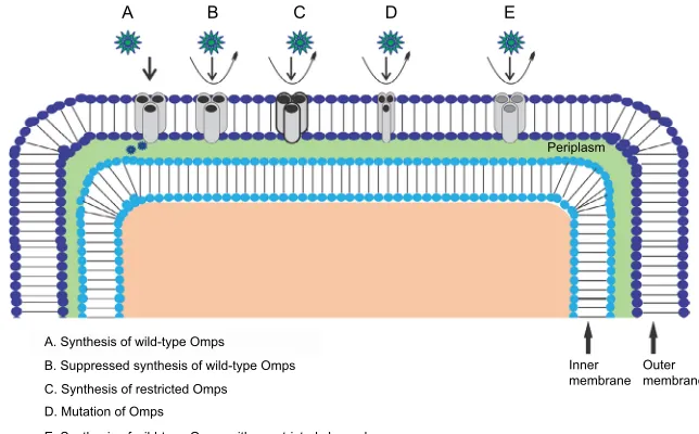
target site of bacteria which is mainly effected by specific
alteration of outer membrane permeability (Figure 2). Efflux
pumps effectively contribute towards resistance mechanism
by antibiotic expulsion. In addition, antibiotic target proteins,
for example, penicillin-binding proteins, are altered inside
the bacterial cells, leading to antibiotic resistance.
2,3,5,6,15–21In this review, we present a systemic overview of the role
of different membrane protein transporters responsible for
antibiotic transport, present in the outer membrane of
Gram-negative bacteria.
4–6,22We highlight the different achievements
of the scientific community in understanding the uptake of
dif-ferent solutes including antibiotics.
7,17,22This active knowledge
of the role of outer membrane influx in antibiotic transport
in Gram-negative bacteria can be useful for antibiotic drug
development in the future, where the computed data can be
employed toward understanding the detailed mechanism of
bacterial membrane transport, and to further design novel
antibiotics with an effective permeability profile.
Gram-negative bacteria
Gram-negative bacteria have a multifaceted cell envelope
comprising an outer membrane that restricts the access to the
periplasm by acting as a molecular filter, thus forming an
effi-cient selective permeation barrier.
4–6,23,24This outer membrane,
like other biological membranes, is fundamentally built up of
a bilayer of lipids.
6,18,25,26As such, this lipid bilayer membrane
is mostly impermeable to hydrophilic molecules including
nutrients.
22,25,27The effective intake of hydrophilic molecules
is mainly controlled by specific water-filled open channels
termed as outer membrane proteins (Omps) or porins.
22,27–29These Omps are intensively characterized in Gram-negative
bacteria and are further distinguished as nonspecific and
specific Omps in accordance with their functional structure
(monomeric or trimeric),
6,7,22,24–26,28substrate specificity,
regulation, and expression.
15,18,29,30These membrane proteins
do not show any hydrophobic stretches in their amino acid
sequences and majorly form hollow
β
-barrel structures
with a hydrophobic outer surface.
28,31The barrel structure
encompasses the transmembranous pore-type structure with a
crucial function of facilitating the passive flux of hydrophilic
substances
22,28and further acting as a functional diffusional
barrier for nonpolar solutes.
6,28These proteins might show
specific selectivity in general for either cations or anions.
5,22,28Bacterial adaptation to reduce influx through these
Omps is an increasing problem that contributes, together
with efflux systems, to antibiotic resistance.
3–5,20,23,32–34An
existing challenge for drug design is to interpret membrane
permeability at molecular level to get a better insight into the
role of membrane transport (Figure 2) in bacterial resistance
mechanism.
4–7,20,35Like other hydrophilic molecules, polar
antibiotics including
β
-lactam antibiotics and
fluoroquino-Figure 2 Antibiotic resistance mechanism associated with Omps modification. Antibiotic β-lactam molecules are represented by green stars, and Omps as trimers by gray
cylinder. The width of the straight arrows imitating the level of β-lactam penetration via Omps. The curved arrows exemplify the uptake failure/reduce uptake occurring with
the following: B: decrease in the level of wild-type Omps expression; C: expression of restricted-channel Omps; D: mutation or modification of the functional properties of a porin channel; and E: synthesis of modified Omps with significant constriction.
Abbreviation: Omps, outer membrane proteins.
A. Synthesis of wild-type Omps B. Suppressed synthesis of wild-type Omps C. Synthesis of restricted Omps D. Mutation of Omps
E. Synthesis of wild-type Omps with constricted channel
Inner membrane Periplasm E
D C
B A
Outer membrane
Infection and Drug Resistance downloaded from https://www.dovepress.com/ by 118.70.13.36 on 22-Aug-2020
Dovepress
Ghai and Ghai
lones majorly sneak into Gram-negative bacteria using these
Omps.
5,31,33Any slight modification by the bacteria in the
responsible Omps can significantly affect the antibiotic drug
therapy.
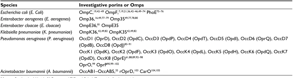
33Many clinically pertinent bacterial species
includ-ing
Enterobacter aerogenes
,
Escherichia coli
,
Enterobacter
cloacae
,
Klebsiella
pneumoniae
,
Pseudomonas aeruginosa
,
and
Acinetobacter baumannii
have been sequenced for
deter-mining the effective key Omps (Table 1) present in the outer
membrane.
3–6,23,28,31–33,36,37Further, bacterial bugs including
Pseudomonas aeruginosa,
and
Acinetobacter baumannii
possess an innate low vulnerability toward
β
-lactams, through
reduced outer membrane permeability.
5,6,20,22,38For instance,
reduced membrane permeability in
Pseudomonas aeruginosa
as compared to
Enterobacteriaceae
mainly occurs due to less
number of Omps present in the outer membrane and their
distinct physicochemical properties.
22,38–41In other
Gram-negative bugs, for example,
Escherichia coli, Enterobacter
and
Klebsiella pneumoniae
, susceptibility toward
β
-lactam
molecules is closely related to the presence of nonspecific
diffusion Omps, for example, OmpF and OmpC.
5,6,22Previous works showing the effective role of different
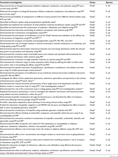
Omps (Table 1) in molecular influx of different antibiotics are
shown in Table 2. We discuss the achievements of the
scien-tific community in this area by studying the role of different
Omps in outer membrane permeability, using separate set of
theoretical and experimental techniques including molecular
simulation (MS), electrophysiology, minimum inhibitory
concentration assay, liposome swelling assay, X-ray
crystal-lography, and fluorescence resonance energy transfer.
Discussion
Computing influx
Typical antibiotic activity toward bacterial cell occurs in
micromolar concentration range, thereby representing
val-ues that are approximately limited to a thousand molecules
inflowing the cell in few minutes to hours.
7,22Such numbers
are considerably beneath the detection limit of most of the
techniques and thus require significant amplification of the
signal.
4,7,22,110Measuring the flux of small molecules across
the outer cell membrane can be possibly achieved by
differ-ent approaches including whole-cell assays, which require
computation of flux using genetically engineered bacterial
cell.
7,111,112These methods involve soaking bacteria in
anti-biotics for a fixed time followed by a separation process to
remove the external media from the internalized antibiotics.
7However, the quality of the separation method is crucial for
improving permeability.
7,111,112There are several published
studies employing whole-cell assays to quantify the uptake,
and their quality has been intensively compared.
7,110–116Once
the separation technique allows collecting sufficient amounts
of internalized antibiotics, several biophysical methods can
be used to quantify the intracellular antibiotics.
7,113–118One of
the promising tools for studying intracellular accumulation is
mass spectrometry. The technique was successfully applied
in measuring the uptake of antibiotics;
117,118for example, a
work demonstrated cellular uptake of linezolid by
E. coli
using liquid chromatography–mass spectrometry.
118The discussed methods allow quantifying the total
turn-over of a cell uptake which represents the relevant actual
effective concentration seen by the bacteria. On the contrary,
the comprehensive flux depends on a multitude of parameters
and renders the molecular understanding difficult.
7,22To
understand the molecular origin of the antibiotic uptake, we
need information on the role of each individual involved
com-ponent. For example, the so-called liposome swelling assay
provides information on a model system.
35,52,55,60,80,97,105The
method involves reconstitution of batches of purified Omps
into (multilamellar) liposomes.
7,22Under isosmotic
addi-tion, the diffusion of substrate inside the liposome results in
alteration of the light-scattering pattern. The effective change
Table 1 Crucial Omps studied in different Gram-negative bacterial species
Species Investigative porins or Omps
Escherichia coli (E. Coli) OmpC,19,42–49 OmpF,7,19,21,36,42–46,49–74 Phoe75–76
Enterobacter aerogenes (E. aerogenes) Omp36,16,49,77–79 Omp3549,77,78,80
Enterobacter cloacae (E. cloacae) Ompe36,81 Ompe35
Klebsiella pneumoniae (K. pneumoniae) OmpK36,42,49,82 OmpK3542,49,82
Pseudomonas aeruginosa (P. aeruginosa) OccD1 (OprD), OccD2 (OpdC), OccD3 (OpdP), OccD4 (OpdT), OccD5 (OpdI), OccD6 (OprQ), OccD7 (OpdB), OccD8 (OpdJ)83–91
OccK1 (OpdK), OccK2 (OpdF), OccK3 (OpdO), OccK4 (OpdL), OccK5 (OpdH), OccK6 (OpdQ), OccK7 (OpdD), OccK8 (Opre)61,88,89,92–98
OprO,99 OprP90,99–102
Acinetobacter baumannii (A. baumannii) OccAB1–OccAB5,35 rOprD,103 CarO104,105
Notes: Studies by Nikaido,5,6 Pages et al,22,42 and Schulz28 provide further insight.
Abbreviation: Omps, outer membrane proteins.
Infection and Drug Resistance downloaded from https://www.dovepress.com/ by 118.70.13.36 on 22-Aug-2020
Dovepress exploring bacterial outer membrane barrier to combat bad bugs
Table 2 Conclusive investigations with different Omps studied in different Gram-negative bacterial species
Conclusive investigation Omps Species
Measured the flux of charged β-lactamase inhibitors sulbactam, tazobactam, and avibactam using eTP zero-current assay and MS36
OmpF E. coli
Measured the transport of charged β-lactamase inhibitors sulbactam, tazobactam, and avibactam using eTP zero-current assay59
OmpC E. coli
Measured the permeability of carbapenems via different mutant proteins from different clinical isolates using eTP and LSA52
OmpC E. coli
Quantified norfloxacin uptake using semiquantitative optofluidic assay56 OmpF E. coli
Quantified and explained the mechanism of small antibiotic molecule enrofloxacin uptake using ETP and MS7 OmpF E. coli
Quantified and demonstrated the translocation of imipenem, ceftazidime, and cefepime using ETP74 OmpF E. coli
Demonstrated the interaction and binding of antibiotic meropenem with channel using eTP53 OmpF E. coli
Demonstrated the translocation of polypeptides using eTP64 OmpF E. coli
Demonstrated the permeation of enrofloxacin across the OmpF channel and modulation of the affinity site
in the presence of magnesium using eTP and MS54,66,72
OmpF E. coli
Demonstrated the transport of ampicillin and benzylpenicillin using eTP, MS, MIC, and LSA60 OmpF E. coli
Demonstrated the role of charged residues in channel constriction, channel conductance, ion selectivity, and voltage gating using eTP and MS71
OmpF E. coli
Demonstrated important electrostatic interactions between ions and charge distribution within the channel that govern ion permeation and selectivity using MS62
OmpF E. coli
Demonstrated the ionization states of titratable amino acid residues and calculated self-consistently the electric potential distribution within channel using MS50
OmpF E. coli
Demonstrated the movement of single ampicillin molecule via channel using eTP and MS67 OmpF E. coli
Demonstrated the molecular origin of cation selectivity within Omps by defining the effect of alkali metal ions atomic radii on the binding-site affinity using ETP and MS58
OmpF E. coli
Demonstrated the specific interaction of grepafloxacin, ciprofloxacin, moxifloxacin, and nalidixic acid with
pore using Uv–visible spectroscopic measurements68
OmpF E. coli
Demonstrated the permeation of moxifloxacin across membrane channel and protein–antibiotic interaction
using eTP, MS and FReT65
OmpF E. coli
Investigated the effects of four polyamines (putrescine, cadaverine, spermidine, and spermine) on the activity of bacterial porins using eTP patch clamp106
OmpF E. coli
Demonstrated the channel functional characteristics of four single amino acid substitutions and effect of deletion mutant in constriction loop L3 using eTP107 and crystallographic analysis108
OmpF E. coli
Demonstrated the role of the constriction loop in voltage gating using eTP and crystallographic analysis109 OmpF E. coli
Employed fluorescence quenching as a tool to investigate the antibiotic interactions with bacterial protein, using nalidixic acid and moxifloxacin, within the pore69
OmpF E. coli
Demonstrated ampicillin translocation through the bacterial pore, and described the effect of mutations within pore affecting molecule passage using eTP and MS61
OmpF E. coli
Provided a descriptive explanation about pathways of ions along channel surface using MS70 OmpF E. coli
Probed the interaction of peptides, magainin 2, and HPA3P with the pore, and displayed the effect of electric
field on pore and peptide geometry using ETP51
OmpF E. coli
Demonstrated the translocation of ampicillin using multiscale approach combined with MS63 OmpF E. coli
Demonstrated the effective binding of carbenicillin, ertapenem, and ampicillin within the pore using X-ray crystallography and MS21
OmpF E. coli
Demonstrated the interaction involved in translocation of ampicillin, amoxicillin, carbenicillin, azlocillin, and piperacillin using eTP and MS57
OmpF E. coli
Demonstrated the effect of specific acid residue D113A substitution on susceptibility to cefepime,
cefpirome, cefotaxime, ceftazidime, cefoxitin, and ampicillin using MIC and MS73
OmpF E. coli
Demonstrated the effective role of anti-loop 3 (Lys-16) residue in cefepime diffusion using LSA, eTP, and MS55
OmpF E. coli
Demonstrated the effect of ion concentration and charged residues at constriction zone on gating behavior of channel using eTP47
OmpC E. coli
established the effect of three mutations within porins isolated from multidrug-resistant E. coli on transport of cefotaxime using MIC, eTP, and MS48
OmpC E. coli
Studied the interaction strengths of ceftriaxone, cefpirome, and ceftazidime using effective fluorescence quenching and ETP44
OmpC, OmpF E. coli
Demonstrated the influx of ceftriaxone, cefepime, ceftazidime, norfloxacin, ciprofloxacin, and enrofloxacin using a chip-based automated patch clamp technique based on ETP45
OmpC, OmpF E. coli
(Continued)
Infection and Drug Resistance downloaded from https://www.dovepress.com/ by 118.70.13.36 on 22-Aug-2020
Dovepress
Ghai and Ghai
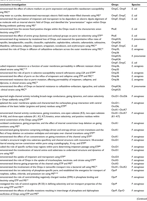
Conclusive investigation Omps Species
Demonstrated the effect of culture medium on porin expression and piperacillin–tazobactam susceptibility using MIC46
OmpC, OmpF E. coli
Using water as a probe, demonstrated macroscopic electric field inside water-filled channels using MS43 OmpF, OmpC E. coli
Demonstrated the permeation of imipenem and meropenem to be dependent on electric dipole alignment of
the molecule with an internal electric field of Omps, and identified the “preorientation” region within Omps
affecting antibiotic pathway using MS19
OmpF, OmpC E. coli
Demonstrated how the excess fixed positive charges within the Omps result in the characteristic anion
selectivity using eTP75
Phoe E. coli
Demonstrated the effect of amino group (lysines) and carboxyl groups on pore ion selectivity using eTP76 Phoe E. coli
Demonstrated drug resistance by mutational loss of Omps, and measured the quantitative influx rates of
ampicillin, benzylpenicillin, oxacillin, cloxacillin, cephalothin, cephaloridine, cefoxitin, cefamandole, cefotaxime, ceftazidime, ceftriaxone, cefepime, imipenem, ertapenem, novobiocin, and erythromycin using MIC49
OmpK35, OmpK36
K. pneumoniae
OmpF, OmpC E. coli
examined the role of Omps in diffusion of ceftazidime–avibactam across the outer membrane using MIC42 Omp35,
Omp36
E. aerogenes
OmpK35, OmpK36
K. pneumoniae
OmpF, OmpC E. coli
Studied imipenem resistance as a function of outer membrane permeability in different resistant clinical isolated strains using MIC77,78
Omp36, Omp35
E. aerogenes
Demonstrated the role of porin in selective susceptibility toward ceftriaxone using LSA and eTP80 Omp35 E. aerogenes
Demonstrated the effect of porin on the influx of ertapenem and cefepime using ETP and MIC16 Omp36 E. aerogenes
Demonstrated resistance due to porin mutation, affecting permeability of imipenem, cefepime, and cefpirome, in clinical strains using MIC79
Omp36 E. aerogenes
Demonstrated the effect of Omps on bacterial resistance to ceftazidime–avibactam, tigecycline, and colistin in clinical strains using MIC82
OmpK36, OmpK35
K. pneumoniae
Reported single-channel activity including broad-range conductance, gating dynamics, and cation selectivity for Omps subfamily using eTP84
OccD1–OccD6 P. aeruginosa
explained the outer membrane uptake and characterized the carboxylate group interaction with central residues of the basic ladder (arginine and lysine) residues using eTP83
OccD1– OccD6, OccK1–OccK7
P. aeruginosa
Demonstrated channel activity conductance, gating transitions, one-open substate (K3), two-open substate (K4–K6), and three-open substate (K1, K2, K7) kinetics, anion selectivity, and positive residues within central constriction of the Omps using eTP89
OccK1–OccK7 (K1–K7)
P. aeruginosa
elucidated conductance, gating properties, and the effect of internal constriction loop deletion on gating transitions using MS96
OccK1 P. aeruginosa
Demonstrated gating dynamics comprising enthalpy-driven and entropy-driven current transitions and the effect of loop deletion on activation enthalpies and entropies over channel transitions using eTP93
OccK1 P. aeruginosa
Demonstrated the effect of ion concentrations on gating transitions of the channel using eTP94 OccK1 P. aeruginosa
Provided a structural insight into substrate specificity and channel structure with monomeric 18-stranded
β-barrel ensuing narrow constriction within pore using crystallography, X-ray, and eTP85
OccD1 P. aeruginosa
Studied the role of specific surface loop regions within pore determining imipenem passage using ETP86 OccD1 P. aeruginosa
Demonstrated the translocation of natural amino acid substrates to understand structure and dynamics of pore using MS87
OccD1 P. aeruginosa
Demonstrated the uptake of imipenem and meropenem using eTP91 OccD3 P. aeruginosa
Demonstrated the role of Omps in the uptake of tricarboxylate, isocitrate, and citrate using eTP98 OccK5 P. aeruginosa
Demonstrated diverse gating properties of the channel using eTP and MS95 OccK5 P. aeruginosa
Demonstrated the involvement of the Omps in temocillin transport into a bacterial cell using MIC92 OccK1, OccK2 P. aeruginosa
Demonstrated the ion selectivity of phosphate-specific pore, and established the energetics for transport of
phosphate, sulfate, chloride, and potassium ion using MS90,102
OprP P. aeruginosa
Demonstrated the role of central-binding negatively charged residue (D94) in phosphate binding and selectivity using eTP and MS101
OprP P. aeruginosa
Investigated the role of central arginine (R133) in defining selectivity and ion transport properties of the
pore using eTP and MS100
OprP P. aeruginosa
Demonstrated the effects of double mutations resulting in interchange of phosphate and diphosphate
specificities of Omps using ETP and MS99
OprP, OprO P. aeruginosa
(Continued) Table 2 (Continued)
Infection and Drug Resistance downloaded from https://www.dovepress.com/ by 118.70.13.36 on 22-Aug-2020
Dovepress exploring bacterial outer membrane barrier to combat bad bugs
in light-scattering signal is then correlated with the relative
permeability of the molecules. The main disadvantage of
this method is that it requires a large quantity of material
and is only effective for uncharged molecules, whereas for
charged molecules, the effect of counterion flow affects the
quality of the measurement. Moreover, the assay can only
determine average turnover numbers and often does not
provide conclusive values.
7Moreover, using conventional electrophysiology,
compu-tation of rate of flux of discrete small molecules across Omps
present in bacterial outer cell membrane involves
measure-ment of flux values at single molecular level.
7,36,45,52,56,66,67Here, electrophysiological measurement using single Omps
provides the best high-resolution (Figure 3) signal-to-noise
ratio,
7,18,40,73,74,83thereby suggesting the higher efficacy of this
method in sensing and understanding uptake at molecular
level.
7,15,22The method includes reconstitution of a single or
multiple Omps into an artificial planar lipid bilayer and
fur-ther uses transmembrane potential-driven ion current across
the channel as a detection probe.
7,67Using ion current as a
probe specifically demonstrates very well-characterized
elec-trophysiological properties of the Omps,
15,34,45,65,66,84,106,119–121including size,
122,123single-channel conductance, channel
ion selectivity,
58,75,76,90,99–101channel gating dynamics, and
more.
47,95,109Likewise, the size of Omps is a key factor
defin-ing transport through the channel.
107,108This factor plays a key
role in antibiotic susceptibility.
72–74Determination of the size
of Omps using electrophysiology provides a crucial insight
into the maximum size of molecule they can transport.
122,123This, further, helps in evaluating the inner structure including
constriction site.
122–125Further, single-channel conductance of
Omps, ion selectivity,
58,75,76,84,89and gating dynamics
35,47,94,95,109give an insight into the channel–substrate binding and
chan-nel–substrate interactions.
35,71,83,85,97,99,101An insight into the
channel conductance can be obtained, specifically using
staircase electrophysiology (Figure 3A and B), where
real-time insertions of single channels at constant voltage can be
attained.
59,123The conductance of any channel can be termed
as its unique characteristic. This allows a better
understand-ing of the open/close states of the channel and its gatunderstand-ing
dynamics which can then be employed in studying channel
structure–activity relationship.
35,71,107,108Using these functions, a proper insight into the channel
interaction with different substrates can be obtained
includ-ing substrate-induced partial or full blockage ( Figure 3C)
of channel
52,53,67and substrate-induced gating.
67The
func-tion of these pores has been well documented on the basis
of pore characteristics, chemical modification, and genetic
mutations.
15These parameters were further used to
elabo-rate transport of the following antibiotics: meropenem,
52imipenem,
52cefotaxime,
48cefpirome,
44ceftriaxone,
44,45cefepime,
45ceftazidime,
44,45ciprofloxacin,
45norfloxacin,
45and
enrofloxacin
45through OmpC; imipenem,
74meropenem,
53ceftazidime,
44,45,74cefepime,
45,55,74ceftriaxone,
44,45cefpi-rome,
44ampicillin,
57,60,61,67benzylpenicillin,
60amoxicillin,
57carbenicillin,
57azlocillin,
57piperacillin,
57ciprofloxacin,
45norfloxacin,
45,56,126enrofloxacin,
7,45,54,66,72moxifloxacin,
65different poly arginines,
64polyamines,
106and antimicrobial
peptides
51through OmpF; ceftriaxone
80through Omp35;
cefepime
16through Omp36; imipenem
91and meropenem
91through OccD3; imipenem
86through OccD1; and
merope-nem,
104glutamic acid,
104arginine,
104and imipenem
104through
CarO Omp (Table 2).
In contrast, single-channel recording provides the best
signal-to-noise ratio and intrinsic data on Omp–substrate
interaction.
40,45,65–67But the interpretation of molecule
Conclusive investigation Omps Species
Demonstrated the structural features responsible for transport of amino acid residues via substrate-specific
channel using LSA, eTP, and MS97
OccK8 P. aeruginosa
Demonstrated Omps uptake of glycine and ornithine and no uptake of glutamic acid, glucose, and imipenem using LSA and MS105
CarO isoforms CarO1, CarO2, CarO3
A. baumannii
Demonstrated channel conductance, cationic selectivity, and specificity toward meropenem, glutamic acid,
arginine, and imipenem using eTP104
CarO A. baumannii
Demonstrated the function of the Omps in imipenem, meropenem, colistin, ceftazidime, and ciprofloxacin
uptake using MIC103
rOprD homologue
A. baumannii
Demonstrated Omps substrate specificities toward glycine, ornithine, arginine, putrescine, glutamic acid,
glucose, maltose, benzoic acid, phenylalanine, tryptophan, imipenem, meropenem, ceftazidime, ampicillin, and fosfomycin using LSA and eTP35
OccAB1– OccAB4
A. baumannii
Abbreviations: Omps, outer membrane proteins; eTP, electrophysiology; MS, molecular simulation; LSA, liposome swelling assay; MIC, minimum inhibitory
concentration; FRET, fluorescence resonance energy transfer.
Table 2 (Continued)
Infection and Drug Resistance downloaded from https://www.dovepress.com/ by 118.70.13.36 on 22-Aug-2020
Dovepress
Ghai and Ghai
translocation cannot be made directly as the chances of
molecule exit on the entry side are almost identical when
compared to the transport of the molecule across the pore.
7Whereas in the case of charged molecules, direct conclusion
of translocation can be made as the increasing voltage will
reduce the residence time of the molecules inside the Omp,
which might provide some evidence of transport across the
Omp. In addition, using channel selectivity, that is, channel
inherent selection of either anion or cation, a quantitative flux
assessment of the charged molecules can be made using
elec-trophysiological reversal potential measurements.
36,59Using
this approach, flux of
β
-lactamase inhibitors across OmpF
and OmpC was estimated, showing the role of Omps in their
transport across bacterial biobarrier.
36,59However, most of the
molecules did not carry a net charge or show low intrinsic
solubility which makes them trivial to measure and thus
excludes them from screening via this method. Furthermore,
the finite time resolution of electrophysiology also makes
the method limited in screening of antibiotics uptake.
7,45,66,67Molecular simulation
In the current scenario, MS is well suited to obtain a
particu-lar information at an atomic scale.
121Thus far, knowledge of
the antibiotic translocation problem has pointed essentially
toward three mechanisms including diffusion with molecule
binding, a mechanism based on pore dehydration induced by
the permeating molecule, and slow diffusion with molecule
binding.
50,61,62,70,71,97,99,121Further, to discriminate among
these mechanisms, and to attain a better description of the
Omps behavior and their role in substrate transport,
under-standing the communication between pore and substrate is
essential.
119–121,127,128Thanks to the high-resolution, molecular
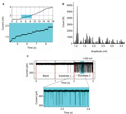
Figure 3 (A) Current recorded using staircase electrophysiology. A graphical representation depicting insertion of Omp over real time under applied potential. Recording
time: 18 seconds. (B) Current histogram for the trace with each peak resembling a single Omp, showing, in total, approximately 45 Omps. (C) OmpF single channel–substrate interaction comparison: without substrate (blank), substrate 1 depicting no blockages, and substrate 2 inducing well-resolved channel blockage; a clear difference between the two substrates can be seen.
Abbreviation: Omp, outer membrane protein.
4
A B
C
2
2.0
Current (nA)
1.5 0 2
5 6 7
Time (s)
8 4 6 8 10 12 14 16 18
6000
5000
4000
3000
2000
1000
0
1.0 1.5 2.0 2.5 3.0
Amplitude (nA)
Blank 450
300
400
Current (pA)
Current (pA)
300
Substrate 1 Substrate 2 +100 mV
3 2
2.5 2.6
Time (s)
Time (s) 1
0
Count (N
)
3.5
Infection and Drug Resistance downloaded from https://www.dovepress.com/ by 118.70.13.36 on 22-Aug-2020
Dovepress exploring bacterial outer membrane barrier to combat bad bugs
modeling simulations, detailed characterization is possible
in terms of energetics (Figure 4 from Ghai et al)
36and bond
formation including hydrogen bonds, hydrophobic contacts,
and more.
50,62,71,121The complete control over the characteristics of the
sys-tem allows MS to explain the impact of pinpoint mutations
and the effects that arise due to different domains of the same
proteins.
95,100,101Further, MS significantly allows
understand-ing and interpretunderstand-ing available experimental data.
50,61,62,70,121When combined with experimental approach, MS proves
to be a complementary method. For instance, together with
electrophysiology,
36,48,54,55,57,58,60,61,65–67,71–73,95,97,99–101MS was
used for understanding the transport of
β
-lactamase inhibitors
(Figure 4), interaction of substrates with Omps (enrofloxacin,
moxifloxacin, ampicillin, benzylpenicillin, carbenicillin,
amoxicillin, azlocillin, piperacillin, ertapenem, imipenem,
meropenem, cefepime, cefpirome, cefotaxime,
ceftazi-dime, cefoxitin, and cefepime with OmpF;
7,19,21,36,50,54,55,57,58, 60–63,65–67,70–73cefotaxime, imipenem, and meropenem with
OmpC;
19,48natural amino acids with OccD1),
87ion transport
including transport of phosphate potassium and chloride
ion via OprP
90,100–102and OprO,
99and interaction of glycine
ornithine, glucose, and imipenem with CarO isoforms.
105Fur-ther, for liposome swelling
55,60,97,105and minimum inhibitory
concentration assay
48,60,73(not described), MS was helpful
for understanding and interpreting the experimental results.
Figure 4 (A) Intrinsic depiction of the two-dimensional free energy of translocation of β-lactamase inhibitor (avibactam), reassembled from metadynamic simulations. (B)
Lateral view and (C) topmost view of the avibactam inside OmpF pore in the two lowest minima near the constriction region and at the subsequent transition state. Reprinted with permission from Ghai I, Pira A, Scorciapino MA, et al. General method to determine the flux of charged molecules through nanopores applied to beta-lactamase
inhibitors and OmpF. J Phys Chem Lett. 2017;8(6):1295–1301.36 Copyright (2017) American Chemical Society.
40
A B
C
30
20
10
0
Z (Å)
∆
G
(kcal/mol)
–10
–20
–30
–40
0 50 100
Dipole moment projection onto z axis 150
40
35
30
25
20
15
10
5
0
Rationalizing the process of permeation of antibiotics
into Gram-negative bacteria via MS requires an accurate and
exhaustive description of some key molecular properties of
the antibiotic molecule.
121MS is the best alternative tool to
obtain homogenously derived physical–chemical descriptors
for molecules with or without experimental approach.
121,127,128MS based on all-atom empirical force fields with the
resolu-tion in microsecond time range and beyond could potentially
provide a good level of description of the structural and
dynamical properties of biological systems.
119,121,127,128Toward translational research
Translational research on understanding antimicrobial
resis-tance has led to implausible development in recent years
4,129together with the expansion of novel techniques including
proteomic analyses, high-sensitivity mass spectrometry,
com-putational bioinformatics, and many more approaches.
4For the
most part, the discovery of novel technologies, the development
of new infrastructures, along with the training of budding
sci-entists have reinforced this evolution.
1,4,129,130But the transition
is still not complete, and roadblocks still exist on the path to
scientific progress, for example, combining different data into
a shared database that can be intrinsically used to understand
how Omps located in the outer membrane of Gram-negative
bacteria are able to filter molecular influx.
24The imperative need
for new, effective Gram-negative antibacterial drugs comes at
Infection and Drug Resistance downloaded from https://www.dovepress.com/ by 118.70.13.36 on 22-Aug-2020
Dovepress
Ghai and Ghai
a time when techniques needed for innovative assays can
pro-vide significant crucial data over understanding the effective
bottleneck.
4Ideally, the overall penetration–efflux puzzle
4will
form part of a larger understanding of the Gram-negative cell
envelope as well as direction on how to create small molecules
that can easily penetrate across the outer membranes.
4This
information should move the antibacterial research community
toward more rational approaches, which may enable the delivery
of new agents to treat life-threatening infections.
1,4,129,130Conclusive remarks
This review summarizes the progressive scientific evidence
explaining the role of Omps in membrane permeability of
Gram-negative bacteria. The control of bacterial membrane
permeability is a complex process that is strongly
struc-tured by an intricate network of arrangements that senses
and retorts to pH, osmotic shock, temperature, and external
chemical stress. Bacteria majorly make use of cultured
regu-lated cascades that perceive and distinguish toxic compounds
and respond through various resistance mechanisms
includ-ing regulation of Omps.
6,7,15,18,22The information on the role
of effective Omps in substrate uptake and their structural
relationship associated with their role in transport highlights
the efforts of the scientific upfront in the direction of
under-standing the bacterial resistance.
6,7,15,18,22Translocation across
the Omps can be assumed as the first step in the journey of an
antibiotic along the defined pathway toward its target.
Con-sequently, interpretation of antibiotic translocation through
porins at the molecular level is crucial for understanding the
correlation between influx and antibiotic activities within
bacteria. The function of the general diffusion pores has
been well studied based on pore characteristics, chemical
modification, and genetic mutations. Our understanding of
the structure of the pore-forming complex has tremendously
improved over the last decade with the emergence of MS,
state-of-the-art X-ray data, mass spectrometry assay
pro-tocols, and novel high-resolution experimental approaches
including electrophysiology. However, a better understanding
of the transportation mechanism by outer membrane pores
is required. The molecular basis of the antibiotic transport
via specific porins is still completely open at present, and
further rigorous studies are needed to give insight into the
structure–activity relationship of pores associated with
anti-biotic transport. The data computed for these Omps can be
further employed to elucidate the antibiotic uptake pathway
through Omps at molecular level, which could possibly
empower rational drug design to further enhance permeation
and support novel strategies to dodge
“impermeability”-mediated resistance mechanism.
Acknowledgments
The publication of this article was funded by the Open Access
fund of Leibniz Universität Hannover. The authors would like
to thank Prof. Dr Mathias Winterhalter and Prof. Dr Richard
Wagner for their constructive comments.
Disclosure
The authors report no conflicts of interest in this work.
References
1. Kostyanev T, Bonten MJ, O’Brien S, et al. The Innovative Medicines Initiative’s New Drugs for Bad Bugs programme: European public-private partnerships for the development of new strategies to tackle antibiotic resistance. J Antimicrob Chemother. 2016;71(2):290–295. 2. Ventola CL. The antibiotic resistance crisis: part 1: causes and threats.
P T. 2015;40(4):277–283.
3. Gootz TD. The global problem of antibiotic resistance. Crit Rev Immunol. 2010;30(1):79–93.
4. Stavenger RA, Winterhalter M. TRANSLOCATION project: how to get good drugs into bad bugs. Sci Transl Med. 2014;6(228):228ed7. 5. Nikaido H. Role of permeability barriers in resistance to beta-lactam
antibiotics. Pharmacol Ther. 1985;27(2):197–231.
6. Nikaido H. Molecular basis of bacterial outer membrane permeability revisited. Microbiol Mol Biol Rev. 2003;67(4):593–656.
7. Winterhalter M, Ceccarelli M. Physical methods to quantify small antibiotic molecules uptake into Gram-negative bacteria. Eur J Pharm Biopharm. 2015;95(Pt A):63–67.
8. Chattopadhyay MK, Jaganandham MV. Vesicles-mediated resistance to antibiotics in bacteria. Front Microbiol. 2015;6:758.
9. Kulkarni HM, Jagannadham MV. Biogenesis and multifaceted roles of outer membrane vesicles from Gram-negative bacteria. Microbiology. 2014;160(10):2109–2121.
10. Chattopadhyay MK, Jagannadham MV. Corrigendum: vesicles-medi-ated resistance to antibiotics in bacteria. Front Microbiol. 2015;6:758. 11. McBroom AJ, Kuehn MJ. Release of outer membrane vesicles by
Gram-negative bacteria is a novel envelope stress response. Mol Microbiol. 2007;63(2):545–558.
12. Maredia R, Devineni N, Lentz P, et al. Vesiculation from Pseudomonas aeruginosa under SOS. Scientific World J. 2012;2012:18.
13. Kulkarni HM, Swamy ChV, Jagannadham MV. Molecular charac-terization and functional analysis of outer membrane vesicles from the antarctic bacterium Pseudomonas syringae suggest a possible response to environmental conditions. J Proteome Res. 2014;13(3): 1345–1358.
14. Schwechheimer C, Kuehn MJ. Outer-membrane vesicles from Gram-negative bacteria: biogenesis and functions. Nat Rev Microbiol. 2015;13(10):605–619.
15. Benz R. Structure and function of porins from gram-negative bacteria. Annu Rev Microbiol. 1988;42:359–393.
16. James CE, Mahendran KR, Molitor A, et al. How beta-lactam antibiotics enter bacteria: a dialogue with the porins. PLoS One. 2009;4(5):e5453. 17. Schirmer T. General and specific porins from bacterial outer
mem-branes. J Struct Biol. 1998;121(2):101–109.
18. Schulz GE. The structure of bacterial outer membrane proteins. Bio-chim Biophys Acta. 2002;1565(2):308–317.
19. Scorciapino MA, D’Agostino T, Acosta-Gutierrez S, Malloci G, Bodrenko I, Ceccarelli M. Exploiting the porin pathway for polar compound delivery into Gram-negative bacteria. Future Med Chem. 2016;8(10):1047–1062.
20. Singh H, Thangaraj P, Chakrabarti A. Acinetobacter baumannii: a brief account of mechanisms of multidrug resistance and current and future therapeutic management. J Clin Diagn Res. 2013;7(11):2602–2605. 21. Ziervogel BK, Roux B. The binding of antibiotics in OmpF porin.
Structure. 2013;21(1):76–87.
Infection and Drug Resistance downloaded from https://www.dovepress.com/ by 118.70.13.36 on 22-Aug-2020
Dovepress exploring bacterial outer membrane barrier to combat bad bugs
22. Pages JM, James CE, Winterhalter M. The porin and the permeating antibiotic: a selective diffusion barrier in Gram-negative bacteria. Nat Rev Microbiol. 2008;6(12):893–903.
23. Dupont H, Choinier P, Roche D, et al. Structural alteration of OmpR as a source of ertapenem resistance in a CTX-M-15-producing Escherichia coli O25b:H4-ST131 clinical isolate. Antimicrob Agents Chemother. 2017;61(5):e00014–e00017.
24. Chakradhar, S. Breaking through: How researchers are gaining entry into barricaded bacteria. Nat Med. 2017;23(8):907–910.
25. Nakae T. Outer membrane of Salmonella. Isolation of protein complex that produces transmembrane channels. J Biol Chem. 1976;251(7):2176–2178.
26. Nikaido H, Vaara M. Molecular basis of bacterial outer membrane permeability. Microbiol Rev. 1985;49(1):1–32.
27. Nguyen-Disteche M, Pollock JJ, Ghuysen JM, et al. Sensitivity to ampicillin and cephalothin of enzymes involved in wall peptide crosslinking in Escherichia coli K12, strain 44. Eur J Biochem. 1974;41(3):457–463.
28. Schulz GE. Porins: general to specific, native to engineered passive pores. Curr Opin Struct Biol. 1996;6(4):485–490.
29. Guillier M, Gottesman S, Storz G. Modulating the outer membrane with small RNAs. Genes Dev. 2006;20(17):2338–2348.
30. Fahie MA, Yang B, Mullis M, Holden MA, Chen M. Selective detec-tion of protein homologues in serum using an OmpG nanopore. Anal Chem. 2015;87(21):11143–11149.
31. Li H, Luo YF, Williams BJ, Blackwell TS, Xie CM. Structure and function of OprD protein in Pseudomonas aeruginosa: from antibi-otic resistance to novel therapies. Int J Med Microbiol. 2012;302(2): 63–68.
32. Dupont H, Gaillot O, Goetgheluck AS, et al. Molecular characterization of carbapenem-nonsusceptible enterobacterial isolates collected during a prospective interregional survey in France and susceptibility to the novel ceftazidime-avibactam and aztreonam-avibactam combinations. Antimicrob Agents Chemother. 2015;60(1):215–221.
33. Hancock RE, Woodruff WA. Roles of porin and beta-lactamase in beta-lactam resistance of Pseudomonas aeruginosa. Rev Infect Dis. 1988;10(4):770–775.
34. Iredell J, Brown J, Tagg K. Antibiotic resistance in Enterobacteriaceae: mechanisms and clinical implications. BMJ. 2016;352:h6420. 35. Zahn M, Bhamidimarri Satya P, Baslé A, Winterhalter M, van den
Berg B. Structural insights into outer membrane permeability of Acinetobacter baumannii. Structure. 2016;24(2):221–231.
36. Ghai I, Pira A, Scorciapino MA, et al. General method to determine the flux of charged molecules through nanopores applied to beta-lactamase inhibitors and OmpF. J Phys Chem Lett. 2017;8(6):1295–1301. 37. Kretschmer N, Damianakos H, Chinou I, et al. Comparison of the
cyto-toxicity and antimicrobial activity of several isohexenylnaphthazarins. Planta Med. 2011;77(12):PM199.
38. Hancock RE, Brinkman FS. Function of pseudomonas porins in uptake and efflux. Annu Rev Microbiol. 2002;56:17–38.
39. Fito-Boncompte L, Chapalain A, Bouffartigues E, et al. Full virulence of Pseudomonas aeruginosa requires OprF. Infect Immun. 2011;79(3): 1176–1186.
40. Nestorovich EM, Sugawara E, Nikaido H, Bezrukov SM. Pseudomo-nas aeruginosa porin OprF: properties of the channel. J Biol Chem. 2006;281(24):16230–16237.
41. Sugawara E, Nestorovich EM, Bezrukov SM, Nikaido H. Pseudomonas aeruginosa porin OprF exists in two different conformations. J Biol Chem. 2006;281(24):16220–16229.
42. Pages JM, Peslier S, Keating TA, Lavigne JP, Nichols WW. Role of the outer membrane and porins in susceptibility of beta-lactamase-producing Enterobacteriaceae to ceftazidime-avibactam. Antimicrob Agents Chemother. 2015;60(3):1349–1359.
43. Acosta Gutierrez S, Bodrenko I, Scorciapino MA, Ceccarelli M. Macroscopic electric field inside water-filled biological nanopores. Phys Chem Chem Phys. 2016;18(13):8855–8864.
44. Lovelle M, Mach T, Mahendran KR, Weingart H, Winterhalter M, Gameiro P. Interaction of cephalosporins with outer membrane chan-nels of Escherichia coli. Revealing binding by fluorescence quenching and ion conductance fluctuations. Phys Chem Chem Phys. 2011;13(4): 1521–1530.
45. Mahendran KR, Kreir M, Weingart H, Fertig N, Winterhalter M. Permeation of antibiotics through Escherichia coli OmpF and OmpC porins: screening for influx on a single-molecule level. J Biomol Screen. 2010;15(3):302–307.
46. Pinet E, Franceschi C, Davin-Regli A, Zambardi G, Pages JM. Role of the culture medium in porin expression and piperacillin-tazobactam susceptibility in Escherichia coli. J Med Microbiol. 2015;64(11):1305–1314.
47. Liu N, Samartzidou H, Lee KW, Briggs JM, Delcour AH. Effects of pore mutations and permeant ion concentration on the spontaneous gating activity of OmpC porin. Protein Eng. 2000;13(7):491–500. 48. Lou H, Chen M, Black SS, et al. Altered antibiotic transport in OmpC
mutants isolated from a series of clinical strains of multi-drug resistant E. coli. PLoS One. 2011;6(10):e25825.
49. Sugawara E, Kojima S, Nikaido H. Klebsiella pneumoniae major porins OmpK35 and OmpK36 allow more efficient diffusion of beta-lactams than their Escherichia coli homologs OmpF and OmpC. J Bacteriol. 2016;198(23):3200–3208.
50. Aguilella-Arzo M, Garcia-Celma JJ, Cervera J, Alcaraz A, Aguilella VM. Electrostatic properties and macroscopic electrodiffusion in OmpF porin and mutants. Bioelectrochemistry. 2007;70(2):320–327. 51. Apetrei A, Asandei A, Park Y, Hahm KS, Winterhalter M, Luchian T.
Unimolecular study of the interaction between the outer membrane protein OmpF from E. coli and an analogue of the HP(2–20) antimi-crobial peptide. J Bioenerg Biomembr. 2010;42(2):173–180. 52. Bajaj H, Scorciapino MA, Moynie L, et al. Molecular basis of filtering
carbapenems by porins from beta-lactam-resistant clinical strains of Escherichia coli. J Biol Chem. 2016;291(6):2837–2847.
53. Bodrenko I, Bajaj H, Ruggerone P, Winterhalter M, Ceccarelli M. Analysis of fast channel blockage: revealing substrate binding in the microsecond range. Analyst. 2015;140(14):4820–4827.
54. Brauser A, Schroeder I, Gutsmann T, et al. Modulation of enrofloxacin binding in OmpF by Mg2+ as revealed by the analysis of fast flickering single-porin current. J Gen Physiol. 2012;140(1):69–82.
55. Bredin J, Saint N, Mallea M, et al. Alteration of pore properties of Escherichia coli OmpF induced by mutation of key residues in anti-loop 3 region. Biochem J. 2002;363(Pt 3):521–528.
56. Cama J, Bajaj H, Pagliara S, et al. Quantification of fluoroquinolone uptake through the outer membrane channel OmpF of Escherichia coli. J Am Chem Soc. 2015;137(43):13836–13843.
57. Danelon C, Nestorovich EM, Winterhalter M, Ceccarelli M, Bezrukov SM. Interaction of zwitterionic penicillins with the OmpF channel facilitates their translocation. Biophys J. 2006;90(5):1617–1627. 58. Danelon C, Suenaga A, Winterhalter M, Yamato I. Molecular origin
of the cation selectivity in OmpF porin: single channel conductances vs. free energy calculation. Biophys Chem. 2003;104(3):591–603. 59. Ghai I, Winterhalter M, Wagner R. Probing transport of charged
beta-lactamase inhibitors through OmpC, a membrane channel from E. coli. Biochem Biophys Res Commun. 2017;484(1):51–55.
60. Hajjar E, Bessonov A, Molitor A, et al. Toward screening for antibi-otics with enhanced permeation properties through bacterial porins. Biochemistry. 2010;49(32):6928–6935.
61. Hajjar E, Mahendran KR, Kumar A, et al. Bridging timescales and length scales: from macroscopic flux to the molecular mechanism of antibiotic diffusion through porins. Biophys J. 2010;98(4):569–575. 62. Im W, Roux B. Ion permeation and selectivity of OmpF porin: a theo-retical study based on molecular dynamics, Brownian dynamics, and continuum electrodiffusion theory. J Mol Biol. 2002;322(4):851–869. 63. Kumar A, Hajjar E, Ruggerone P, Ceccarelli M. Molecular simulations reveal the mechanism and the determinants for ampicillin translocation through OmpF. J Phys Chem B. 2010;114(29):9608–9616.
Infection and Drug Resistance downloaded from https://www.dovepress.com/ by 118.70.13.36 on 22-Aug-2020
Dovepress
Ghai and Ghai
64. Lamichhane U, Islam T, Prasad S, Weingart H, Mahendran KR, Winterhalter M. Peptide translocation through the mesoscopic chan-nel: binding kinetics at the single molecule level. Eur Biophys J. 2013;42(5):363–369.
65. Mach T, Neves P, Spiga E, et al. Facilitated permeation of antibiotics across membrane channels—interaction of the quinolone moxi-floxacin with the OmpF channel. J Am Chem Soc. 2008;130(40): 13301–13309.
66. Mahendran KR, Hajjar E, Mach T, et al. Molecular basis of enro-floxacin translocation through OmpF, an outer membrane channel of Escherichia coli—when binding does not imply translocation. J Phys Chem B. 2010;114(15):5170–5179.
67. Nestorovich EM, Danelon C, Winterhalter M, Bezrukov SM. Designed to penetrate: time-resolved interaction of single antibiotic mol-ecules with bacterial pores. Proc Natl Acad Sci U S A. 2002;99(15): 9789–9794.
68. Neves P, Berkane E, Gameiro P, Winterhalter M, de Castro B. Inter-action between quinolones antibiotics and bacterial outer membrane porin OmpF. Biophys Chem. 2005;113(2):123–128.
69. Neves P, Sousa I, Winterhalter M, Gameiro P. Fluorescence quenching as a tool to investigate quinolone antibiotic interactions with bacterial protein OmpF. J Membr Biol. 2009;227(3):133–140.
70. Pezeshki S, Chimerel C, Bessonov AN, Winterhalter M, Kleinekathofer U. Understanding ion conductance on a molecular level: an all-atom modeling of the bacterial porin OmpF. Biophys J. 2009;97(7): 1898–1906.
71. Phale PS, Philippsen A, Widmer C, Phale VP, Rosenbusch JP, Schirmer T. Role of charged residues at the OmpF porin channel constriction probed by mutagenesis and simulation. Biochemistry. 2001;40(21):6319–6325.
72. Singh PR, Ceccarelli M, Lovelle M, Winterhalter M, Mahendran KR. Antibiotic permeation across the OmpF channel: modulation of the affinity site in the presence of magnesium. J Phys Chem B. 2012; 116(15):4433–4438.
73. Vidal S, Bredin J, Pages JM, Barbe J. Beta-lactam screening by specific residues of the OmpF eyelet. J Med Chem. 2005;48(5):1395–1400. 74. Weichbrodt C, Bajaj H, Baaken G, et al. Antibiotic translocation
through porins studied in planar lipid bilayers using parallel platforms. Analyst. 2015;140(14):4874–4881.
75. Benz R, Darveau RP, Hancock RE. Outer-membrane protein PhoE from Escherichia coli forms anion-selective pores in lipid-bilayer membranes. Eur J Biochem. 1984;140(2):319–324.
76. Darveau RP, Hancock RE, Benz R. Chemical modification of the anion selectivity of the PhoE porin from the Escherichia coli outer membrane. Biochim Biophys Acta. 1984;774(1):67–74.
77. Bornet C, Davin-Regli A, Bosi C, Pages JM, Bollet C. Imipenem resistance of Enterobacter aerogenes mediated by outer membrane permeability. J Clin Microbiol. 2000;38(3):1048–1052.
78. Lavigne JP, Sotto A, Nicolas-Chanoine MH, Bouziges N, Pages JM, Davin-Regli A. An adaptive response of Enterobacter aerogenes to imipenem: regulation of porin balance in clinical isolates. Int J Anti-microb Agents. 2013;41(2):130–136.
79. Thiolas A, Bornet C, Davin-Regli A, Pages JM, Bollet C. Resistance to imipenem, cefepime, and cefpirome associated with mutation in Omp36 osmoporin of Enterobacter aerogenes. Biochem Biophys Res Commun. 2004;317(3):851–856.
80. Bornet C, Saint N, Fetnaci L, et al. Omp35, a new Enterobacter aerogenes porin involved in selective susceptibility to cephalosporins. Antimicrob Agents Chemother. 2004;48(6):2153–2158.
81. Arunmanee W, Pathania M, Solovyova AS, et al. Gram-negative tri-meric porins have specific LPS binding sites that are essential for porin biogenesis. Proc Natl Acad Sci U S A. 2016;113(34):E5034–E5043. 82. Castanheira M, Mendes RE, Sader HS. Low frequency of
ceftazidime-avibactam resistance among Enterobacteriaceae isolates carrying blaKPC collected in U.S. hospitals from 2012 to 2015. Antimicrob Agents Chemother. 2017;61(3).
83. Eren E, Parkin J, Adelanwa A, et al. Toward understanding the outer membrane uptake of small molecules by Pseudomonas aeruginosa. J Biol Chem. 2013;288(17):12042–12053.
84. Liu J, Wolfe AJ, Eren E, et al. Cation selectivity is a conserved feature in the OccD subfamily of Pseudomonas aeruginosa. Biochim Biophys Acta. 2012;1818(11):2908–2916.
85. Biswas S, Mohammad MM, Patel DR, Movileanu L, van den Berg B. Structural insight into OprD substrate specificity. Nat Struct Mol Biol. 2007;14(11):1108–1109.
86. Huang H, Hancock RE. The role of specific surface loop regions in determining the function of the imipenem-specific pore protein OprD of Pseudomonas aeruginosa. J Bacteriol. 1996;178(11):3085–3090. 87. Samanta S, Scorciapino MA, Ceccarelli M. Molecular basis of substrate translocation through the outer membrane channel OprD of Pseudomo-nas aeruginosa. Phys Chem Chem Phys. 2015;17(37):23867–23876. 88. Eren E, Vijayaraghavan J, Liu J, et al. Substrate specificity within a fam-ily of outer membrane carboxylate channels. PLoS Biol. 2012;10(1): e1001242.
89. Liu J, Eren E, Vijayaraghavan J, et al. OccK channels from Pseudomo-nas aeruginosa exhibit diverse single-channel electrical signatures but conserved anion selectivity. Biochemistry. 2012;51(11):2319–2330. 90. Modi N, Benz R, Hancock RE, Kleinekathofer U. Modeling the ion
selectivity of the phosphate specific channel OprP. J Phys Chem Lett. 2012;3(23):3639–3645.
91. Soundararajan G, Bhamidimarri SP, Winterhalter M. Understanding carbapenem translocation through OccD3 (OpdP) of Pseudomonas aeruginosa. ACS Chem Biol. 2017;12(6):1656–1664.
92. Chalhoub H, Pletzer D, Weingart H, et al. Mechanisms of intrinsic resistance and acquired susceptibility of Pseudomonas aeruginosa isolated from cystic fibrosis patients to temocillin, a revived antibiotic. Sci Rep. 2017;7:40208.
93. Cheneke BR, Indic M, van den Berg B, Movileanu L. An outer mem-brane protein undergoes enthalpy- and entropy-driven transitions. Biochemistry. 2012;51(26):5348–5358.
94. Cheneke BR, van den Berg B, Movileanu L. Analysis of gating transitions among the three major open states of the OpdK channel. Biochemistry. 2011;50(22):4987–4997.
95. Pothula KR, Dhanasekar NN, Lamichhane U, et al. Single residue acts as gate in OccK channels. J Phys Chem B. 2017;121(12):2614–2621. 96. Pothula KR, Kleinekathofer U. Theoretical analysis of ion conduc-tance and gating transitions in the OpdK (OccK1) channel. Analyst. 2015;140(14):4855–4864.
97. Samanta S, D’Agostino T, Ghai I, et al. How to get large drugs through small pores? Exploiting the porins pathway in Pseudomonas aerugi-nosa. Biophys J. 2017;112(3 Suppl 1):416a.
98. Tamber S, Maier E, Benz R, Hancock RE. Characterization of OpdH, a Pseudomonas aeruginosa porin involved in the uptake of tricarboxyl-ates. J Bacteriol. 2007;189(3):929–939.
99. Modi N, Ganguly S, Barcena-Uribarri I, et al. Structure, dynamics, and substrate specificity of the OprO porin from Pseudomonas aeruginosa. Biophys J. 2015;109(7):1429–1438.
100. Modi N, Barcena-Uribarri I, Bains M, Benz R, Hancock RE, Kleinekathofer U. Role of the central arginine R133 toward the ion selectivity of the phosphate specific channel OprP: effects of charge and solvation. Biochemistry. 2013;52(33):5522–5532.
101. Modi N, Barcena-Uribarri I, Bains M, Benz R, Hancock RE, Kleinekathofer U. Tuning the affinity of anion binding sites in porin channels with negatively charged residues: molecular details for OprP. ACS Chem Biol. 2015;10(2):441–451.
102. Pongprayoon P, Beckstein O, Wee CL, Sansom MS. Simulations of anion transport through OprP reveal the molecular basis for high affinity and selectivity for phosphate. Proc Natl Acad Sci U S A. 2009;106(51):21614–21618.
103. Catel-Ferreira M, Nehme R, Molle V, et al. Deciphering the function of the outer membrane protein OprD homologue of Acinetobacter baumannii. Antimicrob Agents Chemother. 2012;56(7):3826–3832.
Infection and Drug Resistance downloaded from https://www.dovepress.com/ by 118.70.13.36 on 22-Aug-2020
Dovepress
Infection and Drug Resistance
Publish your work in this journal
Submit your manuscript here: https://www.dovepress.com/infection-and-drug-resistance-journal
Infection and Drug Resistance is an international, peer-reviewed open-access journal that focuses on the optimal treatment of infection (bacte-rial, fungal and viral) and the development and institution of preventive strategies to minimize the development and spread of resistance. The journal is specifically concerned with the epidemiology of antibiotic
resistance and the mechanisms of resistance development and diffusion in both hospitals and the community. The manuscript management system is completely online and includes a very quick and fair peer-review system, which is all easy to use. Visit http://www.dovepress.com/ testimonials.php to read real quotes from published authors.
Dovepress
exploring bacterial outer membrane barrier to combat bad bugs
104. Catel-Ferreira M, Coadou G, Molle V, et al. Structure-function rela-tionships of CarO, the carbapenem resistance-associated outer mem-brane protein of Acinetobacter baumannii. J Antimicrob Chemother. 2011;66(9):2053–2056.
105. Zahn M, D’Agostino T, Eren E, Basle A, Ceccarelli M, van den Berg B. Small-molecule transport by CarO, an abundant eight-stranded beta-barrel outer membrane protein from Acinetobacter baumannii. J Mol Biol. 2015;427(14):2329–2339.
106. Iyer R, Delcour AH. Complex inhibition of OmpF and OmpC bacterial porins by polyamines. J Biol Chem. 1997;272(30):18595–18601. 107. Saint N, Lou KL, Widmer C, Luckey M, Schirmer T, Rosenbusch JP.
Structural and functional characterization of OmpF porin mutants selected for larger pore size. II. Functional characterization. J Biol Chem. 1996;271(34):20676–20680.
108. Lou KL, Saint N, Prilipov A, et al. Structural and functional char-acterization of OmpF porin mutants selected for larger pore size. I. Crystallographic analysis. J Biol Chem. 1996;271(34):20669–20675. 109. Phale PS, Schirmer T, Prilipov A, Lou K-L, Hardmeyer A, Rosen-busch JP. Voltage gating of Escherichia coli porin channels: role of the constriction loop. Proc Natl Acad Sci U S A. 1997;94(13): 6741–6745.
110. Stock JB, Rauch B, Roseman S. Periplasmic space in Salmonella typhimurium and Escherichia coli. J Biol Chem. 1977;252(21): 7850–7861.
111. Testa CA, Johnson LJ. A whole-cell phenotypic screening platform for identifying methylerythritol phosphate pathway-selective inhibi-tors as novel antibacterial agents. Antimicrob Agents Chemother. 2012;56(9):4906–4913.
112. Mortimer PGS, Piddock LJV. A comparison of methods used for measuring the accumulation of quinolones by enterobacteriaceae, Pseudomonas aeruginosa and Staphylococcus aureus. J Antimicrob Chemother. 1991;28(5):639–653.
113. Goessens WH, van der Bij AK, van Boxtel R, et al. Antibiotic trap-ping by plasmid-encoded CMY-2 beta-lactamase combined with reduced outer membrane permeability as a mechanism of carbape-nem resistance in Escherichia coli. Antimicrob Agents Chemother. 2013;57(8):3941–3949.
114. Shin JR, Lim KJ, Kim DJ, Cho JH, Kim SC. Display of multimeric antimicrobial peptides on the Escherichia coli cell surface and its application as whole-cell antibiotics. PLoS One. 2013;8(3):e58997. 115. Croxen MA, Law RJ, Scholz R, Keeney KM, Wlodarska M, Finlay
BB. Recent advances in understanding enteric pathogenic Escherichia coli. Clin Microbiol Rev. 2013;26(4):822–880.
116. Fox DT, Schmidt EN, Tian H, et al. Sub-inhibitory fosmidomycin exposures elicits oxidative stress in Salmonella enterica serovar Typhimurium LT2. PLoS One. 2014;9(4):e95271.
117. Davis TD, Gerry CJ, Tan DS. General platform for systematic quantita-tive evaluation of small-molecule permeability in bacteria. ACS Chem Biol. 2014;9(11):2535–2544.
118. Zhou Y, Joubran C, Miller-Vedam L, et al. Thinking outside the “bug”: a unique assay to measure intracellular drug penetration in gram-negative bacteria. Anal Chem. 2015;87(7):3579–3584.
119. Bezrukov SM, Berezhkovskii AM, Szabo A. Diffusion model of solute dynamics in a membrane channel: mapping onto the two-site model and optimizing the flux. J Chem Phys. 2007;127(11):115101. 120. O’Shea R, Moser HE. Physicochemical properties of
antibacte-rial compounds: implications for drug discovery. J Med Chem. 2008;51(10):2871–2878.
121. Scorciapino MA, Acosta-Gutierrez S, Benkerrou D, et al. Rational-izing the permeation of polar antibiotics into Gram-negative bacteria. J Phys Condens Matter. 2017;29(11):113001.
122. Krasilnikov OV, Sabirov RZ, Ternovsky VI, Merzliak PG, Muratk-hodjaev JN. A simple method for the determination of the pore radius of ion channels in planar lipid bilayer membranes. FEMS Microbiol Immunol. 1992;5(1–3):93–100.
123. Barcena-Uribarri I, Thein M, Maier E, Bonde M, Bergstrom S, Benz R. Use of nonelectrolytes reveals the channel size and oligomeric con-stitution of the Borrelia burgdorferi P66 porin. PLoS One. 2013;8(11): e78272.
124. Krasilnikov OV, Da Cruz JB, Yuldasheva LN, Varanda WA, Nogueira RA. A novel approach to study the geometry of the water lumen of ion channels: colicin Ia channels in planar lipid bilayers. J Membr Biol. 1998;161(1):83–92.
125. Krasilnikov OV. Sizing channels with neutral polymers. In: Kasianow-icz JJ, Kellermayer M, Deamer DW, editors. Structure and Dynamics of Confined Polymers. Dordrecht: Springer; 2002:97–115.
126. Bajaj H, Acosta Gutierrez S, Bodrenko I, et al. Bacterial outer mem-brane porins as electrostatic nanosieves: exploring transport rules of small polar molecules. ACS Nano. 2017;11(6):5465–5473. 127. Schwarz G, Danelon C, Winterhalter M. On translocation through a
membrane channel via an internal binding site: kinetics and voltage dependence. Biophys J. 2003;84(5):2990–2998.
128. Tran QT, Williams S, Farid R, Erdemli G, Pearlstein R. The transloca-tion kinetics of antibiotics through porin OmpC: insights from struc-ture-based solvation mapping using WaterMap. Proteins. 2013;81(2): 291–299.
129. Fontana JM, Alexander E, Salvatore M. Translational research in infectious disease: current paradigms and challenges ahead. Transl Res. 2012;159(6):430–453.
130. Goldman M. The innovative medicines initiative: a European response to the innovation challenge. Clin Pharmacol Ther. 2012;91(3): 418–425.
Infection and Drug Resistance downloaded from https://www.dovepress.com/ by 118.70.13.36 on 22-Aug-2020