1
February 24, 2020
Asians do not exhibit elevated expression or unique genetic polymorphisms for ACE2, the cell-entry receptor of SARS-CoV-2
Ying Chen1,2, Kejia Shan1,2,3, and Wenfeng Qian1,2,3,*
1 State Key Laboratory of Plant Genomics, Institute of Genetics and Developmental
Biology, Chinese Academy of Sciences, Beijing 100101, China
2 Key Laboratory of Genetic Network Biology, Institute of Genetics and
Developmental Biology, Chinese Academy of Sciences, Beijing 100101, China
3 University of Chinese Academy of Sciences, Beijing 100049, China
* Correspondence to: Wenfeng Qian
Institute of Genetics and Developmental Biology Chinese Academy of Sciences
Beijing 100101, China
Email: [email protected]
2 ABSTRACT
The recurrent coronavirus outbreaks in China (SARS-CoV and its relative, SARS-CoV-2) have raised speculations that perhaps Asians are somehow more susceptible to these coronaviruses. Here, we test this possibility based on an analysis of the lung-specific expression of ACE2, which encodes the known cell-entry receptor of both SARS-CoV and SARS-CoV-2. We show that ACE2 expression is not affected during tumorigenesis, supporting that the abundant transcriptomes in cancer genomic studies can be informatively used to study ACE2 expression among diverse
individuals without cancer. We find that ACE2 expression in the lung increases with age, but is not associated with sex. Further, Asians do not differ from other
populations for ACE2 expression and do not harbor unique genetic polymorphisms in the ACE2 locus. Thus, beyond illustrating an innovative method for assessing the potential impacts of demographic factors for non-cancer diseases from large-scale cancer sample datasets, our statistically robust findings emphasize that individuals of all races require the same level of personal protection against SARS-CoV-2.
KEYWORDS
3 INTRODUCTION
The outbreak of coronavirus disease 2019, which is caused by SARS-CoV-2, has
led to significant illness and death and has been designated a global health emergency
by the World Health Organization. It is clear that SARS-CoV-2 is a close relative of
SARS-CoV (Xu et al., 2020), which caused the well-studied severe acute respiratory
syndrome in 2003. However, any demographic factors which may predict differential
susceptibility to these coronaviruses remain poorly understood. In particular, since
both SARS-CoV and SARS-CoV-2 epidemics broke out in China, it has been
speculated that East Asians may be relatively more susceptible to these coronaviruses
(Zhao et al., 2020).
Biologically, research from around the time of the SARS epidemic in 2003
revealed that SARS-CoV enters cells through a protein known as
Angiotensin-converting enzyme 2 (ACE2) (Kuba et al., 2005; Li et al., 2003), whose native
function is to play a role in the renin-angiotensin system regulating blood pressure
and so forth (Donoghue et al., 2000). Susceptibility to SARS-CoV was positively
associated with the ACE2 expression level among cells in the lung, as well as among
nine diverse cell lines (Hofmann et al., 2004; Jia et al., 2005). Another study showed
that, on the one hand, overexpressing ACE2 in cell lines promoted efficient replication
of SARS-CoV, while on the other hand use of neutralizing antibodies against ACE2
inhibited viral replication, and did so in a dose-dependent manner (Li et al., 2003). In
humans, profiling revealed ACE2 expression in alveolar epithelial cells (Hamming et
al., 2004), which are understood as the primary site of SARS-CoV infection in the
lung (Kuiken et al., 2003). And the study using an ACE2 knockout mouse model
showed that mice lacking functional ACE2 exhibited reduced SARS-CoV levels in
the lung (Kuba et al., 2005).
Very recent work has indicated that ACE2 is also used by SARS-CoV-2 to enter
cells, and structural analyses revealed apparently strong binding affinity between the
4
2020; Yan et al., 2020). Furthermore, it was demonstrated that HeLa cells were only
susceptible to infection by SARS-CoV-2 when ACE2 was expressed (Zhou et al.,
2020). Thus, ACE2 expression in lung cells is understood as by far one of the most
promising indicators for susceptibility to infection by SARS-CoV-2.
An analysis using the data of the genotype-tissue expression (GTEx) project
failed to identify any quantitative trait loci associated with ACE2 expression in the
lung (https://gtexportal.org/home/gene/ACE2); Asian-biased expression for any genes
was not detected (Mele et al., 2015), potentially owing to the small number of
samples from Asians (1.3% of all samples) in the GTEx project. In contrast, we
reasoned that data for thousands of lung samples from a great diversity of individuals
of various ages, sexes, and races are present in the database of The Cancer Genome
Atlas (TCGA) and other cancer genomics studies. Pursuing this, we here investigated
potential demographic factors affecting ACE2 expression based on cancer
transcriptome data. We show that ACE2 expression is not affected during
tumorigenesis, and find that Asians do not have higher ACE2 expression than other
populations in a number of organs, including the lung. Furthermore, we did not detect
any genetic polymorphisms in the ACE2 locus specifically enriched amongst Asians.
Thus, beyond helping to better understand the basic biology of coronavirus infection,
our study can inform global efforts to develop efficient protection strategies against
SARS-CoV-2.
RESULTS AND DISCUSSION
TCGA includes data for lung transcriptomes of two cancer types: lung
adenocarcinoma (LUAD) and lung squamous cell carcinoma. We focused on the
LUAD samples in this study because LUAD is known to be more likely developed
from alveolar cells (Lin et al., 2012; Xu et al., 2012). The expression level of ACE2
5
S1C–D), indicating that ACE2 expression is not apparently affected during tumorigenesis.
Pursuing this further with additional cancer types, we determined that the
expression level of ACE2 in primary tumor samples did genuinely reflected the levels
of adjacent normal tissues across 19 cancer types in the TCGA (Pearson’s correlation
coefficient R = 0.8, P = 3×10⁻⁵, Figure S1E and Table S1). These findings support that the LUAD transcriptome data can be used to study ACE2 expression among
cancer-free individuals. However, there were only seven Asian LUAD samples in the
TCGA; to address this paucity of relevant samples, we expanded the sample size for
Asians by including 260 Chinese LUAD transcriptomes (Chen et al., 2020a).
To determine if cancer transcriptome data can detect differential gene expression
with sufficient sensitivity among cancer-free individuals, we used the 38 genes
previously reported from the GTEx project that showed sex- or race-specific
expression in the lung (Mele et al., 2015) as the gold standard. Among them, 37 genes
are expressed in the LUAD samples, and all of these (37 out of 37) showed the same
trends for significant differential expression as reported using the GTEx data (Figure S2), findings highlighting that our approach is highly sensitive for detecting
demographic factors affecting gene expression in lungs.
Analysis in LUAD samples revealed that ACE2 expression was positively
correlated with age among middle-aged and older adults (Pearson’s correlation
coefficient R = 0.13, P = 2×10⁻⁴, N = 835, Figure 1A), increasing by ~ 1.2 times with every 10-year increase in age. This result may help partially explain the observation
that the elderly are more susceptible to SARS-CoV-2 (Chen et al., 2020b; Huang et
al., 2020; Li et al., 2020; The Novel Coronavirus Pneumonia Emergency Response
Epidemiology Team, 2020). Sex was not associated with ACE2 expression (P = 0.24,
the two-tailed Mann-Whitney U test, Figure 1B), echoing reported epidemiological findings that both sexes are equally likely to be infected by SARS-CoV-2 among
6
Epidemiology Team, 2020).
The average expression level of ACE2 among Asians was not significantly
different from that among individuals of African or European ancestry (hereafter
referred to as African or European, P = 0.42 and 0.96, respectively, assessed using
two-tailed Mann-Whitney U tests, Figure 1C). Nor was any race-specific difference detected by a linear model which predicted ACE2 expression levels based on multiple
demographic factors including age, sex, and race (Table S2); the variance in the log2-transformed ACE2 expression level among all LUAD samples was 3.53 (i.e., standard
deviation = 1.88), and race explained only ~ 0.1% of this variance (P = 0.64, the
analysis of variance). These findings and data-driven predictions from our study
refute a previously reported conclusion about the biased ACE2 expression that was
based on a single Asian sample (Zhao et al., 2020).
There are some caveats to bear in mind with the present study. First, we focused
on ACE2 expression in the lung, but ACE2 is expressed in other tissues/organs as well
(Figure S3), with especially strong expression in the gastrointestinal tract and kidney. These tissues/organs were also reported to be infected by SARS-CoV (Gu et al.,
2005), and SARS-CoV-2 has been detected in patients’ stool (Holshue et al., 2020).
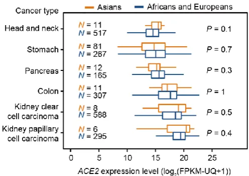
To address this, we further examined the potential Asian-biased expression of ACE2
in other cancer types. No such Asian-bias was detected for any cancer types with
higher ACE2 expression levels than LUAD, including stomach, colon, and kidney
cancers (Figure 2). Nevertheless, we cannot rule out the possibility that maybe ACE2 expresses at higher levels in Asians in some other as-yet-unexamined tissues.
A second caveat relates to our exclusive focus at the gene expression level; it is
possible that some Asian-specific genetic variations in the coding sequence of ACE2
may also affect the cell-entry efficiency of viruses. To explore the potential impact of
population-specific genetic variations, we retrieved all of the genetic variation data
available for the ACE2 locus from the 1000 Genomes Project (The 1000 Genomes
7
East Asians, Europeans, and South Asians. We focused on the polymorphisms in East
Asians, since both coronavirus epidemics broke out in East Asia. All 22 of the
missense or stop-gained variations were present at a low frequency among East
Asians (< 1%, Figure 3). Other variants (N = 690, Table S3) are mostly located in introns, and the frequencies of these variants among East Asians were highly
correlated with those among all populations (Pearson’s correlationcoefficient R =
0.996, P < 2.2×10⁻¹⁶, Figure 3). For instance, we found that among the 37 detected polymorphisms having an alternate allele frequency > 10% in East Asians, none (0
out of 37) had reached a frequency two times higher than all populations (Figure 3). We reached a similar conclusion about the lack of any East-Asian-specific enrichment
for ACE2 genetic variants based on analyzing data from the Genome Aggregation
Database (gnomAD, Figure S4) (Karczewski et al., 2019).
Another possibility is that other layers of regulation (e.g., translational efficiency,
protein modification, folding, or subcellular localization) exist in addition to
regulation at the mRNA level. While ACE2 protein levels are largely correlated with
mRNA levels among tissues (
https://www.proteinatlas.org/ENSG00000130234-ACE2/tissue) (Uhlen et al., 2015), whether these ACE2 proteins are located on the
cell membrane remains unclear. Therefore, future investigations should examine
potential race-specific bias for the accumulation of functional ACE2 on the plasma
membrane. It would also be informative to investigate if co-receptors of SARS-CoV-2
exist and, if so, to determine if their abundances vary among populations.The
identification of host cell features that affect the efficiency of viral amplification will
also help improve accuracy for predicting susceptibility to coronaviruses in the future.
Based on the available data, we conclude that Asians do not express ACE2 at a
higher level than other populations and do not bear unique genetic polymorphisms in
ACE2. The recurrent coronavirus outbreaks in China may be better explained by the
high diversity of coronaviruses and their animal hosts, or perhaps by Chinese food
culture (Fan et al., 2019). Our study, therefore, cautions against any use of race to
8
races require the same level of personal protection against SARS-CoV-2.
METHODS
All available open RNA-seq data and clinical data for LUAD samples in TCGA
were retrieved from https://www.cancer.gov/tcga. The RNA-seq data were obtained in
the FPKM-UQ (fragments per kilobase of transcript per million mapped reads of the
upper quartile gene) format, which is known to perform better in cross-sample
comparisons and differential expression analyses. Transcriptomes of additional East
Asian samples (Chen et al., 2020a) were retrieved fromOncoSG
(https://src.gisapps.org/OncoSG/) under the dataset “Lung Adenocarcinoma” (GIS,
2019); FPKM-UQ values for these samples were calculated from the numbers of
reads mapped to individual genes. Notably, the FPKM-UQ values of expressed genes
were globally higher in the data from OncoSG than from the TCGA data (Figure S5A); therefore, median normalization was further performed to help ensure
comparability of the expression levels from these two data sources (Figure S5B). The annotation of demographic information for each sample and statistical analyses were
performed using in-house R scripts. TCGA contains seven Asian LUAD samples and
does not further distinguish East Asians from South Asians in its annotation.
Nevertheless, after including the 260 Chinese LUAD samples, the majority (> 97%)
of Asian samples used in this study are from East Asians. Kaplan-Meier curves were
generated with UCSC Xena (http://xena.ucsc.edu) (Goldman et al., 2019).
Genetic variation data for the ACE2 locus from 26 different populations around
the globe were retrieved from the 1000 Genomes Project (The 1000 Genomes Project
Consortium, 2015) at the National Center for Biotechnology Information
(https://www.ncbi.nlm.nih.gov/variation/tools/1000genomes/). The allele frequencies
among East Asians were calculated from samples of the following populations:
Chinese Dai in Xishuangbanna, Han Chinese in Beijing, Southern Han Chinese,
Japanese in Tokyo, and Kinh in Ho Chi Minh City. The annotation for the genomic
9
consequences of variants (e.g., synonymous, missense, stop-gained, etc.) was
retrieved from the Ensembl Genome Browser (https://www.ensembl.org/index.html).
The allele frequencies for ACE2 in East Asians and in all populations were also
retrieved from gnomAD (https://gnomad.broadinstitute.org/).
DECLARATIONS Acknowledgments
We thank Drs. Di Liu (Wuhan Institute of Virology, CAS), Weiwei Zhai (Institute of
Zoology, CAS), Yuting Zhao (UT Southwestern Medical Center), and Guanlin Wang
(MRC WIMM Centre for Computational Biology, University of Oxford) for their
critical comments and suggestions on the manuscript. The results shown in this study
are in part based upon data generated by the TCGA Research Network
(https://www.cancer.gov/tcga), the GTEx Project (https://gtexportal.org/home/), the
1000 Genomes Project (https://www.internationalgenome.org/), and the Genome
Aggregation Database (https://gnomad.broadinstitute.org/).
Funding
This work was done by the authors during their holiday break and was not supported
by grants.
Authors’ contributions
Y.C., K.S., and W.Q. designed the study, performed data analyses, and wrote the
manuscript.
Competing interests
The authors declare that they have no competing interests. Code availability
All scripts used to analyze the data and to generate the figures are available at
10 REFERENCES
Chen, J., Yang, H., Teo, A.S.M., Amer, L.B., Sherbaf, F.G., Tan, C.Q., Alvarez, J.J.S., Lu, B., Lim, J.Q., Takano, A., et al. (2020a). Genomic landscape of lung
adenocarcinoma in East Asians. Nat Genet.
Chen, N., Zhou, M., Dong, X., Qu, J., Gong, F., Han, Y., Qiu, Y., Wang, J., Liu, Y., Wei, Y., et al. (2020b). Epidemiological and clinical characteristics of 99 cases of 2019 novel coronavirus pneumonia in Wuhan, China: a descriptive study. Lancet. Donoghue, M., Hsieh, F., Baronas, E., Godbout, K., Gosselin, M., Stagliano, N., Donovan, M., Woolf, B., Robison, K., Jeyaseelan, R., et al. (2000). A novel angiotensin-converting enzyme-related carboxypeptidase (ACE2) converts angiotensin I to angiotensin 1-9. Circ Res 87, E1-9.
Fan, Y., Zhao, K., Shi, Z.L., and Zhou, P. (2019). Bat Coronaviruses in China. Viruses 11.
Goldman, M., Craft, B., Hastie, M., Repečka, K., McDade, F., Kamath, A., Banerjee, A., Luo, Y., Rogers, D., Brooks, A.N., et al. (2019). The UCSC Xena platform for public and private cancer genomics data visualization and interpretation. bioRxiv, 326470.
Gu, J., Gong, E., Zhang, B., Zheng, J., Gao, Z., Zhong, Y., Zou, W., Zhan, J., Wang, S., Xie, Z., et al. (2005). Multiple organ infection and the pathogenesis of SARS. J Exp Med 202, 415-424.
Hamming, I., Timens, W., Bulthuis, M.L., Lely, A.T., Navis, G., and van Goor, H. (2004). Tissue distribution of ACE2 protein, the functional receptor for SARS
coronavirus. A first step in understanding SARS pathogenesis. J Pathol 203, 631-637. Hofmann, H., Geier, M., Marzi, A., Krumbiegel, M., Peipp, M., Fey, G.H., Gramberg, T., and Pohlmann, S. (2004). Susceptibility to SARS coronavirus S protein-driven infection correlates with expression of angiotensin converting enzyme 2 and infection can be blocked by soluble receptor. Biochem Biophys Res Commun 319, 1216-1221. Holshue, M.L., DeBolt, C., Lindquist, S., Lofy, K.H., Wiesman, J., Bruce, H.,
Spitters, C., Ericson, K., Wilkerson, S., Tural, A., et al. (2020). First Case of 2019 Novel Coronavirus in the United States. N Engl J Med.
Huang, C., Wang, Y., Li, X., Ren, L., Zhao, J., Hu, Y., Zhang, L., Fan, G., Xu, J., Gu, X., et al. (2020). Clinical features of patients infected with 2019 novel coronavirus in Wuhan, China. Lancet.
Jia, H.P., Look, D.C., Shi, L., Hickey, M., Pewe, L., Netland, J., Farzan, M., Wohlford-Lenane, C., Perlman, S., and McCray, P.B., Jr. (2005). ACE2 receptor expression and severe acute respiratory syndrome coronavirus infection depend on differentiation of human airway epithelia. J Virol 79, 14614-14621.
Karczewski, K.J., Francioli, L.C., Tiao, G., Cummings, B.B., Alföldi, J., Wang, Q., Collins, R.L., Laricchia, K.M., Ganna, A., Birnbaum, D.P., et al. (2019). Variation across 141,456 human exomes and genomes reveals the spectrum of loss-of-function intolerance across human protein-coding genes. bioRxiv, 531210.
11
Kuiken, T., Fouchier, R.A., Schutten, M., Rimmelzwaan, G.F., van Amerongen, G., van Riel, D., Laman, J.D., de Jong, T., van Doornum, G., Lim, W., et al. (2003). Newly discovered coronavirus as the primary cause of severe acute respiratory syndrome. Lancet 362, 263-270.
Li, Q., Guan, X., Wu, P., Wang, X., Zhou, L., Tong, Y., Ren, R., Leung, K.S.M., Lau, E.H.Y., Wong, J.Y., et al. (2020). Early Transmission Dynamics in Wuhan, China, of Novel Coronavirus-Infected Pneumonia. N Engl J Med.
Li, W., Moore, M.J., Vasilieva, N., Sui, J., Wong, S.K., Berne, M.A., Somasundaran, M., Sullivan, J.L., Luzuriaga, K., Greenough, T.C., et al. (2003). Angiotensin-converting enzyme 2 is a functional receptor for the SARS coronavirus. Nature 426, 450-454.
Lin, C., Song, H., Huang, C., Yao, E., Gacayan, R., Xu, S.M., and Chuang, P.T. (2012). Alveolar type II cells possess the capability of initiating lung tumor development. PLoS One 7, e53817.
Mele, M., Ferreira, P.G., Reverter, F., DeLuca, D.S., Monlong, J., Sammeth, M., Young, T.R., Goldmann, J.M., Pervouchine, D.D., Sullivan, T.J., et al. (2015). Human genomics. The human transcriptome across tissues and individuals. Science 348, 660-665.
The 1000 Genomes Project Consortium (2015). A global reference for human genetic variation. Nature 526, 68-74.
The Novel Coronavirus Pneumonia Emergency Response Epidemiology Team (2020). The epidemiological characteristics of an outbreak of 2019 novel coronavirus diseases (COVID-19) in China. Chinese Journal of Epidemiology 41, 145-151.
Uhlen, M., Fagerberg, L., Hallstrom, B.M., Lindskog, C., Oksvold, P., Mardinoglu, A., Sivertsson, A., Kampf, C., Sjostedt, E., Asplund, A., et al. (2015). Proteomics. Tissue-based map of the human proteome. Science 347, 1260419.
Wrapp, D., Wang, N., Corbett, K.S., Goldsmith, J.A., Hsieh, C.L., Abiona, O.,
Graham, B.S., and McLellan, J.S. (2020). Cryo-EM structure of the 2019-nCoV spike in the prefusion conformation. Science.
Xu, X., Chen, P., Wang, J., Feng, J., Zhou, H., Li, X., Zhong, W., and Hao, P. (2020). Evolution of the novel coronavirus from the ongoing Wuhan outbreak and modeling of its spike protein for risk of human transmission. Sci China Life Sci, 1674-7305. Xu, X., Rock, J.R., Lu, Y., Futtner, C., Schwab, B., Guinney, J., Hogan, B.L., and Onaitis, M.W. (2012). Evidence for type II cells as cells of origin of K-Ras-induced distal lung adenocarcinoma. Proc Natl Acad Sci U S A 109, 4910-4915.
Yan, R., Zhang, Y., Li, Y., Xia, L., and Zhou, Q. (2020). Structure of dimeric full-length human ACE2 in complex with B<sup>0</sup>AT1. bioRxiv,
2020.2002.2017.951848.
Zhao, Y., Zhao, Z., Wang, Y., Zhou, Y., Ma, Y., and Zuo, W. (2020). Single-cell RNA expression profiling of ACE2, the putative receptor of Wuhan 2019-nCov. bioRxiv, 2020.2001.2026.919985.
Zhou, P., Yang, X.L., Wang, X.G., Hu, B., Zhang, L., Zhang, W., Si, H.R., Zhu, Y., Li, B., Huang, C.L., et al. (2020). A pneumonia outbreak associated with a new
12 FIGURES
Figure 1. ACE2 expression levels are associated with age but not with sex or race,
among LUAD samples.
(A) Relationship between ageand ACE2 expression level. Pearson’s correlation coefficient (R) and the corresponding P-value are shown.
(B) Comparison of ACE2 expression between sexes. P-value was calculated with a two-tailed Mann-Whitney U test.
(C) Comparison of ACE2 expression among races. P-values were calculated with two-tailed Mann-Whitney U tests. The single sample of American Indian or Alaska Native in TCGA was not shown.
13
Figure 3. Allele frequencies of ACE2 variants detected by the 1000 Genomes Project, for East Asians (y-axis) or all populations (x-axis). Each dot represents a polymorphic site, and the frequency of the alternate allele is shown. Only dimorphic genetic variations are shown. To avoid negative infinity during the logarithm