Leigh A Maguire.
Processing and Characterisation of Liposomes for
Use in Gene Delivery.
Leigh Anthony Maguire.
University College London Department of Biochemical Engineering
All rights reserved
INFORMATION TO ALL USERS
The quality of this reproduction is dependent upon the quality of the copy submitted.
In the unlikely event that the author did not send a complete manuscript and there are missing pages, these will be noted. Also, if material had to be removed,
a note will indicate the deletion.
uest.
ProQuest U643231
Published by ProQuest LLC(2016). Copyright of the Dissertation is held by the Author.
All rights reserved.
This work is protected against unauthorized copying under Title 17, United States Code. Microform Edition © ProQuest LLC.
ProQuest LLC
789 East Eisenhower Parkway P.O. Box 1346
Leigh A Maguire.
Abstract
In recent years liposomes have received much attention for use in gene therapy and DNA vaccines. This project aims to investigate issues involved with the processing of liposome based gene delivery vectors and in doing so propose a controllable, reliable and scaleable method for processing such a vector. With this in mind a high velocity jet homogeniser was investigated for use in the preparation of small unilamellar vesicles (SUV). The device was able to produce SUV with a distribution having a mean of approximately 140nm following a single pass at 103.4 mNm'^: this size is suitable for sterilisation by filtration and in vivo gene delivery. By increasing either the number of passes through the device or the operating pressure it was possible to control the final size of the SUV between 80 and 140nm.
The liposomes produced were used to encapsulate plasmid DNA that had been condensed with poly-L-lysine (PLL). Charge ratio was determined to be a key processing criteria with a liposome: PLL/DNA charge ratio of 4 or above being required to form stable complexes. The liposomes encapsulated between 85 and 95% of the PLL/DNA with smaller liposomes being more effective.
charge. These effects were shown to result in reduced PLL/DNA encapsulation and stability.
Leigh A Maguire. Acknowledgements.
The acknowledgements page is often the most read part of a thesis and is therefore a source of much distress to the author. With this in mind I would firstly like to thank those whom actually read on. If you enjoy what you read why not tell your fiiends.
I would like to take this opportunity to thank Prof Peter Dimnill for giving me the opportunity to work in such an exciting field as gene therapy and Prof Parvis Ayazi Shamlou for his guidance over the past three years. I would also like to thank Dr Susana Levy, Dr Nick Murrell, Lee Kim Lee (now Dr) and Claire Mount for their advice and training especially in the early days of my studies.
Many people have made the last three years an enjoyable one and there is not enough space here to mention everyone but I would especially like to thank the members of the Colonnades office, especially Alison Kay for all those cups of tea, and my long suffering flat mates, Paul Griffiths, Ed Hibbert and Sam Pickering (thanks for listening but I found your work equally boring). I would like to apologise to anyone who may feel that they should have been included in this section but whom I have failed to mention, your contribution however small has been appreciated. Finally I would like to thank my parents without whose support none of this would have been possible. I love you both.
Contents
1. Introduction and Literature review,
1.1 What is gene therapy L 1. L Potential of gene therapy 1.1.2. DNA Vaccines
Page
17 17 17 19
1.2. Methods of delivery 21
1.3. Obstacles to gene delivery. 22
1.3.1. Access to target cells. 23
1.3.1.1. Surface charge 24
1.3.1.2. Size 25
I.3.I.3. Site of administration 25
1.3.2. Complement System 26
1.3.3. Crossing the plasma membrane 26
1.3.4. Endosomes 28
1.3.5. Vector unpacking and stability in the cytoplasm. 29
1.4. Transfection vectors 30
1.4.1. Viral Vectors 30
1.4.1.1. Retroviruses 31
1.4.1.2. Adenoviruses 32
Leigh A Maguire.
1.4.1.4. Disadvantages of viral vectors. 33
1.4.2. Non-viral vectors 34
1.4.3. Plasmids. 36
1.4.3.1. Production of plasmids. 36
1.4.3.2. Characterisation of plasmids 38
1.4.4. Types of non viral vector. 41
1.4.4.1. Cationic lipids. 42
1.4.4.2. Self assembling vectors. 43
1.5. Liposomes 45
1.5.1. Types of Liposomes. 49
1.5.2. Preparation of Liposomes 53
1.5.2.1. Sonication. 53
1.5.2.2. Homogenisation. 54
1.5.2.3. Microfluidisation 54
1.5.2.4. Extrusion 55
1.5.3. Complexing DNA with liposomes. 56
1.6. Immimoliposomes 59
1.6.1. Antibody Fragments. 59
1.6.1.1. Fermentation of Fab’ 61
1,6.1,2. Processing of Fab’ 63
1.6.2. Covalent attachment of Fab’ to liposomes. 65
1.7. Characterisation of Liposomes and Immimoliposomes 69
1.7.1. Size and Zeta potential 69
1.7.2. Lamellarity and Chemical composition. 70
1.8. Stability. 71
1.8.1. Steric stabilisation. 72
1.9. Project backgroimd and aims. 74
2, Materials and Methods. 77
2.1. Plasmid DNA 77
2.2. Pico Green Double Stranded DNA assay 77
2.3. Fab’ 78
2.4. ELISA 79
2.5. Preparation of PLL/DNA 80
2.6. Preparation of MLV 80
2.7. Sonication 81
2.8. The High Velocity jet homogeniser 81
2.9. Mixing liposomes and PLL/DNA 82
2.10. Preparation of Immunoliposomes 82
Leigh A Maguire.
2.12. Determination of zeta potential 83
2.13. Determination of encapsulation efficiency 84
2.14. Determination of surface tension 84
3. Characterisation of a high velocity
Jet homogeniser for downsizing MT V, 86
3.1 Introduction 86
3.2 Suitability of high velocity jet homogeniser 89 for preparation of SUV
3.3 Control of liposome size 96
3.4 Potential Draw backs 102
3.5 Mechanism of disruption 105
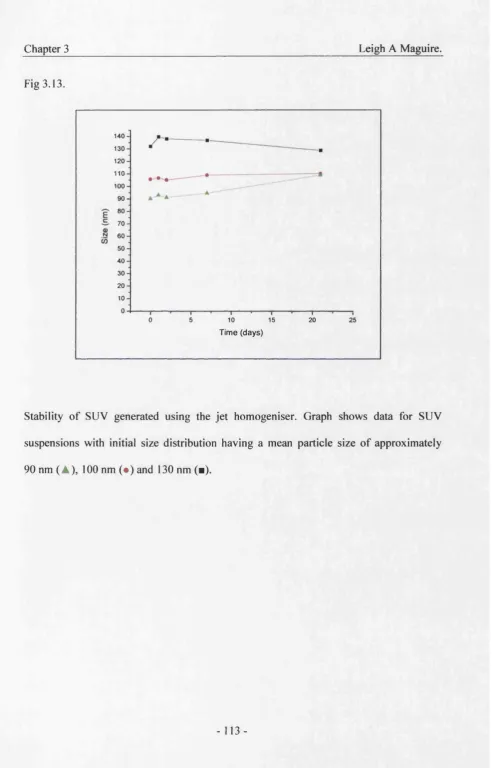
3.6 Stability 111
3.7 Summary 114
4. Liposome encapsulated poly-L-lysine 115
condensed plasmid DNA complexes.
4.1. Introduction 115
4.2. Effects of Charge ratio 117
4.3. Mixing speed 123
4.4. Maturation effects. 125
4.6. Stability 132
4.7. Summary 136
5. Effect of attachment of humAb4d5 Fab’ 138
to liposomes upon processing.
5.1. Introduction 138
5.2. Effect of Fab’ upon size. 140
5.3. Effect of Fab’ upon zeta potential 146
5.4. Effect of Fab’ upon encapsulation efficiency. 150
5.5. Effect of Fab’ upon stability. 153
5.6. Stability of Immunoliposomes in serum 158
5.7. Summary 161
6. Addition of PEG to liposomes. 163
6.1. Introduction. 163
6.2. Effect of PEG upon size. 165
6.3. Zeta potential 167
6.4. Effect of PEG upon encapsulation of PLL/DNA. 170 6.5. Effect of PEG upon surface tension of liposomes. 178
6.6. Summary. 183
7. Discussion and Conclusions. 184
Leigh A Maguire. 7.2. Downsizing MLV
73. Encapsulation of PLL/DNA. 7.4. Storage,
7.5. Gene transfer. 7.6. Conclusion.
186 189 196 199
201
8, Future Work, 205
9. Appendix,
A1. Standard Curves
A2. Calculation of charge ratio.
A3. Fig 4.4 as a function of Lipid: DNA ratio rather than charge ratio
A4. Table of surface tension of biopolar buffers. A5. Publications
208 209 212 213 214 215
10. References. 225
Tables,
1.1 Monogenic diseases being investigated.
1.2 Ligands being investigated for gene therapy
13 Characterisation ofcontaminants of plasmid processing 38
1.4 Liposomal drugs 46
1.5 Properties of commonly used lipids 48
1.6 Purification of proteins 64
A l. Calculation of Charge ratio
A2. Surface tension of biopolar buffers.
Figures.
1.1. Ex Vivo vs in vivo gene therapy 22
1.2. Barriers to gene delivery 23
1.3. Cationic lipids 43
1.4. Bolasome 44
1.5. Common lipids used to make liposomes 50
1.6. Types of Immunoliposome 52
1.7. Complexing DNA with anionic liposomes 58
1.8. An Antibody 60
1.9. Formation of an anchor lipid 67
1.10. Attachment of proteins to liposomes 68
3.1. Schematic of the high pressure jet homogeniser, 88 3.2. Distribution profile of MLV and SLTV 90
Leigh A Maguire.
3.4. Cycles of sonication on turbidity of suspension 93 3.5. Distribution profile following filtration. 95
3.6. Multiple passes at 103.4mNm'^ 98
3.7. Multiple passes at 135.9mNm‘^ 99
3.8. Multiple passes at 172.4mNm'^ 100
3.9. Reproducibility of control 101
3.10. Loss of lipid 104
3.11. CFD of jet energy dissipation rate 107
3.12. liposome size as a fimction of energy dissipation rate 110
3.13. Stability of liposomes 113
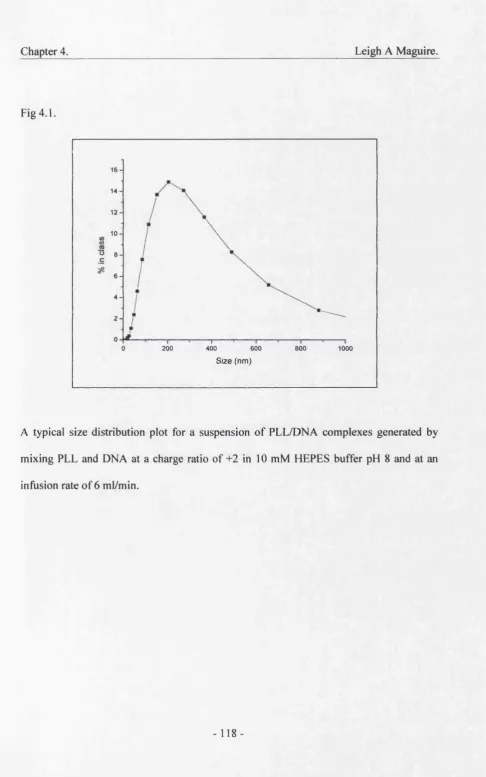
4.1. Distribution of PLL/DNA 118
4.2. Charge ratio upon size distribution 120
43. Charge ratio upon stability 121
4.4. Charge ratio upon zeta potential 122
4.5. Mixing speed 124
4.6. Maturation of PLL/DNA 126
4.7. Effect of initial liposomes size upon complex size 129
4.8. Effect of initial liposome size upon encapsulation efficiency 131
4.9. Effect of serum upon mean size 133
4.10. Effect of serum upon distribution, 134
5.2. Effect of complex formation 145
5.3. Effect of Fab upon zeta potential 148
5.4. Effect of Fab’ upon complex zeta potential 149 5.5. Effect of Fab’ upon encapsulation of PLL/DNA 151 5.6. Theoretical predictions of complex stability 155
5.7. Stability of complexes 157
5.8. Stability in serum 159
5.9. distribution following addition of serum 160
6.1. Effect of PEG upon size distribution of SUV 166 6.2. Effect of PEG upon zeta potential of SUV 168
6.3. Stability of PEG 2000 liposomes 169
6.4. Size d istributions of complexes 171
6.5. Stability of complexes short term 172
6.6. Stability of complexes long term 173
6.7. Zeta potential of complexes 176
6.8. Encapsulation efficiency of PEGylated liposomes 177
6.9. Surface tension of liposomes 179
6.10. Effect of ionic strength on liposomes 182 7,1, Energy plots showing effect of Fab’ upon 195
Leigh A Maguire. A3. Standard curve for protein assay.
Abbreviations.
CF, Cystic fibrosis Choi, Cholesterol ctDNA, Calf thymus DNA DNA, Deoxyribonucleic acid DOPC, Diolyoil phosphotidylcholine DOPE, Diolyoil phosphotidylethanolamine EIAV, Equine Infectious-Anaemia Virus ELISA, Enzyme linked Immunosorbant Assay. EMCA, European Medicines Control Agency FDA, American food and drug administration HA, Haemagluttinin
HIV, Human Immunodeficiency Virus
humAb4D5 Humanised version of the monoclonal antibody 4D5 . LUV, Large Unilamellar Vesicle.
MCA, Medicines Control Agency MLV, Multilamellar Large Vesicle.
munAb4D5 Murine version of the monoclonal antibody 4D5,
OA, Oleic acid
PCS, Photon Correlation Spectroscopy,
p DNA Plasmid DNA
PE, Phosphotidylethanolamine PEG, Polyethylene glycol
PE-MPB, Phosphotidylethanolamine-maleimido-phenyl butyrate PLL, Poly-L-lysine
RES, Reticuloendothelial system RNA, Ribonucleic acid
SDS, Sodium dodecyl sulphate
SIV, Simian Immunodeficiency Virus
SMPB, N-succinimidyl (-4-[P-maleimido-phenyl] butyrate) SUV, Small Unilamellar Vesicle
Tc, Transition temperature
tEM, Transmission electron microscopy .
Notation. Go 7 ; n p ai, a:
extinction coefficient of proteins at A isonm surface/interfacial tension (mNm"^)
zeta potential (mV)
Kolmogoroff length scale (m) viscosity (Kgm‘*)
density (Kgm'^s'^^
Leigh A Maguire. A Hamaker constant (J)
c ionic concentration (M)
e charge of an electron (1.6 x 10'^^ C)
H separation distance between two primary particles (m) k Baltzmann’s constant (1.381 xlO'^^ J/K)
T absolute temperature (K)
Va» Vr, Vt interaction energies (Van der Waal’s attraction, electrical repulsion and
total), (J)
X dimensionless (= (H/ai + a:) y dimensionless (= ai + a:)
z valence (charge niunber) of the ionic species
yi, 72 dimensionless functions of zeta potentials of particles 1 and 2 in Eq 5.3
p permittivity, dimensionless
1. Introduction and Literature Review.
1.1. What is Gene Therapy?
Gene therapy is defined as a technology aimed at modifying the genetic component of cells for a therapeutic benefit (Kim et al, 2002). In the case of monogenic heritable disorders, such as cystic fibrosis (CF), if the normal gene product could be expressed the abnormal biological phenotype of the individual could be corrected (Crystal, 1995).
1.1.1. Potential of Gene Therapy.
Currently clinical trials are underway in areas as diverse as HIV infection, venous leg ulcer treatment, screening for genetic disorders, several different cancers, neuroblastoma, Gaucher’s disease and sickle cell disease to name a few (see http://clinicaltrials.gov/ct/gui).
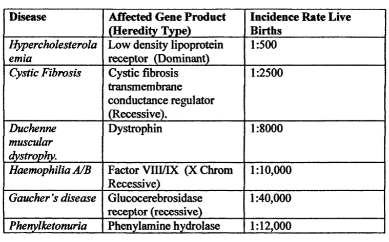
Chapter 1, Leigh A Maguire. Table 1.1. Single gene disorders under investigation for somatic gene therapy.
Disease Affected Gene Product (Heredity Type)
Incidence Rate Live Births
Hypercholesterola emia
Low density lipoprotein receptor (Dominant)
1:500
Cystic Fibrosis Cystic fibrosis transmembrane conductance regulator (Recessive). 1:2500 Duchenne muscular dystrophy. Dystrophin 1:8000
Haemophilia A/B Factor VIII/IX (XChrom Recessive)
1:10,000
Gaucher's disease Glucocerebrosidase receptor (recessive)
1:40,000
Phenylketonuria Phenylamine hydrolase 1:12,000 Adapted from Gottschalk & Chan (1998).
Mutations of the tumour suppressor gene p53 are among the most common in many human cancers. Due to its critical involvement in the cell cycle and apoptotic signalling p53 is the most extensively studied tumour suppressor gene and therefore appears to be an appealing target for gene therapy trials (Zeimet et al, 2000). The re-expression of functional p53 in tumour cells has been shown to suppress tumour growth and induce apoptosis.
One of the more recent uses of gene therapy is in the treatment of cardiovascular disease. Of the 36 protocols currently registered with the National Institutes of Health Office of Biotechnology Activities the vast majority involve the delivery of vascular endothelial growth factor or fibroblast growth factor to enhance angiogenesis for the treatment of coronary artery and peripheral vascular diseases (Liau et al, 2001).
1.1.2. DNA Vaccines.
Traditional vaccines consist of primarily killed, weakened or components of pathogens. They function by priming the immune system to fight dangerous viruses, bacteria and parasites before they can get a foothold by tricking the immune system into behaving as though the body has already been attacked by the pathogen.
Chapter 1._______________________________________________ Leigh A Maguire. The second element, the cellular response, is carried out primarily by cytotoxic T-lymphocytes. These recognise antigens expressed on the surface of cells infected by a pathogenic organism and kill the cell. Once an antigen has been encountered by the immune system some B-lymphocytes differentiate into “memory cells” which remember the encounter so that if the pathogen is encountered again the response will be quicker and more effective.
For an efficient immune response both arms are required. However, with traditional vaccines composed of killed pathogens or their components, only the humoral response is generated, as there is no cellular infection. Using live attenuated pathogens does generate a cellular response in addition to a humoral one, however, they can still generate disease in immune deficient individuals and there is also a small chance that a mutation may occur potentially resulting in the organism regaining its pathogenicity.
Genetic vaccines under development are composed of plasmids containing genes that encode antigenic proteins. They are delivered to cells, usually muscle cells, by microinjection and once in the nucleus synthesise the antigen which may be either secreted out of the cell, resulting in a humoral response, or are displayed on the surface of the cell thus generating a cellular response. In this way DNA vaccines can possess the advantages of attenuated vaccines without the risks (Weiner & Kennedy, 1999).
possess outer capsules composed of polysaccharides. As DNA only codes for proteins, DNA vaccines cannot be substitutes for polysaccharide based vaccines (e.g. pneumococcus) (Robinson et al, 1996). Many proteins are modified during their synthesis (e.g. by glycosylation) and these modifications are specific to the type of organism. Therefore animal cells may not be able to correctly manufacture the proteins of parasites and bacteria. However, as viruses use the host cells machinery for protein synthesis DNA vaccines will result in the correct production of the viral protein (Robinson a/., 1996).
1J2, Methods of Delivery.
Gene therapy requires the successful delivery and expression of an “expression cassette” incorporating the gene plus promoter and regulation sequences, to the target cell (Gottschak & Chan, 1998; Crystal, 1995). Two methods have been developed, in vivo
Chapter 1. Leigh A Maguire.
Figl. l.
Comparison ot ex vivo and in vivo gene therapy
E x iivo transfection
□
n
PatientbionsvD D
Producer cell line
WÎ 0 difie d retr ovirus
O C)
I Cells rem eve d ft om p atient and transfected w ith modified virus.
Implantation
Plasmid DNA f f vivo
transfection
Ini acted into patient.
Adapted from M arquetef a t, (1995)
O
DD
1.3. Obstacles to the Effectiveness of Gene Delivery Vectors.
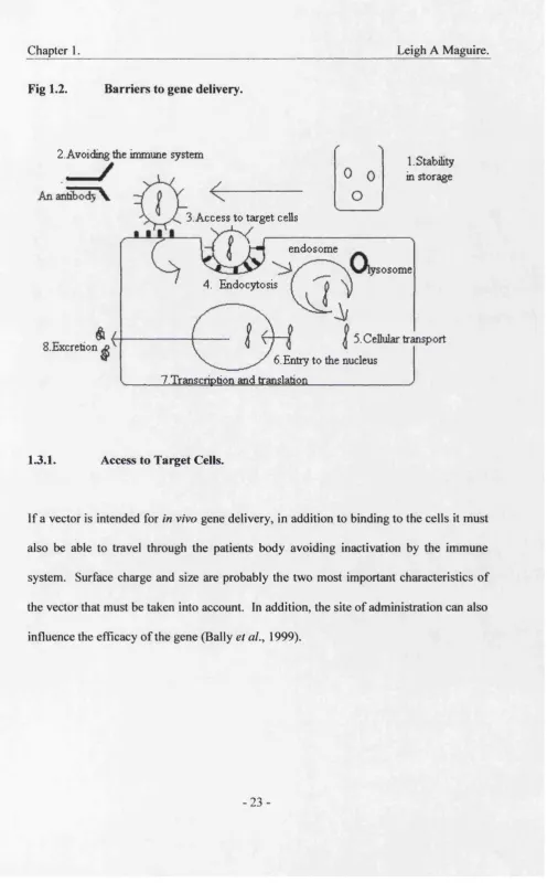
The delivery of DNA to ceils usually requires a vector (see 1.4 and 1.5). The purpose of vectors is to improve the therapeutic activity of the gene by aiding in overcoming barriers that block the eventual successful expression of the therapeutic gene in the desired cells. These barriers include agents that will damage the DNA, such as nucleases; access to target cells; the plasma membrane; endosomes; nuclear transport; transcription and translation ( see fig 1.2.).
-Fig 1.2. Barriers to gene delivery.
2. Avoiding the immune system
1. Stability in storage
3. Access to target cells
endosome
sosome 4. Endocytosis
5. Cellular transport .Excretion
6. Entry to the nucleus 7.Transcription and translation
1.3.1. Access to Target Cells.
Chapter 1._______________________________________________ Leigh A Maguire.
13.1.1. Surface Charge.
1.3.1.2. Size.
The most obvious effect of size is that vectors of a large size (>200 nm) will be trapped in the small capillaries of organs such as the lungs and liver, resulting in their accumulation in them. Also vectors greater than 200 nm in size will be rapidly taken up by the cells of the RES following administration by intravenous injection (Bally et al, 1999). For vectors taken up into cells by endocytosis the size of the endosome is of importance. The endosome of lymphocytes is approximately 100 nm. However it has been shown that in vitro larger lipoplexes (DNA/cationic liposome complexes) tend to associate and be taken up by cells more efficiently than smaller lipoplexes resulting in greater levels of transfection (Ross & Hui, 1999).
13.1.3. Site of Administration.
Chapter 1._______________________________________________ Leigh A Maguire.
al., 1997). Tumours with leaky blood vessels will also accumulate systemically administered vectors especially if they are long circulating.
13.2. The Complement System.
The complement system is group of blood proteins involved in the lysis of foreign cells and the removal of particles by phagocytes, which may pose a barrier to gene delivery by increasing vector elimination from the blood stream. The action of the complement system is through an enzyme cascade triggered either by antibodies attached to the surface (the classical pathway) or certain initiating surfaces. The complement system can be activated by cationic materials such a poly-L-lysine, dendrimers, and polyethyleneimine. The potency of the activation is chain length dependent. Coating vectors with polyoxyethylene has been shown to prevent activation of the complement system (Pouton & Seymour 2001).
13.3. Crossing the Plasma Membrane
1994). A variety of such ligands are currently under investigation for use in gene therapy (see Table 1.2.).
Table 1.2. Ligands currently being investigated for use in receptor mediated gene delivery.
Ligand Target Cells
Asialoglycoprotein Hepatocytes
Hepatoma HepG2
Transferin Various
Insulin Hepatoma
Polymeric Immunoglobulin Respiratory Hepatic epithelium
EGF*^ Carcinoma
Adapted from Mahato et al, (1997).
The process by which the ligand/DNA complexes are internalised is by receptor mediated endocytosis into clathrin coated vesicles (Perales et a l, 1994), Receptor mediated gene transfer is not completely selective. Specific receptors are often found on more than one type of cell. This means that any strategy using receptors should consider the possibility of expression of the gene in tissues other than the desired target tissue (Perales et a l,
Chapter 1._______________________________________________ Leigh A Maguire. 13.4. Endosomes.
Most molecules taken up by cells via receptor mediated endocytosis are subject to the cell’s degradation mechanisms. This bottleneck in gene delivery can be responsible for the degradation of over 99 % of internalised DNA (Ogris & Wagner 2002). The cell internalises the molecule into clathrin coated endosomes. Lysosomes fuse with the endosome and the enzymes within them enter the endosome and digest its contents. The optimum pH of lysosomal enzymes is pH 5. In order to lower the pH of the endosome to pH 5, protons (H^ enter the endosome causing its internal pH to fall. Stability to lysosomal enzymes and an ability to escape the endosome have been found to be critical for the expression of the therapeutic gene ( Perales et al., 1994 ).
The study of viral escape mechanisms fmm endosomes may help in the design of synthetic vectors. Wagner and co-workers (Wagner et al, 1992) have demonstrated that incorporating the haemaglutinin (HA) subunit HA-2 from Influenza viruses into a DNA- transferin-poly-L-lysine) complex increases gene expression. However, a potential drawback to using viral proteins in non-viral vectors is the possibility of introducing immunogenicity to the vector complex (Perales et al., 1994).
proteins or other macromolecules for fusion to other membranes. The fusion of the liposome and endosomal membrane is triggered when the pH in the endosome is about pH 5 and is believed to be due to changes in the surface charge of the liposome enabling it to bind to and disrupt the endosomal membrane allowing release of its contents into the cytoplasm (Wang & Huang, 1987). Poly-L-lysine) complexes have been shown to act in a similar way (Perales et al., 1994).
1.3.5. Vector Unpacking and Stability in the Cytoplasm.
Plasmid unpacking has recently been shown to be a limiting factor in gene expression. Short term expression has also been shown to be significantly enhanced by using short chain polycations that dissociate fiom the plasmid DNA more quickly than larger molecular weight polymers (Schaffer et al, 2000).
Chapter 1._______________________________________________ Leigh A Maguire.
1.4. Transfection Vectors.
The ideal vector should be: 1. Stable in vitro and in vivo,
2. Efficient at delivering the gene.
3. Able to protect the gene from degradation. 4. Specific to the target cell.
5. Non toxic and non immunogenic
6. Available in large quantities.
7. Susceptible to control.
Unfortunately no vector developed to date fulfils all of these requirements (Deshmukh & Huang, 1997). Two strategies have been developed, viral and non-viral gene delivery.
1.4.1. Viral Vectors.
1.4.11. Retroviruses.
Retroviruses such as murine leukeimis virus are RNA viruses that possess the enzyme reverse transcriptase. Retroviruses transfer their genetic material directly into the genome of the host cell. This makes them useful for the treatment of chronic disorders. However, it also makes them potentially oncogenic (Crystal, 1995). Because they incorporate DNA into the host genome, retroviruses require DNA replication (and therefore dividing cells) for the integration of the provirus genome. For this reason they have mostly been used in ex vivo trials (Perales et al, 1994). Research on the development of vectors based on lentiviruses, a sub family of retroviruses which do not integrate their DNA into the host genome and can infect non dividing cells, (such as Human Immunodeficiency Virus (HTV), Simian Immunodeficiency Virus (SIV) and Equine Infectious-Anaemia Virus (EIAV)) may allow in vivo use of retroviral vectors (Robbins er ût/., 1998).
Chapter 1.________________________________________________Leigh A Maguire. 1.41.2. Adenoviruses.
Adenoviruses are DNA viruses and deliver their genome in an episomal form (i.e. they do not incorporate their genome into host chromosomes), they therefore do not require cells to be dividing in order for the genome to be expressed. They can also be produced by cell lines in grater titres than retroviruses (Crystal, 1995). Adenoviruses have a high tropism for cells of the respiratory system making them attractive as vectors for these cells (Gottschalk & Chan, 1998).
The genome of adenoviruses can be classified into early (E1-E4) and Late (L1-L5) genes. The early genes are involved in regulating viral and host cell gene expression, viral replication and inhibition of apoptosis while the late genes are required for encapsulation of the virus (Ribbins et al, 1998). The gene required by the virus to direct production of other adenoviruses is El. For the production of adenoviral vectors the El gene is deleted and replaced by the therapeutic gene and the replication defective virus can be propagated in a packing cell line that provides the El gene polypeptides.
1.4.13. Other Viral Vectors.
Adeno associated viruses have also been studied for use as gene therapy vectors. These viruses are attractive as they insert their genome into a specific region of chromosome 19. However, the size of the expression cassette that can be delivered is small, ~5kb. Also in order to infect cells they require co-infection by adenoviruses or herpes viruses (Ribbins
e ta l, 1998).
Herpes-simplex virus type I and II are also of interest in gene therapy. Herpes-simplex viruses are large DNA virus that can infect a wide variety of cells including neurones, muscle, tumours, liver and pancreatic islets. One advantage of herpes-simplex is that it has a large capacity for the insertion of the therapeutic gene (up to 50kb) (Ribbins et al,
1998).
1.4.14. Disadvantages of using Viral Vectors.
There are several disadvantages to using viral vectors. These
include:-1. Generation of an immune response resulting in an inability to administer more than once.
2. Random integration of viral genes into host chromosome presenting risk of oncogenesis.
3. Clearance of systemically delivered viruses by the immune system.
Chapter 1._______________________________________________ Leigh A Maguire. 5. Possible recombination of viral vector DNA with wild type viral DNA resulting in
generation of infectious viruses.
6. High cost of producing large quantities.
7. Can only deliver small expression cassette, approximately 10 kbp due to the large amount of DNA that must remain for the virus to still be able to infect cells.
(Templeton & Lasic, 1999).
1AJ2. Non Viral Vectors.
Non-viral vectors are man made and include chemical (calcium phosphate precipitation); mechanical (micro-injection); membrane fusion (liposomes) and direct uptake into cells by receptor mediated endocytosis (Morgan & Anderson, 1993).
The advantages of non-viral vectors
include:-1. Reduced immunogenicity;
2. Reduced clearance by complement system; 3. Can deliver a larger expression cassette;
4. Greater potential to perform multiple administrations in vivo than viral vectors; 5. Potential to target cells;
The main disadvantages of non viral vectors are their relatively low levels of delivery and short duration of gene expression compared to viral vectors (Templeton & Lasic, 1999), while many are not suitable for clinical applications due to their lack of regulated and sustained expression of foreign genes in targeted cells (Gottschalk & Chan, 1998).
Even though non-viral vectors are regarded as being potentially safer than viral vectors they still posses some safety concerns. Most of the health risks surrounding non-viral gene therapy are to do with the potential to induce cytotoxic and immune reactions in the recipients. Both the DNA itself and its vector may have the potential to be responsible. The unmethylated CpG motifs of bacterial DNA (plasmids are produced in bacteria) have been shown to trigger an immune response and production of cytokines (Mclachlan et al
2000). Some liposomal drugs have been shown to trigger a hypersensitivity reaction, which includes cardiopulmonary distress. Studies on miniature pigs have shown that the reaction is complement mediated and has been termed “complement activation-related pseudoallergy” (Szebeni et al, 1999). Cationic lipids are not found in nature and cationic liposome/DNA complexes remain quite cytotoxic, as they appear to be incompatible with the physiological environment, which contains mostly anionic molecules (Lee & Huang,
Chapter 1.________________________________________________Leigh A Maguire.
1.43. Plasmids.
Before discussing non-viral vectors further it is worth introducing plasmids, as it is these entities that the majority of non viral vectors have been developed to deliver to the target cells. Plasmids are circular duplex DNA molecules found in bacteria which encode extra genes for none essential proteins but which may convey an advantage upon the possessor such as antibiotic resistance. Plasmids can be easily manipulated to encode none bacterial products. At present most gene therapy trials with plasmid DNA (pDNA) are using plasmids in the order of 10 kbp. However, it is reasonable to predict that this size will increase over the coming years to several hundred or more kilo base pairs enabling delivery of larger genes, multiple genes or even whole pathways.
1.43.1. Production of Plasmid DNA.
Because of the effects of shear, chemical lysis of the cells to release the plasmids is usually used in preference to mechanical methods. A typical method is the lysis of cells using a solution of SDS and sodium hydroxide (Schleef, 1999). This alkaline suspension is subsequently neutralised by addition of acidic potassium acetate (pH 5.5). This high potassium acetate concentration causes renaturation of the plasmids and aggregation of chromosomal DNA, proteins, membrane components and SDS, which forms a floe (Schleef, 1999). Heat treatment for 5 min at 95 “C has also been shown to give DNA release comparable to alkaline lysis. This method has the added advantage of producing a lysate that is less viscous than that obtained by alkaline treatment (Wang et al., 2002).
Centrifugation can be used in the laboratory to remove the floe or alternatively purification can be carried out using a caesium chloride/ethidium bromide density gradient ultra centrifugation. This method however is unacceptable for production of clinical material because it uses mutagenic materials and is unscaleable (Marquet et al,
Chapter 1. Leigh A Maguire. 1.4 J.2 . Characterising pDNA
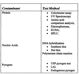
In order to proceed to advanced clinical trials the pharmaceutical development of gene therapy products will have to pass stringent characterisation criteria. The American Food and Drug Administration (FDA) have prepared guidelines that can be found at their web site (http://www.fda.gov/). Important points to consider include the levels of contaminants such as bacterial chromosomal DNA, RNA, Proteins, endotoxins/pyrogens, viruses as well as contaminants picked up during the fermentation and purification processes (some of the testing methods for these contaminants are summarised in Table
1.3.). Other characteristics include the stability and the physical properties of the pDNA itself.
Table 1.3. Characterisation of Process Contaminants.
Contaminant
Test Method
Protein • Colorimeter assay
• UV Spectroscopy • Amino acid
comparison analysis, • Electrophoreses, • ELISA,
• HPLC.
Nucleic Acids DNA hybridisation
• Southern blot • Dot blot
Polymerase chain reaction
Pyrogens • USP pyrogen test
• LAL
assay
Volatile substances
Residuals • Residual Moisture
Solvents
• Gas chromatography mass spectroscopy Metals/Salts
• plasma emission spectroscopy. • atomic absorption
spectroscopy. Adapted from Warqm tetal, 1997b
It is recommended that the final plasmid DNA product should contain no more than 100 pg cellular DNA per dose as determined by a method with a sensitivity of 10 pg (DiPaolo et al, 1999). The monitoring of nucleic acid contamination is generally carried out by hybridisation analysis. The levels of host cell DNA can be monitored by Southern blot (Marquet e/a/., 1997b).
Chapter 1.________________________________________________Leigh A Maguire. Pyrogens are molecules that induce the histamine response resulting in symptoms such as fever and chills. Endotoxins are pyrogenic lipopolysaccharides derived fix>m gram negative bacteria. E. coli is a gram negative bacteria and any system using it must therefore be assessed for contamination by endotoxins. The usual assays for endotoxin levels are the United States Pharmacopeia rabbit pyrogen assay and the limulus
amebocyte lysate assay. The rabbit pyrogen assay will detect any pyrogen and often gives false results, so both assays are used (Dipaolo et al, 1999).
Any process that utilises materials derived from animal or especially human sources poses the risk of contamination by viruses, which may be harmful to the recipient. Virus removal can be accomplished by chromatography or filtration while viruses can be inactivated by low pH. Viral contamination can be assessed by transmission electron microscopy (tEM), polymerase chain reaction, cytopathic effect and reverse transcriptase assays (Dipaolo et al, 1999). It is necessary for the purification to be shown to be able to clear more virus than is actually estimated to be present in a single dose. Viral removal must still be monitored in processes that do not include miimal products due to risk of contamination by personel.
(Dipaolo, et al, 1999). The FDA in “points to consider on plasmid DNA vaccines for preventative infectious disease indications” point out that different plasmid forms may be less effective than the supercoiled form and that a minimum amount should be agreed,
1.4.4. Types of Non-Viral Vectors.
The majority of non-viral vectors utilise molecules with the ability to condense DNA into small particles able to enter cells through endocytosis, for example with poly cations such as poly-L-lysine (PLL) (Choi et al, 1999; Deshmukh & Huang, 1997). Cellular membranes are either negatively or neutrally but never positively charged. Therefore such positively charged complexes will bind unspecificly to cellular membranes (Deshmukh & Huang, 1997). One advantage of PLL over other polycations is that it can be cross-linked to a ligand or antibody so that the complex can target specific cells (Perales et al, 1994). This is known as receptor mediated gene transfer. However, the polydispersity of PLL synthesis leads to variable DNA delivery and difficulty in forming DNA complexes (Lou & Saltzman, 2000).
Chapter 1._______________________________________________ Leigh A Maguire. Imidazole containing polymers such as polyhistadine have also shown some promise (Pack et al, 2000).
1.4.41. Cationic Lipids.
Cationic lipids have also been extensively studied and have reached clinical studies for cancer and cystic fibrosis. They have proven to be safe in relatively low doses, however no long term safety studies have been carried out (Li & Huang, 2000). The invention of cationic lipids has lead to the development of cationic liposomes, which are of great interest in gene therapy. The cationic charge of the liposome neutralises the anionic charge of the DNA condensing the DNA into a more compact structure. Cationic liposomes form complexes with, rather than encapsulate DNA (see 1.5.3.).
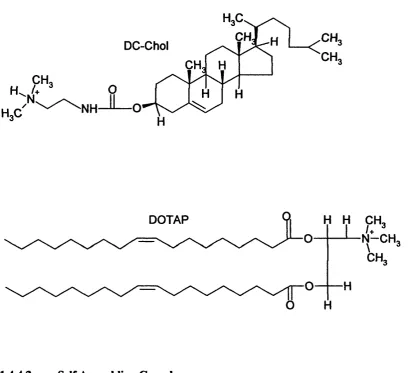
Fig 1.3. Cationic lipids.
OH OH;
CH. OH
CM
NH MX
DOTAP H H
N -C H
1.4.4.2. Self Assembling Complexes.
A self assembling vector is attractive for a number of reasons. Firstly, self assembly implies the innate ability to form discrete ordered structures of a homogenous population of defined units. Secondly such properties are compatible with a reproducible and scaleable process. A novel range of bipolar amphiphilic (BOLA) lipids (Eaton et al,
Chapter 1._______________________________________________ Leigh A Maguire. as plasmid DNA, to form stable complexes in aqueous solutions. The lipid forms a hydrophobic layer to condense the DNA and stabilise the resulting particle while the hydrophilic group provides a shell around the particle, which reduces non specific interactions. The hydrophilic head may be PEG, which is often used to sterically stabilise vectors (see 1.8.1.) or a polysaccharide. An example of a BOLA lipid can be seen in fig 1.3. below. In addition the BOLAsome can be targeted to specific cells by the attachment to the distal end of the hydrophilic tail of a targeting moiety such as an antibody fingment.
Fig 1.4.
u cationic head
Hydrophilic tail ^H ydrophobic back bone
"
à''M'
An exam ple of a pegylated BOLA lipid
1.5. Liposomes.
Liposomes are colloidal particles that can entrap macromolecules such as nucleic acids (Templeton & Lasic, 1999). They are composed of lipids (most commonly phospholipids) arranged in bilayers enclosing an aqueous space. Liposomes are able to encapsulate a wide variety of water soluble molecules ranging in size fiom small ions to macromolecules such as proteins and polynucleotides ( Sulivan et al, 1986 ).
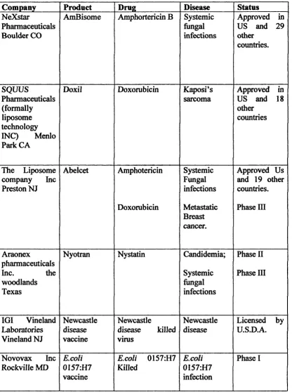
Chapter 1. Leigh A Maguire. Table 1.4. Adapted from Flourence, (1999)
Company Product Drug Disease Status
NeXstar
Pharmaceuticals Boulder CO
AmBisome Amphortericin B Systemic hingal infections
Approved in US and 29 other countries. SQUUS Pharmaceuticals (formally liposome technology INC) Menlo Park C A
Doxil Doxorubicin Kaposi’s
sarcoma
Approved in US and 18 other
countries
The Liposome company Inc Preston NJ Abelcet Amphotericin Doxorubicin Systemic Fungal infections Metastatic Breast cancer.
Approved Us and 19 other countries. Phase III
Araonex
pharmaceuticals
Inc. the
woodlands Texas
Nyotran Nystatin Candidemia;
Systemic fungal infections
Phase II Phase in
IGI Vineland Laboratories Vineland NJ Newcastle disease vaccine Newcastle
disease killed virus
Newcastle disease
Licensed by U.S.D.A.
A wide variety of lipids can be used to form liposomes. Some examples are shown in Table 1.5. Phospholipids form stable structures that undergo a characteristic gel-liquid crystalline phase transition at a lipid specific transition temperature (Tc). Below this temperature the liposomes are solid while when above the Tc they are considered fluid. The Tc of a phospholipid is a function of its chain length. In forming liposomes it is necessary to hydrate the lipid layer (see 1.5.2) above the temperature of the phospholipid with the highest Tc. This is because it can be difficult to remove the lipid layer from the sides of a vessel (Szoka & Papahadjopoulos, 1980). Preparation of liposomes with a defined Tc is important especially in drug delivery, as the liposomes will have a lower permeability below the Tc of the bilayer (Szoka & Papahadjopoulos, 1980).
Chapter 1. Leigh A Maguire. Table 1.5. Properties of some communally used lipids in liposome manufacture.
Lipid Abbreviation Charge/mol Tc (at pH7)
Egg
phosphatidylcholine
EPC 0 -15 to —7
Dimyristoylphosphatidy choline (C16:0)
DMPC 0 23
l-palmitoyl-2-myristoyl
phosphotidylcholine (C16:0 14:0)
PMPC 0 35
Dioleoylphosphatidylch oline (Cl 8:1)
DOPC 0 - 2 2
Dipalmitoyl phosphatidylethanolami ne DPPE (partially titrated at pH7) 60 Dioleoylphospotidylgly cerol
DOPG -1 -18
Dimyristoyl phosphatic acid
DMPA - 2 45
Dipalmitoyl phosphotidylserine
DPPS No data 38
Dipalmitoyl sphingomylin
DPSP 0 41
Oleic acid OA -1 (conveys
pH
sensitivity)
Cholesterol Choi -1
1.5.1. Types of Liposomes.
Liposomes are classified according to their size and number of bilayers. MidtilameUar large vesicles (MLV) (the terms liposome and vesicles are often used interchangeably in the literature) are composed of many bilayers, ranging in size from a half to several microns, with an entrapped aqueous space between each bilayer of between 1 and 4 1/mol
of lipid. Small unilamellar vesicles (SUV) possess a single bilayer, are approximately 100 nm and have an entrapping volume of 0.2 to 1.5 1/mol lipid. Large unilamellar vesicles (LUV) are also composed of a single bilayer, however they are greater than 100 nm and have an entrapping volume of 9 to 15 1/mol lipid (Sulivan et al, 1986).
One of the characteristics of liposomes that give them such potential for use in gene therapy is the ability to alter their characteristics by changing their lipid composition or through incorporation of other molecules into their structure. This has led to an additional classification as defined below.
Chapter 1. Leigh A Maguire.
Fig 1.5. Lipids commonly used in liposome formulation.
X
H-N^'H
HO
DOPE
M X
CH. CH.
CH.
CH
Cholesterol HO
H-N^'
H
HO
0_ □MFC
results from the local surface concentration of highly hydrated PEG groups that creates a steric barrier (see 1.8.) against interactions with molecular and cellular components in the
biological systems (Storm & Cronunelin, 1998).
Targeted liposomes possess ligands on their surface which enables the liposome to target the cells of a specific tissue. Antibody or antibody fragments may also be attached to the liposome (immunoliposomes). In order to increase the circulation time of immunoliposomes, attachment of the antibody (or antibody fi*agment) to the end of PEG molecules already incorporated into the structure has been demonstrated (Allen et al,
1995). Immunoliposomes can be referred to as being either type A (has only antibody attached to liposome), B (PEG and antibody attached to liposome or C (Antibody attached to the distal terminus of PEG) (see Fig 1.5.).
Chapter 1. Leigh A Maguire. Fig 1.6, Types of Immunoliposomes.
Type B immunoliposome Type A Immunoliposome
Type C immunoliposome
1.5.2. Preparation of Liposomes
Several methods have been developed for the production of liposomes of various types. The emphasis of these methods is not on the formation of the bilayer itself, as phospholipids will form bilayers spontaneously as a result of unfavourable interactions with water, but on forming liposomes of the desired size and number of bilayers. The methods can be broken down into three or four phases: the drying down of lipids from organic solvents; dispersion of the lipids in aqueous media; purification of liposomes and analysis of the final product The simplest form of dispersion is mechanical (New, 1990). MLV can be formed by drying down the lipids on the surface of a round-bottomed flask followed by hydration by the addition of an aqueous medium. The liposomes are formed when the fiask is agitated. Small glass beads can be added to the flask to aid in the agitation. The resulting MLV can be converted to SUV by exposing them to a high amount of energy. Several methods have been utilised for converting MLV to SUV including the use of sonication, membrane extrusion and exposing the MLV to a high pressure change, e.g. in a French press or homogeniser (New, 1990).
1.5.2.1. Sonication.
Chapter 1._______________________________________________ Leigh A Maguire. be scaled up while probe sonication, although potentially scaleable, is problematic due to metal contamination, lipid degradation and generation of heat and aerosols (New, 1990).
1.5.2.2. Homogenisation.
Homogenisers can be used to down size liposomes in a reproducible and scaleable manner. It has been reported that the maximum size reduction of cholesterol containing formulations resulted after 5-10 passes through a mini lab 8.30 high pressure homogeniser (Bachmann et al, 1993). Further cycles through the device resulted in re growth of the liposomes. A similar situation was observed by Tsai, (1999), in experiments using the Mini Lab 40 homogeniser. Here it was observed that after 5 passes liposomes were smaller than after 8 passes through the device. The reason for this
regrowth has not yet been determined. A single step method of liposome preparation that avoids the need for MLV preparation via the film method has been developed using the Gaulin micron lab 40 high pressure homogeniser (Brandi et al, 1990). This method involves the mixing of lyophilised lipids in aqueous media followed by homogenisation. However it has proven difficult to reproduce in studies at UCL (Tsai, 1999).
1.5.23. Microfluidisation.
which pressurised streams of MLV are directed through each of two micro-channels and accelerated to a laminar velocity of 100 m/s. The two streams are made to collide and the resulting turbulence and cavitation forces cause the disruption of the liposomes (Watwe & Bellare, 1995). The average size of the resulting liposomes can be controlled by altering the operating pressure used and/or the number of passes through the device. It has been found that a maximum reduction is reached after 5 to 10 passes in the case of cholesterol containing liposomes (Sorgi & Huang, 1996). One of the major advantages of this technique over other methods of liposome down sizing, e.g. sonication and extrusion, is that high lipid concentrations (up to 400 pmol lipid/ml) may be processed (Talsma e ta l, 1989).
1.5.24. Extrusion
Chapter 1._______________________________________________ Leigh A Maguire.
1.5.3. Completing DNA with Liposomes
Cationic liposomes form liposome/DNA complexes. They have been shown to condense plasmid DNA in the same way as cationic polymers. This can be shown by ethidium bromide fluorescence experiments used to show DNA condensation profiles (Hope et al,
1998). These liposome/DNA complexes form by way of a self assembly process and involve the large scale rearrangement of the liposomes into a variety of polymorphic structures such as non-bilayer arrangements e.g. honeycomb type structures as well as bilayer structures which resemble spaghetti and meatball type structures, spherical and invaginated particles, oligolamellar structures and map-pin structures (Sternberg et al,
1994; Sternberg et al, 1998). The biological significance of this has yet to be understood (Li & Huang, 2000).
Papahadjopoulos, 1980). Condensing the DNA with a protein or small organic molecules prior to encapsulation has also been shown to improve encapsulation (Hug & Sleight, 1991).
The drying down of the lipids by rotary evaporation followed by the hydration of the resulting lipid film is not the only method for the preparation of liposomes. These alternative methods are mostly suitable for the production LUV of various sizes and have been developed to improve encapsulation of macromolecules. However, together with one of the methods of downsizing described above they can also be used to form SUV. Detergent removal, ethanol infusion, ether infusion, reverse phase evaporation and calcium induced fusion are examples that have been extensively reviewed in the literature (New, 1990; Szoka, & Papahadjopoulos, 1980).
Chapter 1. Leigh A Maguire. Fig 1.7. Possible mechanism for the encapsulation of cationic PLL/DNA complexes by excess anionic liposomes.
“H
DNA -F
+ -F
-F Poly cation
-F -F
-F -FDNA/Polycation complex
+ -F + Anionic Liposome
Negative charge in excess
;
®
A nionic1.6. Immunoliposomes.
In order to improve the ability of liposomes to target specific cells it is possible to couple receptor ligands or antibodies on the liposome surface. Liposomes possessing antibody or antibody fragments are known as immunoliposomes.
1.6.1 Antibody Fragments.
Paul Ehrlich first proposed the concept that antibodies could be used as “magic bullets” for the treatment of disease almost 100 years ago. However, it was not until 1975, with the development of hybridoma cell technology that the concept became possible in principle (Holliger & Hoogenboom, 1998; Abrams, 1995).
The basic structure of an antibody is a Y shaped tetramer of polypeptides composed of two heavy and two light chains joined by disulphide bonds. See Fig 1.7, for structure.
Chapter 1, Leigh A Maguire. Most antibody fragments are derived from the Fv and Fab region (see fig 1.7). Fv fragments are composed of the Vh and Vl domains and possess the entire antigen binding site. They are quite unstable having a tendency to dissociate upon dilution (Glockshube
et al, 1990). However their stability can be improved by covalently attaching the two domains together, for example by a polypeptide to form single chain Fv fragments (scFv) or by a disulphide bond to form disulphide Fv fragments (dsFv).
Fig 1.8. An Antibody.
Vh
C h i
Vh
Vh
Vh
scFv fragment Ch i
Fab Fragment
Fab fragments consist of the entire light chain plus the variable and first constant domain of the heavy chain. The presence of disulphide bonds increases the stability of the Fab compared to Fv fragments. Fab’ fragments have been extended to include one or more hinge cystine residues, while (Fab) ’2 contain two Fab’ arms linked by a hinge cystine.
Due to the presence of the cystine residue Fab’ possess a free thiol group in their hinge region. This group provides a singular site for the covalent attachment of the antibody fragment to the surface of liposomes and ensures correct orientation of the Fab’ (see
1.6.2.).
The Fab’ used in this project is based upon the humanised version of the monoclonal antibody 4D5 (humAb4D5) which is a recombinant of the murine monoclonal antibody, munAb4D5. This antibody is directed against the growth factor tyrosine kinase receptor, PI8 5HER2, jg product of the HER2 protooncogene. This receptor is found over
expressed in 25-30 % of human primary breast and ovarian cancers and appears to contribute to cell transformation and tumour progression ( Slamon et al, 1989 ).
1.6.11. Fermentation of Antibody Fragments.
Chapter 1.________________________________________________Leigh A Maguire. antibody fragments in E. coli including, cytoplasmic, periplasmic, cell surface and extra cellular expression.
Initial experiments used cytoplasmic expression. However, this technique has the problem of forming insoluble, inactive inclusion bodies. Even though refolding strategies have been developed recovery remains inefficient (Buchner & Rudolf, 1991).
Recombinant proteins may be expressed in the periplasm of gram negative bacteria such as E. coli, by coupling it to a bacterial protein expressed in this way or by adding the appropriate signal sequence. Periplasmic expression has many advantages over cytoplasmic and other expression systems. This is because the periplasm can be regarded as being frmctionally equivalent to the lumen of the endoplasmic recticulum of eukaryotic cells (Skerra & Pluckthun, 1988), which provide a reducing environment necessary for correct disulphide bond formation. This means that recombinant proteins expressed in this way are more likely to possess their correct folding configuration therefore their full activity. An additional advantage of periplasmic expression is that fewer proteases are found in the periplasm when compared to the cytoplasm. The problem with periplasmic expression is the risk of leakage fix>m the periplasm into the extracellular medium.
tac promoter. In order to direct secretion to the periplasm each antibody chain was proceeded by the E. coli omp A signal sequence (Bowering, 2000).
1.6.1^. Downstream Processing of Fab*.
A variety of methods may be used for the recovery and purification of product from fermentations as summarised in Table 1.6. The methods used will depend on where the product has been expressed.
Chapter 1. Leigh A Maguire.
Table 1.6. The stages and methods involved in the down stream processing of a recombinant protein.
Stage location Step Typical Methods
Primary recovery Extracellular medium Cell removal. Cell associated proteins. Cell recovery. Cell disintegration Cell Debris removal. Centrifugation • Tubular, • Bowl,
• Multichamber • Disk.
Microfiltration • Dead-end • Cross-flow
filtration. Centrifugation Microfiltration. Mechanical disruption
• Homogenisation • Bead milling. Non mechanical
• Osmotic shock. Chemical lysis • Enzymatic lysis. Centrifugation
Microfiltration
Purification All Chromatography
• Affinity, • Ion exchange, • Hydrodynamic
1.6.1.3. Characterisation of Fab’.
As with plasmid DNA the concentrations of bacterial DNA, proteins, pyrogens/endotoxins, etc, need to be assessed. The same assay methods as outlined in section 1.4.3.2, may be used for the monitoring of such contaminates.
It is important to show that the Fab’ posses the correct immunological folding configuration in order for it to be able to bind to its target protein. This can be determined by circular dichromic spectroscopy (Kelly et ah, 1992). Differential scanning calorimetry has been used to show that the humanised 4D5 Fab’ is thermodynamically stable at a temperature of up to 76°C (Kelly et al, 1992).
1.6.2. Covalent Attachment of Fab’ to Liposomes.
As mentioned in section 1.6.1, Fab’ is composed of the entire light chain plus the variable and first constant chain of the heavy chain along with a free cystine residue. This sulphur containing amino acid residue can be reduced, e.g. by 2-mercaptoethylamine, in order to
produce a free thiol group. This free thiol group can be grafted to liposomes by covalent attachment to, for example a malimide (to produce a thioether bond) or pyridyldithiol (to produce a disulphide bond) terminated lipid (linker lipid) (Martin et a l, 1990).
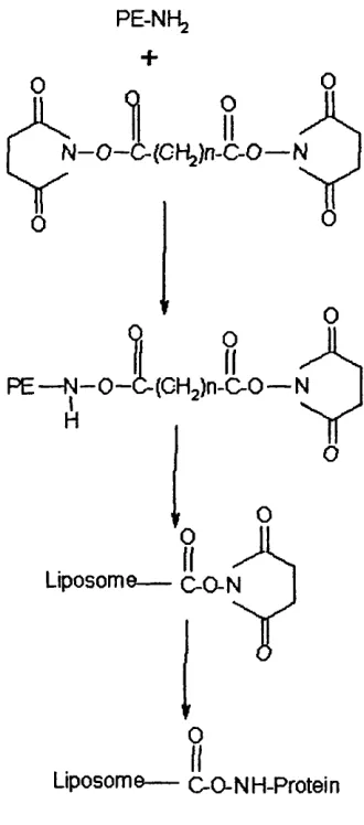
Chapter 1._______________________________________________ Leigh A Maguire. produce a malemide terminated linker lipid, N-succinimidyl (-4-[P-maleimido-phenyl] butyrate) (SMPB) is reacted with PE to give PE-MPB (Martin et al, 1990). Fig 1.8, below shows the reaction for the formation of a linker lipid incorporating a reactive carbonyl group suitable for complexing proteins (Martin et al, 1990).
In order to improve in vivo half-life of liposomes, PEG has been attached to the surface of liposomes (see chapter 6). PEG can also be incorporated into the structure of
Fig 1.9. Reaction scheme for the formation of a linker lipid.
PE-NHg +
1
OII
o
N—O—C- (Cl"^)n-C-0— N
O
0
P E — N—0 —(
H
Liposom(
O
II
(CHjln-C^O— N
O
LA
T
0Liposome— C-O-NH-Protein
Chapter 1. Leigh A Maguire. Fig 1.10. Methods for the attachment of antibody fragments to liposomes.
M P B -D O P ^ O
Ab— SH +
R e d u ce d
Antibody 'Ô
Ab— S
(CHjlg — il— NH— D O PE — L iposom e
M PB-PE L iposom es
'( ^ ^2^3 — — NH— D O P E — L iposom e Im m unoliposom es
PDP-D O PE
Ab— t
j"
N ft+ S — S — (GHg); — fl— NH— D O PE — LiLiposom e
O
MPB an tibidy
PD P -P E G -P E
P D P -P E liposom e
- 2 — M H n n P F ---1 ir o S — (C H jL — U—NH— d o p e — Liposom e
Im m unoliposom e
Ab-P
O MPB-AbAb-^ O
{— s — (CHg)^ — U— NH— {(CHjla-Oln— U— NH— D S P E — L iposom e1 P D P -P E G -P E liposom e
S — (CM,) MU 1
o
1.7. Characterisation of Liposomes and Immunoliposomes.
The physiochemical properties of liposomes strongly influence their behaviour. Characterisation in terms of. Fab’ coupling efficiency (in the case of immunoliposomes), particle size, zeta potential and long term stability is required if they are to be successful (Nassandere/fl/., 1995). Also encapsulation of pDNA needs to be well characterised.
Important factors that should be taken into consideration when preparing immunoliposomes for use as vectors are;
1. A sufficient quantity of antibody must bind to liposomes in a reproducible way; 2. The coupling process must not affect the integrity of the liposome;
3. The liposome antibody complex should be stable on storage and administration; 4. The homing capacity of the antibody should be preserved after binding to liposomes; (Nâssander e/a/., 1995).
1.7.1. Size and Zeta Potential
The size and zeta potential of liposomes are not only important to their behaviour in vivo
Chapter 1.________________________________________________Leigh A Maguire. size are being considered. However, size will effect processing. Smaller liposomes will possess a reduced aqueous volume, which would restrict the loading capacity and reduce drug encapsulation efficiency. Another factor that should be taken into account is that the bilayers of small liposomes have a greater degree of curvature. This would result in increased instability (Sullivan et al, 1986). Size and zeta potential can both be studied by dynamic light scattering.
1.7.2. LameUarity and Chemical Composition.
The Lamellarity of liposomes can be assessed by NMR spectroscopy using labelled phospholipids. The addition of external Mn^^ quenches the NMR signal of the labelled phospholipids in the outer most monolayer. Thus unilamellar vesicles (which have two monolayers) will show a 50 % reduction in the ^*P NMR signal (Mayer, et al,
It is important to prevent the chemical degradation of the lipid components of the liposome. The oxidation of phospholipids may be prevented through the handling and storage of the lipids under an inert atmosphere such a nitrogen or argon (Szoka, & Papahadjopoulos, 1980). If for example DOPE is hydrolysed the permeability of the liposome could be increased which would manifest itself as increased instability and release of encapsulated contents.
1.8. Stability.
Aggregation of gene therapy vectors is one of the major problems hindering their development. Formulations tend to aggregate under physiological conditions, which severely reduces their effectiveness. Recently the aggregation of PLL/DNA complexes has been analysed theoretically using established colloidal theory here at UCL (Lee et al,
2001). They show that aggregation is a complex function of the physiochemical properties of a system with initial distribution, solution pH, ionic strength and temperature being the most important parameters. Low pH, low ionic strength, low temperature and a narrow initial size distribution are process conditions that may help in the formation of stable complexes (Lee et al, 2001).
Chapter 1._______________________________________________ Leigh A Maguire.
et al, (2001). The most favoured method from the literature would be steric stabilisation, usually through the incorporation of polyethylene glycol (PEG) onto the surface of the vector.
1.8.1. Steric Stabilisation.
Two methods have been developed for the stabilisation of colloidal suspensions. Electrostatic stabilisation involves the absorption of ionic species on the surface of the particle. Steric stabilisation is the stabilisation of a suspension of colloidal particles through the addition of a hydrophilic polymer to the surface of the particles, which prevents the particles from aggregating. Steric stabilisation is exploited both industrially and biologically because it offers many advantages over electrostatic stabilisation (Napper, 1983). For example steric stabilisation is relatively insensitive to the presence of electrolytes while the addition of electrolytes, such as salts, to an electrostatically stabilised suspension will result in the aggregation of the particles (Napper, 1983). This is undesirable in preparations designed for introduction into the human body, which has relatively high concentrations of electrolytes. Several polymers have been investigated for use in the steric stabilisation of colloidal gene delivery vectors however the majority in the literature exploit polyethylene glycol (See Chapter 6). In addition to improving the
stability of the liposome PEG has also been shown to shield the liposome against damage
complement system. The addition of 15 mol % of PE-PEG2000 has recently been shown
Chapter 1._______________________________________________ Leigh A Maguire. 1.9. Project Background and Aims.
For gene therapy and DNA vaccination to become a reality the development of vectors capable of the delivery of the therapeutic gene to the required cells in an effective and safe manner is necessary. There are two schools of thought as to what will make the best vectors. The viral camp believe the best vectors will be viruses which have been modified in such a way as to remove their ability to replicate in host cells but which are able to infect and deliver the therapeutic gene to the nucleus of the target cell. However there are a number of major health risks involved with viral gene delivery which have been highlighted by the recent death in 1999 of the American teenager Jessie Gelsinger during a clinical trial with an adenovirus vector, and more recently by the case of a boy in France being treated for X-SCID with a modified retrovirus using the ex vivo gene delivery method developing leukaemia.
therapy for accurate delivery is greater than in cystic fibrosis where transfection of non targeted cells will be less likely to cause severe side effects (Miller & Vile, 1995). Each different target disease treated by gene therapy will therefore require the development of a specific method and vector formulation tailored to the specific needs of that disease. However what is evident from the literature is that these vectors will follow a basic plan, which will be modified for the target.
The aims of this project are to investigate the issues involved with the processing of a multi-component non viral gene delivery vector complex and in doing so propose a controllable, reliable, and reproducible method of processing such a vector with the view of future scale up. There are many vector formulations in the literature with significant inconsistencies between them. However it is generally agreed that the characteristics of a general vector include, a plasmid in which the therapeutic gene or genes are encoded, a means of condensing the plasmid DNA, protection for the DNA both in vivo and in vitro^
incorporate a means of cell targeting and be easy to produce in large quantities.