Neuroblastoma Immunotherapy Using
A Novel Vector System
K
o n s t a n t i n a
E
l e n a
S
i a p a t i
A thesis submitted for the degree o f Doctor o f Philosophy
2001
ProQuest Number: U643742
All rights reserved
INFORMATION TO ALL USERS
The quality of this reproduction is dependent upon the quality of the copy submitted.
In the unlikely event that the author did not send a complete manuscript and there are missing pages, these will be noted. Also, if material had to be removed,
a note will indicate the deletion.
uest.
ProQuest U643742
Published by ProQuest LLC(2016). Copyright of the Dissertation is held by the Author.
All rights reserved.
This work is protected against unauthorized copying under Title 17, United States Code. Microform Edition © ProQuest LLC.
ProQuest LLC
789 East Eisenhower Parkway P.O. Box 1346
A
b s t r a c t
Neuroblastoma is one of the commonest paediatric solid tumours and its treatment
using conventional therapy has so far been of limited success. Immunotherapy in the
form of tumour vaccination is an alternative therapeutic approach which aims at
enhancing the immunogenicity of the tumour and involves the ex vivo manipulation of tumour cells to express certain immunomodulatory molecules. The aim of this project
was to develop a cell-based neuroblastoma vaccine expressing Interleukin-2 and/or
Interleukin-12, and examine its effect in a murine model for the disease. A non-viral
vector system (LID) consisting of an integrin-targeting peptide and Lipofectin was
used to deliver the cytokine genes into neuroblastoma cells. Optimisation of the vector
components resulted in transfection of neuroblastoma cells at high efficiency (20-
60%) utilising an aspi integrin-targeting peptide. Examination of the intracellular
trafficking of LID complexes in these cells suggested that the LID vector may employ
a non-coated pit endocytic or phagocytic pathway to mediate cell entry. Despite the
transient nature of transfection, expression of biologically active cytokines persisted
above potentially therapeutic levels for at least 10 days, suggesting a sufficient time
window for an immune response to be elicited. Indeed, transfection of the mouse
Neuro-2A cell line with IL-2 and/or IL-12 completely abrogated its tumourigenicity in
a syngeneic mouse model for neuroblastoma. Pre-vaccination with irradiated Neuro-
2A cells conferred protective immunity against subsequent challenge with parental
cells, which was, however, irrespective of the cytokine expression by the cells. There
was a clear effect of IL-12 with reduced tumour growth in the presence of IL-2. In vivo immunodepletion experiments suggest that CD8^ T cells may be responsible for the initial rejection of Neuro-2A cells expressing both IL-2 and IL-12, while tumour
regression seems to be mediated by leukocyte infiltration and necrosis. These results
indicate that the complex nature of anti-tumour immune responses may require multi
modality treatments, such combination of cytokines, for efficient tumour eradication
and generation of systemic immunity. The improved anti-tumour immunity of co
transfection of IL-2 and IL-12, therefore, provides a promising immunotherapeutic
C
o n t e n t s
Title page
1
Abstract
2
Contents
3
Figures
9
Tables
12
Abbreviations
13
Amino Acids
16
Publications
17
Acknowledgements
18
1 INTRODUCTION
19
1.1 NEUROBLASTOMA 20
1.1.1 Features of the disease 20
1.1.2 Conventional therapy 22
1.2 GENE THERAPY OF CANCER 23
1.2.1 Drug sensitivity 23
1.2.2 Oncogenes and tumour-suppressor genes 24
1.2.3 Anti-angiogenesis 25
1.2.4 Immunotherapy 27
1.2.4.1 Tumour immune evasion 27
1.2.4.1.1 Down-regulation of MHC molecules 27
1.2.4.1.2 Role of Fas/FasL 28
1.2.4.1.3 Inhibition of apoptosis 30
1.2.4.1.4 T cell anergy 30
1.2.4.1.5 Loss of immunodominant antigens 32
1.2.4.2 Cytokine vaccines 33
1.2.4.2.1 Interleukin-2 33
1.2.4.2.3 Lymphotactin 39
1.2,4.3 Dendritic cell vaccines 40
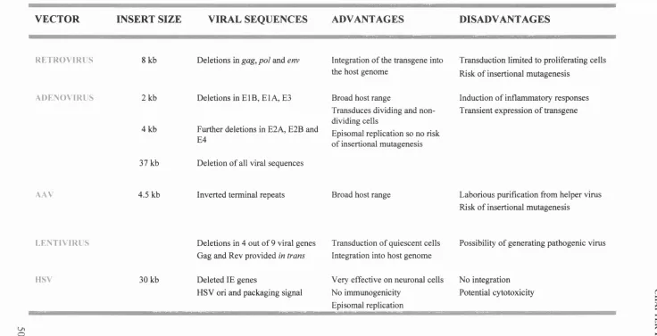
1.3 GENE THERAPY VECTORS 42
1.3.1 Retrovirus vectors 43
1.3.2 Lentivirus vectors 44
1.3.3 Adenovirus vectors 45
1.3.4 Adeno-Associated Virus (AAV) vectors 47
1.3.5 Herpes simplex virus (HSV) vectors 48
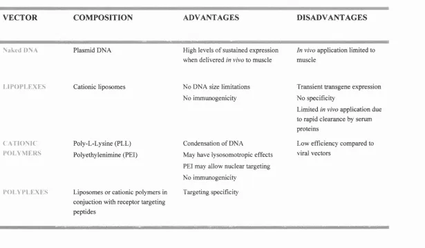
1.3.6 Non-viral vectors 49
1.3.6.1 Naked DNA 49
1.3.6.2 Lipoplexes 51
1.3.6.3 Polyplexes 52
1.4 BARRIERS TO NON-VIRAL GENE DELIVERY 52
1.5 INTEGRINS 57
1.5.1 Integrins as receptors for cell entry 57
1.5.2 Integrin-targeting peptides 58
1.5.3 Integrin-targeted vector systems 5 9
1.5.4 Integrins and neuroblastoma 60
1.6
AIMS AND OBJECTIVES 622 MATERIALS AND METHODS
63
2.1 Reagents 64
2.2 Cell culture 64
2.2.1 Primary cells 64
2.2.2 Cell lines 64
2.2.3 Antibody purification 65
2.3 LID transfection 66
2.3.1 Preparation of LID complexes 66
2.3.2 Calculation of charge o f transfection complexes 68
2.3.3 Luciferase assay 68
2.3.4 Cytokine ELISA 68
2.4 Nucleic acids 70
2.4.1 Preparation of competent cells 70
2.4.2 Transformation of plasmid DNA 70
2.4.3 Plasmid DNA 70
2.4.4 Plasmid DNA purification 71
2.4.5 Enzyme digestion of plasmid DNA 71
2.4.6 Agarose gel electrophoresis 71
2.4.7 Gel purification of DNA fragments 72
2.4.8 DNA ligation 72
2.5 Flow cytometry 72
2.5.1 BGFP expression 72
2.5.2 FACS antibody staining 73
2.5.3 Annexin-V/PI staining 74
2.6 Western analysis 75
2.6.1 Preparation of cell lysates 75
2.6.2 SDS-PAGE analysis 75
2.6.3 Transfer to nitrocellulose 75
2.6.4 Immunoblotting 76
2.7 Animal studies 76
2.7.1 Transfection of Neuro-2A cells 76
2.7.2 Animal inoculation 77
2.8 Immunohistochemistry 77
2.8.ITissue embedding and sectioning 77
2.8.2 Immunohistochemical staining 78
2.8.3 Histology 79
2.9 Statistical analysis 79
3 LID VECTOR OPTIMISATION
82
3.1 INTRODUCTION 83
3.2 RESULTS 84
3.2.1 Enhanced transfection efficiency by incorporation of a lipid 84 component
3.2.2 Titration of the LID vector components 84
3.2.2.1 DNA 86
3.2.2.2 Lipofectin 86
3.2.2.3 Integrin-targeting peptide 89
3.2.2.4 Alternative Lipids 93
3.2.3 Maturation of LID complexes 93
3.3 DISCUSSION 97
4 MECHANISM OF THE LID VECTOR
103
4.1 INTRODUCTION 104
4.2 RESULTS 106
4.2.1 Integrin expression 106
4.2.2 Competitive inhibition of LID transfection 106
4.2.2.1 Integrin-binding antibodies 106
4.2.2.2 Soluble RGD/RGE peptides 111
4.2.2.3 Integrin-targeting peptides transfect neuroblastoma cells 111 more efficiently than Ki6
4.2.3 Inhibition of endocytosis 113
4.2.3.1 Phosphatidylinositol 3-kinase Inhibitors 113
4.2.3.2 Chloroquine 117
4.2.3.3 Disruption of clathrin molecules 117
4.2.4 Inhibition of phagocytosis 119
5
IN VITRO
CYTOKINE EXPRESSION
125
5.1 INTRODUCTION 126
5.1.1 Cytokine bioactivity assays 126
5.1.2 y-Irradiation 127
5.1.3 Apoptosis 128
5.2 RESULTS 129
5.2.1 Cloning of cytokine genes 129
5.2.2 Cytokine Expression 132
5.2.2.1 Interleukin-2 132
5.2.2.2 Interleukin-12 134
5.2.3 Cytokine Bioassay 134
5.2.4 Lymphotactin expression by mouse Neuro-2 A cells 138
5.2.5 y-Irradiation 140
5.2.5. lAnnexin-V/ PI staining for detection of apoptosis 140
5.2.5.2 In vitro cell proliferation 142
5.2.5.3 IL-2 and IL-12 Expression 142
5.2.5.4 Lymphotactin Expression 144
5.3 DISCUSSION 147
6 In Viv o S t u d i e s 150
6.1
INTRODUCTION 1516.2
RESULTS 1526.2.1 In vivo tumourigenicity of cytokine-transfected neuroblastoma 152
cells
6.2.2 Prophylactic application of the vaccine 155 6.2.3 Eradication of established tumours 159 6.2.4 Inoculation into yc/RAG2 knockout mice 165
6 .2 .5/« v/vo immunodepletion 168
6.2.6 Histology of murine neuroblastoma tumours 172
7 DISCUSSION
180
7.1 LID transfection 182
7.2 Internalisation pathway of the LID vector 184
7.3 In vivo anti-tum our effect of vaccination with IL-2 and IL-12 187 transfected neuroblastoma cells
7.4 Potential problems 192
7.5 F uture directions 194
REFERENCES
198
APPENDICES
223A. Bacterial plasmids used in LID transfection experiments 224
B. Culturing of primary neuroblastoma cells 227
C. Cytokine expression by transfected Neuro-2 A cells used in mouse 230
F
ig u r e s
Ch a p t e r 1
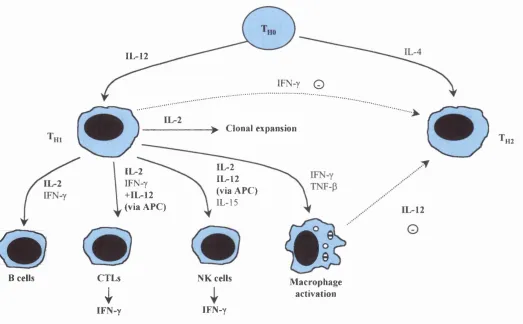
Fig 1.1 Effects of IL-2 and IL-12 on the immune system 34
Fig 1.2 Potential mechanisms of tumour antigen presentation 41
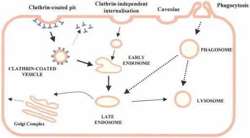
Fig 1.3 Endocytosis in mammalian cells 55
Chapter 3
Fig 3.1 Effect of Lipofectin on transfection efficiency of the LID vector 85
Fig 3.2 Titration of the DNA component of the LID vector on neuroblastoma cells
87
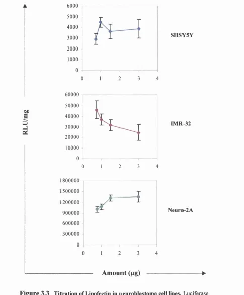
Fig 3.3 Titration of Lipofectin in neuroblastoma cell lines 88
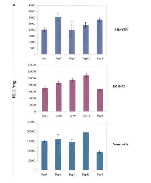
Fig 3.4 Optimisation of the integrin-targeting peptide component of the LID vector
90
Fig 3.5 Influence of the peptide/DNA charge ratio on the transfection efficiency of LID complexes
92
Fig 3.6 Transfection of neuroblastoma cell lines using Tfx Reagent 1 94
Fig 3.7 Correlation between luciferase gene activity and efficiency of transfection as a function of duration of complex formation
96
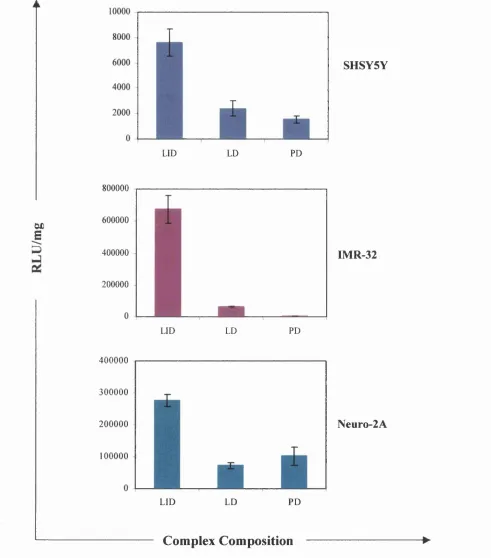
Fig 3.8 Transfection efficiency of neuroblastoma cells using the LID vector 98
Fig 3.9 Photographs of LID-transfected neuroblastoma cell lines 99
Chapter 4
Fig 4.1 Integrin expression on human neuroblastoma cell lines 107
Fig 4.2 Blocking of LID transfection with a4 integrin antibodies 109
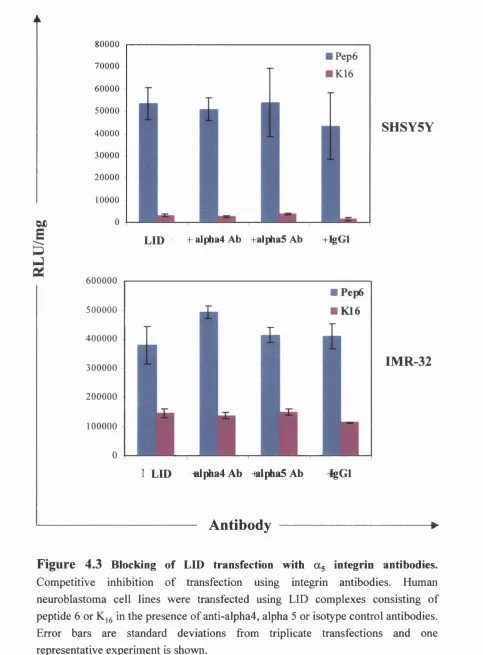
Fig 4.3 Blocking of LID transfection with as integrin antibodies 110
Fig 4.4 Competitive inhibition of LID transfection 112
Fig 4.7 The importance of clathrin-coated pits on LID internalisation 118
Fig 4.8 Cytochalasin-B inhibits transfection with the LID vector 120
Ch a p t e r 5
Fig 5.1 Cloning of cytokine genes 131
Fig 5.2 lnterleukin-2 expression by neuroblastoma cell lines 133
Fig 5.3 In vitro expression of Interleukin-12 by neuroblastoma cell lines 135
Fig 5.4 Biological activity of lL-2 and IL-12 expressed by transfected 137
1 neuroblastoma cell lines
Fig 5.5 Expression of murine lymphotactin by Neuro-2 A cells 139
Fig 5.6 Apoptosis of y-irradiated Neuro-2 A cells 141
Fig 5.7 Effect of y-irradiation on proliferation of LID-transfected Neuro-2 A 143 cells
Fig 5.8 Comparison of cytokine expression by irradiated, necrotic and normal 145 transfected Neuro-2 A cells
Fig 5.9 Expression of murine lymphotactin by y-irradiated transfected Neuro- 146 2A cells.
Ch a p t e r 6
Fig 6.1 In vivo tumourigenicity of cytokine-transfected Neuro-2 A cells in 153 syngeneic mice
Fig 6.2 In vitro proliferation of cytokine-transfected Neuro-2 A cells used in 154 syngeneic mice
Fig 6.3 Effect of vaccine application on systemic anti-tumour immunity 157
Fig 6.4 Titration of the number of Neuro-2 A cells used in pre-vaccination 158
experiments
Fig 6.5 Effect of vaccination on established murine tumours 160
Fig 6.6 Effect of intratumoural vaccination on the growth and survival of 161
animals with established tumours
Fig 6.7 Tumour growth of individual animals used in a second eradication 163
Fig 6.8 Average tumour growth and survival of animals from the second 164 eradication experiment
Fig 6.9 In vivo tumourigenicity of N2A-IL-2+IL-12 cells in yc/RAG2 knockout 166 mice
Fig 6.10 In vitro proliferation of Neuro-2A cells used to inoculate yc/RAG2 167 knockout mice
Fig 6.11 In vivo CD4 and CDS immunodepletion 169
Fig 6.12 CD4 and CDS staining of splenocytes from immunodepleted animals 170
Fig 6.13 In vitro proliferation of Neuro-2 A cells used in the in vivo 171 immunodepletion experiment
Fig 6.14 Histology of wild-type murine neuroblastoma tumours 173
Fig 6.15 Histology of N2A-IL-2+IL-12-vaccinated neuroblastoma tumours 175
T
a b l e s
Table 1.1 International Neuroblastoma Staging System (INSS) 21
Table 1.2 Viral vectors for gene therapy applications 50
Table 1.3 Synthetic vector systems for gene therapy applications 53
Table 2.1 Peptides used in LID transfection experiments 67
Table 2.2 Lipids used in LID transfection experiments 67
Table 2.3 Antibodies used for integrin expression 73
Table 2.4 Antibodies used for FACS staining of mouse splenocytes 74
Table 2.5 Antibodies used in Western blotting 76
Table 4 Effect of various drugs on transfection efficiency of the LID vector
122
A
b b r e v ia t io n s
APC APS ATP BM bp BSA CDA cDNA CNS CTL dHzO DC DMEM DMSG DNA DOPE DOTMA DTT ECL E.coli EDTA ELISA FACS PCS FITC GM-CSF hIL-2Antigen presenting cell
Ammonium persulphate
Adenosine triphosphate
Bone marrow
Base pair(s)
Bovine serum albumin
Cytosine deaminase
Complementary DNA
Central nervous system
Cytotoxic T lymphocyte
Distilled water
Dendritic cell
Dulbecco's Modified Eagle medium
Dimethyl sulphoxide Deoxyribonucleic acid dioleoyl phosphatidylethanolamine N-[l(-2,3,-dioleyloxy)propyl]-N,N,N-trimethylammonium chloride Dithiothreitol Enhanced chemiluminescence Escherichia coli Ethylenediaminetetraacetic acid
Enzyme linked immunosorbent assay
Fluorescence activated cell sorting
Fetal calf serum
Fluoroscein isothiocyanate
Granulocyte macrophage cell stimulating factor
Human Interleukin-2
HRP Horseradish peroxidase
HSV-tk Herpes Simplex Virus thymidine kinase
ICE Interleukin-Ip converting enzyme
kb Kilobase pair(s)
kDa Kilo Daltons
LB Luria-Bertani (media)
LD Lipid-DNA
LID Lipid-integrin targeting peptide-DNA
LOH Loss of heterozygosity
MDR Multi-drug resistance
MHC Major histocompatibility complex
mIL-12 Murine IL-12
MRP Multi-drug resistance associated protein
NCAM Neural cell adhesion molecule
NK Natural killer cell
OD Optical density
PAGE Polyacrylamide gel electrophoresis
PBS Phosphate buffered saline
PD Peptide-DNA
PEA Paraformaldehyde
PHA-L Phaseolus leucoagglutinin
PI3K Phosphatidylinositol 3-kinase
PEL Polylysine
RNA Ribonucleic acid
rpm Revolutions per minute
RT Room temperature
SDS Sodium dodecyl sulphate
TAE Tris acetate EDTA
TAP Transporters associated with antigen processing
TCR T cell receptor
TE Tris/EDTA buffer
Tris 2-amino-2-[hydroxymethyl]-1,3 propandiol
Tween 20 Polyethylene-sorbitan monolaurate
UV Ultraviolet
VEGF Vascular endothelial growth factor
v/v Volume per volume
w/v Weight per volume
A
m in o
A
c id s
Alanine Ala A
Arginine Arg R
Asparagine Asn N
Aspartic acid Asp D
Cysteine Cys C
Glutamic acid Glu E
Glutamine Gin
Q
Glycine Gly G
Histidine His H
Isoleucine He I
Leucine Leu L
Lysine Lys K
Methionine Met M
Phenylalanine Phe F
Proline Pro P
Serine Ser S
Threonine Thr T
Tryptophan Trp W
Tyrosine Tyr Y
P
u b l ic a t io n s
This thesis has contributed to the following publications:
K.£.Siapati,
C.Kinnon, A.M ichalski, R.A nderson, A .Thrasher and S.L.Hart.Improved anti-tumour immunity in murine neuroblastoma with a combination o f IL-2 and IL-12. Manuscript in process
Published abstracts:
K.£.Siapati,
P.Brickell, C.Kinnon, A .M ichalski, A .Thrasher and S.Hart (2000). Neuroblastoma Immunotherapy Using a Novel Vector System. Molecular Therapy 1: 270A
c k n o w l e d g e m e n t s
Firstly, I would like to thank my supervisors Dr Steve Hart and Dr Adrian Thrasher
for their help and support throughout this project. Special thanks also to Prof
Christine Kinnon for her encouragement and invaluable advice on the preparation of
this thesis. I am grateful to Mike Blundell, Kate Parseley and Susie Barker for their
great help with the animal work and the long trips to the Royal Veterinary College! I
would like to acknowledge Dr Anthony Michalski and Dr John Anderson for the
patient neuroblastoma samples and Caroline Marshall for her assistance and helpful
practical tips on the preparation and histology of tumour specimens. I would also like
to thank Dr Richard Parkes for critical review and heated discussions on certain parts
of this thesis.
Last but not least I’m indebted to my family for the enormous psychological support
--- CHAPTER 1
1.1 NEUROBLASTOMA
1.1.1 Features of the disease
Neuroblastoma is an extracranial solid tumour that accounts for approximately 10% of
the solid malignancies occurring in children. It arises from precursor cells of the
sympathetic nervous system and primary sites include the adrenal gland and the
abdomen. Localisation of the tumour in the thorax and the neck are less common
(reviewed in Ninane and Pearson, 1997). Recently, a number of studies have reported
spread of the disease at CNS sites (Kramer et a l, 2001). Neuroblastoma has the highest rate of spontaneous regression but one of the poorest outcomes when it occurs
as disseminated disease. Stage 4S neuroblastoma is a special form of the malignancy
characterised by localised primary tumour with métastasés that exclude involvement
o f the bone (Table 1.1). The prognosis for stage 4S, even in the absence of any
therapy, is very good, with the majority of tumours undergoing spontaneous
regression. Due to its high incidence, neuroblastoma has been extensively studied and
various therapeutic approaches have been tested.
Neuroblastoma is a genetically heterogeneous disease with the majority of tumours
demonstrating deletions or rearrangements of several genomic regions. These include
loss of heterozygosity at lp36 (Takeda et a i, 1996) where a number of tumour suppressor genes such as p73 have been identified (Jost et a l, 1997; Kaghad et a l,
1997). In addition, LOH for chromosome 14q and l l q, and chromosome 17
aberrations have been reported (Takayama et a l, 1992; Lastowska et a l, 1997; Hoshi
et a l, 2000). LOH at chromosome 19ql3 is associated with aggressive disease (Mora
et a l, 2001). Another very common genetic abnormality in neuroblastoma is amplification of the N-myc oncogene, which is located at chromosome 2p23-24. The
family of myc genes encodes transcription factors that control cell proliferation
(Bouchard et a l, 1998). Transgenic mice over-expressing N-myc in neural crest cells develop neuroblastoma tumours a few months after birth strongly implicating N-myc
---CHAPTER 1
phenomenon could be due to a failure to repress telomerase during development or
because of reactivation of telomerase activity at a later stage accompanied by other
genetic abberations leading to neuroblastoma development (Hiyama et ah, 1995).
A tumour-associated marker has not been identified for neuroblastoma. Many studies
have used the disialoganglioside GD2 as a target antigen either for the generation of
specific cytotoxic T lymphocytes (CTL) (Zhao and Cheung, 1995) or for the direction
of certain immunostimulatory molecules such as Interleukin-2 fused to monoclonal
antibodies (Lode et a l, 1998a). In addition, monoclonal antibodies targeted to GD2
display antibody-dependent cytotoxicity against neuroblastoma cells when used
together with lymphokine activated killer cells in vitro (Honsik et a l, 1986). This suggests that GD2 may be an effective immunotherapeutic target for neuroblastoma.
Table
1.1 International Neuroblastoma Staging System (INSS)^Stage 1 Localised tumour with complete surgical resection; distant lymph nodes
negative for tumour (microscopically)
Stage 2A Localised tumour with incomplete resection; lymph nodes negative for
tumour
Stage 2B As in Stage2A but ipsilateral lymph nodes positive for tumour while
contralateral lymph nodes must be negative for tumour
Stage 3 Unresectable tumour with or without regional lymph node involvement
or localised tumour with contralateral lymph node involvement
or tumour crossing the vertebral column by infiltration or by lymph node
involvement
Stage 4 Primary tumour with métastasés in bone, bone marrow, lymph nodes, liver
and/or other organs
Stage 4S Localised primary tumour with métastasés limited to skin, liver or bone
marrow. Limited to children under 1 year of age
Adapted from (Castel et al , 1999).
--- CHAPTER 1
GD2 is highly expressed in many human tumours including neuroblastoma and is
believed to play a role in the clinical phenotype of neuroblastoma. Gangliosides are
membrane-bound glycosphingolipids that are prominent in neuronal tissue.
Biosynthesis of gangliosides from ceramide occurs in two distinct pathways, a and b,
with b being associated with early brain development. Comparison between
neuroblastoma patients younger than 1 year of age with a group of patients older than
1 year, revealed that b pathway gangliosides were prominent in 92% of patients in the
former group compared to just 40% of the latter group (Kaucic et a l, 2001). This suggests that the pathway of ganglioside synthesis is important for the clinical
differences observed among different stages of neuroblastoma biology and that the b
pathway is probably associated with better prognosis.
1.1.2 Conventional therapy
The type of therapy chosen to treat patients with neuroblastoma depends on the stage
of the disease but has mainly consisted of chemotherapy followed by surgical
resection of the tumour or radiotherapy (Pinkerton et a l, 2000). Although the disease is responsive to therapy, survival of high-risk group patients at 2 years is 50% and
survival at 5 years is 33% (Philip et a l, 1997). The main drawback of such approaches is the acquired drug resistance after exposure to chemotherapeutic agents. Patient cell
lines exhibit higher than 1000-fold resistance after etoposide and doxorubicin
treatment (Keshelava et a l, 1997). Multi-drug resistance depends on the expression of two genes, multi-drug resistance (MDR) (Haber et a l, 1997) or multi-drug resistance- associated protein (MRP) genes that encode ATP-dependent membrane transport
proteins. There is a strong correlation between the expression patterns of these genes
in neuroblastoma tumours lacking N-myc amplification (Haber et a l, 1997). Although expression of the MDR 1 gene is associated with poor outcome, it is found to inversely
correlate with amplification of N-myc. Another drawback of chemotherapy is its non
specific nature, which does not target the minimal residual disease and causes patients
to relapse. Strategies to address that problem have included high-dose induction
--- CHAPTER 1
following initial surgical resection or chemotherapy have an 80% chance of becoming
long-term survivors if they receive bone marrow transplantation (Philip et a l, 1997).
In order to increase the specificity of the treatment, the use of anti-neuroblastoma IgM
antibodies found in healthy individuals has been investigated. Administration of such
antibodies in a metastatic neuroblastoma model in nude rats completely abrogated the
tumourigenicity o f human LAN-1 neuroblastoma cells. In addition, there was a 90%
reduction in the initial volume of established adrenal gland tumours following
treatment. The main anti-tumour functions of these antibodies are achieved through
complement activation and apoptosis (Engler et a l, 2001).
1.2
GENE THERAPY FOR CANCER
The concept of gene therapy for treating cancer arose with the recognition that all
tumours arise as a result of genetic lesions. It has been proposed that anti-tumour
therapy can be achieved by genetic intervention, a process that can induce selective
killing o f tumour cells and eliminate cytotoxic side effects caused by
chemotherapeutic agents and radiotherapy. Although numerous approaches have been
devised, the common aim is to specifically eradicate tumour cells. Limitations of the
available gene transfer vectors make it impossible to achieve 100% transduction
efficiency of the tumour cells. However, due to a process known as the ‘bystander
effect’, whereby the cytotoxic effect of a treatment is diffused to neighbouring cells,
100% transduction efficiency is not always required. This effect can be local, by
release o f cytotoxic agents to neighbouring cells, immune, where tumour-specific
immune responses are generated, or angiostatic.
1.2.1 Drug sensitivity
Transduction of tumour cells with a gene whose product can convert a non-toxic
administered prodrug to a toxic metabolite is a very efficient approach of inducing cell
--- CHAPTER 1
death. Herpes Simplex Virus thymidine kinase (HSV-tk) is a well-studied suicide gene
that converts ganciclovir (GV) to the toxic metabolite ganciclovir-triphosphate once
inside a cell expressing HSV-tk. Cell death is caused by incorporation o f the toxic
metabolite into the DNA chain and termination of DNA synthesis. The advantage of
this system is that adjacent untransduced cells are rendered sensitive to ganciclovir.
This effect occurs only when cells are in direct contact with HSV-tk-transduced cells
and has been attributed to the transfer of the toxic metabolite through gap junctions
(Bi et a l, 1993). Retroviral transfer of HSV-tk into murine neuroblastoma tumours and subsequent administration o f ganciclovir results in tumour regression and
extensive necrosis as indicated by TUNEL staining (Cho et ah, 1999). Another well- studied suicide gene is cytosine deaminase (CDA) that converts 5-fluorocytosine (5-
FC) to 5-fluorouracil (5-FU). This is then converted to 5-FU monophosphate and 5-
FU triphosphate that interact with DNA and RNA synthesis respectively and lead to
cell death. The ‘bystander effect’ occurs with CDA as well but probably by a different
mechanism where 5-FU freely diffuses through the cell membrane. Adenoviral
transfer o f the CDA gene to colon carcinoma cells increases expression of CD95 and
leads to apoptotic cell death upon treatment with 5-FC (Ju et a l, 2000). Combination of other treatments such as administration of lymphotactin, results in greater inhibition
o f tumour growth, which is mediated by CD4^/CD8^ T cells and natural killer (NK)
cells.
1.2.2 Oncogenes and tumour suppressor genes
Transformation o f a normal to a malignant phenotype is the result o f genetic
mutations causing loss of tumour suppressor function or acquisition o f oncogenic
phenotype. As a result of this observation, gene therapy strategies have been devised
that involve the introduction or restoration o f tumour-suppressor function by
correcting for the mutated allele. The tumour suppressor gene p53 is crucial in
induction of apoptosis and its absence is believed to render cells resistant to mutations
caused by irradiation and chemotherapeutic compounds. Loss o f p53 is associated
--- CHAPTER 1
50% of human cancers either express a mutant p53 or a non-functional protein
emphasising the need for effective therapies for tumours that lack p53 function. In a
human non-small cell lung cancer study, tumour biopsies following retroviral
administration o f p53 gene show apoptotic death of tumour cells and tumour
regression in 33% of patients (Roth et a l, 1996). The majority of neuroblastoma cells lack p53 function or its closely related p73 tumour suppressor gene, implicating it in
the process o f malignant transformation. Expression of the human polyomavirus BK
(BKV) large T antigen by neuroblastoma interferes with p53 function. Antisense
oligonucleotides against BKV large T antigen restore p53 expression and induce
apoptosis (Jorgensen et a l, 2000). In addition, p53 inactivation may be a consequence o f antagonistic function of the glucocorticoid receptor (GR) on p53-downstream
targets such as p21 and bcl2 (Sengupta et a l, 2000).
Lack of p53 expression in many tumour cells has been exploited as a target for tumour
therapy. A number of viral proteins bind to and inactivate p53 including the protein
encoded by E lB gene of adenovirus. A virus lacking E lB should, in theory, he able to
infect and lyse p53-deficient tumour cells. Administration of ONYX-15, an E lB gene-
attenuated adenovirus, in nude mice engrafted with p53 mutant tumours (C33A
cervical cancer) or tumours expressing defective form of p53 (HLaC laryngeal cancer)
resulted in tumour regression. No cytolysis was observed in tumours expressing a
fully functional p53 gene (Heise et a l, 1997). Phase I-II clinical trials with ONYX-15 have been initiated in a number of cancers. However, administration of this
replication-selective adenovirus alone failed to exhibit any anti-tumour effects unless
it was combined with chemotherapy. The virus was well tolerated in all doses and
routes administered, but further modifications are required to enhance its efficacy in
certain tumours (Kim, 2001).
1.2.3 Anti-angiogenesis
Generation o f new blood vessels is a prerequisite for tumour growth and metastasis
formation. Human neuroblastoma cell lines secrete metalloproteinases, MMP-2 and
--- CHAPTER 1
MMP-9 that degrade the extracellular matrix, induce proliferation of human vascular
endothelial cells and promote angiogenesis in vivo (Ribatti et al., 1998). Activin A, a member of the TGF-p superfamily, is present during embryonic development and
suppresses angiogenesis in vivo. An inverse correlation of activin A with N-wyc expression has been demonstrated in human neuroblastoma cell lines. The down-
regulation involves interaction of the N-myc with the activin A promoter implicating
the former in the angiogenesis process (Breit et a l, 2000).
In a clinical context, high vascular density correlates with N-myc amplification and
poor prognosis in neuroblastoma patients (Meitar et a l, 1996). Expression of angiogenic factors such as vascular endothelial growth factor (VEGF), VEGF-B,
VEGF-C, transforming growth factor (TGF-a), angiopoietin-1 and -2 , and platelet-
derived growth factor (PDGF) is higher in patients with more advanced stages of the
disease (stages 3 and 4S) (Eggert et a l, 2000). The fact that several angiogenic factors are co-expressed in most patients, suggests that they act in synergy to create highly
vascularised neuroblastoma tumours. Therefore, multi-modality treatments may be
required that target a number of different factors. Treatment of experimental
neuroblastoma tumours with the angiogenesis inhibitor TNP-470 [0-(A-chloroacetyl-
carbamoyl)-fumagillol significantly reduces the tumour growth rate but has no effect
in animals with large tumour burdens (Katzenstein et a l, 1999). Although this angiostatic agent acts on endothelial proliferation independently of angiogenic factor
expression, reducing the vascularisation of large tumours has not been possible.
Retroviral transduction of murine fibroblasts with the gene for the soluble VEGFR-2
(flk-1) and co-administration with neuroblastoma cells into SCID mice significantly
decreased murine tumour growth after 25 days (Davidoff et a l, 2000). In a similar way, VEGFR-2 transduction of neuroblastoma cells reduced their tumourigenicity in
SCID mice so that the resulting tumours were about one third the volume of those of
--- CHAPTER 1
Expression o f certain integrins such as ayps by endothelial cells is implicated in the
formation o f blood vessels (Brooks et a l, 1994). Cytokines such as TNF and IFN-y do not only induce an immune response but also demonstrate angiostatic properties.
Treatment with either of these cytokines inhibits avps-mediated endothelial cell
adhesion and survival in vitro. Their administration in melanoma patients disrupt tumour vascularisation by causing the detachment of ttvPa -positive endothelial cells
(Ruegg et a l, 1998).
1.2.4 Immunotherapy
Cancer immunotherapy depends on the proposition that most tumours possess specific
antigens that are either of embryonic origin and are absent from ‘mature’ tissues, or
are expressed as a result of mutations, or other genetic aberrations, and are thus
attractive targets for immunotherapy. However, malignant transformation may be
accompanied by a number of phenotypic changes in the cancer cells that allow
tumours to evade the immune system. Although those changes depend largely on the
individual neoplasm, certain common mechanisms have been identified.
1.2.4.1 Tumour immune evasion
1.2.4.1.1 Down-regulation of MHC molecules
Down-regulation of MHC molecules is a frequent observation in malignant cells and
over-expression of MHC class I reverses the oncogenic effects (Tanaka et a l, 1986). Cytotoxic T cells (CTLs) require antigen presentation through MHC class I molecules
while NK cells recognise MHC class I via their inhibitory receptors (KIRs). Down-
regulation of MHC class I molecules may cause tumour evasion o f immune attack
either by insufficient antigen presentation to CTLs or inhibition of NK cell function.
In neuroblastoma, down-regulation of MHC class I molecule expression is associated
with 'H-myc amplification. This occurs through suppression of the p50 subunit of the
--- CHAPTER 1
transcription factor NF-kB, which is involved in up-regulation of IL-2 expression by
T cells and their subsequent proliferation. Transfection of neuroblastoma cells with
p50 restores surface expression of MHC class I molecules (van't Veer et a l, 1993).
Neuroblastoma immunogenicity is related to MHC class II expression. Retroviral
transfer of syngeneic, allogeneic or xenogeneic MHC class II molecules, causes
murine neuroblastoma cells to lose their tumourigenicity. However, cells transduced
with human DR (xenogeneic) or I-A^ (allogeneic) induce a more rapid immune
response that correlates with enhanced stimulation of naive splenocytes compared to
the syngeneic (I-A^) MHC expressing vaccines (Hock et a l, 1996). The speed of the observed immune response may be due to the foreign nature of the transduced antigen
that may result in an initial alloreactive response.
Defects in the assembly pathway of MHC molecules, such as the TAP (transporters
associated with antigen processing) complex have been reported in cervical
carcinomas. TAP consists of TAPI, TAP2 and tapasin and mediates the transportation
of the antigenic peptides from the proteosome to the endoplasmic reticulum. In
neuroblastoma cell lines with N-myc amplification and deletion at Ip, low expression
of HLA class I molecules correlates with absence of TAPI. Expression of all three
members of the TAP complex as well as surface HLA class I molecule expression can
be restored by treatment with IFN-y (Corrias et ah, 2001). The importance of TAP in tumour immunogenicity has been demonstrated by incorporation of a functional TAP
gene in a recombinant vaccinia virus in a small-cell lung carcinoma model. TAP-
transduced cells exhibited improved immunogenicity and enhanced antigen
presentation (Alimonti et a l, 2000).
1.2.4.1.2 Role of Fas/FasL
Cytotoxic T lymphocytes and NK cells are among the most effective mediators of
anti-tumour immunity. Killing of target tumour cells is initiated by cell contact
---CHAPTER 1
cause apoptotic death. Apoptosis can also be induced by interaction of CD95L (FasL)
on the effector cell with the ‘death receptor’ CD95 (Fas) on the target cell. Ligation of
the receptor triggers a cascade of caspase proteins such as caspase 1 or interleukin-ip-
converting enzyme (ICE). The latter cleaves interleukin-Ip to its active form or
activates IL-18. Both of these cytokines have pro-inflammatory effects and can trigger
an anti-tumour immune response. Expression of ICE mRNA has been detected in 69%
of stage 1, 2 and 4S neuroblastoma tumours while only 19% of patients with poorer
prognosis (stage 3 and 4) express ICE. In addition, ICE expression inversely correlates
with N-myc amplification (Ikeda et ah, 1997).
Reports on Fas/FasL interaction and its importance in tumour immune evasion are
quite controversial. Although most neuroblastoma cell lines express very low levels of
Fas and FasL (Bemassola et a l, 1999), expression of FasL induces T cell death in vitro indicating a potential mechanism of T cell elimination at the tumour environment (Shurin et a l, 1998). Indeed, melanoma specific T cells after MHC-class-I recognition undergo apoptosis, a process that is inhibited with anti-Fas antibodies. FasL
expression, in that particular study, could not be detected on tumour cells but rather on
T lymphocytes following activation (Zaks et a l, 1999). The observed T cell apoptosis could be due to Fas/FasL interaction among T cells after prolonged antigen activation.
Alternatively, Fas upregulation can occur in tumours in vivo, suggesting that Fas/FasL interaction plays an important role in immune escape especially at the early stages of
tumour growth (Rose et a l, 2000). On the other hand, vaccination with FasL expressing neuroblastoma tumour cells showed efficacy in neuroblastoma
immunotherapy. FasL-expressing cells induced a strong inflammatory response
consisting mainly of neutrophils and provided a CD8^ T cell tumour-specific
protective immunity in a murine model. This vaccine also proved very effective in
eradicating established tumours (Shimizu et a l, 1999).
In a similar way, other tumour-associated molecules may act as a protection by
tumours against immune surveillance. For example, RCASl (receptor-binding cancer
antigen expressed in SiSo cells) expression has been observed in uterine, ovarian,
colon and lung cancers and recently its receptor has been identified on T, B and NK
---CHAPTER 1
cells. Upon binding with RCASl, receptor-expressing cells cease to proliferate and
undergo apoptotic death (Nakashima et a l, 1999).
1.2.4.1.3 Inhibition of apoptosis
Resistance to apoptosis can be an outcome of inactivation or functional loss of certain
members of the apoptotic pathway. Caspase 8 inactivation has been reported in
neuroblastomas with N-myc oncogene amplification (Teitz et a l, 2000). Recently, a number of viral proteins that inhibit transmission of apoptotic signals through Fas or
other death receptors have been identified (FLIPs). These inhibitors are produced by
herpes viruses and inhibit interaction of the cytoplasmic death domain o f Fas receptor
(FADD) with caspase proteins. Transduction of A20 lymphoma cells with Kaposi’s
sarcoma herepesvirus FLIP protein enhances their tumourigenicity in vivo and protects them from Fas-induced apoptosis, demonstrating the tumourigenic effect of such
inhibitors (Djerbi et a l, 1999). However, T cell-mediated lysis of CD95/FLIP- transfected lymphoma cells can still take place in vitro through the perforin pathway. The importance of this pathway is demonstrated in perforin knockout mice, where
administration of cells expressing high levels of FLIP results in progressive tumour
growth. In contrast, cells that express low levels of FLIP are cleared. Therefore, as
CD95/FLIP expression in both wild-type and perforin-knockout mice results in
tumour growth, CD95-dependent apoptosis seems to be the most prominent
mechanism of tumour eradication in vivo (Medema et a l, 1999).
1.2.4.1.4 T cell anergy
Alternative explanations of failure of immune surveillance include the phenomenon of
T cell anergy. This unresponsive state of T cells is characterised by lack of particular
responses despite optimal stimulation through their T cell receptor (TCR). T cell
unresponsiveness may also be a consequence of lack of co-stimulatory molecules such
---CHAPTER 1
their CD28 receptor in order to become activated and absence of a simultaneous co
stimulatory signal leads to T cell anergy. Secretion of immunomodulatory molecules,
such as IL-10, TGF-P or other cytokines, from the tumour cells or their surrounding
environment may also contribute to T cell anergy. Anergic T cells exhibit reduced
production of certain cytokines such as IL-2 and GM-CSF whereas IFN-y levels
remain unchanged. The decreased IL-2 levels and reduced cellular proliferation can be
caused by the production of certain inhibitors such as Nil-2a that block cytokine
transcription or p21 activation. These effects can be reversed in the presence of co
stimulatory signals during antigen presentation or by signalling through the IL-2
receptor (reviewed in Schwartz, 1996). Also, in vivo administration of activating antibody against CD40, which activates antigen-presenting cells, is capable of
reversing the peptide-induced T cell unresponsiveness in a renal cell carcinoma model
expressing influenza haemaglutinnin (Sotomayor et a l, 1999).
Groux et al, (1996) have reported that human CD4^ T cells fail to proliferate or secrete any cytokines in a mixed lymphocyte reaction culture in the presence of IL-10.
The induced anergic state cannot be reversed with anti-CD3 or anti-CD28 antibodies
suggesting that IL-10 impairs signalling through the TCR/CD3 complex. However,
after treatment with PMA these T cells proliferate normally indicating that a direct
activation of protein C kinase is required (Groux et a l, 1996). Secretion of IL-10 and TGF-P may also explain why animals with big tumour burdens fail to respond to
immunotherapeutic treatments. Hsieh et al, (2000) show that the expression of these two cytokines at the tumour site correlates with its progression stage. Addition of IL-
10 and TGF-P during in vitro T cell function assays from vaccinated animals inhibits the proliferative and cytolytic activity of these T cells (Hsieh et a l, 2000). IFN-y has been widely used in many cancer animal models to enhance the immunogenicity of
the cells. A recent report, however, demonstrates its involvement in promoting
immune escape in a CT26 murine colon carcinoma model. The decreased
immunogenicity of the tumour cells is due to infiltrating CD8^ cells secreting IFN-y
and down-regulating the immunodominant antigen gp70 (Beatty and Paterson, 2000).
--- CHAPTER 1
1.2.4.1.5 Loss of immunodominant antigens
Tumours can also escape immune surveillance by shutting down genes expressing
immunodominant antigens. MAGE-1, -3 and - 6 are genes encoding melanoma
rejection antigens and are presented to CTLs in an HLA-I context, 59% of clinical
neuroblastoma samples and 43% of cell lines express those antigens and in clinical
tumours expression of MAGE antigens inversely correlated with clinical stage (Ishida
et a l, 1996). This may suggest that during neuroblastoma progression, silencing of MAGE genes comprises a mechanism of immune escape that provides neuroblastoma
with a growth advantage. Furthermore, shedding of gangliosides that have an
immunosuppressive function by neuroblastoma tumours may be an alternative way of
destroying the host immune response. Tumour derived gangliosides inhibit IL-2
mediated proliferation of T cell lines and inhibit production of TNF-a by monocytes
(Lu and Sharom, 1995). In addition, generation of DCs from murine bone marrow
progenitors incubated with neuroblastoma cell lines is significantly impaired.
Furthermore, when DCs are cultured in the presence of neuroblastoma cells, their
antigen presenting capacity is inhibited in an allogeneic mixed leukocyte reaction
(MLR) (Shurin et a l, 2001). Treatment of neuroblastoma cell lines with D-PDMP
(D-threo-1 -phenyl-2-decanoylamino-3 -morpholino-1 -propanol), an inhibitor of
glycosphingolipid synthesis, significantly reduces the production and shedding of
gangliosides (Li and Ladisch, 1996). This could potentially restore immune
suppression and increase the immunogenicity of the tumour.
In conclusion, a number of potential mechanisms that allow tumours to escape
immune surveillance have been identified. The precise mechanism varies for each
type of cancer and is probably a combination of more than one. In order to increase
the immunogenicity of many cancers and by-pass any immune suppressive effects, a
number of studies have been performed using immunomodulatory molecules such as
cytokines or chemokines systemically or in the form of a vaccine. In the latter case,
patient tumour cells or established cell lines have been genetically modified to secrete
these immunomodulatory molecules. The main advantages of this approach are the
---CHAPTER 1
at the tumour site thereby avoiding toxicity caused by systemic administration of
cytokines.
1.2.4.2 Cytokine vaccines
Certain cytokines such as interferons have direct anti-tumour effects through localized
tumour killing while others work by augmenting the T cell-mediated immunity against
the tumour as a result of localised expression at the tumour site. For example, IL-4
and TNF-a induce inflammatory responses by attracting eosinophils and macrophages
at the tumour site and by regulating the expression of certain adhesion molecules on
the surface of endothelial cells, so that the transmigration of inflammatory cells is
enabled (reviewed in Tepper and Mule, 1994). GM-CSF augments the immunity for a
number of tumours and its mechanism is thought to be CD4^ and CD8^ T-cell
dependent. An alternative mechanism of action could be through the activation and
differentiation of DCs (reviewed in Pardoll, 1995). Other cytokines demonstrated to
augment T cell-mediated tumour immunity are IL- 6 and IL-7. IL- 6 enhances tumour-
specific cytotoxic T-lymphocytes and NK cell activity, while the actions o f IL-7 are
less clear.
1.2.4.2.1 Interleukin-2
Interleukin-2 (IL-2) is a 15.5 kDa protein, which is produced mainly by activated T
cells and in certain cases by B cells. Immature human T cells express low affinity IL-2
receptors consisting of IL2Ra chain alone or dimeric interactions among the a, p and
Y chains. Upon antigenic activation, the three chains form a complex that comprises
the high-affinity IL-2 receptor. The main function of IL-2 on T cells is their clonal
expansion following antigenic stimulation (Fig 1.1). IL-2 production by CD4^ T cells
(Thi) also stimulates the growth of B cells and increases the cytotoxic activity of NK
cells. Myeloid cells such as macrophages and peripheral blood monocytes also express
n
IL-12
IFN-y Q
^ C lonal expansion
IL-2 IL-12 (via A PC) IL-15 IL-2
IFN-y +IL-12
(via A PC) IL-12
#
B cells CTLs N K cells
;
IFN-y IFN-y
M acrophage activation
H2
4^
Figure 1.1
Effects of IL-2 and IL-12 on the immune system. The anti-tumour activity of IL-2 has been demonstrated in a number of studies mainly due to its ability to activate and enhance the cytotoxic activity of NK cells and CTLs (Fearon etal, 1990; Gansbacher et al, 1990; Arienti et al, 1994). IL-12 is a potent activator of natural killer and T cells and promotes
the development of CD4^ T^j inducing anti-tumour immunity (Trinchieri, 1998). Many of its functions are mediated
through the actions of IFN-y but it was initially identified as an NK cell stimulatory factor (Gately et al, 1991).
n
X
--- CHAPTER 1
IL2-Rp and respond to IL-2. Also, proliferation and differentiation of CD8^ T cells
may be induced by direct ligation of IL-2 secreted by CD4^ T cells (reviewed in
Gaffen et a l, 1998).
IL-2 was among the first cytokines to be used in immunotherapeutic studies. Initially,
it was used to expand tumour-infiltrating lymphocytes (TIL) ex vivo followed by adoptive transfer to melanoma patients. Detection was enabled after TIL retroviral
transduction with the neomycin resistance gene and circulation of TILs in treated
patients persisted up to 2 months (Rosenberg et a l, 1990). The introduction of IL-2 into tumour cells has been based on the speculation that the failure of the immune
system to eliminate tumours is due to defective T cell help. At first, IL-2 was
administered systemically a route that has, however, been associated with numerous
side effects such as TNF and nitric oxide (NO) production (Fraker et a l, 1989; Kasid
et a l, 1989). Co-administration of lymphokine-activated killer (LAK) cells together with IL-2 enhanced survival of immunodeficient mice compared to recombinant IL-2
alone and inhibited liver métastasés formation (Sabzevari et a l, 1994). Studies utilising IL-2 have shown infiltration of CD8'*’ and NK cells but not CD4^ T cells,
suggesting that the need for helper T cells can be bypassed (Fearon et a l, 1990). It is important to note that each experimental model exhibits distinct patterns of anti
tumour immunity with different effector cells playing key role in tumour eradication.
Alternative strategies to achieve specific delivery o f the cytokine to the tumour site
include fusion o f IL-2 to an anti-GDa antibody. The GD2-IL- 2 fusion protein exhibits
an NK cell-mediated immunity in a murine model of neuroblastoma metastasis and
treatment o f murine splenocytes with anti-MHC I antibody enhance their cytolytic
activity against NXS2 target cells (Lode et a l, 1998a). In the same study, recombinant IFN-y administration failed to activate T cells and resulted in tumour formation when
treated animals were challenged subcutaneously. This could be attributed to T cell
anergy induced by IL-10 and TGF-p 1 secreted by NXS2 cells, which have an
activating effect on NK cells.
One of the first studies to utilise IL-2-transduced tumour cells was in a murine
fibrosarcoma model. Vaccination of animals inhibited tumour formation and
--- CHAPTER 1
specific cytolytic activity could be detected. Subsequent investigations, however,
showed that initial rejection of IL-2-secreting cells was not T cell-mediated. Although
a strong T cell response was induced upon re-challenge with parental cells, T cells
could not be detected at the site of injection (Gansbacher et a l, 1990). However, in a murine melanoma model, pre-vaccination of immunocompetent animals not only
offered protective immunity upon challenge with wild-type melanoma cells, but had
no effect in athymic mice. This suggests that the immune response to IL-2-transduced
tumour cells is T cell-mediated (Zatloukal et a l, 1995). Moreover, transfer experiments of the T lymphocyte-enriched population from immunised mice, indicates
that IL-2 needs both CD4^ and CD8^ T cells for an effective anti-tumour response.
However, different tumours may elicit distinct immune responses. In a murine model
for neuroblastoma the predominant infiltrating cells were C D llb^ and CD90^ upon
vaccination with IL-2- or GMCSF-expressing C l300 cells (Yoshida et a l, 1999).
Failure to identify a neuroblastoma-specific antigen has directed immunotherapeutic
strategies to using whole-tumour cell vaccines engineered to express certain
immunomodulatory molecules. The main cytokine used to transduce neuroblastoma
cells has been IL-2 (Leimig et a l, 1994; Corrias et a l, 1998). Neuroblasts producing IL-2 have been used in patient clinical trials (Brenner, 1992). Bowman et a l, (1998) have used an adenoviral vector to deliver the IL-2 gene in autologous tumour cells and
a retroviral vector in allogeneic patient cell lines. These IL-2-transduced neuroblasts
have been used to vaccinate 10 and 12 children, respectively with neuroblastoma. In
the autologous study, five patients showed tumour responses with anti-tumour
antibody production and enhanced MHC-restricted CTL activity against autologous
tumour cells. The results from the allogeneic study are, however, less striking. No
direct CTL killing against the immunising cell line could be detected, but 3 of the 5
patients studied exhibited an increased frequency o f tumour reactive CTL precursors.
Only one child showed response and 7 had stable disease. These data suggest that the
IL-2 tumour vaccination approach generates immunomodulatory responses consisting
of cytotoxic T cell activity that correlate with tumour responses (Bowman et a l,
1998a, b). However, such responses were more evident when autologous cells were
--- CHAPTER 1
immunisation of neuroblastoma patients with IL-2 transduced autologous tumour cells
induced a tumour-specific B cell response. This was revealed by isolating patient B
cells and cloning anti-tumour antibody fragments from IgG scFv phage display
libraries (Rossig et ah, 2000). A high percentage of the isolated clones (13%) were specific for neuroblastoma cells and although the target antigens were not identified,
the antibodies exhibited a different pattern of reactivity compared to known
neuroblastoma antigens such as GD2 and N-CAM.
It is becoming apparent, that due to the complexity of the immune response against a
parental tumour and the number of steps required to achieve long-lasting immunity, a
combination of therapies may be necessary. In order to achieve therapeutic efficacy,
treatments that target different compartments of tumour biology can be combined.
Lode et al., (1999) report the synergistic effect o f targeted immunotherapy with an anti-GD] antibody-IL2 fusion protein and anti-angiogenic therapy using an ay integrin
antagonist. While each therapy alone results in a delay in tumour growth,
simultaneous application of both treatments induces regression of experimental
tumours o f neuroblastoma, colon carcinoma and melanoma. Furthermore, there is
significant reduction in tumour vasculature and enhancement of infiltrating cells
(Lode et a l, 1999a).
1.2.4.2.2 Interleukin-12
There has been an increasing interest in Interleukin-12 (IL-12) as a potent anti-tumour
agent. IL-12 was originally identified as an NK cell stimulatory factor and cytotoxic T
cell maturation factor (Perussia et a l, 1992) (Fig 1.1). In addition, it induces CD4^ T cell-specific immune responses by shifting the differentiation of T h o cells towards the
T h i pathway (Manetti et a l, 1993; Tsung et a l, 1997; Trinchieri, 1998). IL-12 is an
inflammatory cytokine secreted by macrophages at the site o f infection. It is also
involved in the proliferation of activated T cells. Upon antigenic stimulation,
expression o f IL-12 receptor on the surface of T cells is up-regulated enabling them to
proliferate in response to this cytokine independently o f IL-2 (Desai et a l, 1992).
---CHAPTER 1
Administration of IL-12 in mice with pulmonary métastasés of melanoma results in
prolonged survival by augmenting the NK and lymphokine activated killer cell (LAK)
cytotoxicity and inducing IFN-y production. CD8^ T cells are the cellular infiltrate and
the anti-tumour response is sensitive to CD8^ and CD4^ T cell depletion, suggesting
that CD4^ T cell-help is important for the anti-tumour function of IL-12 (Brunda et a l, 1993; Nastala et a l, 1994). CD8^ T cells were also the dominant effector cell type
in a murine model for neuroblastoma metastasis as revealed by in vivo
immunodepletion experiments (Lode et a l, 1998b). Furthermore, vaccination with IL-
1 2-secreting cells induced protective immunity and resulted in complete eradication of
established liver métastasés. Direct intra-tumoural administration of a vector carrying
the cytokine transgene is an alternative approach o f inducing a local immune
response. Adenoviral in situ transduction of murine tumours with IL-12 results in 75% partial or complete tumour regression. The immune response consists of CD4^ and
CD8^ T cells and protective immunity can be generated (Davidoff et a l, 1999).
However, in a clinical context the drawback o f this approach would be its limited use
on patients with localised tumour.
IFN-y is thought to be one of the most important mediators of the IL-12 anti-tumour
effects. Although IFN-y is a potent stimulator of macrophages and can therefore result
in direct tumour killing, it has been shown to up-regulate tumour cell expression of
MHC class I and MHC class II molecules. This is believed to enhance their ability to
present tumour antigens. In addition, it has angiostatic effects mediated by
chemokines such as interferon IP-10 (inducible protein 10). Indeed, the anti-tumour
effect of murine IL-12 was completely abrogated when antibodies against IP-10 were
used during the immunisation phase (Pertl et a l, 2001). In addition, generation of tumour specific CD8^ T cells was inhibited.
Many tumours are thought to express tumour-specific antigens that are, however, not
efficiently presented to T cells since tumour cells do not express co-stimulatory
molecules. Co-expression of B7-1 and IL-12 by adenocarcinoma cells is very effective
in a murine model resulting in complete tumour regression in 70% of the animals
--- CHAPTER 1
1.2.4.2.3 Lymphotactin
Lymphotactin belongs to the family of chemokines whose biological function is to
primarily chemoattract lymphocytes at sites of inflammation. To date, there are four
chemokine groups, CC, C, CXC and CX3C, depending on the number and positioning
of the four conserved cysteine residues found in their amino termini. However,
lymphotactin lacks two of those four cysteines and, as a result, maintains only one
disulfide bridge within the molecule.
Lymphotactin was initially discovered and cloned from a mouse progenitor T cell
cDNA library but was shown to be expressed by activated CD8^ T cells, and also
mouse intraepithélial yô T cells (Kelner et a l, 1994; Boismenu et a l, 1996). Parallel studies have resulted in the identification of two molecules designated AT AC
(activation-induced, T cell-derived, and chemokine-related) and single cysteine motif-
1, which are in essence the same as lymphotactin.
Lymphotactin has proven to be an attractive candidate for immunotherapy due to its
role in lymphocyte trafficking and inflammatory responses. On its own, lymphotactin
does not have any anti-tumour effects although it induces infiltration of CD4^ and
CD8^ T cells. When combined with IL-2 or IL-12, however, it results in tumour
regression of experimental breast cancer tumours (Emtage et a l, 1999). Transduction of DCs with lymphotactin induces protective and therapeutic anti-tumour immunity
against murine lung carcinoma tumours. Administration of lymphotactin-transduced
peptide-pulsed DCs as a vaccine gives rise to more potent cytotoxic T cells compared
to peptide-pulsed untransduced DCs. The immune response depends largely on CD8^
and NK cells while depletion of CD4^ T cells at the immunization stage abrogates any
anti-tumour effects. This demonstrates the requirement of CD4^ T cells for cross
priming of CD8^ T cells (Cao et a l, 1998).
--- CHAPTER 1
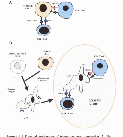
1.2.4.3 Dendritic cell vaccines
Initially it was speculated that the tumour cells were capable o f presenting tumour-
associated antigens to T cells via MHC class I and/or MHC class II molecules (Fig 1.2
A). However, since most tumours are MHC class II negative and in the case of
neuroblastoma MHC class I low, it was not clear how T cells could be primed. Recent
studies on the immune system function have provided a better appreciation of the
mechanism of antigen presentation and the role o f antigen-presenting cells (APC) in
the initiation of cell-mediated immune responses. Increasing evidence that MHC class
I-negative tumour cells are not capable of acting as APCs but priming of naïve CD8^
cells is actually carried out by dendritic cells (DCs) favours the second model of
antigen presentation (Fig 1.2B). This hypothesises that DCs are actually responsible
for presenting tumour specific antigens to CD4^ or CD8^ T cells in the context of
MHC class II and MHC class I complexes (Albert et a l, 1998). These cells are thought to circulate in the tissues, phagocytose antigens from apoptotic cells and
travel to the draining lymph nodes where they cross-present antigen to CD4^ and/or
CD8^ T cells (Banneiji et a l, 1994). The current view is, therefore, that cytokines
produced by modified tumour cells in vaccine studies attract DCs or other
inflammatory cells at the vaccination site and enhance the uptake of apoptotic material
by DCs. Furthermore, generation of CTLs by DCs does not require CD4^ T cell help
if signalling through the CD40 molecule on DCs occurs (Ridge et a l, 1998).
A number of subsequent investigations have focused on the use of DCs in novel
therapies for the treatment of cancer. Since in most cancers identification o f tumour-
specific antigens has not been possible, DCs are pulsed with tumour peptides acid-
eluted from MHC class I molecules on the surface o f tumour cells. Vaccination results
in the eradication o f established murine tumours generating a T cell-dependent anti
tumour response that requires the delivery o f co-stimulatory signals (Zitvogel et a l,
1996). An alternative approach exploits DCs fused with carcinoma cells providing
DCs with the full repertoire of tumour cell antigens. The fused cells exhibit DC