Developmental changes in large-scale network connectivity in autism
Jason S. Nomi
a,1, Lucina Q. Uddin
a,b,⁎
a
Department of Psychology, University of Miami, Coral Gables, FL, USA b
Neuroscience Program, University of Miami Miller School of Medicine, Miami, FL, USA
a b s t r a c t
a r t i c l e i n f o
Article history:
Received 5 December 2014
Received in revised form 19 February 2015 Accepted 28 February 2015
Available online 6 March 2015 Keywords:
Autism spectrum disorder Independent component analysis Resting state fMRI
Functional connectivity Salience network
Background:Disrupted cortical connectivity is thought to underlie the complex cognitive and behavior profile ob-served in individuals with autism spectrum disorder (ASD). Previous neuroimaging research has identified pat-terns of both functional hypo- and hyper-connectivity in individuals with ASD. A recent theory attempting to reconcile conflicting results in the literature proposes that hyper-connectivity of brain networks may be more characteristic of young children with ASD, while hypo-connectivity may be more prevalent in adolescents and adults with the disorder when compared to typical development (TD) (Uddin et al., 2013). Previous work has examined only young children, mixed groups of children and adolescents, or adult cohorts in separate studies, leaving open the question of developmental influences on functional brain connectivity in ASD.
Methods:The current study tests this developmental hypothesis by examining within- and between-network resting state functional connectivity in a large sample of 26 children, 28 adolescents, and 18 adults with ASD and age- and IQ-matchedTD individuals for thefirst time using an entirely data-driven approach. Independent component analyses (ICA) and dual regression was applied to data from three age cohorts to examine the effects of participant age on patterns of within-networkwhole-brain functional connectivity in individuals with ASD compared with TD individuals. Between-network connectivity differences were examined for each age cohort by comparing correlations between ICA components across groups.
Results:Wefind that in the youngest cohort (age 11 and under), children with ASD exhibit hyper-connectivity within large-scale brain networks as well as decreased between-network connectivity compared with age-matchedTD children. In contrast, adolescents with ASD (age 11–18) do not differ from TD adolescents in within-network connectivity, yet show decreased between-network connectivity compared with TD adoles-cents. Adults with ASD show no within- or between-network differences in functional network connectivity compared with neurotypical age-matched individuals.
Conclusions:Characterizing within- and between-network functional connectivity in age-stratified cohorts of in-dividuals with ASD and TD inin-dividuals demonstrates that functional connectivity atypicalities in the disorder are not uniform across the lifespan. These results demonstrate how explicitly characterizing participant age and adopting a developmental perspective can lead to a more nuanced understanding of atypicalities of functional brain connectivity in autism.
© 2015 The Authors. Published by Elsevier Inc. This is an open access article under the CC BY-NC-ND license (http://creativecommons.org/licenses/by-nc-nd/4.0/).
1. Introduction
Early neuroimaging research comparing functional brain connectiv-ity in individuals with autism spectrum disorder (ASD) and typically de-veloping (TD) individuals led to the hypo-connectivity hypothesis proposing that fronto-posterior connectivity deficits are partly respon-sible for cognitive deficits in ASD(Belmonte et al., 2004;Just et al., 2004). Previous task-based functional magnetic resonance imaging
(fMRI) studies primarily using region-of-interest (ROI) analyses found support for the hypo-connectivity theory (Just et al., 2012;Minshew and Williams, 2007). These experiments found hypo-connectivity with-in the temporal–parietal junction in a theory of mind task (Kana et al., 2009), the limbic system in a face perception task (Kleinhans et al., 2008), between the frontal and parietal regions in a working memory task (Koshino et al., 2005), and between the frontal, parietal and occip-ital regions in a cognitive control task (Solomon et al., 2009). However, later task-based fMRI studies found hyper-connectivity in connections involving the posterior superior temporal sulcus in visual search tasks (Shih et al., 2011), the medial temporal lobe in face perception tasks (Welchew et al., 2005), within the left hemisphere in a source recognition task (Noonan et al., 2009), between the inferior frontal gyrus, and between the inferior parietal lobule and the superior temporal sulcus in * Corresponding author at: Department of Psychology, University of Miami, P.O. Box
248185, Coral Gables, FL 33124, USA. Tel.: +1 305 284 3265; fax: +1 305 284 3402. E-mail address:[email protected](J.S. Nomi),[email protected](L.Q. Uddin). 1
Department of Psychology, University of Miami, P.O. Box 248185, Coral Gables, FL 33124, USA.
http://dx.doi.org/10.1016/j.nicl.2015.02.024
2213-1582/© 2015 The Authors. Published by Elsevier Inc. This is an open access article under the CC BY-NC-ND license (http://creativecommons.org/licenses/by-nc-nd/4.0/). Contents lists available atScienceDirect
NeuroImage: Clinical
semantic judgments and letter decision tasks (Shih et al., 2010). These results suggested that ASD could also be characterized by hyper-connectivity, providing evidence against a pure hypo-connectivity ac-count (Kana et al., 2011).
Recently, resting-state fMRI (rsfMRI), has emerged as a powerful tool for examining intrinsic functional brain connectivity in clinical pediatric populations. Resting-state fMRI offers two advantages over task-based fMRI. First, it allows easier data collection from special pop-ulations such as young children with ASD, who have difficulties with long task-based fMRI experiments (Yerys et al., 2009). Second, it identifies underlying intrinsic functional networks that are not confounded by dif-ferences in task performance or strategy difdif-ferences commonly found be-tween individuals with ASD and neurotypical controls. Resting-state fMRI typically involves instructing participants to rest for 5–8 min while blood oxygen level dependent (BOLD) signals are acquired with MRI. By focus-ing on temporal correlations of the BOLD signal between functionally coupled brain regions, it is possible to identify intrinsically connected functional networks that are not confounded by cognitive tasks (Biswal et al., 1995;Bressler and Menon, 2010).
Resting state fMRI studies in neurotypical individuals have identified several major intrinsically connected networks related to visual, motor, auditory, memory and executive processes (Damoiseaux et al., 2006). Re-search examining individuals with ASD has recently focused on hypo- and hyper-connectivity differences observed in two major large-scale brain networks. The default mode network (DMN) consists of key nodes in the posterior cingulate cortex (PCC), the medial temporal lobes (MTL) and the medial prefrontal cortex (MPFC) and is active in self-related tasks such as autobiographical memories or social tasks such as theory of mind (Fox and Raichle, 2007;Spreng et al., 2009). The salience network (SN) involves the anterior insula (AI) and the anterior cingulate cortex (ACC) and is thought to regulate switching of endogenous and exogenous attention to relevant stimuli that helps in guiding behavior (Uddin, 2015). Studies using rsfMRI have found both hypo- and hyper-connectivity of these and other functional networks when comparing individuals with ASD with neurotypical (NT) individuals. Hypo-connectivity in ASD com-pared with TD individuals has been identified in connections between the insula and amygdala (Ebisch et al., 2011) and most consistently be-tween connections of nodes within the DMN(Assaf et al., 2010;Ebisch et al., 2011;Kennedy and Courchesne, 2008a;Monk et al., 2009;Weng et al., 2010). Hyper-connectivity in ASD compared with TD individuals has been identified in motor and visual networks, as well as the DMN and SN(Uddin et al., 2013;Washington et al., 2014), and between striatal areas and the insula (Di Martino et al., 2011). Finally, one study has even demonstrated extremely small to no differences in functional connectivi-ty in ASD compared with neuroconnectivi-typical adults (Tyszka et al., 2014), pro-ducing additional conflicting evidence.
Although methodological differences surely contribute to the mixed
findings in the literature (Muller et al., 2011), a more recent idea attempting to reconcile these discrepantfindings proposes that hyper-connectivity may be more characteristic of young children with ASD, while hypo-connectivity may begin to emerge in adolescence and persist into adulthood. In a review of the rsfMRI functional connectivity literature in ASD it is suggested that studies demonstrating hyper-connectivity have typically examined children less than 12 years of age, while studies demonstrating hypo-connectivity have typically ex-amined adolescents and adults over the age of 12 (Uddin et al., 2013). The authors proposed that a developmental account hypothesizing early childhood hyper-connectivity and later adolescent and adult hypo-connectivity in ASD compared with TD could partially account for the mixed functional connectivityfindings in the literature.
Previous functional connectivity studies have focused on a single age group (e.g., childhood, adolescence, or adulthood), mixed age groups (e.g., combining childhood and adolescence, or adolescence and adult-hood), or used a linear regression correlational approach across a single group of subjects containing various age ranges (Assaf et al., 2010;
Kennedy and Courchesne, 2008b;Monk et al., 2009). Several studies
exploring whole-brain connectivity have included subjects with a wide range of ages, allowing for the possibility that a certain age group was driving the functional connectivityfindings in the results (Assaf et al., 2010;Gotts et al., 2012). Therefore, an important gap in the literature concerns the principled examination of functional connec-tivity alterations in ASD across different developmental stages. In the current work we stratify individuals into age cohorts to directly test if some of the mixedfindings throughout the literature can be accounted for by explicitly sorting groups of ASD and TD participants according to their ages.
An additional aspect of network connectivity that has received less at-tention in ASD research is how correlations between networks compare to that observed in the neurotypical population. Previous research has shown that the DMN, referred to as a‘task-negative network’(TNN), typ-ically exhibits negative correlations with task-positive networks (TPN) such as the dorsal attention network (DAN) (Fox et al., 2005). The TNN nomenclature refers to the fact that nodes of the DMN typically show re-ductions in activity when a participant is focused on a task demanding ex-ogenous attention while TPN refers to the fact that nodes of the DAN typically show increases in activity during such a task. Thus these net-works are often referred to as“anti-correlated”because when a TPN is ac-tive the TNN is not, and vice versa. The relationship between these networks relates to behavioral performance in the neurotypical popula-tion (Kelly et al., 2008), but is not well understood in ASD. Characteriza-tion of relaCharacteriza-tionships between these two networks may have important implications for understanding brain dynamics in ASD.
A challenge to synthesizing the functional connectivity literature in autism is that several studies have used ROI-based analyses that are
dif-ficult to compare with each other, as they are often linked to hypotheses about specific functional circuits (Abrams et al., 2013;Kennedy and Courchesne, 2008b;Lynch et al., 2013). The current study sought to compare whole-brain functional connectivity in ASD and TD individuals using an entirely data-driven approach. We explored the nature and ex-tent of functional differences both within- and between-networks when comparing ASD and TD individuals across three age groups— chil-dren (under 11), adolescents (11–18), and adults (over 18). In order to assess within-network group differences in functional connectivity, we used independent component analysis (ICA) (Beckmann et al., 2005) across three different age groups to examine if the developmental tra-jectory of hyper- to hypo-connectivity in children to adults respectively, as predicted by Uddin and colleagues (Uddin et al., 2013) would be present. To assess between-network group differences in functional connectivity, we applied a network analysis to examine how correla-tions between networks potentially differ across the three age groups. This approach of exploring within- and between-network functional connections was adapted to elucidate how large-scale brain networks in ASD compare to those observed in age-matchedTD individuals across development.
2. Methods 2.1. Participants
We used data from the Autism Brain Imaging Data Exchange (ABIDE), a publicly available data set (http://fcon_1000.projects.nitrc. org/indi/abide/) (Di Martino et al., 2014). Only data collected at the New York University Langone Medical Center were utilized to avoid cross-study methodological acquisition differences. To explore the ef-fects of participant age on functional connectivity, we divided the data into three age groups of ASD and TD participants: young children under 11 years of age (n= 52), adolescents from 11–18 years of age (n= 56), and adults over 18 years of age (n= 36;Table 1). Individuals with ASD had a clinical DSM-IV diagnosis of Autistic Disorder, Asperger's syndrome, Pervasive Developmental Disorder Not-Otherwise Specified (PDD-NOS) while TD participants were required to have no Axis-I disorders based on the KSADS-PL questionnaire.
Within each of the three cohorts, there were no significant group dif-ferences in age and full-scaleIQ between participants with ASD and TD participants (psN.09). A 2 × 3 mixed-modelANOVA on IQ showed that there was a marginally significant main effect of age (p= .06) but no main effect of group (p= .1) and no age × group interaction (p= .8). Fol-low upt-tests showed that the children (M = 110.40) had higher IQ scores than adolescents (M = 104.38;p= .03), and adults (M = 112.08) had higher IQ scores than adolescents (p= .01), with no differ-ences between children and adults (p= .6). As is typical in young chil-dren with ASD, these chilchil-dren had greater RMS relative and absolute motion than TD individuals (Supplementary Table 1; relative ASD = .10, TD = .06,p= .001; absolute ASD = .39, TD = .23,p= .008). There were no motion differences between the adolescent and adult groups (psN.15) (Supplementary Table 1). There were no differences between individuals with ASD across the three age groups for all ASD diagnostic questionnaires (ADI and ADOS; one way ANOVAs,pN.2).
For the overall group ICA used to create templates for subsequent analyses, we randomly selected 18 participants from each age group (18 × 6 = 108) to derive the independent components (ICs). This was done in order to avoid non-network noise differences across the three age groups by creating a common template of ICs for use in the dual re-gression analysis. There were no differences in full-scaleIQ for any group of 18 participants (2 × 3 ANOVA:p= .59), nor were there any differ-ences for all ASD diagnostic questionnaires (ADI and ADOS) across the three age cohorts (one way ANOVAs,pN.38).
2.2. Data acquisition
Resting state fMRI data were collected on a 3 T Siemens Allegra scan-ner using an echo-planscan-ner imaging (EPI) sequence (TR = 2000 ms;
TE = 15 ms; flip angle = 90°; FOV = 240 mm; voxel size = 3 × 3 × 4 mm; number of slice = 33, 4 mm slice thickness). Each resting-state scan lasted for 6 min, consisting of 180 volumes collected while participants were asked to relax with their eyes open andfixate on a projection screen displaying a white cross hair on a black background.
Anatomical images were acquired using a magnetization prepared gradient echo sequence (TR = 2530 ms; TE = 3.25 ms; inversion time = 8.07 min;flip angle = 7°; 128 slices; 1 volume; FOV = 256 mm) (Di Martino et al., 2014).
2.3. Image preprocessing
Functional MRI data were preprocessed using FSL 5.06 (http://fsl. fmrib.ox.ac.uk/fsl/fslwiki/). Thefirst three volumes of each data set were deleted. Preprocessing steps included motion correction, inter-leaved slice-timing correction, spatial smoothing (full width at half maximum = 5 mm), and high pass temporalfiltering using a localfit of a straight line (Gaussian-weighted least-squares straight linefitting with sigma = 100 s). Images were then normalized to the Montreal Neurological Institute (MNI) 152 stereotactic space (2 mm) using the default settings in FSL3s FEAT toolbox by applying a linear transforma-tion with 12 degrees of freedom. Global signal regression was not ap-plied (Saad et al., 2012).
2.4. Within-network connectivity: dual regression ICA
In order to examine within-network differences in functional connectivity, the dual regression approach in FSL (v 5.06) was applied to preprocessed images (Beckmann and Smith, 2004). Cur-rently, dual regression is the preferred data-driven approach for exploring between-population differences in large-scale functional connectivity patterns (Filippini et al. 2009). The overall group preprocessed data consisting of 108 subjects were concatenated and subjected to an ICA using MELODIC(http://fsl.fmrib.ox.ac.uk/ fsl/fslwiki/MELODIC) in FSL. Next, 25 ICs were created representing large-scalegroup-level functional networks. Visual inspection of these group-level ICs was used to identify those best representing previously identified functional networks (Damoiseaux et al., 2006). The remaining components were considered noise or artifacts such as movement, white matter, or ventricles and were not subject-ed to further analysis.
To account for head motion differences between the child ASD and TD groups, and keep analyses consistent between age groups, the time courses of all 144 subjects were subjected to a covariate regression of the Friston 24 motion parameters (6 typical motion parameters for each volume, the preceding volume, and each of the 12 derivatives) using the DPARSF-A toolbox (http://rfmri.org/DPARSF). This 24-parameter model has been shown to better reduce the influence of motion effects than other models (Satterthwaite et al., 2012;Yan et al., 2013). The covar-iate regression was not applied to the initial ICA because the initial ICA creates motion components that were then discarded from further analysis.
The ICs of interest were then compared to motion-regressedparticipant-specific time courses and spatial maps with a dual regression algorithm (http://fsl.fmrib.ox.ac.uk/fsl/fslwiki/DualRegression) producing group difference maps for each component within each age group. Permutation testing using the randomize feature in FSL was conducted using the de-fault settings (5000 permutations) to create difference maps between groups for each component of interest. The resulting group difference maps were thresholded using threshold-free cluster enhancement with an alpha level of .05 (corrected). Correction for multiple component test-ing was not applied in this case, as in previous similar studies (Uddin et al., 2013). Table 1 Participant demographics. ASD TD pvalue Children (b11) Mean age 9.51 (1.12) 9.10 (1.32) .22 Age range 7.15–10.96 6.47–10.86 Gender 24M/2F 19M/7F Full IQ 107.77 (16.16) (76–142) 113.04 (13.67) (80–136) .10 ADI social scorea
19.4 (5.42) (7–27) ADI verbal scorea
16.16 (3.92) (8–22) ADI RRBa 5.92 (2.27) (3–10) ADOS communication 3.31 (1.85) (0–7) ADOS social 7.58 (2.67) (4–14) Adolescent (11–18) Mean age 13.71 (1.79) 14.01 (1.74) .53 Age range 11.01–17.88 11.32–16.93 Gender 23M/5F 23M/5F Full IQ 103.57 (15.45) (78–132) 105.18 (9.90) (80–121) .65 ADI social scoreb
20.46 (5.53) (13–28) ADI verbal scorea
15.78 (4.06) (8–23) ADI RRBa 6.07 (2.66) (0–12) ADOS communication 3.64 (1.52) (1–6) ADOS social 8.64 (2.98) (2–14) Adults (N18) Mean age 24.13 (3.92) 25.41 (5.87) .45 Age range 18.58–39.1 18.59–31.78 Gender 14M/4F 14M/4F Full IQ 108.06 (13.86) (80–137) 116.11 (14.20) (81–139) .09 ADI social scorec
18 (6.14) (9–27) ADI verbal score 6.46 (5.95) (8–25) ADI RRB score 4.62 (2.36) (2–9) ADOS communication 3.72 (1.36) (2–6) ADOS social 7.44 (3.14) (2–12) a
Score missing for 1 participant. b
Score missing for 2 participants. c
2.5. Between-network connectivity: FSL Nets
To examine between-network differences in functional connectivity for each age cohort, the FSL Nets analysis package was implemented in Matlab (http://fsl.fmrib.ox.ac.uk/fsl/fslwiki/FSLNets). This analysis takes the participants3time courses from the dual regression analysis and subjects them to between-network comparisons that determine how independent components from the overall ICA are correlated with each other (Smith et al., 2013). Between-subject testing is then con-ducted across correlation values acquired for pairs of independent components.
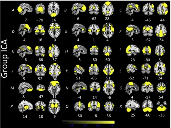
3. Results 3.1. Group ICA
The group ICA from the subset of 108 subjects produced 25 ICs. Of these 25 ICs, 7 were determined to be noise-related artifacts representing cerebral spinal fluid, ventricles, and head motion. These ICs were discarded from further analysis, leaving 18 ICs of interest used in each dual regression analysis (Fig. 1). Group ICAs were also conducted for each age cohort separately for comparison purposes. Each age cohort ex-hibited similar components to the overall ICA (Supplementary Figs. 1–3). 3.2. Overall between-network comparisons
The overall FSL Nets analysis was conducted on each age group (Fig. 2). Boxes below the diagonal line represent full correlation com-parisons, while boxes above the diagonal line represent partial correla-tion values. Full correlacorrela-tion comparisons allow for the influence of other network values on pairs of interest, while partial correlations are a more direct measure of the relationship between pairs of networks. Positive correlations for each age group can be seen between the DAN and
higher-level visual areas (labeled 1) while negative correlations be-tween the DAN and two DMN components (labeled 2 and 3) are also present in both the full and partial correlation matrices.
3.3. Within-network connectivity: children with ASD vs. TD children For the within-network comparison, two components showed sig-nificant hyper-connectivity in ASD compared with TD (ASDNTD,
Fig. 3). ComponentCrepresenting a network that included nodes of both the DMN and central-executive network showed hyper-connectivity in the right frontal pole. ComponentPrepresenting the insula and subcortical areas showed hyper-connectivity in bilateral areas that included the insula, thalamus, hippocampus, and amygdala. No TDNASD functional connectivity differences were observed in any of the networks examined.
3.4. Between-network connectivity: children with ASD vs. TD children The between-network comparison showed only one significant dif-ference for partial correlation values between componentsFandB representing the DMN(Fig. 2, labeled 4; FWE corrected:p= .017). The difference was such that children with ASD showed a significantly smaller correlation between these two networks compared with TD children. No other differences emerged for full or partial correlation comparisons (FWE corrected:pN.07.).
3.5. Within-network connectivity: adolescents with ASD vs. TD adolescents There were no significant within-network differences between ado-lescents with ASD and TD adoado-lescents, in either direction (ASDNTD or TDNASD).
3.6. Between-network connectivity: adolescents with ASD vs. TD adolescents The between-network comparison showed one significant difference for full correlation values between componentsBandPrepresenting the DMN and a subcortical/insula network. The difference was such that indi-viduals with ASD had a significantly smaller correlation between the two components (Fig. 2, labeled 5; FWE corrected:p= .004). No other differ-ences emerged for between-network comparisons (FWE corrected: pN.08).
3.7. Adults with ASD vs. NT adults
No significant group differences in within- or between-network con-nectivity were observed for any of the networks examined, in either di-rection (ASDNTD or TDNASD).
4. Discussion
The results of the current study demonstrate age-specific patterns of whole-brain functional connectivity atypicalities in ASD. Hyper-connectivity within large-scale brain networks in ASD was observed in young children under the age of 11, with no within-network differences in functional connectivity in adolescents and adults with ASD compared with neurotypical individuals. Abetween-network analysis showed that
children with ASD had a smaller correlation between two DMN networks, while adolescents with ASD had a smaller correlation between the DMN and a subcortical/insula network compared with TD individuals. Adults with ASD showed no differences in either within- or between-network functional connectivity compared to neurotypical controls. Overall, the re-sults demonstrate that children with ASD exhibit atypical within- and between-network functional connectivity, adolescents with ASD show atypical between-network functional connectivity, and adults with the disorder do not differ from their age-matched peers on either of these measures.
4.1. Within-network functional connectivity
The results from this data-driven developmental approach to inves-tigating whole-brain functional connectivity between ASD and TD indi-viduals are an importantfirst step in helping to resolve discrepancies in the ASD rsfMRI literaturefinding hyper- and hypo-connectivity of in-trinsically functional networks across different studies. Importantly, these results are partially in accord with the developmental trajectory hypothesis proposed by Uddin et al. (2013) predicting hyper-connectivity in young children with ASD and hypo-hyper-connectivity in adults with ASD. Although hypo-connectivity was not found in the adult group, the general developmental trajectory shows widespread hyper-connectivity in children with ASD that reduces with age. The
Fig. 2.FSL Nets between network correlations for each age group. Full correlations are shown below the diagonal line with partial correlations shown above the diagonal line. Letters on each axis indicate specific components fromFig. 1. Number 1 within each correlation matrix represents a positive correlation between the dorsal attention network (K) and higher order visual areas (I). Numbers 2 and 3 represent negative correlations between default mode networks (B and F) and the dorsal attention network (K). Number 4 represents a significant dif-ference in partial correlations between default mode networks (B and F) for children (TDNASD). Number 5 represents a significant difference in full correlations between default mode (B) and subcortical/insula networks (P) for adolescents (TDNASD). Groupings on top of each matrix represents hierarchical clustering of component timeseries.
results also confirm thefindings of previous rsfMRI studies showing hyper-connectivity in young children with ASD(Di Martino et al., 2011; Lynch et al., 2013;Supekar et al., 2013;Uddin et al., 2013;
Washington et al., 2014) along with no differences in whole-brain func-tional connectivity in adults with ASD (Tyszka et al., 2014).
The current study found hyper-connectivity within the DMN in chil-dren with ASD. This replicatesfindings ofUddin et al. (2013), and other studies showing hyper-connectivity of DMN nodes in children with ASD(Lynch et al., 2013;Washington et al., 2014). Although the exact function of the DMN is still not clear, activity in key nodes of the DMN such as the MPFC and PCC is found to decrease during tasks with a high cognitive demand (Raichle et al., 2001) and increase during rest and also during tasks of a social nature involving self-reflection(Buckner et al., 2008;Harrison et al., 2008;Northoff et al., 2006). Previously, most studiesfinding aberrant DMN function in ASD have found hypo-connectivity across nodes of the DMN(Kennedy and Courchesne, 2008a;Monk et al., 2009;von dem Hagen et al., 2013;
Weng et al., 2010). However, these studies were conducted in adoles-cents and adults. Thus, a developmental account predicting within-networkhyper-connectivity in children with ASD that gradually recedes over time would be a plausible explanation as to why some previous studies have found hyper- opposed to hypo-connectivity across nodes of the DMN.
The mechanisms underlying the observed widespread functional hyper-connectivity in children with ASD are still unclear. Previous re-search has shown that ASD is characterized by increased head circumfer-ence in childhood (Lainhart et al., 1997) while in-vivo(Courchesne et al., 2003) and post-mortem studies (Courchesne et al., 2011) have shown in-creased neuronal growth in children with ASD from 2–5 years and 2–16 years respectively. Additionally, another post-mortem study has
shown both increased spine density and decreased synaptic pruning across childhood and adolescence (Tang et al., 2014). Although no direct evidence exists that links neuronal density to functional connectivity within neural networks in the context of rsfMRI studies, it is plausible that changes in neural density are related to changes in functional con-nectivity. Thus, increased neural density found early in life in children with ASD in these post-mortem studies could be related to increased functional connectivity found in rsfMRI studies.
Additionally, mouse models have also demonstrated that decreases in synaptic pruning (Gogolla et al., 2014;Tang et al., 2014) and increases in synaptic turnover (Isshiki et al., 2014) during critical developmental periods are related to atypical behavior similar to those found in human individuals with ASD. These studies suggest that critical periods of synaptic development may influence cortical function in ASD, making the study of developmental trajectories imperative for research examin-ing the disorder in humans. Within the context of the current study, changes in synaptic pruning could be related to changes in functional connectivity found across the lifespan such that increased synaptic pruning could be responsible for normalizing the increased neuronal growth found in children with ASD. Thus, increased synaptic pruning of extra neuronal growth could help to normalize the amount of neu-rons present in individuals with ASD during development that would result in a more typical pattern of functional connectivity of individuals with ASD as they get older. Unfortunately, a large amount of ASD re-search is conducted on adolescents and adults, probably due to the difficulty in acquiring artifact-free fMRI data from younger children, es-pecially young children with psychiatric disorders such as ASD(Yerys et al., 2009). The few studies that have examined rsfMRI functional con-nectivity in young children with ASD have generally found hyper-connectivity between brain areas (Di Martino et al., 2011;Lynch et al.,
Fig. 3.Functional networks showed greater connectivity for children with ASD compared with TD children in 2 out of 18 networks examined: default mode (top,C), and insula/subcortical (bottom,P).
2013;Uddin et al., 2013;Washington et al., 2014) demonstrating the importance of exploring child populations in ASD research.
One previously proposed explanation for thefindings of changing functional connectivity across development in ASD is that the pubertal period during adolescence is responsible for changes in underlying brain organization, and that pubertal hormones may differentially affect developmental trajectories in the disorder (Uddin et al., 2013). Hor-monal changes during puberty have been linked with changes in both gray and white matter (Herting et al., 2012). However, there have been no cross-sectional or longitudinal studies examining changes in functional connectivity that accompany the pubertal transition in humans, in either typical or atypical development.
Previous studies showing hypo-connectivity in adults with ASD using rsfMRI data have mostly used seed ROI-based functional connec-tivity analysis while also including mixed groups of participants youn-ger than 18 (Cherkassky et al., 2006;Ebisch et al., 2011;Kennedy and Courchesne, 2008a;Monk et al., 2009) whereas the current study exam-ined resting state data using a whole brain ICA approach with subjects 18 years and older in the adult group. The current study and the previ-ous studyfinding a lack of hypo-connectivity in adults (Tyszka et al., 2014) used only participants older than 18 years in a data-drivenICA analysis. However, two other studies using ICA with participants over 18 have found hypo-connectivity in ASD(Mueller et al., 2013;von dem Hagen et al., 2013). Thus, it is still unclear why some studies have found hypo-connectivity in adults with ASD, compared with the results of the current study and the previous study (Tyszka et al., 2014) which found no differences in functional connectivity between adults with ASD and neurotypical adults. In addition to the proposed developmental account whichfinds empirical support from the current study, others have suggested that hypo-connectivity is generally found in task-driven studies using seed-based analysis approaches compared to studies notfinding hypo-connectivity that have used whole brain analysis approaches combined with task-regression analysis to re-move task-related activity (Muller et al., 2011). These and other methodological factors may contribute to some of the inconsis-tencies in the literature.
Three earlier studies examined functional connectivity in ASD using linear regression correlations between functional connectivity and age.Wiggins et al. (2011)examined the DMN in groups of ASD and TD adolescents (10.25–18.97) and found that connectivity be-tween posterior DMN nodes such as the inferior parietal lobule had decreased connectivity with the superior frontal gyrus as age in-creased in ASD, but connectivity inin-creased with age in the TD group.Padmanabhan et al. (2013)examined cortico-striatal connec-tivity in groups of ASD and TD individuals (8–36 years) and found that both groups showed general decreases in cortico-striatal con-nections as age increased. Additionally, the ASD group had increased connectivity of the striatum with the cerebellum, fusiform gyrus, and inferior/superior temporal gyri with age while these connections de-creased in TD individuals. However, when controlling for age, there was ASD striatal hyper-connectivity with the parietal cortex but hypo-connectivity with the pre-frontal cortex compared with TD in-dividuals. Bos et al. (2014) found that adolescents with ASD (8.3–15.1) had little to no within-network connectivity differences compared to TD adolescents (6.4–15.8) using a group ICA, but found that the ASD group had connectivity between the right insula and other nodes of the DMN that increased with age, while decreas-ing connectivity with age was observed in the TD group.
Although previous studies have also found hyper-connectivity in the DMN and insula (Bos et al., 2014), these results were found using a linear correlation approach as opposed to the whole brain ICA used in the cur-rent study. AlthoughBos et al. (2014)found no group differences be-tween ASD and TD in an overall group ICA, they used age ranges that combined children with adolescents (6.4–15.8) while the current study separated children under 11 and adolescents from 11–18 and found hyper-connectivity in children, and no differences in adolescents. Thus,
future research is needed to further explore the effects of stratifying sam-ples by age.
4.2. Between-network functional connectivity
The overall between-network results in the current study showed that both ASD and TD groups for all ages had similar anti-correlations between task-positive and task-negative networks represented by the DAN and DMN respectively. Additionally, both ASD and TD groups for all ages had positive correlations between the DAN and higher-levelvisual areas. This shows that individuals with ASD show typical anti-correlations between the DAN and DMN and typical positive corre-lations between the DAN and higher-level visual areas. Both of these results replicate previous work exploring between-network connectiv-ity of resting state data in the neurotypical population (Smith et al., 2013).
Previously,Kennedy and Courchesne (2008b) utilized a hypothesis-driven seed ROI-based approach to demonstrate that adults with ASD show typical anti-correlations between the DMN and DAN. The current study extends this work by using a completely data-driven method to demonstrate typical anti-correlations between the DMN and DAN, in addition to typical positive correlations between the DAN and higher-level visual areas. In addition, the novel contribution of the current study demonstrates these results apply to children and adolescents with ASD in addition to adults.
Between-network differences were observed in the current study between DMN components for the child cohort and between a DMN and subcortical/insula component for the adolescent cohort. The difference for children (ASDbTD) was found in the partial-correlationcomparison, while the difference for adolescents was found in the full-correlation comparison. The partial-correlation difference for children suggests that DMN networks had less direct functional rela-tionships with each other, demonstrating atypical cooperation between DMN components in children with ASD. The full-correlation difference in adolescents would suggest that a weaker functional relationship be-tween the DMN and subcortical/insula component for children with ASD is moderated by another network.
Starck et al. (2013) used an ICA approach to examine the DMN and its subnetworks in adolescents with ASD compared with TD adoles-cents. Their study found no differences in within-networkDMN func-tional connectivity compared with TD adolescents, consistent with the current findings. They did find significantly reduced correlations between anterior and posterior DMN subnetworks in ASD.
Additionally, the current results are in accord with previous ideas suggesting that increased within-network connectivity in children with ASD could be responsible for reduced between-network connec-tivity as tighter coupling within networks could lead to reduced cou-pling between networks (Uddin et al., 2013). In the current study, this may explain the reduced coupling between DMN components for chil-dren with ASD. The lack of within-networkhyper-connectivity in ado-lescents suggests that reductions in between-network coupling can still occur in the absence of within-network hyper-connectivity. In the case of adolescents, the between-network differences were found between the DMN and a subcortical/insula network. Both of these net-works are related to cognitiveflexibility and may contribute to some of the behavioral patterns found in ASD, as discussed in the following section. However, it is not possible to determine from the current study if there is a direct relationship that relates within- and between-network coupling.
4.3. Insula and the default mode network
Although the current study did notfind any functional connectivity group differences within the context of the salience network, there were within- and between-network differences involving a subcorti-cal/insula component. Thus, although the currentfindings implicate
the insula as a source of atypical functional connectivity in ASD, this does not occur within the context of the SN. The exact reason for this di-vergence is unclear. One possible reason is that insula activation within the SN in the context of the current study was generally related to ho-mogeneous activity across the entire insula—afinding typical in ICA based studies of the SN (Seeley et al., 2007;Uddin et al., 2013;Uddin et al., 2011). However, other work suggests that insula subregions such as the dorsal anterior, ventral anterior, and posterior insula regions vary substantially in their functional roles and patterns of connectivity (Deen et al., 2011;Uddin et al., 2014). The ventral anterior insula has been shown to have direct structural connections with subcortical areas in primate studies (Mesulam and Mufson, 1982) as well as sharing task-based subcortical activations in fMRI studies of humans (Uddin et al., 2014;Uddin, 2015). Additionally, the ventral anterior insula has been shown to have connections to subcortical limbic areas in task-based activation studies (Uddin et al., 2014). Thus, it may be possible that different insula subregions may be driving the functional connec-tions of the insula with the ACC in the context of the SN and subcortical areas with the insula in the context of the subcortical/insula component found in the current study. Thus, different insula subregions may be driving the atypical functional connections found in the current study compared to previous studiesfinding atypical functional connections of the insula within the context of the SN(Uddin et al., 2013).
Thefindings of the current study and previous studies strongly im-plicate atypical within- and between-network functional connectivity of the DMN and insula as possible brain markers of ASD. The major nodes of the DMN include the posterior parietal and lateral prefrontal cortices while the nodes of the SN include the anterior cingulate and an-terior insular cortices; areas that are important inflexibly switching be-tween internal cognitive and external information (Cole et al., 2013;
Dosenbach et al., 2007;Uddin et al., 2014;Uddin et al., 2011). Accord-ingly,Uddin et al. (2014)have demonstrated that atypical effective con-nectivity of these brain areas is correlated with restrictive and repetitive patterns of behavior in children with ASD such that greater atypical con-nectivity is related with more severe behavior deficits. This suggests that the atypical functional connectivity of these networks that are im-portant in cognitiveflexibility contribute to the restrictive and repeti-tive behaviors that often characterize the disorder. The current study shows both within- and between-network differences involving the DMN and insula that further implicate the atypical functional connec-tions of the two as a possible biomarker of brain function in ASD. 5. Conclusions
In sum, the currentfindings support adopting a developmental perspective to help reconcile the heterogeneousfindings of functional hypo- and hyper-connectivity observed in the rsfMRI literature in ASD. These results demonstrating group differences specific to certain age cohorts highlight the utility of carefully considering developmental stage in studies of functional brain connectivity in ASD. Wefind that while children show atypical within and between-network functional relationships, adolescents exhibit fewer such differences and adults are indistinguishable from age-matched neurotypical peers on such measures. The fact that both within- and between-network differences diminish across the lifespan could offer an explanation for some of the improved function often found in adults with ASD compared to children with ASD. These results also highlight the importance of considering within- and between-network whole brain functional connections in conjunction with a developmental approach in order to better charac-terize brain connectivity in ASD.
Although the current study is an importantfirst step in taking a devel-opmental approach to investigating differences in functional connectivity between individuals with ASD and neurotypical individuals, future stud-ies should explore the influence of various age groupings to more precise-ly determine where differences in hyper- and hypo-connectivity begin to emerge between specific brain areas. As an increasing awareness of the
impact of development on brain function in ASD has begun to emerge (Picci and Scherf, 2014;Uddin et al., 2013) more studies that explore the impact of age on brain-based biomarkers in ASD are needed in order to provide a better picture of the developmental maturation of func-tional connectivity patterns that emerge across the lifespan in individuals with ASD.
Conflicts of interest None.
Acknowledgments
This work was supported by a National Institute of Mental Health Career Development award (K01MH092288), a NARSAD Young Investi-gator Award, and a Slifka/Ritvo Innovation in Autism Research Award from the International Society for Autism Research to LQU. The content is solely the responsibility of the authors and does not necessarily repre-sent the official views of the NIMH or the NIH.
Appendix A. Supplementary data
Supplementary data to this article can be found online athttp://dx. doi.org/10.1016/j.nicl.2015.02.024.
References
Abrams, D.A., Lynch, C.J., Cheng, K.M., Phillips, J., Supekar, K., Ryali, S., Uddin, L.Q., Menon, V., 2013. Underconnectivity between voice-selective cortex and reward circuitry in children with autism. Proc. Natl. Acad. Sci. U S A 110 (29), 12060–12065.http://dx. doi.org/10.1073/pnas.130298211023776244.
Assaf, M., Jagannathan, K., Calhoun, V.D., Miller, L., Stevens, M.C., Sahl, R., O’Boyle, J.G., Schultz, R.T., Pearlson, G.D., 2010. Abnormal functional connectivity of default mode sub-networks in autism spectrum disorder patients. Neuroimage 53 (1), 247–256. http://dx.doi.org/10.1016/j.neuroimage.2010.05.06720621638.
Beckmann, C.F., DeLuca, M., Devlin, J.T., Smith, S.M., 2005. Investigations into resting-state connectivity using independent component analysis. Philos. Trans. R. Soc. Lond., B, Biol. Sci. 360 (1457), 1001–1013.http://dx.doi.org/10.1098/rstb.2005.163416087444. Beckmann, C.F., Smith, S.M., 2004. Probabilistic independent component analysis for
func-tional magnetic resonance imaging. I. E.E.E. Trans. Med. Imaging 23 (2), 137–152. http://dx.doi.org/10.1109/TMI.2003.82282114964560.
Belmonte, M.K., Allen, G., Beckel-Mitchener, A., Boulanger, L.M., Carper, R.A., Webb, S.J., 2004. Autism and abnormal development of brain connectivity. J. Neurosci. 24 (42), 9228–9231.http://dx.doi.org/10.1523/JNEUROSCI.3340-04.200415496656. Biswal, B., Yetkin, F.Z., Haughton, V.M., Hyde, J.S., 1995. Functional connectivity in the
motor cortex of resting human brain using echo-planar MRI. Magn. Reson. Med. 34 (4), 537–541.http://dx.doi.org/10.1002/mrm.19103404098524021.
Bos, D.J., van Raalten, T.R., Oranje, B., Smits, A.R., Kobussen, N.A., Belle, J.v, Rombouts, S.A., Durston, S., 2014. Developmental differences in higher-orderresting-state networks in autism spectrum disorder. Neuroimage Clin. 4, 820–827.http://dx.doi.org/10. 1016/j.nicl.2014.05.00724936432.
Bressler, S.L., Menon, V., 2010. Large-scale brain networks in cognition: emerging methods and principles. Trends Cogn. Sci. 14 (6), 277–290.http://dx.doi.org/10. 1016/j.tics.2010.04.00420493761.
Buckner, R.L., Andrews-Hanna, J.R., Schacter, D.L., 2008. The brain3s default network: anat-omy, function, and relevance to disease. Ann. N. Y. Acad. Sci. 1124 (1), 1–38.http:// dx.doi.org/10.1196/annals.1440.01118400922.
Cherkassky, V.L., Kana, R.K., Keller, T.A., Just, M.A., 2006. Functional connectivity in a base-line resting-state network in autism. Neuroreport 17 (16), 1687–1690.http://dx.doi. org/10.1097/01.wnr.0000239956.45448.4c17047454.
Cole, M.W., Reynolds, J.R., Power, J.D., Repovs, G., Anticevic, A., Braver, T.S., 2013. Multi-task connectivity revealsflexible hubs for adaptive task control. Nat. Neurosci. 16 (9), 1348–1355.http://dx.doi.org/10.1038/nn.347023892552.
Courchesne, E., Carper, R., Akshoomoff, N., 2003. Evidence of brain overgrowth in thefirst year of life in autism. JAMA 290 (3), 337–344.http://dx.doi.org/10.1001/jama.290.3. 33712865374.
Courchesne, E., Mouton, P.R., Calhoun, M.E., Semendeferi, K., Ahrens-Barbeau, C., Hallet, M.J., Barnes, C.C., Pierce, K., 2011. Neuron number and size in prefrontal cortex of chil-dren with autism. JAMA 306 (18), 2001–2010.http://dx.doi.org/10.1001/jama.2011. 163822068992.
Damoiseaux, J.S., Rombouts, S.A., Barkhof, F., Scheltens, P., Stam, C.J., Smith, S.M., Beckmann, C.F., 2006. Consistent resting-state networks across healthy subjects. Proc. Natl. Acad. Sci. U S A 103 (37), 13848–13853.http://dx.doi.org/10.1073/pnas. 060141710316945915.
Deen, B., Pitskel, N.B., Pelphrey, K.A., 2011. Three systems of insular functional connectiv-ity identified with cluster analysis. Cereb. Cortex 21 (7), 1498–1506.http://dx.doi. org/10.1093/cercor/bhq18621097516.
Di Martino, A., Kelly, C., Grzadzinski, R., Zuo, X.N., Mennes, M., Mairena, M.A., Lord, C., Castellanos, F.X., Milham, M.P., 2011. Aberrant striatal functional connectivity in chil-dren with autism. Biol. Psychiatry 69 (9), 847–856.http://dx.doi.org/10.1016/j. biopsych.2010.10.02921195388.
Di Martino, A., Yan, C.G., Li, Q., Denio, E., Castellanos, F.X., Alaerts, K., Anderson, J.S., Assaf, M., Bookheimer, S.Y., Dapretto, M., Deen, B., Delmonte, S., Dinstein, I., Ertl-Wagner, B., Fair, D.A., Gallagher, L., Kennedy, D.P., Keown, C.L., Keysers, C., Lainhart, J.E., 2014. The autism brain imaging data exchange: towards a large-scale evaluation of the intrinsic brain architecture in autism. Mol. Psychiatry 19 (6), 659–667.http://dx.doi.org/10. 1038/mp.2013.7823774715.
Dosenbach, N.U., Fair, D.A., Miezin, F.M., Cohen, A.L., Wenger, K.K., Dosenbach, R.A., Fox, M.D., Snyder, A.Z., Vincent, J.L., Raichle, M.E., Schlaggar, B.L., Petersen, S.E., 2007. Distinct brain networks for adaptive and stable task control in humans. Proc. Natl. Acad. Sci. U S A 104 (26), 11073–11078.http://dx.doi.org/10.1073/pnas.070432010417576922.
Ebisch, S.J., Gallese, V., Willems, R.M., Mantini, D., Groen, W.B., Romani, G.L., Buitelaar, J.K., Bekkering, H., 2011. Altered intrinsic functional connectivity of anterior and posterior insula regions in high-functioning participants with autism spectrum disorder. Hum. Brain Mapp. 32 (7), 1013–1028.http://dx.doi.org/10.1002/hbm.2108520645311. Filippini, N., MacIntosh, B.J., Hough, M.G., Goodwin, G.M., Frisoni, G.B., Smith, S.M.,
Matthews, P.M., Beckmann, C.F., Mackay, C.E., 2009. Distinct patterns of brain activity in young carriers of the APOE-epsilon4 allele. Proc. Natl. Acad. Sci. U S A 106 (17), 7209–7214.http://dx.doi.org/10.1073/pnas.081187910619357304.
Fox, M.D., Raichle, M.E., 2007. Spontaneousfluctuations in brain activity observed with functional magnetic resonance imaging. Nat. Rev. Neurosci. 8 (9), 700–711.http:// dx.doi.org/10.1038/nrn220117704812.
Fox, M.D., Snyder, A.Z., Vincent, J.L., Corbetta, M., Van Essen, D.C., Raichle, M.E., 2005. The human brain is intrinsically organized into dynamic, anticorrelated functional net-works. Proc. Natl. Acad. Sci. U S A 102 (27), 9673–9678.http://dx.doi.org/10.1073/ pnas.050413610215976020.
Gogolla, N., Takesian, A.E., Feng, G., Fagiolini, M., Hensch, T.K., 2014. Sensory integration in mouse insular cortex reflects GABA circuit maturation. Neuron 83 (4), 894–905. http://dx.doi.org/10.1016/j.neuron.2014.06.03325088363.
Gotts, S.J., Simmons, W.K., Milbury, L.A., Wallace, G.L., Cox, R.W., Martin, A., 2012. Frac-tionation of social brain circuits in autism spectrum disorders. Brain 135 (9), 2711–2725.http://dx.doi.org/10.1093/brain/aws16022791801.
Harrison, B.J., Pujol, J., López-Solà, M., Hernández-Ribas, R., Deus, J., Ortiz, H., Soriano-Mas, C., Yücel, M., Pantelis, C., Cardoner, N., 2008. Consistency and functional specialization in the default mode brain network. Proc. Natl. Acad. Sci. U. S. A. 105 (28), 9781–9786. http://dx.doi.org/10.1073/pnas.071179110518621692.
Herting, M.M., Maxwell, E.C., Irvine, C., Nagel, B.J., 2012. The impact of sex, puberty, and hormones on white matter microstructure in adolescents. Cereb. Cortex 22 (9), 1979–1992.http://dx.doi.org/10.1093/cercor/bhr24622002939.
Isshiki, M., Tanaka, S., Kuriu, T., Tabuchi, K., Takumi, T., Okabe, S., 2014. Enhanced synapse remodelling as a common phenotype in mouse models of autism. Nat. Commun. 5, 4742.http://dx.doi.org/10.1038/ncomms574225144834.
Just, M.A., Cherkassky, V.L., Keller, T.A., Minshew, N.J., 2004. Cortical activation and synchro-nization during sentence comprehension in high-functioning autism: evidence of underconnectivity. Brain 127 (8), 1811–1821.http://dx.doi.org/10.1093/brain/ awh19915215213.
Just, M.A., Keller, T.A., Malave, V.L., Kana, R.K., Varma, S., 2012. Autism as a neural systems disorder: a theory of frontal-posterior underconnectivity. Neurosci. Biobehav. Rev. 36 (4), 1292–1313.http://dx.doi.org/10.1016/j.neubiorev.2012.02.00722353426. Kana, R.K., Keller, T.A., Cherkassky, V.L., Minshew, N.J., Just, M.A., 2009. Atypical
frontal-posterior synchronization of theory of mind regions in autism during mental state attribution. Soc. Neurosci. 4 (2), 135–152.http://dx.doi.org/10. 1080/1747091080219851018633829.
Kana, R.K., Libero, L.E., Moore, M.S., 2011. Disrupted cortical connectivity theory as an ex-planatory model for autism spectrum disorders. Phys. Life Rev. 8 (4), 410–437.http:// dx.doi.org/10.1016/j.plrev.2011.10.00122018722.
Kelly, A.M., Uddin, L.Q., Biswal, B.B., Castellanos, F.X., Milham, M.P., 2008. Competition be-tween functional brain networks mediates behavioral variability. Neuroimage 39 (1), 527–537.http://dx.doi.org/10.1016/j.neuroimage.2007.08.00817919929.
Kennedy, D.P., Courchesne, E., 2008a. Functional abnormalities of the default network during self- and other-reflection in autism. Soc. Cogn. Affect. Neurosci. 3 (2), 177–190.http://dx.doi.org/10.1093/scan/nsn01119015108.
Kennedy, D.P., Courchesne, E., 2008b. The intrinsic functional organization of the brain is altered in autism. Neuroimage 39 (4), 1877–1885.http://dx.doi.org/10.1016/j. neuroimage.2007.10.05218083565.
Kleinhans, N.M., Richards, T., Sterling, L., Stegbauer, K.C., Mahurin, R., Johnson, L.C., Greenson, J., Dawson, G., Aylward, E., 2008. Abnormal functional connectivity in au-tism spectrum disorders during face processing. Brain 131 (4), 1000–1012.http:// dx.doi.org/10.1093/brain/awm33418234695.
Koshino, H., Carpenter, P.A., Minshew, N.J., Cherkassky, V.L., Keller, T.A., Just, M.A., 2005. Functional connectivity in an fMRI working memory task in high-functioning autism. Neuroimage 24 (3), 810–821.http://dx.doi.org/10.1016/j.neuroimage.2004.09. 02815652316.
Lainhart, J.E., Piven, J., Wzorek, M., Landa, R., Santangelo, S.L., Coon, H., Folstein, S.E., 1997. Macrocephaly in children and adults with autism. J. Am. Acad. Child Adolesc. Psychiatry 36 (2), 282–290.http://dx.doi.org/10.1097/00004583-199702000-000199031582. Lynch, C.J., Uddin, L.Q., Supekar, K., Khouzam, A., Phillips, J., Menon, V., 2013. Default mode
network in childhood autism: posteromedial cortex heterogeneity and relationship with social deficits. Biol. Psychiatry 74 (3), 212–219.http://dx.doi.org/10.1016/j. biopsych.2012.12.01323375976.
Mesulam, M.M., Mufson, E.J., 1982. Insula of the old world monkey. III: efferent cortical output and comments on function. J. Comp. Neurol. 212 (1), 38–52.http://dx.doi. org/10.1002/cne.9021201047174907.
Minshew, N.J., Williams, D.L., 2007. The new neurobiology of autism: cortex, connectivity, and neuronal organization. Arch. Neurol. 64 (7), 945–950.http://dx.doi.org/10.1001/ archneur.64.7.94517620483.
Monk, C.S., Peltier, S.J., Wiggins, J.L., Weng, S.J., Carrasco, M., Risi, S., Lord, C., 2009. Abnormal-ities of intrinsic functional connectivity in autism spectrum disorders. Neuroimage 47 (2), 764–772.http://dx.doi.org/10.1016/j.neuroimage.2009.04.06919409498. Mueller, S., Keeser, D., Samson, A.C., Kirsch, V., Blautzik, J., Grothe, M., Erat, O., Hegenloh,
M., Coates, U., Reiser, M.F., Hennig-Fast, K., Meindl, T., 2013. Convergentfindings of altered functional and structural brain connectivity in individuals with high function-ing autism: a multimodal MRI study. PLOS One 8 (6), e67329.http://dx.doi.org/10. 1371/journal.pone.006732923825652.
Müller, R.A., Shih, P., Keehn, B., Deyoe, J.R., Leyden, K.M., Shukla, D.K., 2011. Underconnected, but how? A survey of functional connectivity MRI studies in autism spectrum disorders. Cereb. Cortex 21 (10), 2233–2243.http://dx.doi.org/10.1093/cercor/bhq29621378114. Noonan, S.K., Haist, F., Müller, R.A., 2009. Aberrant functional connectivity in autism:
ev-idence from low-frequencyBOLD signalfluctuations. Brain Res. 1262, 48–63.http:// dx.doi.org/10.1016/j.brainres.2008.12.07619401185.
Northoff, G., Heinzel, A., de Greck, M., Bermpohl, F., Dobrowolny, H., Panksepp, J., 2006. Self-referential processing in our brain—a meta-analysis of imaging studies on the self. Neuroimage 31 (1), 440–457.http://dx.doi.org/10.1016/j.neuroimage.2005.12. 00216466680.
Padmanabhan, A., Lynn, A., Foran, W., Luna, B., O’Hearn, K., 2013. Age related changes in striatal resting state functional connectivity in autism. Front. Hum. Neurosci. 7, 814. http://dx.doi.org/10.3389/fnhum.2013.0081424348363.
Picci, G., Scherf, K.S., 2014.A two-hit model of autism: adolescence as the second hit. Clin-ical PsychologClin-ical Science.
Raichle, M.E., MacLeod, A.M., Snyder, A.Z., Powers, W.J., Gusnard, D.A., Shulman, G.L., 2001. A default mode of brain function. Proc. Natl. Acad. Sci. U S A 98 (2), 676–682. http://dx.doi.org/10.1073/pnas.98.2.67611209064.
Saad, Z.S., Gotts, S.J., Murphy, K., Chen, G., Jo, H.J., Martin, A., Cox, R.W., 2012. Trouble at rest: how correlation patterns and group differences become distorted after global signal regression. Brain Connect. 2 (1), 25–32.http://dx.doi.org/10.1089/brain.2012. 008022432927.
Satterthwaite, T.D., Wolf, D.H., Loughead, J., Ruparel, K., Elliott, M.A., Hakonarson, H., Gur, R.C., Gur, R.E., 2012. Impact of in-scanner head motion on multiple measures of func-tional connectivity: relevance for studies of neurodevelopment in youth. Neuroimage 60 (1), 623–632.http://dx.doi.org/10.1016/j.neuroimage.2011.12.06322233733. Seeley, W.W., Menon, V., Schatzberg, A.F., Keller, J., Glover, G.H., Kenna, H., Reiss, A.L.,
Greicius, M.D., 2007. Dissociable intrinsic connectivity networks for salience process-ing and executive control. J. Neurosci. 27 (9), 2349–2356.http://dx.doi.org/10.1523/ jneurosci.5587-06.200717329432.
Shih, P., Keehn, B., Oram, J.K., Leyden, K.M., Keown, C.L., Müller, R.A., 2011. Functional dif-ferentiation of posterior superior temporal sulcus in autism: a functional connectivity magnetic resonance imaging study. Biol. Psychiatry 70 (3), 270–277.http://dx.doi. org/10.1016/j.biopsych.2011.03.04021601832.
Shih, P., Shen, M., Ottl, B., Keehn, B., Gaffrey, M.S., Müller, R.A., 2010. Atypical net-work connectivity for imitation in autism spectrum disorder. Neuropsychologia 48 (10), 2931–2939.http://dx.doi.org/10.1016/j.neuropsychologia.2010.05. 03520558187.
Smith, S.M., Beckmann, C.F., Andersson, J., Auerbach, E.J., Bijsterbosch, J., Douaud, G., Duff, E., Feinberg, D.A., Griffanti, L., Harms, M.P., Kelly, M., Laumann, T., Miller, K.L., Moeller, S., Petersen, S., Power, J., Salimi-Khorshidi, G., Snyder, A.Z., Vu, A.T., Woolrich, M.W., 2013. Resting-state fMRI in the Human Connectome Project. Neuroimage 80 (0), 144–168.http://dx.doi.org/10.1016/j.neuroimage.2013.05.039 http://dx.doi.org/10. 1016/j.neuroimage.2013.05.039.
Solomon, M., Ozonoff, S.J., Ursu, S., Ravizza, S., Cummings, N., Ly, S., Carter, C.S., 2009. The neural substrates of cognitive control deficits in autism spectrum disorders. Neuropsychologia 47 (12), 2515–2526.http://dx.doi.org/10.1016/j.neuropsychologia. 2009.04.01919410583.
Spreng, R.N., Mar, R.A., Kim, A.S., 2009. The common neural basis of autobiographical memory, prospection, navigation, theory of mind, and the default mode: a quantita-tive meta-analysis. J. Cogn. Neurosci. 21 (3), 489–510.http://dx.doi.org/10.1162/jocn. 2008.2102918510452.
Starck, T., Nikkinen, J., Rahko, J., Remes, J., Hurtig, T., Haapsamo, H., Jussila, K., Kuusikko-Gauffin, S., Mattila, M.L., Jansson-Verkasalo, E., Pauls, D.L., Ebeling, H., Moilanen, I., Tervonen, O., Kiviniemi, V.J., 2013. Resting state fMRI reveals a default mode dissoci-ation between retrosplenial and medial prefrontal subnetworks in ASD despite mo-tion scrubbing. Front. Hum. Neurosci. 7, 802.http://dx.doi.org/10.3389/fnhum.2013. 0080224319422.
Supekar, K., Uddin, L.Q., Khouzam, A., Phillips, J., Gaillard, W.D., Kenworthy, L.E., Yerys, B.E., Vaidya, C.J., Menon, V., 2013. Brain hyperconnectivity in children with autism and its links to social deficits. Cell Rep. 5 (3), 738–747.http://dx.doi.org/10.1016/j. celrep.2013.10.00124210821.
Tang, G., Gudsnuk, K., Kuo, S.H., Cotrina, M.L., Rosoklija, G., Sosunov, A., Sonders, M.S., Kanter, E., Castagna, C., Yamamoto, A., Yue, Z., Arancio, O., Peterson, B.S., Champagne, F., Dwork, A.J., Goldman, J., Sulzer, D., 2014. Loss of mTOR-dependent macroautophagy causes autistic-like synaptic pruning deficits. Neuron 83 (5), 1131–1143.http://dx.doi.org/10.1016/j.neuron.2014.07.04025155956.
Tyszka, J.M., Kennedy, D.P., Paul, L.K., Adolphs, R., 2014. Largely typical patterns of resting-state functional connectivity in high-functioning adults with autism. Cereb. Cortex 24, 1894–1905.http://dx.doi.org/10.1093/cercor/bht04023425893.
Uddin, L.Q., 2015. Salience processing and insular cortical function and dysfunction. Nat. Rev. Neurosci. 16 (1), 55–61.http://dx.doi.org/10.1038/nrn385725406711. Uddin, L.Q., Kinnison, J., Pessoa, L., Anderson, M.L., 2014. Beyond the tripartite cognition–
emotion–interoception model of the human insular cortex. J. Cogn. Neurosci. 26 (1), 16–27.http://dx.doi.org/10.1162/jocn_a_0046223937691.
Uddin, L.Q., Supekar, K., Lynch, C.J., Cheng, K.M., Odriozola, P., Barth, M.E., Phillips, J., Feinstein, C., Abrams, D.A., Menon, V., 2014. Brain state differentiation and behavioral in-flexibility in autism. Cereb. Cortexhttp://dx.doi.org/10.1093/cercor/bhu16125073720. Uddin, L.Q., Supekar, K., Lynch, C.J., Khouzam, A., Phillips, J., Feinstein, C., Ryali, S., Menon,
V., 2013. Salience network-based classification and prediction of symptom severity in children with autism. J.A.M.A. Psychiatry 70 (8), 869–879.http://dx.doi.org/10.1001/ jamapsychiatry.2013.10423803651.
Uddin, L.Q., Supekar, K., Menon, V., 2013. Reconceptualizing functional brain connectivity in autism from a developmental perspective. Front. Hum. Neurosci. 7, 458.http://dx. doi.org/10.3389/fnhum.2013.0045823966925.
Uddin, L.Q., Supekar, K.S., Ryali, S., Menon, V., 2011. Dynamic reconfiguration of structural and functional connectivity across core neurocognitive brain networks with develop-ment. J. Neurosci. 31 (50), 18578–18589. http://dx.doi.org/10.1523/JNEUROSCI.4465-11.201122171056.
Von dem Hagen, E.A., Stoyanova, R.S., Baron-Cohen, S., Calder, A.J., 2013. Reduced func-tional connectivity within and between‘social’resting state networks in autism spec-trum conditions. Soc. Cogn. Affect. Neurosci. 8 (6), 694–701.http://dx.doi.org/10. 1093/scan/nss05322563003.
Washington, S.D., Gordon, E.M., Brar, J., Warburton, S., Sawyer, A.T., Wolfe, A., Mease-Ference, E.R., Girton, L., Hailu, A., Mbwana, J., Gaillard, W.D., Kalbfleisch, M.L., VanMeter, J.W., 2014. Dysmaturation of the default mode network in autism. Hum. Brain Mapp. 35 (4), 1284–1296.http://dx.doi.org/10.1002/hbm.2225223334984.
Welchew, D.E., Ashwin, C., Berkouk, K., Salvador, R., Suckling, J., Baron-Cohen, S., Bullmore, E., 2005. Functional disconnectivity of the medial temporal lobe in Asperger3s syndrome. Biol. Psychiatry 57 (9), 991–998.http://dx.doi.org/10.1016/j. biopsych.2005.01.02815860339.
Weng, S.J., Wiggins, J.L., Peltier, S.J., Carrasco, M., Risi, S., Lord, C., Monk, C.S., 2010. Alter-ations of resting state functional connectivity in the default network in adolescents with autism spectrum disorders. Brain Res. 1313, 202–214.http://dx.doi.org/10. 1016/j.brainres.2009.11.05720004180.
Wiggins, J.L., Peltier, S.J., Ashinoff, S., Weng, S.J., Carrasco, M., Welsh, R.C., Lord, C., Monk, C.S., 2011. Using a self-organizing map algorithm to detect age-related changes in functional connectivity during rest in autism spectrum disorders. Brain Res. 1380, 187–197.http://dx.doi.org/10.1016/j.brainres.2010.10.10221047495.
Yan, C.G., Cheung, B., Kelly, C., Colcombe, S., Craddock, R.C., Di Martino, A., Li, Q., Zuo, X.N., Castellanos, F.X., Milham, M.P., 2013. A comprehensive assessment of regional variation in the impact of head micromovements on functional connectomics. Neuroimage 76, 183–201.http://dx.doi.org/10.1016/j.neuroimage.2013.03.00423499792.
Yerys, B.E., Jankowski, K.F., Shook, D., Rosenberger, L.R., Barnes, K.A., Berl, M.M., Ritzl, E.K., Vanmeter, J., Vaidya, C.J., Gaillard, W.D., 2009. The fMRI success rate of children and adolescents: typical development, epilepsy, attention deficit/hyperactivity disorder, and autism spectrum disorders. Hum. Brain Mapp. 30 (10), 3426–3435.http://dx. doi.org/10.1002/hbm.2076719384887.