Ya-Yen Lee
1Pamela Van Tassel
Craig Nauert
Luceil B. North
Baa-Shan
Jing
This article appears in the July/August 1987 issue of AJNR and the September 1987 issue of AJR.
Received October 20, 1986; accepted after re-vision January 21, 1987.
, All authors: Division of Diagnostic Imaging, D e-partment of Diagnostic Radiology, M. D. Anderson Hospital and Tumor Institute, University of Texas System Cancer Center, 1515 Holcombe Blvd., Houston, TX 77030. Address reprint requests to Y-Y Lee.
AJNR 8:665-671, July / August 1987 0195-6108/87/0804-0665
© American Society of Neuroradiology
665
Lymphomas of the Head and
Neck:
CT Findings
at Initial
Presentation
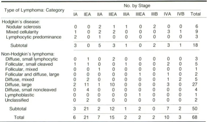
CT findings were reviewed in 68 patients with untreated head and neck lymphoma, More than half of the patients with either Hodgkin's disease or non-Hodgkin's lymphoma were detected in the earlier stages (stage I or II), Four types of abnormalities were identified with CT: nodal involvement alone (type 1), extranodal involvement alone (type 2), a combination of extranodal and nodal disease (type 3), and multifocal extra nodal disease with or without nodal involvement (type 4), In the 18 patients with Hodgkin's disease, a subgroup of mixed cellularity was most common; type 1 was the prevailing CT presentation, and no type 2 or 4 lesions were observed. In the 50 patients with non-Hodgkin's lymphoma, diffuse large-cell lymphoma was the most common histologic subtype, and the most common CT presentation was type 2, followed by type 3.
Lymphomatous nodes may be extensive and confluent, but often they are smaller than 2 cm and rarely are necrotized. The most frequent extranodal sites of head and neck lymphomas are Waldeyer's ring, paranasal sinuses, and nasal cavity. Extranodal lym-phoma cannot be differentiated reliably from the more commonly occurring carcinoma, although it is less often associated with invasion and destruction of adjacent bony structures. Multiple sites of extranoda! involvement, with or without neck lymphadenop-athy, may suggest a diagnosis of non-Hodgkin's lymphoma.
Hodgkin's disease and non-Hodgkin's lymphoma often involve the head and neck region, and many clinical articles have been published on head and neck lymphomas [1-14]. However, to our knowledge there are no thorough radiologic evaluations of head and neck lymphomas, particularly ones that emphasize CT findings. In this paper, we report our analysis of the CT findings of untreated head and neck lymphomas in 68 patients to establish CT patterns and to correlate them with histopathologic and staging classifications.
Materials and Methods
Between October 1981 and July 1986, 68 untreated patients with a diagnosis of lymphoma involving the head and neck region were referred to the Department of Diagnostic Radiology at the University of Texas M. D. Anderson Hospital and Tumor Institute at Houston for CT evaluation of tumor extent in the head and neck. The CT studies were performed with a GE 8800, GE 9800, Siemens Somatom 2, or Siemens DR-3 scanner after an IV bolus injection of contrast medium (either 300 ml of 30% iodine or 150 ml of 60% iodine). Consecutive 4-or 5-mm-thick axial images were obtained routinely from the sphenoid sinus to the angle of the mandible, and sections 8 or 10 mm thick from there to the sternal notch. For those patients with lesions suspected in the orbit or paranasal sinuses, both direct coronal and axial 4-or 5-mm-thick images were obtained.
666
LEE ET AL. AJNR:8, July/August 1987TABLE 1: Classification of Head and Neck Lymphomas
Type of Lymphoma: Category
IA lEA
Hodgkin's disease:
Nodular sclerosis 0 0
Mixed cellularity 1 0
Lymphocytic predominance 2 0
Subtotal 3 0
Non-Hodgkin's lymphoma:
Diffuse, small lymphocytic 0 1 Follicular, small cleaved 1 1
Follicular, mixed 0 0
Follicular and diffuse, large 0 0
Diffuse, mixed 0 2
Diffuse, large 2 11 Diffuse, small noncleaved 0 4
Lymphoblastic 0 0
Unclassified 0 2
Subtotal 3 21
Total 6 21
biopsy. Eighteen patients had Hodgkin's disease and 50 had non-Hodgkin's lymphoma (Table 1).
Results
CT Patterns
Four CT patterns of head and neck lymphomas were identified (Table 2).
In Hodgkin's disease, six were type 1 A, nine were 1 B, and three were 3A. In non-Hodgkin's lymphoma, five were type 1 A, two were 1 B, five were 2A, 20 were 2B, eight were 3A, seven were 3B, two were 4A, and one was 4B (Table 3).
Histopathologic correlation.-In Hodgkin's disease, the
mixed-cellularity subgroup was observed most often (nine of 18 patients), followed by the nodular sclerosis subtype. Nodal
involvement occurred in all 18 cases. Among these, three patients presented with additional extranodal lesions in Wal-deyer's ring (type 3A): one in the nasopharynx, one in the tonsil, and one at multiple sites). No cases were observed of isolated extranodal presentation (type 2).
Twenty-seven (54%) of 50 non-Hodgkin's patients had diffuse large-cell lymphomas. The predominant feature was extranodal presentation, 25 (50%) without (type 2) and 15
(30%) with (type 3) nodal involvement. Isolated nodal
involve-ment (type 1) was observed in only seven (14%) of 50 cases. The most common single CT pattern was type 2B, occurring in 20 patients (40%). The most common sites of extranodal involvement were Waldeyer's ring, the paranasal sinuses, and the nasal cavity (Table 4). Waldeyer's ring involvement in non-Hodgkin's lymphoma was found at more than one site in eight of 13 patients and was associated with nodal disease in eight of 13. Conversely, 12 of 13 patients whose disease involved the paranasal sinuses and nasal cavity were free of nodal involvement.
IIA
2 2 1
5
0 0 1 0 0
1
0 0 0
2
7
No. by Stage
IlEA lilA IIIEA IIIB IVA IVB Total
1 1 0 2 0 0 6
2 0 0 0 3 1 9
0 0 0 0 0 0 3
3 0 2 3 18
2 0 0 0 0 0 3
0 1 0 0 2 0 5
0 0 0 0 0 0 1
0 0 1 0 1 0 2
0 0 0 0 1 2 5
10 0 0 0 3 0 27
0 0 0 0 0 0 4
0 0 1 0 0 0 1
0 0 0 0 0 0 2
12 2 0 7 2 50
15 2 2 2 10 3 68
TABLE 2: CT Patterns of Head and Neck Lymphomas
1, Nodal lymphomas:
1 A, Unilateral
1 B, Bilateral
Type, Description
2, Extranodallymphomas:
2A, Extranodal lesion(s) confined within Waldeyer's ring 2B, Extranodallesion outside Waldeyer's ring (extralymphatic) 3, Nodal and extranodallymphomas (combined types 1 and 2):
3A, Waldeyer's ring lesion(s), with nodal involvement 3B, Extralymphatic lesion, with nodal involvement 4, Multifocal extranodal lymphomas:
4A, Without nodal involvement 4B, With nodal involvement
Staging correlation.-In Hodgkin's disease, more than half
(11 of 18) the patients had earlier staging (stage I or II) at presentation. The same observation was made in
non-Hodg-kin's lymphoma (38 of 50). Most of the patients in both groups
were free of constitutional symptoms (for example, fever, sweats, pruritis, and unexplained weight loss of more than 4.5 kg).
Anatomic Considerations
Nodal involvement.-In 13 (72%) of 18 patients the involved lymph nodes in Hodgkin's disease measured less than 2 cm in greatest diameter on CT (Fig. 1). All cases had involvement
of the deep lymphatic chains, particularly the internal jugular chains, either unilaterally (10 of 18) or bilaterally (eight of 18).
Additional superficial nodes were involved in only one case. One case showed evidence of extracapsular spread and nodal
[image:2.613.129.493.98.312.2] [image:2.613.318.560.370.502.2]AJNR:8. July/August 1987 CT OF HEAD AND NECK LYMPHOMAS 667
TABLE 3: Distribution of Head and Neck Lymphomas by CT Pattern
Type of Lymphoma: Category
1A
Hodgkin's disease:
Nodular sclerosis 2
Mixed cellularity 2
Lymphocytic predominance 2
Subtotal 6
Non-Hodgkin's lymphoma:
Diffuse, small lymphocytic 0 Follicular, small cleaved cell 2 Follicular, mixed cell 1 Follicular and diffuse, large cell 0
Diffuse, mixed cell 0
Diffuse, large cell 2
Diffuse, small noncleaved 0
Lymphoblastic 0
Unclassified 0
Subtotal 5
Total 11
Note.-CT patterns are defined in Table 2.
TABLE 4: Extranodal Sites of Head and Neck Lymphomas
Type of Lymphoma: Site
Hodgkin's disease: Waldeyer's ring:
Nasopharynx Tonsil Multiple sites
Subtotal Total
Non-Hodgkin's lymphoma: Waldeyer's ring:
Nasopharynx Tonsil Multiple sites
Subtotal
Paranasal sinuses and nasal cavity Cheek:
Skin
Buccal mucosa Subtotal Thyroid gland Nose Orbit Parotid gland Multifocal:
Nasopharynx plus submandibular gland Nasopharynx plus paranasal sinus Waldeyer's ring plus cheek
Subtotal Total No. 1 1 1
3
3
3 2 813
13
4 37
'4
'1
'1
'1
1 1 13
43
1B 3 5 1 9 0 0 0 0 1 1 0 0 0 2 11In non-Hodgkin's lymphoma, most (15) of the 23 involved
nodes also measured less than 2 cm. However, in seven (30%) of 23 cases the largest node was more than 5 cm; of these, six had extracapsular and three had nodal necrosis
No. by CT Pattern
2A 2B 3A 3B 4A 4B Total
0 0 1 0 0 0 6
0 0 2 0 0 0 9
0 0 0 0 0 0 3
0 0 3 0 0 0 18
0 1 2 0 0 0 3
2 1 0 0 0 0 5
0 0 0 0 0 0 1
0 2 0 0 0 0 2
0 2 2 0 0 0 5
3 8 3 7 2 1 27
0 4 0 0 0 0 4
0 0 1 0 0 0 1
0 2 0 0 0 0 2
5 20 8 7 2 50
5 20 11 7 2 68
(Fig. 2). Of interest, in three cases very large nodes were
present (greater than 10 cm in the largest), but none showed evidence of central necrosis (Fig. 3). As in Hodgkin's disease
the deep lymphatic chain was always involved in cases in which non-Hodgkin's lymphoma appeared in the neck lymph
nodes unilaterally (13 of 23) or bilaterally (10 of 23). However, the superficial lymphatic chains were involved in nine (39%) of 23 patients because associated disease was common at extralymphatic sites such as the cheek or thyroid gland.
Extranoda/ involvement (Table 4).- Waldeyer's ring was
the site at which extranodal disease occurred most often in
head and neck lymphomas (Fig. 4). In our series there were
three in Hodgkin's disease and 16 in non-Hodgkin's
lym-phoma, and the disease often infiltrated more than one site within the ring. However, in several cases isolated lesions were found in the nasopharynx or tonsil. None of the Waldey-er's-ring lesions had associated destruction of the base of
the skull.
All the extranodal lymphomas outside the Waldeyer's ring (extralymphatic) were in non-Hodgkin's lymphoma,
predomi-nantly the diffuse large-cell subgroup (Table 3). The paranasal sinuses and nasal cavity were the sites at which
extralym-phatic disease was found most often (Fig. 5). Minimal adjacent
bone destruction was seen in nine of 13 cases. Tumor calci-fication occurred in only one case, appearing in the ethmoid
sinus with extensive bone destruction and intracranial exten-sion (Fig. 6).
All four cases of thyroid lymphoma in our series had a
preceding history of goiter with a histologic background of chronic lymphocytic thyroiditis detected microscopically. All of these lymphomas were aggressively infiltrating the adjacent
aerodigestive tract and had extensive nodal involvement (Fig.
668
LEE ET AL.A
B
AJNR:8, July/August 1987
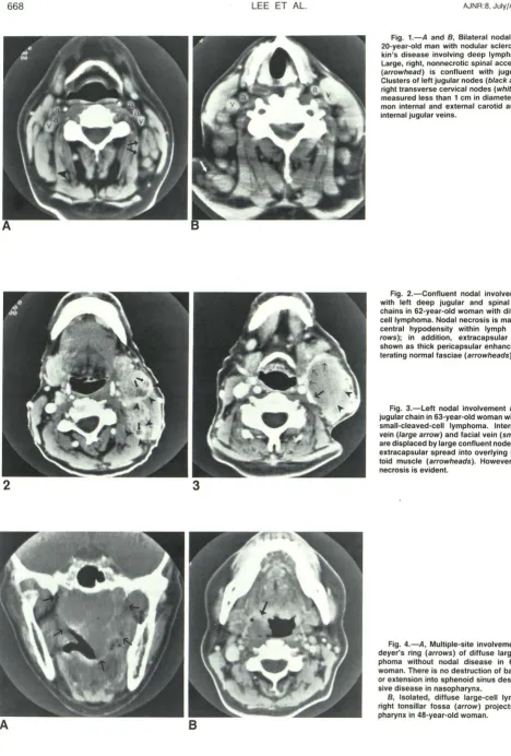
Fig. 1.-A and B, Bilateral nodal disease in
20-year-old man with nodular sclerosing
Hodg-kin's disease involving deep lymphatic chains.
Large, right, nonnecrotic spinal accessory node
(arrowhead) is confluent with jugular nodes.
Clusters of left jugular nodes (black arrows) and right transverse cervical nodes (white arrow) all
measured less than 1 cm in diameter. a = com
-mon internal and external carotid arteries; v
=
internal jugular veins.
Fig. 2.-Confluent nodal involvement along
with left deep jugular and spinal accessory chains in 62-year-old woman with diffuse
large-cell lymphoma. Nodal necrosis is manifested as
central hypodensity within lymph nodes (ar
-rows); in addition, extracapsular spread is
shown as thick pericapsular enhancement
obli-terating normal fasciae (arrowheads).
Fig. 3.-Left nodal involvement along deep
jugular chain in 63-year-old woman with follicular
small-cleaved-cell lymphoma. Internal jugular
vein (large arrow) and facial vein (smaJl arrows) are displaced by large confluent node that shows extracapsular spread into overlying
sternomas-toid muscle (arrowheads). However, no nodal
necrosis is evident.
Fig. 4.-A, Multiple-site involvement of
Wal-deyer's ring (arrows) of diffuse large-cell
lym-phoma without nodal disease in 68-year-old
woman. There is no destruction of base of skull
or extension into sphenoid sinus despite
exten-sive disease in nasopharynx.
B, Isolated, diffuse large-cell lymphoma in
right tonsillar fossa (arrow) projects into
[image:4.613.56.524.61.753.2] [image:4.613.59.390.92.270.2]AJNR:8. July/August 1987 CT OF HEAD AND NECK LYMPHOMAS 669
A
8
C
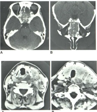
Fig. 5-A, Diffuse large-cell lymphoma involving sphenoid sinus in 43-year-old woman. Minimal bone destruction (arrow) but no intracranial extension. 8 and C, Extensive infiltrative lymphoma (arrows) of diffuse, small, noncleaved subtype (non-Burkitt's) in 4-year-old boy originates from right maxillary sinus and has extensive bone destruction. There is direct extension into right orbit, temporal fossa, infratemporal fossa, and right nasal cavity.
Fig. 6.-Axial (A) and coronal (8) views.
Dif-fuse involvement of nasal cavity and ethnoid sinuses with bone destruction and intracranial extension (arrow) in 49-year-old woman with dif-fuse large-cell lymphoma. Note scattered tu-moral calcifications.
Fig. 7.-A and 8, Diffuse thyroid lymphoma
(arrowheads) of diffuse large-cell subtype with extensive regional extension and nodal disease (N) in 60-year-old woman. Note extension into hypopharynx (H) and esophagus (E). Nodal ne-crosis (large arrows) is seen in confluent nodal involvement. Common carotid artery and internal jugular vein (small arrows) are encased by tumor infiltration.
[image:5.614.56.558.98.282.2] [image:5.614.223.562.341.730.2]670
LEE ET AL. AJNR:8, July/August 1987with bone destruction in three of seven patients. Three pa-tients had multifocal extranodal disease, all from diffuse,
large-cell lymphomas (Fig. 8).
Discussion
The malignant lymphomas are a heterogeneous group of
solid Iymphoreticular neoplasms. Hodgkin's disease is a subgroup of lymphomas that originate from cells of the
mon-ocyte-histiocyte series, and it accounts for 25% of all
malig-nant lymphomas [18]. The other lymphomas are grouped
together as non-Hodgkin's lymphomas.
One of the most common initial presentations of Hodgkin's disease is painless lymph-node enlargement in the head and neck region. Hodgkin's disease spreads in a predictable fash-ion from one lymph-node group to the next contiguous group by lymphatic channels [19]. Extranodal presentation of
Hodg-kin's disease is rare, even with nodal involvement. Con-versely, non-Hodgkin's lymphoma frequently involves e
xtra-nodal structures in the head and neck region, which is the second most common site of extranodal lymphoma after the gastroenterologic tract [7, 11-13]. More than half of the patients with extranodal head and neck lymphomas present with disease localized to the site of origin, and less often associated with nodal involvement [1]. The extranodal sites
most frequently involved are Waldeyer's ring, the parana sal
sinuses, and the nasal cavity. The histologic subtypes are
usually intermediate or high-grade, particularly diffuse,
large-cell lymphomas [1, 3, 10, 14].
When neck nodes are involved in either Hodgkin's or
non-Hodgkin's lymphoma, the disease spreads into multiple
nodes. Whether the nodal involvement is unilateral or bilateral,
the deep lymphatic chains are almost always involved,
partic-ularly the internal jugular chain. The middle and lower jugular
nodes are often diseased when lymphomas present in the nodal pattern (type 1). Isolated lymphomatous involvement of the superior internal jugular chain or superficial nodes is
unusual; this is in contrast to the carcinomatous nodal involve-ment of unknown primary site. The size of the involved lymph
node can be quite variable, with larger ones more than 10 cm
in diameter and smaller ones less than 5 mm. Frequently,
multiple nodes of varying sizes are involved. Sometimes they
are elliptical in shape, although a rounded appearance is most
common. It is of interest to note that nodal necrosis was
found in only one (5%) of our 18 patients with Hodgkin's disease and three (13%) with non-Hodgkin's lymphoma, even when the disease was extensive. Since necrosis is prevalent
in carcinomatous nodes, its absence may warrant a prebiopsy
diagnosis.
The extranodal lesions in Waldeyer's ring can be isolated
by their appearance, which resembles squamous carcinoma;
however, more often there are multiple sites of involvement
within the ring. Rarely does nasopharyngeal lymphoma
pre-sent with skull-base destruction, even with extensive involve-ment. This is in contrast to the frequent bone destruction seen with squamous carcinoma. The extralymphatic lesions in the paranasal sinus and oral cavity can be small or extensive and often are associated with sinus obstruction. This
obser-vation is nonspecific, and these lesions are indistinguishable
from other slow-growing neoplasms. Tumor calcification, pre-viously unreported, was observed in one of our cases, and
its cause is undetermined.
Thyroid lymphoma tends to diffusely infiltrate the adjacent soft tissue, including the aerodigestive tract, and is often accompanied by extensive nodal involvement. As mentioned
[20, 21], all four cases in our series had associated chronic lymphocytic thyroiditis histologically, suggesting a strong
re-lationship between the two. Lymphoma originating from the
cheek, either in the skin or buccal submucosa, tends to extend regionally into the nasal cavity, maxillary sinus, and orbit, and
is often associated with bone destruction. Again, this finding
mimics that of other slow-growing neoplasms.
In summary, the extranodal presentation of head and neck
lymphoma is nonspecific and indistinguishable from the more
commonly occurring carcinoma. However, multiple sites of involvement within or outside Waldeyer's ring, without or with
nodal disease, should alert radiologists to the possible diag-nosis of a Iymphoreticular neoplasm.
Treatment of both Hodgkin's and non-Hodgkin's lymphoma
[image:6.612.52.389.548.729.2]AJNR:8, July/August 1987 CT OF HEAD AND NECK LYMPHOMAS 671
is based on the histologic classification and anatomic staging
of disease, the latter of which has been influenced by modern
diagnostic imaging. The diagnosis and management of head and neck lymphoma, in particular non-Hodgkin's, often varies with the site of extranodal disease. The precise definition of the anatomic extent of disease is critical to effective treatment when radiotherapy is used alone. Therefore, the decision to obtain a CT scan should be made according to the histologic classification and clinical involvement of disease [22]. It is the practice in our institute not to obtain a CT scan of the head and neck routinely unless the findings will dictate the
treat-ment planning of the lymphoma. Generally, nodal lymphoma
does not require a CT evaluation. A CT scan of the head and neck is needed for identifying a possible extranodal primary site when there are isolated superficial nodes positive for
lymphoma. Waldeyer's-ring lymphoma may require a CT scan
for evaluation of exact tumor extent for radiotherapeutic portal definition. A CT evaluation is definitely desired in extranodal
extralymphatic lymphomas for diagnosis and staging
pur-poses as well as for determining radiation portals,
REFERENCES
1. Fienstein JT, Thawley SE. Lymphoma of the head and neck. Laryngoscope 1978;88: 582-593
2. Mill WB, Lee FA, Franssila KO. Radiation therapy treatment of stage I and
II extranodal non-Hodgkin's lymphoma of the head and neck. Cancer
1980;45:653-661
3. Reddy S, Pellettiere E, Saxena V, Hendrickson FR. Extranodal non-Hodgkin's lymphoma. Cancer 1980;46:1925-1931
4. Plantenga KF, Hart G, Van Heerde P, Tierie AH. Non-Hodgkin's malignant lymphomas of upper digestive and respiratory tracts. Int J Radiat On col
Bioi Phys 1981;7: 141 9-1427
5. Evans C. A review of non-Hodgkin's lymphomata of the head and neck.
Clin Oncol 1981;7:23-31
6. Horiuchi J, Okurama T, Matsubara S, Shibura H, Suzuki S, Kamiyama R. Extranodal non-Hodgkin's lymphoma in the head and neck: irradiation and
clinical course. Acta Radiol {Oncol} 1982;21 :393-399
7. Kong JS, Fuller LM, Butler JJ, et al. Stages I and II non-Hodgkin's lymphomas of Waldeyer's ring and the neck. Am J Clin Oncol 1984;7: 629 -639
8. Aozasa K, Nara H, Ikeda H, et al. The influence of histologic type on
survival in early extranodal non-Hodgkin's lymphoma in head and neck. Oncology 1984;41: 164-169
9. Larson DL, Robbins KT, Butler JJ. Lymphoma of the head and neck: a
diagnostic dilemma. Am J Surg 1984;148:433-437
10. Robbins KT, Fuller LM, Vlasak M, et al. Primary lymphomas of the nasal cavity and paranasal sinuses. Cancer 1985;56:814-819
11. Jacobs C, Hoppe R. Non-Hodgkin's lymphomas of head and neck extra-nodal sites. Int J Radiat On col Bioi Phys 1985;11 :357-364
12. Albada J, Hordijk GJ, van Unnik JAM, Dekker AW. Non-Hodgkin's lym -phoma of Waldeyer's ring. Cancer 1985;56: 2911-2913
13. Shidnia H, Hornback NB, Lingeman R, Barlow P. Extranodallymphoma of the head and neck area. Am J Clin Oncol 1985;8:235-243
14. Jacobs C, Weiss L, Hoppe RT. The management of extra nodal head and neck lymphomas. Arch Otolaryngol Head Neck Surg 1986;112:654-658 15. Lukes RJ, Butler JJ, Hicks EB. Natural history of Hodgkin's disease as
related to its pathologic pictures. Cancer 1966;19:317-344
16. National Cancer Institute sponsored study of classification of
non-Hodg-kin's lymphomas. Summary and description of a working formulation for clinical usage. Cancer 1982;49: 2112-2135
17. Carbone PP, Kaplan HS, MusshoH K, Smithers EW, Tubiana M. Report of the committee on Hodgkin's disease staging. Cancer Res 1971 ;31: 1860 -1861
18. Canellos GP. Malignant lymphomas. In: Rubenstein E, Federman DD, eds.
Scientific medicine. New York: Scientific American, 1986:1-15
19. Castellino RA. Hodgkin's disease: practical concepts for the diagnostic
radiologist. Radiology 1986;159:305-310
20. Aozasa K, Inoue A, Yoshimura J, et al. Intermediate lymphocytic lymphoma of the thyroid. Cancer 1986;57: 1762-1767
21. Aozasa K, Inoue A, Tajima K, Mirauchi A, Matsuzuka T, Kuma K. Malignant
lymphomas of the thyroid gland. Cancer 1986;58: 1 00-1 04