Increased osteogenesis with hydroxyapatite
constructs combined with serially-passaged bone
marrow-derived mesenchymal stem cells
Manabu Akahane1*, Tomoyuki Ueha2, Takamasa Shimizu2, Yusuke Inagaki2, Akira Kido2,
Tomoaki Imamura1, Kenji Kawate3, Yasuhito Tanaka2
1Department of Public Health, Health Management and Policy, Nara Medical University School of Medicine, Nara, Japan; *Corresponding Author: [email protected]
2Department of Orthopedic Surgery, Nara Medical University, Nara, Japan
3Department of Artificial Joint and Regenerative Medicine, Nara Medical University, Nara, Japan
Received 18 June 2012; revised 16 July 2012; accepted 17 August 2012
ABSTRACT
We have previously reported on both the os- teogenic potential of hydroxyapatite (HA) com- bined with bone marrow-derived mesenchymal stem cells (BMSCs) and a method involving os- teogenic matrix cell sheet transplantation of BMSCs. In the present study, we assessed the osteogenic potential of serially-passaged BMS- Cs, both in vitro and in vivo. We also assessed whether an additional cell-loading technique can regain the osteogenic potential of the constructs combined with serially-passaged BMSCs. The present study revealed that passage (P) 1 cells cultured in osteogenic-induced medium showed strong positive staining for alkaline phosphat- ase (ALP) and Alizarin Red S, whereas P3 cells showed faint staining for ALP, with no Alizarin Red S staining. Staining of P1, P2 and P3 cells were progressively weaker, indicating that the osteogenic potential of the serially-passaged rat BMSCs is lost after P3 in vitro. The in vivo study showed that little bone formation was observed in the HA constructs seeded with P3 cells, 4 weeks after subcutaneous implantation. How- ever, P3 cell/HA constructs which had incre- ased cell-loading showed abundant bone for- mation within the pores of the HA construct. ALP and osteocalcin mRNA expression in these constructs was significantly higher than that of constructs with regular cell-seeding. The pre- sent study indicates that the osteogenic poten- tial of the constructs with serially-passaged BMSCs is increased by additional cell-loading. This method can be applied to cases requiring hard tissue reconstruction, where BMSCs require
serial expansion of cells.
Keywords:Osteogenesis; Mesenchymal Stem Cells; Serial Passaging; Hydroxyapatite; Tissue Engineering; Bone Reconstruction
1. INTRODUCTION
Bone marrow contains a population of undifferentiated cells known as mesenchymal stem cells (MSCs) [1,2]. Bone marrow-derived MSCs (BMSCs) have the potential for self-renewal and differentiation into several cell types after culture expansion in vitro and in vivo, including os- teoblastic, chondrocytic, adipocytic and neuronal cells [3-7]. Differentiation into osteoblastic lineage cells after subculture in osteogenic-induced media, containing dexa- methasone (Dex), ascorbic acid phosphate and β-gly- cerophosphate (β-GP), has been shown for rat and human BMSCs [8-11]. The osteogenic phenotypic markers, alka- line phosphatase (ALP) and osteocalcin (OC), can be de- tected by days 10 - 12 in passage 1 (P1)-cultured cells, in- cluding mineralization [12]. The combination of BMSCs with a scaffolding material, such as hydroxyapatite (HA), can promote the formation of new bone tissue in vivo after subcutaneous transplantation with freshly isolated bone marrow cells [13] or culture-expanded BMSCs (HA/ BMSCs construct) [2,14-17], Recently, we reported a new cell transplantation technique in which BMSCs were cultured in culture medium containing Dex and ascorbic acid phosphate and transplanted as cell sheets [18]. These cell sheets can be combined with HA, in which they resulted in abundant bone formation compared with conventional techniques [17].
aches, BMSCs are obtained from the patients, following which they generally need to be expanded by serial pass- aging in vitro as the number of cells generated in primary culture is limited. However, the osteogenic potential of BMSCs might be lost by serial passaging in vitro, re- sulting in decreased osteogenesis after subcutaneous transplantation [8]. Therefore, it is important to deter- mine whether HA constructs combined with serially- passaged BMSCs maintain sufficient osteogenic potential after in vivo transplantation. In the present study, we used BMSCs obtained from rat bone marrow and assessed the osteogenic potential of serially-passaged BMSCs, both in vitro and in vivo. We also assessed whether an additional cell-loading technique, where cells are cultured in os- teoblastic cell sheet preparation medium can regain the osteogenic potential of the constructs combined with se- rially-passaged BMSCs.
2. MATERIALS AND METHODS
2.1. Bone Marrow Cell Preparation and Cell Culture
Approval from the animal experimental review board of Nara Medical University was obtained before begin- ning the experiments. Institutional guidelines for the care and use of laboratory animals have been observed. We previously reported the method of bone marrow cell preparation [11,18]. Briefly, bone marrow cells were ob- tained from the femur shafts of 7-week-old male Fischer 344 rats. Both ends of the femur were removed from the epiphysis and the marrow was flushed out using 10 ml of standard culture medium which was expelled from a sy- ringe containing a 20-gauge needle. Standard culture medium consisted of minimal essential medium (MEM; Nacalai Tesque, Kyoto, Japan) containing 15% fetal bo- vine serum (FBS, JRH Bioscience Inc., KS, USA) and antibiotics (100 U/ml penicillin and 100 µg/ml strepto- mycin, Nacalai Tesque).
The obtained cells were collected in two T-75 flasks (Falcon, BD, NJ, USA) containing 15 ml of standard culture medium. These cells were known as passage 0 (P0). Cell culture was maintained in a humidified atom- sphere of 95% air with 5% CO2 at 37˚C. After reaching confluency, cells were released from the culture substra- tum using trypsin/EDTA (Gibco, Invitrogen, CA, USA). The released cells were seeded at density of 1 × 10⁴ cells/cm2 for subculture in T75 flasks (Falcon) for serial passaging to obtain passage 3 (P3) cells. Each passage of cells (P1, P2 and P3) were used for the following ex- periments.
2.2. ALP and Alizarin Red S Staining
To evaluate the osteogenic potential of serially-pass- aged cells in vitro, P1, P2 and P3 cellswere seeded in
6-well plates at density of 1 × 10⁴ cells/cm2 and cultured with or without osteogenic-induced media containing Dex (10 nM), ascorbic acid phosphate (82 μg/mL) and β-GP (10 mM) for 14 days. Each well was stained with Naphtol-AS-MX phosphate sodium salt (Sigma) and Fast Red Violet B (Nacalai Tesque) for ALP staining. 10 mg of each reagent was dissolved in 20 ml of AMP buffer (1.0 mM MgCl2, 10 mM p-nitrophenyl phosphate in 0.056 M 2-amino-2-methyl-propanol). Cells were incu- bated with 2 ml of the substrate solution at room tem- perature for ~2 minutes and rinsed with dH2O. Alizarin Red S (0.5%) was also used to evaluate calcium deposi- tion, following which cells were rinsed with dH2O [12,18]. Each experiment contained 3 replicates and was performed in duplicate.
2.3. ALP Activity, Calcium and DNA
Concentration in Vitro
Cells cultured in 12-well culture plates were used to measure ALP activity, as previously reported [12]. Briefly, P1, P2 and P3 cells, seeded in 12-well plates at a density of 1 × 10⁴ cells/cm2 and cultured for 14 dayswith or without osteogenic-induced media containing Dex, ascorbic acid phosphate and β-GP, were scraped into 1 ml of 0.05 M sodium phosphate buffer, 2 mM EDTA and 2 M NaCl, following which they were homogenized and sonicated. To measure the ALP activity of cultured cells, 0.1 ml of the sonicated cell suspension was added to 0.5 ml of 0.2%Nonidet P-40 containing 1 mM MgCl2and centrifuged at 13,000 rpm for 10 minutesat 4˚C. The supernatant was assayed for ALP activity usingp-nitro- phenylphosphateas a substrate. An aliquot (10 µl)of the supernatant was added to 1 ml of 50 mM p-nitroph- enylphosphatecontaining 1 mM MgCl2and the mixture was incubated for 30 minutesat 37˚C.Subsequently, 2 ml of 0.2 N NaOH was addedto stop the enzymatic reaction and the absorptionat 410 nm was measured by spectro- photometry. ALP activity was represented as p-nitro- phenolrelease (µmol)after 30 minutes of incubation at 37˚C.
2.4. Preparation of HA Constructs Seeded with P3 Cells
We have previously reported the preparation of HA constructs combined with BMSCs (BMSC/HA con- struct) [12,17]. Briefly, after release from the substratum, P3 cells were centrifuged and resuspended at a density of 1 × 10⁶ cells/ml in standard medium. Air was removed from the HA, which were previously soaked in cell sus- pension, by vacuum prior to making the constructs. Then, the HA disks were soaked in the cell suspension for 2 hours in a CO2 incubator at 37˚C to create the P3 cell/HA constructs. After soaking in the cell suspension, the con- struct was transferred into a 12-well plate (Falcon) in 1 mL of osteogenic-induced medium, consisting of stan- dard medium, 82 μg/mL ascorbic acid phosphate (Wako Pure Chemical Industrials, Kyoto, Japan) and 10 nM Dex (Sigma, St. Louis, MO, USA) for further subculture. After 14 days of subculture, the cultured P3 cell/HA constructs were transplanted into a subcutaneous site of syngenic rats for the control group of the in vivo experiments.
Porous HA ceramics (50% average void volume, 5 mm in diameter and 2 mm thickness) were used in this study (Cellyard HA scaffold, Pentax Co., Tokyo, Japan). The solid and porous components of the microstructure are interconnected.
2.5. Additional Cell-Loading of the P3
Cell/HA Constructs
We previously reported that BMSCs cultured in stan- dard medium containing Dex and ascorbic acid phos- phate resulted in the formation of cell sheets [10,18]. In the present study, additional P3 cells for loading were also prepared in cell sheet culture medium. Briefly, P3 cells were seeded at a density of 1 × 10⁴ cells/cm2 in 3.5 cm and 10 cm dishes (Falcon) and cultured in standard medium containing Dex (10 nM) and ascorbic acid phos- phate (82 μg/mL) (cell sheet preparation medium) until confluent. We used a mechanical retrieval technique for the preparation of additional P3 cells for cell loading of the constructs. The cells were rinsed twice with phos- phate-buffered saline (PBS; Gibco) and lifted as sheet structure using a cell-scraper. As P3 cells formed in- comeplete cell sheets compared with P1 cells, they were loaded onto P3 cell/HA constructs by a short centrifuga- tion at 900 rpm for 2 min (Kubota 5100, Kubota Corp., Tokyo, Japan). Subsequently, the constructs were trans- planted into a subcutaneous site as the additional-loading group. Because we used a number of different cell densi- ties for the additional loading of the constructs, we placed them into two subgroups, according to whether they were cultured in 3.5 cm or 10 cm dishes.
Thus, there were three groups in the in vivo study: the
P3 cell/HA construct without additional loading (control group), the P3 cell/HA construct cultured in a 3.5 cm dish of P3 cells (small loading group) and the P3 cell/HA construct cultured in a 10 cm dish of P3 cells (large loading group). Each group contained seven HA disks in two recipient rats (three or four disks per rat), which were implanted at subcutaneous sites on their backs. Three disks were used for histology and four for real- time PCR analysis. The experiment was performed in duplicate.
2.6. Sample Harvest and Histological Analysis
Implanted disks were harvested 4 weeks after implan- tation. HA disks for analysis by real-time PCR were fro- zen until RNA isolation. Disks for histological evaluation were fixed in 10% buffered formalin after trimming off the excess rat tissue around the HA disc. The HA disks fixed in formalin were decalcified in K-CX solution (Falma Co., Tokyo, Japan) and embedded in paraffin. They were cut parallel down the centre of specimens to the round surface of the HA discs and stained with hema- toxylin and eosin (H-E).
2.7. RNA Isolation and Real-Time Quantitative PCR
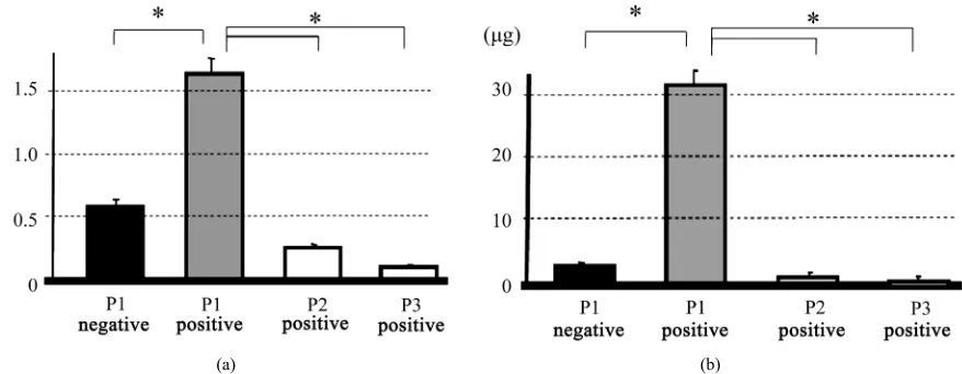
2.8. Statistics Figure 2 shows ALP activity and calcium deposition,
which were standardized to DNA content in each group. The activity of P2 cells cultured in osteogenic-induced medium were significantly lower than that of the positive control, indicating that passaging of cells decreases the osteoblastic phenotype of cultured cells (Figure 2(a)).
There was a significant difference in calcium deposition between the positive control and P2 cells, indicating loss of mineralization ability with increasing passage (Figure 2(b)).
Statistical significant was determined by one-way ANOVA post-hoc multiple comparisons using Tukey’s test. A p value of 0.05 was considered statistically sig- nificant.
3. RESULTS
3.1. Osteogenic Potential of Serial Passaged
Cells in Vitro
Figure 1 shows ALP (Figure 1(a)) and Alizarin Red
S staining (Figure 1(b)) of BMSCs that were cultured
with or without osteogenic-induced medium at P1, P2 and P3. P1 cells cultured with osteogenic-induced me- dium showed strong positive staining for ALP and Aliza- rin Red S, whereas P3 cells showed faint staining for ALP and no Alizarin Red S staining. ALP staining in P2 cells seems weaker than in P1 cells and the size of mi- neralized nodule in P2 cells appears smaller than P1 cells. Progressively weaker staining was seen in P1, P2 and P3 cells, indicating that osteogenic potential of serially- passaged BMSCs is lost by P3. P1, P2 and P3 cells cul- tured without osteogenic-induced medium were negative for ALP and Alizarin Red S staining.
3.2. Bone Ormation of P3-Cell/HA Constructs after Implantation
Figure 3 shows representative histological sections of
the harvested HA constructs 4 weeks after implantation. Little bone formation occurred in the pores of the control group (the P3 cell/HA constructs: Figure 3(a)) and the
small loading group (P3 cell/HA constructs with 3.5 cm dish P3 cell-loading: Figure 3(b)). In contrast, abundant
bone formation, including with osteoblasts and osteo- cytes, was observed in the large loading group (P3 cell/HA constructs with 10 cm dish P3 cell-loading: Fig- ure 3(c)).
(a) (b)
[image:5.595.82.521.86.257.2]
(a) (b)
Figure 2. ALP activity (a) and calcium deposition (b), standardized to DNA content. P1 and P2 represent the passage of cells. Positive and negative indicate cells cultured with or without osteogenic-induced medium, respectively. Asterisk indi- cates p < 0.05.
(a) (b) (c)
[image:5.595.140.457.305.500.2]
(d) (e) (f)
Figure 3. Representative histological sections of the harvested HA constructs 4 weeks after implantation. Little bone formation was observed in the pore of the control group (the P3 cell/HA construct: Figures 3(a) and (b)) and small cell-loading group (the P3 cell/HA construct with 3.5 cm dish cell-loading: Figures 3(c) and (d)). In contrast, abundant bone formation together with osteoblasts and osteocytes was seen in large cell-loading group (the P3 cell/HA construct with 10 cm dish cell-loading: Figures 3(e) and (f)). Arrows indicate bone tissue. Bar = 200 μm.
Figure 4 shows ALP and OC mRNA expression levels
evaluated by real-time PCR. The ALP expression in the large loading group was significantly higher than in the control and small loading groups (Figure 4(a)). OC ex-
pression levels were similar to ALP levels (Figure 4(b)).
4. DISCUSSION
The present study shows that serial passaging of BMSCs decreases the osteogenic potential in vitro. Con- sequently, little bone formation was seen in the pore of the construct when P3 cells were combined with HA disk (P3 cell/HA construct) and implanted into a subcutane-
[image:6.595.96.504.87.255.2]
(a) (b)
Figure 4. Real-time PCR of ALP and OC mRNA expression. OC expression in the large loading group was sig- nificantly higher than those in the control and small loading groups (Figure 4(a)). The tendency of ALP expres- sion (Figure 4(b)) was similar to OC expression. Control: control group, Large loading: large cell-loading group, Small loading: small cell-loading group. Asterisk indicates p < 0.05.
and 10 cm culture dishes. The culture area of 10 cm dish is approximately 5 times larger than the 3.5 cm dish. Consequently, the cell density is also 5 times more in a 10 cm dish. The construct with the 3.5 cm dish P3 cell-loading showed smaller bone formation 4 weeks after implantation compared with the 10 cm dish P3 cell-loading constructs, indicating that the loaded cell number could be an important factor to form bone tissue in the construct combined with serially-passaged cells.
Seeded cells may contain a combination of differen- tiated and undifferentiated cells after passaging. Differ- entiated cells have a limited capacity for proliferation [23,24] and as the cells proliferating from the seeded cells are undifferentiated cells, there is a decrease in the percentage of osteogenic cells in the culture [8,25]. Bone marrow cells are a mixed population of cells, including BMSCs, fibroblasts and endothelial cells. After serial passaging, the population of cultured cells changes. There- fore, we assume that percentage of osteogenic cells might decrease during cell culture and result in less ALP acti- vity and mineralization by P2 and P3 passages.
Kumar et al. [26] have reported a method for the pre- paration of cell sheet/HA constructs using a thermosen- sitive polymer to fabricate the cell sheet. In their report, they showed rapid and complete cellularization to HA by wrapping it with a cell sheet fabricated from a human osteosarcoma cell line. Thus, we think that it is possible to load a lot of cells onto the construct by using the wrapping method with P1 cell sheets. The purpose of the present study was to evaluate the osteogenic potential of serially-passaged cells. We used P3 cells to assess the osteogenic potential of P3 cell/HA constructs. At P3, cell culture with the cell sheet preparation method formed incomplete sheet like fragments. Therefore, we used cen- trifugation to load the incomplete cell sheet onto the
constructs as an additional cell-loading technique. Be- cause little bone formation was seen in the harvested construct with 3.5 cm dish cell-loading, the amount of bone formation could depend on the loading cell number on the HA. The decreased number of osteogenic cells could be compensated by the additional cell-loading from a 10 cm culture dish.
applications for tissue-engineered bone reconstruction. There are a number of limitations of the study that should be acknowledged. First is the use of rat BMSCs in the present study. For the clinical application of this technique, human BMSCs should be used to determine whether our cell-loading method can restore the oseteo- genic potential of HA constructs combined with seri- ally-passaged cells. Second, the culture technique for the preparation of cell sheets formed incomplete sheets at P3. Therefore, further improvement of cell sheet preparation for serially-passaged cells is needed. A complete cell sheet created by serial passaging could load large amounts of cells onto the constructs without centrifugation and resulted in increased bone formation. Third, we used only HA in the present study. Other ceramics, such as β-tri- calcium phosphate (β-TCP), are also commonly used cli- nically [27-29]. Therefore, other types of ceramics should be tested to confirm our results.
5. CONCLUSION
The present study indicated that osteogenic potential of BMSCs decreases by serial passaging. However, bone formation could be regained using the additional cell- loading technique. Owing to its usage of serially-pas- saged cells, this method could be applied in cases of hard tissue reconstruction, which requires the serial passaging of BMSCs to obtain a suitable number of cells. This technique, along with the cell suspension method, can be used to prepare cell-loaded constructs from ceramics, such as HA and β-TCP.
6. ACKNOWLEDGEMENTS
We thank Ms M. Yoshimura and Ms M. Matsumura (Nara Medical University School of Medicine, Japan) for their technical assistance. This study was supported by Takeda Science Foundation.
REFERENCES
[1] Owen, M. (1988) Marrow stromal stem cells. Journal of Cell Science, 10, 63-76.
[2] Ohgushi, H. and Caplan, A.I. (1999) Stem cell technology and bioceramics: From cell to gene engineering. Journal of Biomedical Materials Research, 48, 913-927. doi:10.1002/(SICI)1097-4636(1999)48:6<913::AID-JBM 22>3.0.CO;2-0
[3] Ohgushi, H., Yoshikawa, T., Nakajima, H., Tamai, S., Dohi, Y. and Okunaga, K. (1999) Al2O3 doped apatite- wollastonite containing glass ceramic provokes osteo-genic differentiation of marrow stromal stem cells. Jour-nal of Biomedical Materials Research, 44, 381-388. doi:10.1002/(SICI)1097-4636(19990315)44:4<381::AID-JBM3>3.0.CO;2-E
[4] Sonal, R., Jackson, J.D., Brusnahan, S.K., O’Kane, B. J. and Sharp, J.G. (2012) Characterization of a mesenchy-
mal stem cell line that differentiates to bone and provides niches supporting mouse and human hematopoietic stem cells. Stem Cell Discovery,2, 5-14.
doi:10.4236/scd.2012.21002
[5] Brazelton, T.R., Rossi, F.M., Keshet, G.I. and Blau, H.M. (2000) From marrow to brain: Expression of neuronal phenotypes in adult mice. Science, 290, 1775-1779. doi:10.1126/science.290.5497.1775
[6] Jiang, Y., Jahagirdar, B.N., Reinhardt, R.L., Schwartz, R.E., Keene, C.D., Ortiz-Gonzalez, X.R., Reyes, M., Lenvik, T., Lund, T., Blackstad, M., Du, J., Aldrich, S., Lisberg, A., Low, W.C., Largaespada, D.A. and Verfaillie, C.M. (2002) Pluripotency of mesenchymal stem cells de-rived from adult marrow. Nature, 418, 41-49.
doi:10.1038/nature00870
[7] Krause, D.S. (2002) Plasticity of marrow-derived stem cells.Gene Therapy, 9, 754-758.
doi:10.1038/sj.gt.3301760
[8] Ter Brugge, P.J. and Jansen, J.A. (2002) In vitro osteo-genic differentiation of rat bone marrow cells subcultured with and without dexamethasone. Tissue Engineering, 8, 321-331. doi:10.1089/107632702753725076
[9] Matsushima, A., Kotobuki, N., Tadokoro, M., Kawate, K., Yajima, H., Takakura, Y. and Ohgushi, H. (2009) In vivo
osteogenic capability of human mesenchymal cells cul-tured on hydroxyapatite and on beta-tricalcium phosphate.
Artificial Organs, 33, 474-481. doi:10.1111/j.1525-1594.2009.00749.x
[10] Akahane, M., Shigematsu, H., Tadokoro, M., Ueha, T., Matsumoto, T., Tohma, Y., Kido, A., Imamura, T. and Tanaka, Y. (2010) Scaffold-free cell sheet injection results in bone formation. Journal of Tissue Engineering and Regenerative Medicine, 4, 404-411. doi:10.1002/term.259 [11] Nakamura, A., Akahane, M., Shigematsu, H., Tadokoro,
M., Morita, Y., Ohgushi, H., Dohi, Y., Imamura, T. and Tanaka, Y. (2010) Cell sheet transplantation of cultured mesenchymal stem cells enhances bone formation in a rat nonunion model. Bone, 46, 418-424.
doi:10.1016/j.bone.2009.08.048
[12] Nakamura, A., Dohi, Y., Akahane, M., Ohgushi, H., Na- kajima, H., Funaoka, H. and Takakura, Y. (2009) Osteo- calcin secretion as an early marker of in vitro osteogenic differentiation of rat mesenchymal stem cells. Tissue En-gineering Part C: Methods, 15, 169-180.
doi:10.1089/ten.tec.2007.0334
[13] Akahane, M., Ohgushi, H., Yoshikawa, T., Sempuku, T., Tamai, S., Tabata, S. and Dohi, Y. (1999) Osteogenic phe-notype expression of allogeneic rat marrow cells in po-rous hydroxyapatite ceramics. Journal of Bone and Min-eral Research, 14, 561-568.
doi:10.1359/jbmr.1999.14.4.561
[14] Bianco, P. and Robey, P.G. (2001) Stem cells in tissue engineering. Nature, 414, 118-121.
doi:10.1038/35102181
doi:10.1002/1097-4636(200111)57:2<208::AID-JBM116 0>3.0.CO;2-N
[16] Petite, H., Viateau, V., Bensaid, W., Meunier, A., de Pol- lak, C., Bourguignon, M., Oudina, K., Sedel, L. and Guil- lemin, G. (2000) Tissue-engineered bone regeneration.
Nature Biotechnology, 18, 959-963. doi:10.1038/79449 [17] Shigematsu, H., Akahane, M., Dohi, Y., Nakamura, A.,
Ohgushi, H., Imamura, T. and Tanaka, Y. (2010) Osteo- genic potential and histological characteristics of mesen- chymal stem cell sheet/hydroxyapatite constructs. The Open Tissue Engineering and Regenerative Medicine Journal, 2, 63-70. doi:10.2174/1875043500902010063 [18] Akahane, M., Nakamura, A., Ohgushi, H., Shigematsu,
H., Dohi, Y. and Takakura, Y. (2008) Osteogenic matrix sheet-cell transplantation using osteoblastic cell sheet re- sulted in bone formation without scaffold at an ectopic site. Journal of Tissue Engineering and Regenerative Medicine, 2, 196-201. doi:10.1002/term.81
[19] Wakitani, S., Imoto, K., Yamamoto, T., Saito, M., Murata, N. and Yoneda, M. (2002) Human autologous culture ex- panded bone marrow mesenchymal cell transplantation for repair of cartilage defects in osteoarthritic knees. Os-
teoarthritis and Cartilage, 10, 199-206. doi:10.1053/joca.2001.0504
[20] Ohgushi, H., Kotobuki, N., Funaoka, H., Machida, H., Hirose, M., Tanaka, Y. and Takakura, Y. (2005) Tissue engineered ceramic artificial joint—Ex vivo osteogenic differentiation of patient mesenchymal cells on total an- kle joints for treatment of osteoarthritis. Biomaterials, 26, 4654-4661. doi:10.1016/j.biomaterials.2004.11.055 [21] Kawate, K., Yajima, H., Ohgushi, H., Kotobuki, N., Su-
gimoto, K., Ohmura, T., Kobata, Y., Shigematsu, K., Kawamura, K., Tamai, K. and Takakura, Y. (2006) Tis- sue-engineered approach for the treatment of steroid- induced osteonecrosis of the femoral head: Transplanta- tion of autologous mesenchymal stem cells cultured with beta-tricalcium phosphate ceramics and free vascularized fibula. Artifical Organs, 30, 960-962.
doi:10.1111/j.1525-1594.2006.00333.x
[22] Akahane, M.T.U., Shimizu, T., Shigematsu, H., Kido, A., Omokawa, S., Kawate, K., Imamura, T. and Y. Tanaka. (2010) Cell Sheet Injection as a technique of osteogenic supply. International Journal of Stem Cells, 3, 138-143. [23] McCulloch, C.A., Strugurescu, M., Hughes, F., Melcher,
A.H. and Aubin, J.E. (1991) Osteogenic progenitor cells in rat bone marrow stromal populations exhibit self-re- newal in culture. Blood, 77, 1906-1911.
[24] Aubin, J.E. (1998) Advances in the osteoblast lineage.
Biochemistry and Cell Biology, 76, 899-910. doi:10.1139/o99-005
[25] Kadiyala, S., Young, R.G., Thiede, M.A. and Bruder, S.P. (1997) Culture expanded canine mesenchymal stem cells possess osteochondrogenic potential in vivo and in vitro.
Cell Transplantation, 6, 125-134. doi:10.1016/S0963-6897(96)00279-5
[26] Anil, K.P.R., Varma, H.K. and Kumary, T.V. (2005) Rapid and complete cellularization of hydroxyapatite for bone tissue engineering. Acta Biomaterialia, 1, 545-552. doi:10.1016/j.actbio.2005.05.002
[27] Ogose, A., Hotta, T., Hatano, H., Kawashima, H., Toku-naga, K., Endo, N. and Umezu, H. (2002) Histological examination of beta-tricalcium phosphate graft in human femur. Journal of Biomedical Materials Research, 63, 601-604. doi:10.1002/jbm.10380
[28] Yamamoto, T., Onga, T., Marui, T. and Mizuno, K. (2000) Use of hydroxyapatite to fill cavities after excision of be-nign bone tumours. Clinical results. Journal of Bone &
Joint Surgery, British Volume, 82, 1117-1120. doi:10.1302/0301-620X.82B8.11194
[29] Schindler, O.S., Cannon, S.R., Briggs, T.W. and Blunn, G.W. (2008) Composite ceramic bone graft substitute in the treatment of locally aggressive benign bone tumours.