Polymer Supported Lipid Bilayers
Ian P. McCabe1, Martin B. Forstner1,2
1
Department of Biomedical and Chemical Engineering, Syracuse University, Syracuse, USA
2
Physics Department, Syracuse University, Syracuse, USA Email: [email protected]
Received January 1, 2013; revised February 5, 2013; accepted February 14,2013
ABSTRACT
Lipid bilayers are some of the most fascinating self-assembled structure in living nature. Not only do they serve as the protective boundary of cells and their internal organelles, they also organize and host major parts of the biochemical machinery for cellular communication and transmembrane transport. To study aspects of cellular membranes in a con- trolled manner, solid supported planar bilayers have served as reliable tools for many decades. They have been used in a large variety of studies ranging from fundamental investigations of membranes and their constituents to the dissection of cellular signaling mechanisms. However, there are limitations to these systems and recently a class of new systems in which the lipid bilayer is supported on a soft, polymer cushion has emerged. Here, we review the different polymer cushioned bilayer systems and discuss their manufacture and advantages.
Keywords: Supported Lipid Bilayers; Polymer Supported Bilayers; Membrane Model Systems
1. Introduction
For the last three decades functionalization of interfaces with mimics of biological membranes has been an ongo- ing effort. These model-membrane systems have gar- nered much attention because they provide a useful and interesting interface between the biological world and man-made materials. Thus, they have great potential for basic membrane and cell-biology research as well as a variety of biotechnological and biomedical applications. The simplest of these membrane mimics is the solid sup- ported lipid bilayer that in many ways behaves similar to free lipid membranes. A thin water layer between the substrate and the bilayer serves as lubricant that enables long range lateral diffusion of the lipids. Thus, they pre- serve the fluidity of biological membranes that is so cen- tral to many cellular functions. Since the first fabrication of a solid supported bilayer via successive deposition of two monolayers by Tamm and McConnell [1], solid sup- ported bilayers have been instrumental in a wide variety of studies. This is in part because proteins or other mem- brane constituents can be placed on or in the membrane, thus providing a highly controlled environment for ex- perimentation. One of the great advantages of using a planar solid supported lipid membrane as opposed to lipid bilayer vesicles is the ability to bring to bear a num- ber of light based analytical techniques such as; Förster resonance energy transfer (FRET) [2], fluorescence cor- relation spectroscopy (FCS) [3,4], total internal reflection fluorescence (TIRF) [5,6] or fluorescence recovery after
photo-bleaching (FRAP) [7]. By using a thin (~150 µm) support, such as a glass cover slip, means that high mag- nification optics, with high numerical apertures and small working distances, can be used to bring light to and col- lect light from those bilayer. For example, supported lipid bilayers have been used to investigate membrane bound signaling events of cells [8,9], study protein-lipid interactions on the single molecule level [5] and develop biosensor platforms [10-12].
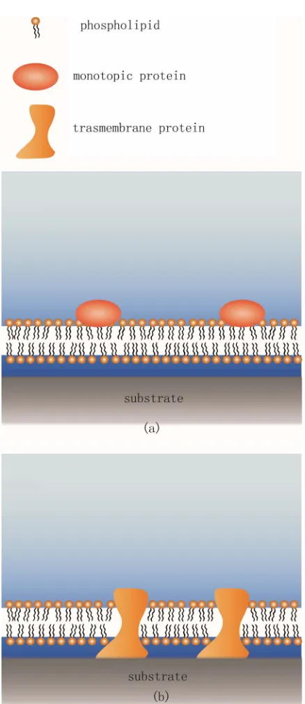
Figure 1. Conventional solid supported lipid bilayer and its limitations. (a) The SLB is able to accommodate monotopic proteins so that they stay functional. Both proteins and lip- ids are laterally mobile in such a scenario; (b) In contrast, transmembrane proteins with extended cytosolic domains have not enough space between the substrate and the bi- layer. Thus, they will contact the substrate, deform and of- ten denature which leads to loss of function. In addition, such proteins are also immobilized.
chemical biosensors [17]. Here, we discuss the options available for polymer bilayer supports and try to under- score the particular strengths and weaknesses of the dif- ferent systems and methods.
2. Polymer Supports for Lipid Bilayers
Most common polymer supports have had their genesis in convenience. Popular biological techniques involve numerous polymerizing substances; consequently some have been adopted for use as membrane cushions. For a successful polymer based lipid bilayer cushion, the poly- mer must have some few specific characteristics. Firstly, they must be capable of forming a thin layer with surface uniformity suitable for bilayer formation. Secondly, they would ideally have a well-defined elastic modulus that can be replicated at every iteration of the experiment. Thirdly, the polymer must be hydrophilic, and they must be relatively chemically inert so as not to cause unwanted reactions and interactions with the membrane. Due to their hydrophilicity such polymers typically have high water contents and are known as hydrogels. Hydrogels have refractive indices that deviate only slightly from that of the liquid used to hydrate them, this allows for good optical coupling between the hydrogel and the aqueous solution, giving aberration free imaging through the gels. Most other light based measurement techniques such as FCS [18] and FRAP [19] are also compatible with these systems. The nature of self-assembly of amphiphillic mo- lecules such as lipids dictates that there must be water present for the formation of a bilayer. Consequently, to avoid de-wetting of the lipid/polymer interface during or after deposition of the bilayer there can be no strong at- tractive forces between a substrate and the membrane. Care must be taken when using polymers that have charged or polarized functional groups to ensure the at- tractive forces between these and the lipids are not too great. Typically polymer wetting ability is characterized by the contact angle of a water droplet on its surface. This can give some indication of a good polymer for a bilayer cushion application. Typical contact angles range from 30 - 70 degrees [20,21].
that are attached to the solid substrate through an inter- mediary binding molecule: alkylsilanes for silica and mica substrates [22], or alkylthiols for GaAs or gold sub- strates [23]. These binding molecules need to have a functionalized domain for polymer attachment and can be either coated over the entire solid substrate when us- ing independent polymer supports [24] or attached to the distal end of each polymer when using lipopolymer sup- ports [25].
[image:3.595.322.519.80.319.2]Yet, in this review we separated polymer supported bi- layers into two main classes: independent polymer to the bilayer, and coupled membrane-polymer systems where all or parts of the polymers are linked to lipids or hydro- phobic molecules that integrate into the bilayer (See Fig- ure 2). A short summary of the different polymer sys- tems are given in Table 1, while the chemical structures are summarized in Figure 3.
Figure 2. Schematic of the two major classes of polymer supported lipid bilayers. (a) The independent support, with- out linkage between the bilayer and the polymers; (b) A coupled membrane-polymer systems where the polymer is covalently linked (red dots) to components of the mem- brane.
(f) (e)
(d) (c)
(b) (a)
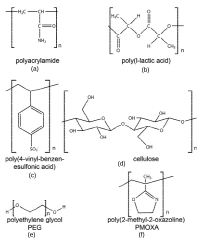
Figure 3. Chemical structure of commonly used polymers for bilayer support. (a) Polyacrylamide; (b) Poylacticacid; (c) The polyelectrolyte poly(4-vinyl-benzen-esulfonic acid); (d) Cellulose; (e) PEG and (f) PMOXA.
2.1. Independent Polymer Support
Independent polymer supports are characterized by the fact that they have no direct linkage with the lipid bilayer. This allows for maximal flexibility with respect to poly- mer choice as well as deposition and manufacture pro- cedures. The polymer in question can be spin coated on [26], deposited by sequential dipping [27] or, for chemi- cally induced polymerization, polymerized while sand- wiched between the substrate and a second solid layer with a nonreactive coating [28]. Following polymer pre- paration the lipid bilayer is deposited using one of three main techniques: Langmuir-Schaefer, Langmuir-Blodgett, or a hybrid monolayer/vesicle fusion system (vide infra).
2.1.1. Polyacrylamide
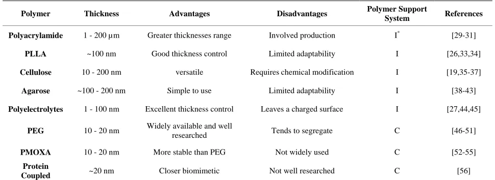
[image:3.595.68.279.298.667.2]Table 1. Polymers used as membrane supports.
Polymer Thickness Advantages Disadvantages Polymer Support
System References
Polyacrylamide 1 - 200 µm Greater thicknesses range Involved production I* [29-31]
PLLA ~100 nm Good thickness control Limited adaptability I [26,33,34]
Cellulose 10 - 200 nm versatile Requires chemical modification I [19,35-37]
Agarose ~100 - 200 nm Simple to use Limited adaptability I [38-43]
Polyelectrolytes 1 - 100 nm Excellent thickness control Leaves a charged surface I [27,44,45]
PEG 10 - 20 nm Widely available and well
researched Tends to segregate C [46-51]
PMOXA 10 - 20 nm More stable than PEG Not widely used C [52-55]
Protein
Coupled ~20 nm Closer biomimetic Not well researched C [56]
*
I refers to independent polymer supports (see Section 2.1), C refers to coupled polymer supports (see Section 2.2).
the unpolymerized solution is sandwiched between the activated solid substrate and another surface coated with a special non-reactive coating, typically a short chain si- lane polymer which renders the surface inert [32]. Once the nonreactive layer is removed the result is a uniform polymer surface suitable for bilayer deposition. This sand- wiching technique is only possible because polymeriza- tion and crosslinking of the polyacrylamide is induced chemically and occurs over the time of minutes. In con- trast to other polymer systems, the thickness of the cross- linked polyacrylamide gel can be easily controlled during production and thickness from tens to hundreds of mi- crometers can be achieved [32]. In comparison, other te- chniques give gel thicknesses in the tens to hundreds of nanometers range [29]. This wide range of thicknesses increases the number of potential applications for such a system. However, it should be noted that the acrylamide monomer is a toxin that should be handled and processed with care in particular if live cells are involved in a study.
2.1.2. Poly-L Lactic Acid (PLLA)
PLLA is another commonly used membrane support (Figure 3(b)). It is hydrophilic and quite inert and thus provides a good substrate for biological studies [26]. It has some promise in the medical field due to its biocom- patibility and biodegradability making it a good candi- date as a scaffold for tissue engineering [33]. PLLA can be formed into a uniform support by spin coating a solu- tion onto a solid substrate. The coating then gets an- nealed before use to complete the polymerization. This yields layer thicknesses in the 100 nm range [26]. Having polymer layers this thin allows the use of sensitive opti- cal techniques that rely on the use of objectives with high numerical aperture, such as; sum frequency generation vibrational spectroscopy [26], total internal reflection
fluorescence and glancing angle illumination [34].
2.1.3. Cellulose
2.1.4. Agarose
Agarose is a polysaccharide most commonly found in agar, the gelatinous substance used for bacterial cell cul- ture. It is derived from certain species of red algae and is used in such things as ice cream, the brewing process, as well as a food item in its own right [38]. In biological studies agarose is used to make a porous gel for micro- organism motility assays [39]; the concentration of aga- rose in solution determines the final viscosity of the sub- stance [40]. Agarose has been used as a polymer support for bilayers for the last 15 years [41,42]. It can be depos- ited on glass by brushing on a solution of agarose type VII in water, this is dried at room temperature, no further modifications are required [43]. This makes agarose ar- guably the simplest polymer supports to work with.
2.1.5. Polyelectrolyte Cushions
Another polymer cushioning system involves polyelec- trolytes. These are polymers whose monomer subunits have an electrolyte group. The electrolyte groups will dis- sociate when exposed to an aqueous solution leaving the polymer with a net charge. To form a bilayer cushion the polyelectrolyte is deposited onto the substrate (which is typically charged) in a layer by layer fashion [44,45]. The substrate is repeatedly dipped between two polye- lectrolyte solutions; one a polycation (such as Poly (dial- lyldimethylammonium chloride)), one a polyanion (such as poly(4-venyl-benzenesulfonic) acid, Figure 3c) [27]. Each dipping causes a monolayer of polyelectrolyte to be adsorbed on the surface through electrostatic attraction and reverses the charge on the surface leaving it ready for the next layer. This layer by layer method is inexpen- sive, easy and gives excellent thickness control, down to single nanometer precision [27]. Bilayer deposition is then dependent on the relative charges in the system, for a positively charged final polyelectrolyte layer negatively charged lipids are required to get total coverage. This electrostatic coupling may make polyelectrolyte cushions a poor choice for membrane dynamics studies but a good choice for ion channel studies. Surface patterning can be carried out by making use of the electrostatics to selec- tively layer certain sections through micro contact print- ing [27]. This approach has the advantage of providing a chemical contrast as opposed to a topographical contrast for membrane patterning [27].
2.2. Coupled Membrane Polymer Systems
Coupling between the bilayer and the polymer support is usually achieved by the use of lipopolymers. These are molecules that have a lipid like structure on one end of the polymer chain allowing that part to insert into a lipid bilayer, while the rest of the polymer is free to form the cushion. In order to get full coverage of the membrane
with a supporting cushion the distal portion of the lipo- polymer needs to have a reactive end domain allowing it to covalently bind to the solid substrate. Without these tethering points the polymers tend to all reside in the upper leaflet of the bilayer and provide no measurable bi- layer/substrate spacing [46]. Even in the presence of the covalent bonding of the polymer to the solid support the system tends to segregate into domains with polymer support and bilayer parts that sit right on top of the solid substrate. The concentration of lipopolymers in the bi- layer and the length of the polymer chain can be used to fine tune bilayer/substrate distance for each application
2.2.1. Polyethylene Glycol (PEG)
PEG is one of the most ubiquitous polymers used for lipopolymer constructs. It is a polyether that can be linear or branched and carries little to no charge [47] (Figure 3(e)). It is non-toxic and has excellent wetting character- istics making it an ideal choice for a wide range of bio- logical applications: PEG is used as an antifouling coat- ing on biomedical devices due to its “protein repellent” characteristics, i.e. flexibility and hydrophilicity [48-50]. It was first bio-functionalized in the 1970s to aid in drug solubility and stability in immunological studies [51]. The process of covalently attaching a PEG to a biomo- lecule (known as pegylation) was developed with pro- teins in mind; however the process is easily adjusted for lipids. The PEG molecule has a hydroxyl group on both ends of the polymer chain that permits hydrogen bonding to a number of end groups suitable for bio reactions such as: amides, esters, and aldehydes. PEGS for lipid studies usually require different reactive groups on either end, some good options include: amine, maleimide, pyridyl di- sulfide, and carboxylic acids [48]. As with other lipopo- lymers, the lateral density of pegylated lipids in a sup- ported lipid bilayer determines whether or not the forma- tion of a polymer cushion will be successful. Too few lipopolymer tethers and the bilayer will sag and contact the solid support. Too many PEGs and free diffusion in the bilayer will be impacted [46].
2.2.2. Poly(2-Methyl-2-Oxazoline) (PMOXA)
2.2.3. Protein Coupled Polymer Cushion
Recently researches have started to employ membrane in- corporated proteins as anchor points to the polymer cu- shion. Presently, the only known such system is based on poly(N-(2-hydroxyethyl)acrylamide-co-5-acrylamido-1- carboxypentyl-iminodiacetate-co-4-benzoylphenyl meth- acrylate) (P(HEAAm-co-NTAAAm-co-MABP)) that has been modified with the nickel chelating nitrilotriacetic acid (NTA) groups. This allows binding of cytochrome c oxidase via a poly histidine-tag to the polymer cushion surface. The bilayer between the proteins is then formed using direct vesicle deposition [56].
This review focused on the popular methods for pro- viding a polymer bilayer support. Other polymer systems such as dextran and polyethyleneimine (PEI) have also been developed as membrane supports, but have so far not seen widespread use [57,58].
3. Bilayer Deposition on Polymer Support
For the deposition of the lipopolymer containing bilayers or of the lipid membrane on the polymer cushion there are three main options that have successfully been used. They all involve the use of a Langmuir film balance to a greater or lesser degree. This is in stark contrast to solid supported bilayers that can often be formed by simple in- cubation of the clean substrate with small unilamellar ve- sicles.
The Langmuir-Blodgett technique [59] requires the lip- ids to be dispersed as a monolayer at the air-water inter- face of a Langmuir film balance. The Langmuir film ba- lance allows the surface density of the lipid monolayer to be adjusted: eukaryotic cells are thought to have a sur-
face pressure of 32 mN/m [60] and surface pressure any- where between this and 20 mN/m have been successfully used for membrane deposition. The surface pressure is adjusted using the parallel barriers and the substrate to be coated is drawn from the liquid phase through the lipid monolayer into the air perpendicular to the surface (Fig- ure 4(a)), this deposits the first lipid monolayer onto the polymer substrate. The substrate is then dipped back through the monolayer, again perpendicular to the sur- face, to deposit the second monolayer creating a bilayer (Figure 4(d)). This technique is suitable for depositing symmetrical as well as asymmetrical bilayers or multi- layers.
The second option is the Langmuir-Schäfer technique [61]. This involves the same first step as for Langmuir- Blodgett transfer (Figure 4(a)), however this time the sec- ond dip is done parallel to the liquid surface (Figure 4(c)), this way lipids are distributed more evenly in the second monolayer as there is no adjustment required to maintain the surface pressure during the dip. Consequently this produces more homogenous bilayers. Langmuir-Schäfer is sometimes regarded as a variant of the Langmuir-Blo- dgett technique however Langmuir-Schäfer deposition has typically a better success rate.
The third option is a hybrid Langmuir-Blodgett/vesicle fusion technique [62]. Here the first monolayer is depos- ited using Langmuir-Blodgett transfer (Figure 4(a)) and the upper leaflet is formed by incubation with vesicles of the desired upper leaflet lipid composition (Figure 4(b)).
[image:6.595.64.533.495.689.2]The preparation of the lipid bilayer in a lipopolymer based support can be achieved using the same techniques as above, but with some small variations. The necessity of binding the polymer to the substrate (typically through
Figure 4. Deposition of a lipid bilayer on a polymer cushion by one of 3 methods. The first step (a) is the same in all schemes: deposition of a monolayer on the substrate via Langmuir-Blodgett transfer. The second monolayer can be created either by incubation with small unilamellar vesicles that fuse to form the top monolayer (b), by horizontal Langmuir-Schäfer transfer
salinization for glass) means that there are in general two
Ob ting a lipid bilayer on a polymer support
su
(e.g. fibronectin, and albumin [66]) that can be deposited
directly onto a polymer substrate providing diffusion
se
su bilayers without them losing form or options: Either the molecule that links the solid support
to the polymer cushion is part of the polymer already [25] or it is separately deposited as a film over the entire solid support [26]. There are also different options for how to attach the polymers to the bilayer components. The poly- mers can either be pre-bound to the solid substrate and then have a modified bilayer element to which they bind [63] or they can be pre attached to the lipid monolayer and then deposited on the pretreated surface [25].
4. Advantages and Limitations of Polymer
Supports
viously, crea
is much more involved and challenging than producing a bilayer on a solid support or most other membrane mim- ics. Thus, it is worthwhile to briefly discuss the benefits of such an undertaking. Originally, the creation of poly- mer supported bilayers was driven by the desire to study membrane bound proteins that have substantially sized cytosolic domain (>10 Å, the typical distance between a solid support and a supported lipid bilayer). Studying such large proteins requires that there be no potentially denaturing interactions between the protein and the solid support. Having an inert polymer spacer solves this pro- blem and still permits the protein to diffuse in the bilayer. The introduction of a space between the bilayer and the substrate also means that a reservoir has been created into which ions can flow through ion channels. Therefore with an electrode at the solid support electrophysiologi- cal experiments can be conducted in a controlled manner. This makes for an interesting alternative to traditional patch clamping [43] and vertical free-standing black lipid membranes. A further advantage for such studies is the enhanced self-healing seen in polymer supported bilayers. The elimination of bilayer defects increases the electrical resistance across the bilayer; a definite advantage for ion channel characterization.
It has furthermore been shown that similar to solid pported membranes independent polymer supports can also easily be patterned using one of two different tech- niques: photo mask lithography or micro contact printing. In the lithographic technique the polymer is chosen such that polymerization or crosslinking can be induced by light [64]. This results in a patterned polymer substrate onto which a bilayer can be easily deposited and con- strained by a physical corral. The micro contact printing system uses a polydimethylsiloxane (PDMS) master stamp to transfer a patterned monolayer of “ink” onto a sub- strate through direct contact [65]. The ink is adsorbed to the substrate leaving an ink design with micrometer fea- ture size. A variety of protein inks have been developed
barriers to a lipid bilayer. Micro-patterns such as these can be used to do direct side by side comparison of dif- ferent lipid species without intermixing, or alternatively to apply different stimuli to different parts of the bilayer without intermixing of the lipids.
When using lipids that have a high charges (such as phosphatidylinositol 4,5-bisphosphate (PIP2) which has
three negative charges on its head group [67]) there is po- tentially electrostatic interactions between the supported lipid bilayer and the substrate. In particular since com- mon glass preparation methods such as piranha etching (75% sulfuric acid (H2SO4) and 25% hydrogen peroxide
(H2O2)), hydroxylate the surface leaving it hydrophilic
and slightly negatively charged [68]. Separating the char- ged lipid bilayer from the charged solid support through an inert polymer support of at least several dozens of na- nometers introduces enough spatial separation to effec- tively screen any electrostatic interactions between the solid substrate and the bilayer considering that the Debye screening length at physiological conditions (~150 mM NaCL) is about 1 nm [69].
A final advantage of polymer membrane supports is that while they effectively overcome many of the prob- lems inherent to solid supported membranes, they con- rve the latter’s compatibility with most of the modern light-based experimental methods. Thus, techniques such as Fluorescence Correlation Spectroscopy (FCS), Föster resonant energy transfer microscopy, fluorescence re- covery after photo bleaching or total internal reflection fluorescence microscopy can readily be used with these systems [4,18,70].
The main disadvantage of polymer supported bilayers is the increased complexity of the production process when compared to solid supported bilayers. Many more steps are required for polymer cushion fabrication and lipid deposition presenting many more opportunities for failure of the system. For the independent polymer sup- ports incorporation of the lipid bilayer requires a Lang- muir trough. Even in its simplest form this machine re- quires a moderate outlay in cost and training and requires much more time and resources than vesicle incubation. The total time required for bilayer production with a polymer support is approximately an order of magnitude more than for a solid supported bilayer. This is a severe disadvantage as it increases the personnel cost as well as the materials expenditure.
5. What Lies Ahead for Polymer Cushioned
Bilayers?
The clear, distinctive advantage of polymer supports is the ability to incorporate integral membrane proteins into
function. The place where this technology advances how- ever will be at the intersection with the techniques dis- cussed above as well as developments that are still on- going. The combination of polymer supported lipid mem- branes and semiconductor supports can for example be used as an organic transistor to reliably detect surface charge on a lipid monolayer [71]. Incorporation of this ability with the proper cultured cells could herald new types of cell based biosensors with an electronic output. Patterned polymer supports could be used to develop whole arrays of different biosensors capable of detecting an enormous range of different properties or reagents on an extremely compact surface.
Polymer cushioned bilayers are new meta-materials that have some features similar to the actin-membrane structure of living cells. Actin is a very dynamic biop m
ve been an ex-l in membrane characterization. They er when it comes to studying
trans-aterial is based upon work supported by the Na- er Grant No. PHY-0955945.
[1] L. K. Tamm and H. M. McConnell, “Supported
Phospho-lipid-Bilayers, ol. 47, No. 1
pp. 105-113. d 85)83882-0
oly- er and it seems natural to utilize polymers for mem- brane support that have some added functionality. Using such polymers as cushion could turn polymer supported bilayers into rather active surfaces. For example the pH dependent properties of hydrophilic poly(acrylic acid) (PAA) has been recently used to create an active polymer cushion for bilayer support [72]. One could also envision the use of hydrophilic shape memory polymers such as polyethylene terephthalate-polyethylene glycol copolymer [73] or poly(N-isopropylacrylamide) [74,75] to actively change the topography of the polymer support which would allow for interesting studies of the active coupling of membrane composition and curvature.
6. Conclusion
In conclusion, supported lipid bilayers ha tremely useful too
are limited howev
membrane proteins. Here we have reviewed some of the options that are available for introducing a polymer cu- shion to support a lipid bilayer and have discussed the major benefits of such systems. As we reach the limits of what the traditional solid supported lipid bilayer is capa- ble of, we expect greater uptake of polymer cushions and further development of the technology in the coming years.
7. Acknowledgements
This m
tional Science Foundation und
REFERENCES
” Biophysical Journal, V oi:10.1016/S0006-3495(
, 1985,
opy,”
[2] G. W. Gordon, G. Berry, X. H. Liang, B. Levine and B.
Herman, “Quantitative Fluorescence Resonance Energy Transfer Measurements Using Fluorescence Microsc Biophysical Journal, Vol. 74, No. 5, 1998, pp. 2702-2713. doi:10.1016/S0006-3495(98)77976-7
[3] M. B. Forstner, C. K. Yee, A. N. Parikh and J. T. Groves, “Lipid Lateral Mobility and Membrane Phase Structure Modulation by Protein Binding,” Journal of the American Chemical Society, Vol. 128, No. 47, 2006, pp. 15221- 15227. doi:10.1021/ja064093h
[4] J. T. Groves, R. Parthasarathy and M. B. Forstner, “Fluo- rescence Imaging of Membrane Dynamics,” Annual Re-view of Biomedical Engineering, Vol. 10, No. 1, 2008, pp.
branes,” Journal of Physical Chemistry B, Vol. 311-338.
[5] S. Rozovsky, M. B. Forstner, H. Sondermann and J. T. Groves, “Single Molecule Kinetics of Enth Binding to Lipid Mem
116, No. 17, 2012, pp. 5122-5131. doi:10.1021/jp210045r
[6] D. Axelrod, T. P. Burghardt and N. L. Thompson, “Total Internal-Reflection Fluorescence,” Annual Review of Bio- physics and Bioengineering, Vol. 13, No. 1, 1984, pp. 247-268.
[7] D. Axelrod, D. E. Koppel, J. Schlessinger, E. Elson and W. W. Webb, “Mobility Measurement by Analysis of Fluorescence Photobleaching Recovery Kinetics,” Bio- physical Journal, Vol. 16, No. 9, 1976, pp. 1055-1069. doi:10.1016/S0006-3495(76)85755-4
[8] K. D. Mossman, G. Campi, J. T. Groves and M. L. Dustin, “Altered Tcr Signaling from Geometrically Repatterned Immunological Synapses,” Science, Vol. 310, No. 5751, 2005, pp. 1191-1193. doi:10.1126/science.1119238
[9] K. Salaita, P. M. Nair, R. S. Petit, R. M. Neve, D. Das, J. W. Gray, “Restriction of Receptor Movement Alters Cel- lular Response: Physical Force Sensing by Epha2,” Sci- ence, Vol. 327, No. 5971, 2010, pp. 1380-1385. doi:10.1126/science.1181729
[10] Y. Shao, Y. D. Jin, J. L. Wang, L. Wang, F. Zhao and S. J. Dong, “Conducting Polymer Polypyrrole Supported Bila-yer Lipid Membranes,” Biosensors & Bioelectronics, Vol. 20, No. 7, 2005, pp. 1373-1379.
doi:10.1016/j.bios.2004.06.001
[11] M. Tanaka and E. Sackmann, “Supported Membranes as Biofunctional Interfaces and Smar
Physica Status Solidia-Applicat
t Biosensor Platforms,” ions and Materials Sci-
, 2004, pp. ence, Vol. 203, No. 14, 2006, pp. 3452-3462.
[12] M. Trojanowicz and A. Mulchandani, “Analytical Appli- cations of Planar Bilayer Lipid Membranes,” Analytical and Bioanalytical Chemistry, Vol. 379, No. 3
347-350. doi:10.1007/s00216-004-2611-4
[13] M. M. Baksh, M. Jaros and J. T. Groves, “Detection of Molecular Interactions at Membrane Surfaces through Colloid Phase Transitions,” Nature, Vol. 427, No. 6970, 2004, pp. 139-141. doi:10.1038/nature02209
[14] M. Stelzle, R. Miehlich and E. Sackmann, “2-Dimensio- nal Microelectrophoresis in Supported Lipid Bilayers,” Biophysical Journal, Vol. 63, No. 5, 1992, pp. 1346-1354. doi:10.1016/S0006-3495(92)81712-5
Patterned Arrays,” Science, Vol. 285, No. 5430, 1999, pp. 1046-1048. doi:10.1126/science.285.5430.1046
[16] M. Fischer, A. Bacher, I. Haase, M. Tristl and E. Sack- mann, “Design of Biofunctional Assemblies on Solids through Recombinant Spherical Bacterial Protein Luma- zine Synthase,” ChemPhysChem, Vol. 2, No. 10, 2001, pp. 623-627.
doi:10.1002/1439-7641(20011015)2:10<623::AID-CPHC 623>3.0.CO;2-R
[17] V. Borisenk
muller, N. Fertig and J. C. Behrends, “Simultaneous Op- tical and Electrica
o, T. Lougheed, J. Hesse, E.
Fureder-Kitz-l Recording of SingFureder-Kitz-le Gramicidin Chan- nels,” Biophysical Journal, Vol. 84, No. 1, 2003, pp. 612- 622. doi:10.1016/S0006-3495(03)74881-4
[18] Y. F. Dufrene and M. F. Garcia-Parajo, “Recent Progress in Cell Surface Nanoscopy: Light and Force in the Near- Field,” Nano Today, Vol. 7, No. 5, 2012, pp. 390-403. doi:10.1016/j.nantod.2012.08.002
[19] M. Tanaka, J. Hermann, I. Haase, M. Fischer and S. G. Boxer, “Frictional Drag and Electrical Manipulation o Recombinant Proteins in Polymer-S
f upported Membranes,” Langmuir, Vol. 23, No. 10, 2007, pp. 5638-5644. doi:10.1021/la0628219
[20] R. J. Good, “Contact-Angle, Wetting, and Adhesion—A Critical-Review,” Journal of Adhesion Science and nology, Vol. 6, No. 12, 1
Tech-992, pp. 1269-1302.
doi:10.1163/156856192X00629
[21] P. F. Rios, H. Dodiuk, S. Kenig, S. McCarthy and A. Dotan, “The Effect of Polymer Surface on th
and Adhesion of Liquid System
e We s,” Journal of Adhesion
tting
Science and Technology, Vol. 21, No. 3-4, 2007, pp. 227- 241. doi:10.1163/156856107780684567
[22] J. Piehler, A. Brecht, R. Valiokas, B. Liedberg and G. Gauglitz, “A High-Density Poly(Ethylene Glycol) Poly- mer Brush for Immobilization on Glass-Type Surfaces,” Biosensors & Bioelectronics, Vol. 15, No. 9-10, 2000, pp. 473-481. doi:10.1016/S0956-5663(00)00104-4
[23] E. Sackmann and M. Tanaka, “Supported Membranes on Soft Polymer Cushions: Fabrication, Characterization and Applications,” Trends in Biotechnology, Vol. 18, No. 2, 2000, pp. 58-64. doi:10.1016/S0167-7799(99)01412-2
[24] M. Kuhner, R. Tampe and E. Sackmann, “Lipid Mono- and Bilayer Supported on Polymer Films: Composite Poly- mer-Lipid Films on Solid Substrates,” Biophysical Jour- nal, Vol. 67, No. 1, 1994, pp. 217-226.
doi:10.1016/S0006-3495(94)80472-2
[25] M. Wagner and L. Tamm, “Tethered Polymer-Supported Planar Lipid Bilayers for Reconstitution o
brane Proteins: Silane-Polyethylenegly
f Integral Mem-col-Lipid as a Cu-shion and Covalent Linker,” Biophysical Journal, Vol. 79, 2000, pp. 1400-1414.
doi:10.1016/S0006-3495(00)76392-2
[26] T. Wang, D. Li, X. Lu, A. Khmaladze, X. Han and S. Ye, “Single Lipid Bilayers
Studied by Sum Frequency Generation
Constructed on Polymer Cushion Vibrational
Spec-,” Journal of Colloid and Inter-
toskele-me Applications,” Pro-
troscopy,” Journal of Physical Chemistry C, Vol. 115, No. 15, 2011, pp. 7613-7620.
[27] N. Kohli, S. Vaidya, R. Ofoli, R. Worden and I. Lee,
“Arrays of Lipid Bilayers and Liposomes on Patterned Polyelectrolyte Templates
face Science, Vol. 301, No. 2, 2006, pp. 461-469. [28] Y. L. Wang and R. J. Pelham, “Preparation of a Flexible,
Porous Polyacrylamide Substrate for Mechanical Studies of Cultured Cells,” Molecular Motors and the Cy ton, Vol. 298, 1998, pp. 489-496.
[29] H. Towbin, T. Staehelin and J. Gordon, “Electrophoretic Transfer of Proteins from Polyacrylamide Gels to Nitro- cellulose Sheets—Procedure and So
ceedings of the National Academy of Sciences of the United States of America, Vol. 76, No. 9, 1979, pp. 4350- 4354. doi:10.1073/pnas.76.9.4350
[30] E. C. Muniz and G. Geuskens, “Compressive Elastic Mo- dulus of Polyacrylamide Hydrogels and Semi-Ipns with Poly(N-Isopropylacrylamide),” Macromolecules, Vol. 34, No. 13, 2001, pp. 4480-4484. doi:10.1021/ma001192l
[31] C. E. Kandow, P. C. Georges, P. A. Janmey and K. A. Beningo, “Polyacrylamidc Hydrogels for Cell Mechanics: Steps toward Optimization and Alternative Uses,” Cell Mechanics, Vol. 83, 2007, pp. 29-46.
doi:10.1016/S0091-679X(07)83002-0
[32] J. Tse and A. Engler, “Preparation of Hydrogel Substrates with Tunable Mechanical Properties,”
in Cell Biology, Vol. 10, No. 16, 2010,
Current Protocols pp. 1-16.
,”
nter-473.
in-Oxide Electrodes,” Langmuir, Vol. 15,
r, Vol. 17, No. 17, 2001, pp. 5129-
ariaceae, Rhodophyta),” Carbohydrate Research, [33] F. Yang, R. Murugan, S. Ramakrishna, X. Wang, Y. X. Ma and S. Wang, “Fabrication of Nano-Structured Porous Plla Scaffold Intended for Nerve Tissue Engineering Biomaterials, Vol. 25, No. 10, 2004, pp. 1891-1900. [34] Y. Duan, J. Liu, H. Sato, J. Zhang, H. Tsuji and Y. Ozaki,
“Molecular Weight Dependence of the Poly(L-Lactide)/ Poly(D-Lactide) Stereocomplex at the Air-Water I face,” Biomacromolecules, Vol. 7, No. 10, 2006, pp. 2728- 2735.
[35] F. Rehfeldt and M. Tanaka, “Hydration Forces in Ultra- thin Films of Cellulose,” Langmuir, Vol. 19, 2003, pp. 1467-1
[36] H. Hillebrandt, G. Wiegand, M. Tanaka and E. Sackmann, “High Electric Resistance Polymer/Lipid Composite Films on Indium-T
1999, pp. 8451-8459.
[37] J. Groves, L. Mahal and C. Bertozzi, “Control of Cell Ad- hesion and Growth with Micropatterned Supported Lipid Membranes,” Langmui
5133.
[38] R. Falshaw, R. H. Furneaux and D. E. Stevenson, “Agars from Nine Species of Red Seaweed in the Genus Curdiea (Gracil
Vol. 308, No. 1-2, 1998, pp. 107-115. doi:10.1016/S0008-6215(98)00049-4
[39] N. K. Jerne and A. A. Nordin, “Plaque Formation in Agar by Single Antibody-Producing Cells,” Science
No. 356, 1963, p. 405.
, Vol. 140, ence.140.3565.405 doi:10.1126/sci
doi:10.1016/S0021-9290(02)00437-2
[41] H. Yuan, A. Leitmannova-Ottova and H. Ti Tien, “An Agarose-Stabilized Blm: A New Method for For
layer Lipid Membranes,” Materials
ming Bi-Science and
Bilayer
le Ion Channels,”
05, pp. 738-743. neering C, Vol. 4, No. 1, 1996, pp. 35-38.
[42] X. Lu, A. Leitmannova-Ottova and H. Tien, “Biophysical Aspects of Agar-Gel Supported Bilayer Lipid Nembranes: A New Method for Forming and Studying Planar Lipid Membranes,” Bioelectroehemistry and Bioenerget- ics, Vol. 39, No. 2, 1996, pp. 285-289.
[43] T. Ide and T. Yanagida, “An Artificial Lipid Bilayer For- med on an Agarose-Coated Glass for Simultaneous Elec- trical and Optical Measurement of Sing
Biochemical and Biophysical Research Communications, Vol. 265, No. 2, 1999, pp. 595-599.
[44] K. Katagiri and F. Caruso, “Monodisperse Polyelectro- lyte-Supported Asymmetric Lipid-Bilayer Vesicles,” Ad- vanced Materials, Vol. 17, No. 6, 20
doi:10.1002/adma.200401441
[45] G. Lee, Y. Lee and B. Kyung, “Layer-by-Layer Assembly of Zeolite Crystals on Glass with Polyelectrolytes as Ion Inkers,” Journal of the Americ
ic an Chemical Society, Vol. 123, No. 40, 2001, pp. 9769-9779.
doi:10.1021/ja010517q
[46] E. B. Watkins, R. J. El-Khouri, C. E. Miller, B. G. Seaby, J. Majewski and C. M. Marques, “St
dynamics of Lipid Bilay
ructure and Thermo- ers on Polyethylene Glycol Cu- shions: Fact and Fiction of Peg Cushioned Membranes,” Langmuir, Vol. 27, No. 22, 2011, pp. 13618-13628. doi:10.1021/la200622e
[47] J. Jimenez, A. Heim, G. Matthews and N. Alcantar, “Con- struction and Characterization of Soft-Supported L Bilayer Membranes for
ipid Biosensors Application,” IEEE
ecules: Delivery Aspects,” Expert Opinion Annual International Conference of the Engineering in Medicine and Biology Society, Vol. 1, No. 1, 2006, pp. 4119-4122.
[48] S. M. Ryan, G. Mantovani, X. X. Wang, D. M. Haddleton and D. J. Brayden, “Advances in Pegylation of Important Biotech Mol
on Drug Delivery, Vol. 5, No. 4, 2008, pp. 371-383. doi:10.1517/17425247.5.4.371
[49] N. Ngadi, J. Abrahamson, C. Fee and K. Morison, “Are Peg Molecules a Universal Protein Repellent?” Inter tional Journal of Biological and
Life Sciences, Vol. 5, No.
ntrolled Release, Vol. 161, No. 161 3, 2009, pp. 106-110.
[50] G. Pasut and F. Veronese, “State of the Art in Pegylation: The Great Versatility Achieved after Forty Years of Re-
search,” Journal of Co ,
2012, pp. 461-472. doi:10.1016/j.jconrel.2011.10.037
[51] F. F. Davis, “Commentary—The Origin of Pegnology,” Advanced Drug Delivery Reviews, Vol. 54, No. 4, 2002, pp. 457-458. doi:10.1016/S0169-409X(02)00021-2
[52] R. Konradi, B. Pidhatika, A. Muhlebach and M. Textor, “Poly-2-Methyl-2-Oxazoline: A Peptide-Like Polymer for Protein-Repellent Surfaces,” Langmuir, Vol. 24, No. 3, 2008, pp. 613-616.
[53] R. Konradi, B. Pidhatika, Q. Li and M. Textor, “Poly(2- Methyl-2-Oxazoline): Protein-Like Polymer for the Fab-
rication of Functional Non-Fouling Surface Coatings,”
oration of Cell Receptors,” ChemPhysChem, Vol. 5, European Cells and Materials, Vol. 14, No. 3, 2007, p. 131.
[54] O. Purrucker, A. Fortig, R. Jordan and M. Tanaka, “Sup- ported Membranes with Well-Defined Polymer Tethers- Incorp
No. 3, 2004, pp. 327-335. doi:10.1002/cphc.200300863
[55] C. A. Naumann, O. Prucker, T. Lehmann, J. Ruhe, W. Knoll and C. W. Frank, “The P
pholipid Bilayer: Tethering as
olymer-Supported Phos- a New Approach to Sub- strate-Membrane Stabilization,” Biomacromolecules, Vol. 3, No. 1, 2002, pp. 27-35. doi:10.1021/bm0100211
[56] A. Kibrom, R. F. Roskamp, U. Jonas, B. Menges, W. Knoll and H. Paulsen, “Hydrogel-Supported Protein-Te- thered Bilayer Lipid Membranes: A New Approach to- ward Polymer-Supported Lipid Membranes,” Soft Matter, Vol. 7, No. 1, 2011, pp. 237-246.
doi:10.1039/c0sm00618a
[57] J. Majewski, J. Y. Wong, C. K. Park, M. Seitz, J. N. Is- raelachvili and G. S. Smith, “Struc
mer-Cushioned Lipid Bilay
tural Studies of Poly- ers,” Biophysical Journal, Vol. 75, No. 5, 1998, pp. 2363-2367.
doi:10.1016/S0006-3495(98)77680-5
[58] K. Adlkofer, M. Tanaka, H. Hillebrandt, G. Wiegand, E. Sackmann and T. Bolom, “Electro
Gallium Arsenide Surface with Organ
chemical Passivation of ic Self-Assembled Monolayers in Aqueous Electrolytes,” Applied Physics Letters,Vol. 76, No. 22, 2000, pp. 3313-3315.
doi:10.1063/1.126636
[59] Y. G. Jin, Y. X. Qiao and X. P. Hou, “The Effects of Chain Number and State of Lipid Derivatives of
sides on Hydrogen Bo
Nucleo- nding and Self-Assembly through the Investigation of Langmuir-Blodgett Films,” Applied Surface Science, Vol. 252, No. 22, 2006, pp. 7926-7929. doi:10.1016/j.apsusc.2005.09.073
[60] H. L. Brockman, M. M. Momsen, J. R. Knudtson, S. T. Miller, G. Graff and J. M. Yanni, “Interactions of Olo- patadine and Selected Antihistamines with Model and Natural Membranes,” Ocular Immunology and Inflamma- tion, Vol. 11, No. 4, 2003, pp. 247-268.
doi:10.1076/ocii.11.4.247.18261
[61] A. V. Hughes, J. R. Howse, A. Dabkowska, R. A. L. Jones, M. J. Lawrence and S. J. Roser, “Floa
Bilayers Deposited on Chemicall
ting Lipid y Grafted Phosphatidyl- choline Surfaces,” Langmuir, Vol. 24, No. 5, 2008, pp. 1989-1999. doi:10.1021/la702050b
[62] E. T. Castellana and P. S. Cremer, “Solid Supported Lipid Bilayers: From Biophysical Studies to Sensor Design,” Surface Science Reports, Vol. 61, No. 10, 2006, pp. 429- 444. doi:10.1016/j.surfrep.2006.06.001
[63] Y. Lin, D. Minner, V. Herring and C. Naumann, “Phy- sisorbed Polymer-Tethered Lipid Bilayer with Lipopoly- mer Gradient,” Materials, Vol. 5, No. 3, 2012, pp. 2243- 2257. doi:10.3390/ma5112243
2001, pp. 172-174.
doi:10.1002/1521-3773(20010105)40:1<172::AID-ANIE 172>3.0.CO;2-G
[65] J. T. Groves, L. K. Mahal and C. R. Bertozzi, “Control of Cell Adhesion and Growth with Micropatterned Sup-ported Lipid Membranes,” Langmuir, Vol. 17, No. 17, 2001, pp. 5129-5133. doi:10.1021/la010481f
[66] C. S. Chen, M. Mrksich, S. Huang, G. M. Whitesides and D. E. Ingber, “Geometric Control of Cell Life and Death,” Science, Vol. 276, No. 5317, 1997, pp. 1425-1428. doi:10.1126/science.276.5317.1425
[67] I. M. Thornell, J. P. Wu, X. F. Liu and M. O. Bevensee, “Pip2 Hydrolysis Stimulates the Electrogenic Na+-Bicar- bonate Cotransporter Nbce1-B and -C Variants Expre in Xenopus Laevis Oocytes,” Journa
ssed l of Physiology-Lon- don, Vol. 590, No. 23, 2012, pp. 5993-6011.
doi:10.1113/jphysiol.2012.242479
[68] K. J. Seu, A. P. Pandey, F. Haque, E. A. Proctor, A. E. Ribbe and J. S. Hovis, “Effect of Surface Treatment on Diffusion and Domain Formation in Supporte
layers,” Biophysical Journal, Vol.
d Lipid Bi-92, No. 7, 2007, pp. 2445-2450. doi:10.1529/biophysj.106.099721
[69] D. Trebotich, G. H. Miller and M. D. Bybee, “A Penalty Method to Model Particle Interactions in DNA-Laden Flows,” Journal of Nanoscience and Nanotechnology, Vol. 8, No. 7, 2008, pp. 3749-3756.
[70] K. P. N. Bruns, L. M. Bergeron, T. A. Whitehead and D.
S. Clark, “Mechanical Nanosensor Based on Fret within a
sitive Detection of Surface Thermosome for Damage-Reporting Polymeric Materi- als,” Angewante Chemie Internatioanl Edition, Vol. 48, No. 31, 2009, pp. 5666-5669.
[71] H. Hillebrandt, M. Tanaka and E. Sackmann, “A Novel Membrane Charge Sensor: Sen
Charge at Polymer/Lipid Composite Films on Indium Tin Oxide Electrodes,” Journal of Physical Chemistry B, Vol. 106, No. 2, 2002, pp. 477-486. doi:10.1021/jp011693o
[72] R. J. El-Khouri, D. A. Bricarello, E. B. Watkins, C. Y. Kim, C. E. Miller, T. E. Patten, “Ph Responsive Polymer Cushions for Probing Membrane Environment Interac- tions,” Nano Letters, Vol. 11, No. 5, 2011, pp. 2169-2172. doi:10.1021/nl200832c
[73] J. S. Leng, X. Lan, Y. J. Liu and S. Y. Du, “Shape-Me- mory Polymers and Their Composites: Stimulus Methods and Applications,” Progress in Materials Science, Vol. 56, No. 7, 2011, pp. 1077-1135.
doi:10.1016/j.pmatsci.2011.03.001
[74] P. T. Mather, X. F. Luo and I. A. mory Polymer Research,” Annual
Rousseau, “Shape Me- Review of Materials Research, Vol. 39, No. 1, 2009, pp. 445-471.
doi:10.1146/annurev-matsci-082908-145419
[75] K. A. Davis, K. A. Burke, P. T. Mather and J. H son, “Dynamic Cell Behavior on Shape Memo