Myotonometry for the evaluation of
Achilles tendon mechanical properties:
a reliability and construct validity study
Alessandro Schneebeli ,1,2 Deborah Falla ,2 Ron Clijsen ,1,3 Marco Barbero 1
To cite: Schneebeli A, Falla D, Clijsen R, et al. Myotonometry for the evaluation of Achilles tendon mechanical properties: a reliability and construct validity study. BMJ Open Sport & Exercise Medicine
2020;6:e000726. doi:10.1136/ bmjsem-2019-000726
Accepted 24 January 2020
1Rehabilitation Research
Laboratory 2rLab, Department of Business Economics, Health and Social Care, University of Applied Sciences and Arts of Southern Switzerland, Manno/Landquart, Switzerland
2Centre of Precision
Rehabilitation for Spinal Pain (CPR Spine), School of Sport, Exercise Rehabilitation Sciences, College of Life Environmental Sciences, University of Birmingham, Birmingham, UK
3University College
Physiotherapy, Thim van der Laan AG, Landquart, Switzerland
Correspondence to
Alessandro Schneebeli; alessandro. schneebeli@ supsi. ch © Author(s) (or their
employer(s)) 2020. Re- use permitted under CC BY- NC. No commercial re- use. See rights and permissions. Published by BMJ.
AbsTrACT
Objective This study evaluates the intra- rater and inter- rater reliability of the MyotonPRO and its construct validity for the assessment of Achilles tendon stiffness.
Design Reliability and construct validity study.
Methods Forty healthy participants were assessed using the MyotonPRO by two raters on two different occasions. Tendon was evaluated in three different positions (relaxed, 0° plantarflexion and standing) and during different isometric contractions (range 0–3 kg). Reliability was calculated using intraclass correlation coefficient (ICC and 95% CI) standard error of measurement and minimal detectable change. Construct validity was evaluated between the different positions and the different contraction intensities using Friedman test.
results Intra- rater reliability was very high ICC2,k 0.87– 0.98. The reliability of the 0.5 kg contraction was moderate with an ICC2,k of 0.59. Inter- rater reliability ranged from high to very high with an ICC2,k of 0.76–0.86. The reliability of the 0.5 kg, 1 kg contraction and the standing position was moderate with an ICC2,k of 0.55, 0.54 and 0.56 respectively. Inter- session reliability ranged from high to very high with an ICC2,k of 0.70–0.89. The reliability of the 0.5 kg contraction was moderate with an ICC2,k of 0.54. Construct validity was demonstrated between different contraction levels and different positions.
Conclusion MyotonPRO is a reliable tool for the evaluation of Achilles tendon stiffness during different contraction levels and in different positions. Construct validity was supported by changes of tendon stiffness during the explored conditions. MyotonPRO can be implemented, as a ready to use device, in the evaluation of tendon tissue mechanical properties.
InTrODuCTIOn
The Achilles tendon (AT) is one of the most commonly injured structures in the lower limb, both in athletes and the general
popu-lation.1 2 Tendinopathy of AT can alter the
mechanical, material and morphological properties of the tendon structure with a decrease of stiffness and young modulus and an increase of tendon cross- sectional area and diameter.3 4
Healthy tendon is highly responsiveness to
mechanical loading.5 Adaptation of tendon
has been shown following different loading protocols, and the intensity of the exercise (ie, high loading magnitude) and the intervention duration (>12 weeks) appear to contribute the most to changes in the mechanical properties of the tendon. The change in tendon stiffness appears to be largely due to adaptations in the material rather than morphological prop-erties of the tendon.5 That is, early changes in
young modulus is apparent following loading protocols whereas changes in tendon hyper-trophy appear as longer- term adaptations to
mechanical loading.6
Tendon responses to loading, unloading and ageing have been evaluated in a recent review revealing adaptability of the tendon tissue with an increase in stiffness when loaded
and reduced stiffness when unloaded.3
Never-theless, conflicting results has been reported when evaluating the in vivo effects of ageing on tendon stiffness, with some authors
suggesting a decrease in modulus3 7–11 and
others reporting unchanged tendon stiffness with ageing.12 13
Given the clinical and research interest in evaluate tendon stiffness as a parameter to describe tendon behaviour and tendon health, different measurement techniques have been developed. Once recent, non- invasive, ready to use technology is the
summary box
► Achilles tendon stiffness can be measured with MyotonPRO. High intra- rater, inter- rater and inter- session reliability has been found in different posi-tion and during isometric contracposi-tions.
► The interest in measuring tendon stiffness is grow-ing for its clinical and research relevance. A reliable and ready to use device has been presented in this study. MyotonPRO could be implemented in the eval-uation of Achilles tendon mechanical properties. ► Stiffness of healthy Achilles tendons has been
as-sessed, reliability in patient with tendinopathies could be different.
copyright.
on May 7, 2020 by guest. Protected by
MyotonPRO (Muomeetria, Tallinn, Estonia). This device is a handheld myotonometer producing a mechanical impulse to the skin overlying the target structure. The oscillation of the tissues underneath the probe permits calculation of the viscoelastic properties of the tissue.14–16 One parameter that can be extracted with this device is dynamic stiffness (N/m), which has been used in different studies to characterise the behaviour of different muscu-loskeletal tissues. This device has recently been used to evaluate tendon stiffness17–20 with a recent work showing
decreased stiffness over the tendinopathic region of the tendon in people with Achilles tendinopathy and a significant difference in tendon stiffness between weight- bearing and non- weight- bearing positions.
The reliability of the MyotonPRO has been
demon-strated for the evaluation of muscle stiffness,15 21–23
however, the reliability of MyotonPRO for measuring AT stiffness requires further investigation,17 18 24 25
espe-cially regarding the capability of this device to measure the tendon in different conditions. In contrast to muscle, values of AT stiffness are extremely high and even higher
when tested under load.19 Being able to quantify AT
mechanical properties with a ready to use and affordable device which provides a reliable and valid evaluation of the tendon under different conditions, would provide new opportunities to characterise the mechanical behaviour of the AT, for example, in people with Achilles tendinopathy or following an intervention.
This study evaluates the intra- rater and inter- rater
reliability of using the MyotonPRO to measure stiffness of asymptomatic AT in different position and during increasing load and to determine the construct validity of this device for measuring tendon stiffness.
MeThODs
The study was conducted between January and July 2019 at the University of Applied Sciences and Arts of Southern Switzerland. The study reporting adheres to the Guide-lines for Reporting Reliability and Agreement Studies.26
A convenience sample of 40 healthy participants (18 women, 22 men, aged 33.8±14.2 years) were prospectively recruited trough public announcement at the University. Their mean (±SD) height and weight were 172.2 (±8.2) cm and 71.7 (±12) kg. Health- related physical activity was monitored using the International Physical Activity
Ques-tionnaires Short form (IPAQ)27; 23 participants were
categorised as high, 15 as moderate and 2 as having low physical activity levels. Participants were excluded if they had a history of tendon or foot injury or surgery; connec-tive tissue, metabolic or endocrine disease. Furthermore, participants with systemic inflammatory disorders, rheumatoid arthritis, spondyloartropathy or hypercho-lesterolaemia were excluded.
Sample size was estimated according to Walter et al28
using the following parameters: two replicates, p0=0.6, p1=0.80, α=0.05 and β=0.2. The optimal sample size was 39.1, and we rounded up to 40 participants.
Procedures
The AT mechanical properties were randomly analysed by two independent operators who were both physio-therapists with a minimum of 4 hours of training with the MyotonPRO prior to the study. Two measurement sessions were planned within 7 days. During the first session, the two operators consecutively performed the measures in order to evaluate the intra- rater and inter- rater reliability. The second session was used to evaluate inter- session reliability.
Testing location and room temperature (21°C–23°C) was kept constant across sessions. Participants were asked to maintain the same activity level between the two testing days.
The resistance of the soft tissues to the deformation induced by the MyotonPRO is calculated from an acceler-ation graph representing the damped natural oscillacceler-ation of the tissues. Dynamic stiffness, calculated in N/m, is computed by multiplying the maximum acceleration of the oscillation graph to the mass of the probe divided by the maximum displacement of the tissue; higher values represent a stiffer tissue.
Both the left and right AT were evaluated at the most central point of the tendon following a trajectory from the most central component of the medial malleolus as this point has been suggested as the most prevalent
region for tendinopathy.29 Furthermore, a previous
study using MyotonPRO to evaluate stiffness of the AT in people with Achilles tendinopathy and asymptomatic controls showed that changes in mechanical parameters are more likely to be detected from 8 to 12 cm from the plantar aspect of the heel, corresponding to the free tendon part.19
MyotonPRO was setup with a multiscan sequence of 5 impulses (1 s apart), a short duration (15 ms) impulse involving minimal mechanical force with a precompres-sion of 0.18 N and an impulse of 0.40 (total=0.58 n) was exerted on the tendon.
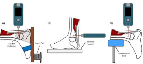
Measurements ware taken in three different posi-tions; standing, a relaxed position and during isometric contractions of the plantar flexors of increasing inten-sities, in order to evaluate construct validity (figure 1). As the triceps surae contracts to increase plantarflexor force, the passive tension within the tendon increases as
shown in previous studies.30 Since tendon stiffness and
tendon displacement are directly related to plantarflexor
strength31 and ankle angle,32 myonotometry values are
expected to increase with increasing muscle contrac-tion and with increased plantarflexion. To illustrate this construct, stiffness of the AT was compared across the five different contraction levels and between the three different positions. Testing in the three different posi-tions as well as the sequence of the isometric contracposi-tions were randomised using a random number table.
Isometric contractions
The participants were positioned in prone with the foot secured within a dynamometer with the ankle in
copyright.
on May 7, 2020 by guest. Protected by
Figure 1 Schematic of the experimental setup. (A) Isometric contractions: the foot was securely attached to a dynamometer with a wooden board at a fixed angle of 0° of dorsiflexion. The force measured by the load cell was proportional to the torque exerted at the ankle level. (B) Standing position: participants were asked to stand on a Wii balance board (Nintendo, Kyoto, Japan) with the centre of mass centred on the centre of board with the feet equally spaced. (C) Relaxed position: the feet were hanging freely over the edge of the examination bed. MyotonPRO was applied to the most central point of the tendon at the level of the medial malleolus.
0° of dorsiflexion/plantarflexion. Different intensities of isometric contractions of the plantar flexors were performed (0, 0.5, 1, 2 and 3 kg). The torque at the ankle joint was evaluated using a dynamometer connected to a force sensor operating in a 0–100 n range (CCT Trans-ducers, Turin, Italy). The force sensor was attached to a wooden board connected to a hinge that enabled natural movement of the ankle joint in dorsiflexion
and plantarflexion (figure 1A). The board was then
fixed in order to permit isometric contractions only at a defined angle of 0° dorsiflexion. The force detected by the sensor was proportional to the torque exerted at the ankle level. Force signals were amplified using MISO- II (OT Bioelettronica, Turin, Italy) and visual feedback was provided to the participants.
A practice phase prior to the recording was conducted by asking the patient to familiarise with the visual feed-back and perform contractions with the instruction to ‘push on the board as if you want to reach a tip- toe position’. During this practice phase, the lift of the heel was controlled. In order to prevent the detachment of the heel from the board, the foot and ankle were tightly secured in the dynamometer with two belts.
standing
For the analysis in the standing position, participants were asked to stand on a Wii- Balance board (Nintendo, Kyoto, Japan) with the centre of mas on the middle of the board with the feet equally spaced (figure 1B). Visual feedback was provided by the BrainBLOX Software (University of Colorado, Boulder, Colorado, USA) to the participant in order to ensure that the centre of mass remained in the same position. Participants were asked to rest their hands on a fixed box which ensured the stability of the standing position and avoided oscillation of the centre of mass during the measurement that could affect the tension of the triceps surae muscle- tendon complex.
relaxed position
The measurements in the relaxed position were performed with the participants in prone on an exam-ination bed and the feet hanging freely over the edge (figure 1C). The participants were instructed not to move their feet during the measurement.
statistical analysis
Statistical Package for the Social Sciences (SPSS V.24) was used to perform statistical analysis. Descriptive statistics (mean±SD) were used to describe the demo-graphic characteristics of the sample. The distribution of the values was assessed using the Shapiro- Wilk test. Given the non- normal distribution of the data, non para-metric, independent samples, Mann- Whitney test was used to assess whether there were significant differences in AT mechanical properties between men and women, between participants with different physical activity levels based on their IPAQ score.
Intra- rater and inter- rater reliability was assessed using intraclass correlation coefficient (ICC2,k) and the abso-lute reliability was calculated using standard error of measurement (SEM) and minimal detectable change
(MDC).33
Furthermore, a cut- off point of ICC >0.6 was used to consider the clinical relevance of the measurements.34 35
Construct validity was tested analysing the correla-tions between the five different contraction levels and the three different positions using Friedman test with post hoc pairwise comparison Bonferroni correction was applied and statistical significance was set to α=0.05. Data are presented as median and IQR.
Patient and public involvement statement
This research was done without patient involvement. Patients were not invited to comment on the study design and were not consulted to develop patient rele-vant outcomes or interpret the results. Patients were not invited to contribute to the writing or editing of this document for readability or accuracy.
resulTs
No significant difference in AT stiffness was observed between men and women (p=0.6) or between partici-pants with different physical activity levels based on their IPAQ score (p=0.9), therefore, the data were pooled and
further analysed across the sample. Table 1 reports the
descriptive statistics for the sample in the different condi-tions.
A total of eighty AT of 40 healthy participants were assessed by the two physical therapists.
Intra- rater reliability was very high with an ICC2,k of 0.95 for 0 kg, 0.87 for 1 kg, 0.95 for 2 kg, 0.94 for 3 kg, and 0.98 and 0.87 for the relaxed and standing positions respectively. The intra- rater reliability of measuring AT stiffness during the 0.5 kg contraction was moderate with an ICC2,k of 0.59.
copyright.
on May 7, 2020 by guest. Protected by
Table 1 Descriptive statistics
N
Sample (mean±SD) Stiffness N/m (median IQR)
Age (year) Height (cm) Weight (kg) 0 kg 0.5 kg 1 kg 2 kg 3 kg Relax Standing
Total 40 33.8 172.2 71.7 1043.0 1096.5 1110.5 1180.5 1381.5 771.0 1596.0
14.2 8.2 12.0 132.3 62.0 121.3 255.5 214.3 105.3 339.3
Men 22 35.6 177.7 77.4 1057.5 1089.5 1086.5 1117.5 1333.0 792.0 1577.5
13.8 5.6 10.6 144.0 65.0 132.3 143.3 198.3 124.0 337.3
Women 18 32.4 167.6 67.0 1041.0 1099.5 1129.5 1284.0 1435.5 753.0 1616.5
15.1 6.9 11.0 139.0 89.5 129.8 279.8 201.3 83.3 363.5
[image:4.595.41.555.595.728.2]Sample characteristics and stiffness values.
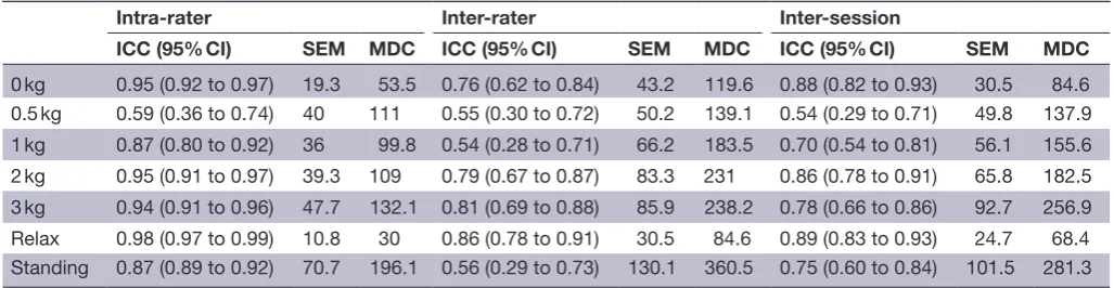
Table 2 Absolute and relative reliability for the different conditions
Intra- rater Inter- rater Inter- session
ICC (95% CI) SEM MDC ICC (95% CI) SEM MDC ICC (95% CI) SEM MDC
0 kg 0.95 (0.92 to 0.97) 19.3 53.5 0.76 (0.62 to 0.84) 43.2 119.6 0.88 (0.82 to 0.93) 30.5 84.6
0.5 kg 0.59 (0.36 to 0.74) 40 111 0.55 (0.30 to 0.72) 50.2 139.1 0.54 (0.29 to 0.71) 49.8 137.9
1 kg 0.87 (0.80 to 0.92) 36 99.8 0.54 (0.28 to 0.71) 66.2 183.5 0.70 (0.54 to 0.81) 56.1 155.6
2 kg 0.95 (0.91 to 0.97) 39.3 109 0.79 (0.67 to 0.87) 83.3 231 0.86 (0.78 to 0.91) 65.8 182.5
3 kg 0.94 (0.91 to 0.96) 47.7 132.1 0.81 (0.69 to 0.88) 85.9 238.2 0.78 (0.66 to 0.86) 92.7 256.9
Relax 0.98 (0.97 to 0.99) 10.8 30 0.86 (0.78 to 0.91) 30.5 84.6 0.89 (0.83 to 0.93) 24.7 68.4
Standing 0.87 (0.89 to 0.92) 70.7 196.1 0.56 (0.29 to 0.73) 130.1 360.5 0.75 (0.60 to 0.84) 101.5 281.3
CI, confidence interval; ICC, intraclass correlation coefficient; MDC, minimal detectable change; SEM, standard error of measurement.
Inter- rater reliability between the two raters ranged
from high to very high with an ICC2,k of 0.76 for 0 kg,
0.79 for 2 kg, 0.81 for 3 kg and 0.86 for the relaxed posi-tion. The inter- rater reliability of measuring AT stiffness during the 0.5 kg, 1 kg contraction and the standing posi-tion was moderate with an ICC2,k of 0.55, 0.54 and 0.56, respectively.
Inter- session reliability ranged from high to very high with an ICC2,k of 0.88 for 0 kg, 0.70 for 1 kg 0.86 for 2 kg, 0.78 for 3 kg, 0.89 and 0.75 for the relaxed and standing positions, respectively. The inter- session reliability of measuring AT stiffness during the 0.5 kg contraction was moderate with an ICC2,k of 0.54.
Table 2 reports the relative reliability with ICC and 95% CI and absolute reliability with SEM and MDC.
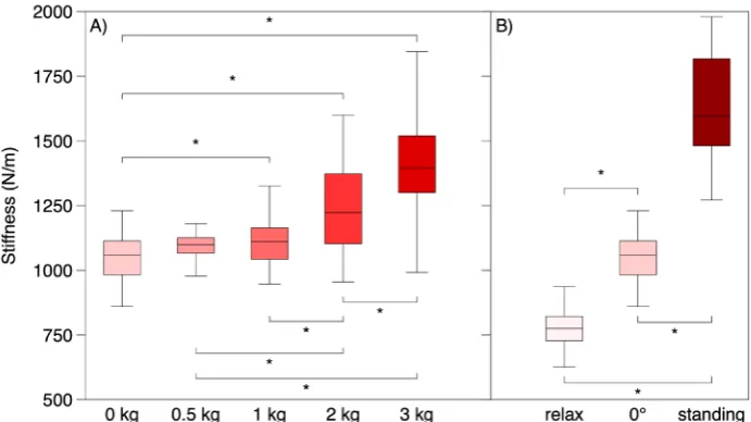
Changes in AT stiffness between the different condi-tions were calculated using Friedman test for related samples. Significant differences (p<0.01) were found between all contraction levels except for 0–0.5 kg (p=0.9); 0.5–1 kg (p=0.9); 1–2 kg (p=0.7) and between the different positions (figure 2).
DIsCussIOn
The present study evaluated the absolute and relative reli-ability of the myotonometry to measure the stiffness of AT in different conditions. Even though the MyotonPRO was developed to evaluate the stiffness and tone of skel-etal muscle, the present study shows the transferability of this technology and that measuring stiffness of tendon
tissue is feasible and reliable. Intra- rater and inter- rater reliability ranged from high to very high in most of the tested conditions. Moreover, stiffness changes between the different contraction intensities and the different positions was detected, demonstrating the construct validity of this device.
One of the conditions in the current study was assessing AT stiffness with the person in a relaxed posi-tion with the feet hanging freely over the edge of the
examination bed. A previous study25 also showed high
intra- rater reliability when the AT was evaluated in a relaxed position (ICC: 0.80 in healthy participants). A further study evaluating the effect of dorsiflexion of the ankle joint on tendon stiffness18 showed high to very high intra- reliability and inter- reliability when measured in a relaxed position (ICC >0.88). Our results strengthen these previous observations by confirming high to very high intra and inter- rater reliability (ICC >0.86), now demonstrated in a larger sample. The stiffness values vary slightly between studies with an average of 873 N/m reported by Finnamore et al,25 776.11±71.70 reported by Liu et al,18 and 771±105.3 N/m observed in the present study. Nevertheless, collectively the current and previous findings confirm that MyotonPRO is a reliable device to evaluate AT stiffness in a relaxed position on the same day, between different days and between different oper-ators.
Stiffness values increased from 1043±132.3 N/m
when tendon was assessed in a 0° ankle dorsiflexion
copyright.
on May 7, 2020 by guest. Protected by
Figure 2 Construct validity. Box plot showing the stiffness median and IQR values of the entire sample for the different contraction levels (A) and for the different positions (B). *p<0.01; statistical significant difference between the different conditions.
position with 0 kg of contraction to 1381±214 N/m for the isometric contraction at 3 kg. The ICC values showed high to very high intra- rater and inter- rater reliability for the majority of the isometric contraction levels (see table 2). Moderate intra- rater and inter- rater reliability was found for the 0.5 kg contraction level and the level of reliability did not exceed the cut- off point of ICC =0.6 considered as the threshold for clinical relevance.34 The
low reliability for the contraction at 0.5 kg is likely due to the difficulty in maintaining the force feedback stable during such a low contraction of the plantar flexors. A previous study18 evaluating AT stiffness at different ankle
angles showed similar results to the present study with an average stiffness of 1143 N/m at 0° of dorsiflexion compared with 1043±132 N/m found in the current study. Liu et al18 did not examine reliability at different
ankle angles, however, the data reported in this article demonstrated a statistically significant increase in tendon stiffness due to the change in ankle dorsiflexion angle from 1143 N/m at 0° to 1329 N/m at 30°. These results are in line with the present study, which showed a signif-icant increase in tendon stiffness with contractions of increasing intensity. Thus, the MyotonPRO can reliably detect changes in AT stiffness whether the tendon is stretched from contraction of the triceps surae muscle or stretched as a result of a change in the angle of the ankle.
Another study20 analysing the tissue properties of the
AT and plantar fascia in healthy volunteers found slightly different stiffness values when the tendon was measured at 0° of dorsiflexion. The article reports a mean (±SD) of 848 (±95.73) N/m and 847.3 (±101.5) N/m for the left and right AT, respectively, whereas we observed higher values with an average stiffness of 1043±132 N/m.
Tendon structural and mechanical adaptations are strongly related to the load magnitude exerted to the
musculotendinous complex3 5 and the majority of
interventions targeting pathological tendon tissue are
loading- based exercise regimens.36–40 The current results
support the evaluation of the tendon tissue behaviour during load with a reliable and easy to use device could be helpful to detect possible mechanical adaptation following different rehabilitation protocol.
Stiffness of the AT was also evaluated in standing posi-tion in this study to verify the feasibility of MyotonPRO to analyse the tendon in a weight- bearing position. Intra- rater reliability was very high and inter- session reliability was high. Conversely inter- rater reliability in the standing position was moderate. It is important to note, however, that in the standing position the average stiffness values were 1596 (±339) N/m which is approaching the upper limit that the device is able to detect. The lower reliability could be a consequence of the measurement threshold of the device such that when it is close to the higher limit, there were difficulties in providing a mechanical impulse strong enough to give a proper oscillation wave. Morgan et al19 found similar results in a population of
healthy participants in a weight- bearing position. Despite the possible technical limitation of analysing tendon stiff-ness in a weight- bearing position, the results demonstrate good intra- rater reliability but only moderate inter- rater reliability.
This is the first study to evaluate changes in AT stiff-ness between different contraction levels using the MyotonPRO. The results confirm a significant difference in tendon stiffness between the different contraction levels. Although stiffness changes can be detected across different contractions, the levels with similar intensity (0–0.5 kg (p=0.9); 0.5–1 kg (p=0.9); 1–2 kg (p=0.7)) showed similar stiffness values likely as the difference between consecutive contraction levels was not sufficient to change the mechanical properties of the tendon. These results are consistent with those reported in a previous
sonoelastography evaluation41 showing an increase of
tendon elastography colour scale index when the AT was
copyright.
on May 7, 2020 by guest. Protected by
gradually put under tension by isometric contractions of increasing intensity. In further support of the construct validity, pairwise comparisons showed a difference in AT stiffness between the different positions (relaxed, 0° ankle angle and standing). Similarly, a previous study
using shear wave sonoelastography42 showed an increase
in wave velocity, indicating a hardening of the AT, when the ankle angle changed from a plantarflexed position to 0° of dorsiflexion.
There are some limitations of this study which should be noted. Healthy participants were recruited and stiff-ness was evaluated in only one point of the tendon and it is therefore unknown whether variation may exist in different regions of the tendon. Another potential limita-tion was the seleclimita-tion of the contraclimita-tion intensities. The upper limit of this ramp was chosen because after a certain amount of load (approximately 5 kg) the device cannot detect stiffness values. The small changes between the different contraction levels could be not sufficient to fully describe the behaviour of the tendon during load.
COnClusIOns
MyotonPRO is a reliable tool to measure AT stiffness in different conditions including when the tendon is in a relaxed position and under load. Reliability is confirmed for the same rater, between different days and between two raters. Construct validity was supported by changes of tendon stiffness during the explored contractions and ankle positions. MyotonPRO can be implemented, as a ready to use device, in the evaluation of tendon tissue mechanical properties.
Acknowledgements We thank the students involved in the present study at the University of Applied Sciences and Arts of Southern Switzerland (SUPSI). We especially thank the Thim van der Laan Foundation, Landquart, Switzerland for financial support.
Contributors AS, DF, RC and MB designed and planned the study. AS and MB collected the data. All authors were involved in the drafting and revision of the manuscript and approved the final version of the article.
Funding The authors have not declared a specific grant for this research from any funding agency in the public, commercial or not- for- profit sectors.
Competing interests None declared.
Patient consent for publication Not required.
ethics approval The study was approved by the Ethics Committee of Canton Ticino (ID:2018–01483/CE3391) and written informed consent was provided by the participants.
Provenance and peer review Not commissioned; externally peer reviewed.
Data availability statement Data are available on reasonable request.
Open access This is an open access article distributed in accordance with the Creative Commons Attribution Non Commercial (CC BY- NC 4.0) license, which permits others to distribute, remix, adapt, build upon this work non- commercially, and license their derivative works on different terms, provided the original work is properly cited, appropriate credit is given, any changes made indicated, and the use is non- commercial. See: http:// creativecommons. org/ licenses/ by- nc/ 4. 0/.
OrCID iDs
Alessandro Schneebeli http:// orcid. org/ 0000- 0002- 8411- 2012 Deborah Falla http:// orcid. org/ 0000- 0003- 1689- 6190 Ron Clijsen http:// orcid. org/ 0000- 0001- 7125- 5332 Marco Barbero http:// orcid. org/ 0000- 0001- 8579- 0686
reFerenCes
1 de Jonge S, van den Berg C, de Vos RJ, et al. Incidence of midportion Achilles tendinopathy in the general population. Br J
Sports Med 2011;45:1026–8.
2 Józsa L, Kvist M, Bálint BJ, et al. The role of recreational sport activity in Achilles tendon rupture. A clinical, pathoanatomical, and sociological study of 292 cases. Am J Sports Med 1989;17:338–43. 3 Magnusson SP, Kjaer M. The impact of loading, unloading, ageing
and injury on the human tendon. J Physiol 2019;597:1283–98. 4 Obst SJ, Heales LJ, Schrader BL, et al. Are the mechanical or
material properties of the Achilles and Patellar tendons altered in tendinopathy? A systematic review with meta- analysis. Sports Med
2018;48:2179–98.
5 Bohm S, Mersmann F, Arampatzis A. Human tendon adaptation in response to mechanical loading: a systematic review and meta- analysis of exercise intervention studies on healthy adults. Sports
Med Open 2015;1:7.
6 Kjaer M, Langberg H, Heinemeier K, et al. From mechanical loading to collagen synthesis, structural changes and function in human tendon. Scand J Med Sci Sports 2009;19:500–10.
7 Couppé C, Svensson RB, Grosset J- F, et al. Life- Long endurance running is associated with reduced glycation and mechanical stress in connective tissue. Age 2014;36:9665.
8 Kubo K, Kanehisa H, Miyatani M, et al. Effect of low- load resistance training on the tendon properties in middle- aged and elderly women.
Acta Physiol Scand 2003;178:25–32.
9 Onambele GL, Narici MV, Maganaris CN. Calf muscle- tendon properties and postural balance in old age. J Appl Physiol
2006;100:2048–56.
10 Quinlan JI, Maganaris CN, Franchi MV, et al. Muscle and tendon contributions to reduced rate of torque development in healthy older males. J Gerontol A Biol Sci Med Sci 2018;73:539–45.
11 Stenroth L, Peltonen J, Cronin NJ, et al. Age- Related differences in Achilles tendon properties and triceps surae muscle architecture in vivo. J Appl Physiol 2012;113:1537–44.
12 Carroll CC, Dickinson JM, Haus JM, et al. Influence of aging on the in vivo properties of human Patellar tendon. J Appl Physiol
2008;105:1907–15.
13 Couppé C, Hansen P, Kongsgaard M, et al. Mechanical properties and collagen cross- linking of the Patellar tendon in old and young men. J Appl Physiol 2009;107:880–6.
14 Agyapong- Badu S, Warner M, Samuel D, et al. Measurement of ageing effects on muscle tone and mechanical properties of rectus femoris and biceps brachii in healthy males and females using a novel hand- held myometric device. Arch Gerontol Geriatr
2016;62:59–67.
15 Fröhlich- Zwahlen AK, Casartelli NC, Item- Glatthorn JF, et al. Validity of resting myotonometric assessment of lower extremity muscles in chronic stroke patients with limited hypertonia: a preliminary study. J
Electromyogr Kinesiol 2014;24:762–9.
16 Gervasi M, Sisti D, Amatori S, et al. Muscular viscoelastic characteristics of athletes participating in the European master indoor athletics Championship. Eur J Appl Physiol
2017;117:1739–46.
17 Feng YN, Li YP, Liu CL, et al. Assessing the elastic properties of skeletal muscle and tendon using shearwave ultrasound elastography and MyotonPRO. Sci Rep 2018;8:17064.
18 Liu CL, Li YP, Wang XQ, et al. Quantifying the stiffness of Achilles tendon: intra- and Inter- Operator reliability and the effect of ankle joint motion. Med Sci Monit 2018;24:4876–81.
19 Morgan GE, Martin R, Williams L, et al. Objective assessment of stiffness in Achilles tendinopathy: a novel approach using the MyotonPRO. BMJ Open Sport Exerc Med 2018;4:e000446. 20 Orner S, Kratzer W, Schmidberger J, et al. Quantitative tissue
parameters of Achilles tendon and plantar fascia in healthy subjects using a handheld myotonometer. J Bodyw Mov Ther
2018;22:105–11.
21 Ortega- Cebrian S, Luchini N, Whiteley R. Dry needling: effects on activation and passive mechanical properties of the quadriceps, pain and range during late stage rehabilitation of ACL reconstructed patients. Phys Ther Sport 2016;21:57–62.
22 Bizzini M, Mannion AF. Reliability of a new, hand- held device for assessing skeletal muscle stiffness. Clin Biomech 2003;18:459–61. 23 Kelly JP, Koppenhaver SL, Michener LA, et al. Characterization
of tissue stiffness of the infraspinatus, erector spinae, and gastrocnemius muscle using ultrasound shear wave elastography and superficial mechanical deformation. J Electromyogr Kinesiol
2018;38:73–80.
24 Sohirad S, Wilson D, Waugh C, et al. Feasibility of using a hand- held device to characterize tendon tissue biomechanics. PLoS One
2017;12:e0184463.
copyright.
on May 7, 2020 by guest. Protected by
25 Finnamore E, Waugh C, Solomons L, et al. Transverse tendon stiffness is reduced in people with Achilles tendinopathy: a cross- sectional study. PLoS One 2019;14:e0211863.
26 Kottner J, Audigé L, Brorson S, et al. Guidelines for reporting reliability and agreement studies (GRRAS) were proposed. J Clin
Epidemiol 2011;64:96–106.
27 Craig CL, Marshall AL, Sjöström M, et al. International physical activity questionnaire: 12- country reliability and validity. Med Sci
Sports Exerc 2003;35:1381–95.
28 Walter SD, Eliasziw M, Donner A. Sample size and optimal designs for reliability studies. Stat Med 1998;17:101–10.
29 Mahieu NN, Witvrouw E, Stevens V, et al. Intrinsic risk factors for the development of Achilles tendon overuse injury: a prospective study.
Am J Sports Med 2006;34:226–35.
30 Kjaer M. Role of extracellular matrix in adaptation of tendon and skeletal muscle to mechanical loading. Physiol Rev 2004;84:649–98. 31 Magnusson SP, Hansen P, Aagaard P, et al. Differential strain
patterns of the human gastrocnemius aponeurosis and free tendon, in vivo. Acta Physiol Scand 2003;177:185–95.
32 Arampatzis A, Karamanidis K, Albracht K. Adaptational responses of the human Achilles tendon by modulation of the applied cyclic strain magnitude. J Exp Biol 2007;210:2743–53.
33 Atkinson G, Nevill AM. Statistical methods for assessing measurement error (reliability) in variables relevant to sports medicine. Sports Med 1998;26:217–38.
34 Chinn S. The assessment of methods of measurement. Stat Med
1990;9:351–62.
35 Munro BH. Statistical methods for health care research. 5th edn. Philadelphia, Penn, London: Lippincott Williams & Wilkins, 2005.
36 Silbernagel KG, Thomeé R, Thomeé P, et al. Eccentric overload training for patients with chronic Achilles tendon pain--a randomised controlled study with reliability testing of the evaluation methods.
Scand J Med Sci Sports 2001;11:197–206.
37 Silbernagel KG, Thomeé R, Eriksson BI, et al. Continued sports activity, using a pain- monitoring model, during rehabilitation in patients with Achilles tendinopathy: a randomized controlled study.
Am J Sports Med 2007;35:897–906.
38 Mafi N, Lorentzon R, Alfredson H. Superior short- term results with eccentric calf muscle training compared to concentric training in a randomized prospective multicenter study on patients with chronic Achilles tendinosis. Knee Surg Sports Traumatol Arthrosc
2001;9:42–7.
39 Kongsgaard M, Kovanen V, Aagaard P, et al. Corticosteroid injections, eccentric decline squat training and heavy slow resistance training in Patellar tendinopathy. Scand J Med Sci Sports
2009;19:790–802.
40 Alfredson H, Lorentzon R. Chronic Achilles tendinosis: recommendations for treatment and prevention. Sports Med
2000;29:135–46.
41 Schneebeli A, Del Grande F, Falla D, et al. A novel application of strain sonoelastography can detect changes in Achilles tendon elasticity during isometric contractions of increasing intensity. J Foot
Ankle Res 2019;12:30.
42 Aubry S, Nueffer J- P, Tanter M, et al. Viscoelasticity in Achilles tendonopathy: quantitative assessment by using real- time shear- wave elastography. Radiology 2015;274:821–9.
copyright.
on May 7, 2020 by guest. Protected by