Original Article
Decreased heart rate variability is associated
with declined renal function in hypertensive patients
Dufang Ma1, Ping Jiang2, Rong Yu2, Yongcheng Wang2, Xiao Li1
1Department of Cardiology, Affiliated Hospital of Shandong University of Traditional Chinese Medicine, Jinan,
Shandong, P. R. China; 2First Clinical Medical College, Shandong University of Traditional Chinese Medicine, Jinan,
Shandong, P. R. China
Received January 30, 2018; Accepted July 6, 2018; Epub October 15, 2018; Published October 30, 2018
Abstract: The aim of the study was to identify the association between heart rate variability (HRV) and renal func-tion in hypertensive patients. A total of 195 nondiabetic hypertensive patients without evident renal impairment were included in the present cross-sectional observational study. HRV was assessed for the entire 24 hours, and biomarkers of renal function including serum levels of creatinine, urea nitrogen, uric acid, and estimated glomerular filtration rate (eGFR) were measured. Association between HRV and renal function parameters was identified by univariate correlation analysis, stepwise multivariate linear regression analysis and binary Logistic regression analy-sis. Patients with depressed HRV (SDNN < 100 ms) had higher serum levels of urea nitrogen, creatinine, and uric acid as well as decreased eGFR values compared with patients with normal HRV (SDNN ≥ 100 ms). After adjusting for compounding factors, only eGFR remained positively correlated with SDNN (beta = 0.534, P < 0.01). Moreover, eGFR is an independent variable predicting the presence of depressed HRV (OR = 0.959 [0.931, 0.989]) according to logistic analysis. Our results indicate that declined HRV parameter SDNN is associated with declined renal func-tion in essential hypertension.
Keywords: Heart rate variability, renal dysfunction, hypertension, autonomic dysfunction, glomerular filtration rate, uric acid
Introduction
Renal damage is one of the leading target organs damaged by hypertension [1, 2]. Au-tonomic dysfunction characterized with increa- sed sympathetic activity and sympathetic-para-sympathetic imbalance is reported to be in- volved in diabetic nephropathy [3]. Relatively speaking, whether autonomic dysfunction is associated with hypertension-induced renal dysfunction has been less clear.
Heart rate variability (HRV) is a practical and convenient way to evaluate cardiac autonomic function using standard electrocardiographic monitoring. Suppressed HRV (SDNN < 100 ms) has been used as a marker of cardiac auto-nomic dysfunction and an independent prog-nostic factor of cardiovascular diseases [4]. Studies have demonstrated that HRV is a pre-dictor of adverse renal outcomes in patients with non-dialysis and dialysis chronic kidney
diseases [5, 6]. Similarly, in diabetic patients, studies have reported that decreased HRV is associated with increased albuminuria and lower estimated glomerular filtration rate (eG-FR) at baseline, which predicted subsequently declined eGFR [7, 8]. However, there has been a paucity of studies on the association between HRV and renal function in essential hyperten-sive patients without evident renal impairment. In this study, we aim to determine the associa-tion between HRV and renal funcassocia-tion in a cohort of 195 nondiabetic hypertensive patients with-out evident renal impairment.
Materials and methods
Study population
and composed of 195 patients with essential hypertension. Hypertension was diagnosed according to guidelines [9], defining as systolic BP (SBP) ≥ 140 or diastolic BP (DBP) ≥ 90 mmHg, and patients on anti-hypertensive treat-ment were also considered as hypertensive, regardless of their BP values. Exclusion criteria for the present analysis were: Patients with secondary hypertension, old or acute myocar-dial infarction, diabetes mellitus, acute and chronic heart failure, cancer, liver cirrhosis and/or failure, moderate-to-severe renal dys-function (eGFR < 60 mL/min/1.73 m2),
narcot-ics abuse, thyroid disease, mental illness (depression, anxiety, schizophrenia and moder-ate to severe insomnia),sleep apnea hypopnea syndrome, history of gastrointestinal bleeding and acute renal injury (within 12 months), and recent history of use of nephrotoxic drugs or contrast agents (within 6 months). All of the patients gave informed consent to participate in this research project, which was approved by the Ethics Committee of Shandong University of Traditional Chinese Medicine.
Clinical parameters
As has been previously described, comprehen-sive demographic characteristics including sex, age, smoking, alcohol intake, past history of diseases, and medications before enrollment were investigated by a designed and pre-tested questionnaire.
Body weight, BP, abdomen ultrasound and car-diac ultrasound were analyzed and recorded at the first day of hospitalization. Measurement of BP was performed after a 10 minute period of rest using an electronic sphygmomanometer (HEM-7071, OMRON Healthcare cooperation, China), and two BP readings were taken from both arms at 30 second intervals between 08:00 and 09:00 AM on the second morning after hospital admission. Simultaneously, fast-ing venous blood was drawn on the second morning after hospital admission for biochemi-cal parameters including fasting plasma glu-cose, creatinine, urea nitrogen, uric acid, lipid profile, homocysteine, and thyroid hormone (Free T3 and T4). Samples were centrifuged and serum was separated within 30 minutes of collection and stored at -20°C until analyzed. The analyses were performed in the clinical bio-chemical laboratory in our hospital by AU5400
automated analyzer (Olympus, Tokyo, Japan), and reference ranges are those used by the laboratory. Serum urea nitrogen and uric acid levels were measured using uric acid enzymatic assay kits and urea nitrogen enzymatic assay kits (Beckman Coulter, Inc, Suzhou, China), and serum creatinine levels were measured using creatinine PAP FS (Diasys diagnostic system co., LTD, Shanghai, China). eGFR was calculat -ed by the Chronic Kidney Disease Epidemiology Collaboration (CKD-EPI) equation [10]:
Heart rate variability
Patients were given 24-hours for the ECG Holter recording. The recorders were applied between 9:00 and 11:00 AM on the second day after hospital admission, and patients were asked to follow as closely as possible their usual daily activities during each monitoring session. As described in our previous study [11], long trac -ings were recorded and independently ana-lyzed by an experienced operator masked to patients’ clinical status using a JincoMed Holter System (MIC-3H-3L, Jinco Medical Equipment Company, Beijing, China). HRV was assessed for the entire 24 hours in both the time- and frequency-domain analysis. After classifying the QRS morphology, the RR intervals were con-firmed manually until no QRS sequence was incorrectly labeled. Traces were checked to ensure R-waves were adequately identified from artifacts and ectopic beats, and sequenc -es with normal QRS characteristics were ana-lyzed for HRV analysis. Time-domain HRV analy-sis parameters were 1) SDNN: standard deviation of all of the RR intervals between two normal QRS complexes; 2) RMSSD: root mean square successive difference between each value and the preceding value. Fast Fourier transformation was used to convert the differ-ent successive RR intervals in the frequency domain. Data were analyzed in 10-minute epochs throughout the 24 hours and the results from all epochs were averaged to form a com-posite spectrum. The amplitudes of the follow-ing frequency-domain HRV parameters were
Serum creatinine
(umol/L) eGFR (mL/min/1.73 m 2)
Female ≤ 62 GFR = 144 × (Scr/62)-0.329 × (0.993)Age
> 62 GFR = 144 × (Scr/62)-1.209 × (0.993)Age
Male ≤ 80 GFR = 144 × (Scr/80)-0.411 × (0.993)Age
parameters (SDNN, RMSSD, LF, HF and TP) and biomarkers of renal function (serum levels of cre-atinine, urea nitro-gen, uric acid, and eGFR. According to the results of univari-ate correlation analy-sis, stepwise multi-variate linear regres- sion analysis (inclu-sion at 0.05 and exclusion at 0.01) was employed to find out the independent predictors of renal function parameters. Binary Logistic regre- ssion analysis was also performed to identify significant in-dependent related factors for suppre- ssed HRV. Possible compounding facto- rs including clinical variables (age, sex, body weight, smok-ing, alcohol drinking), history of ischemic heart disease and stroke, heart rate, SBP and DBP,
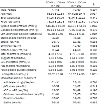
medi-Table 1. Clinical characteristics of patients with SDNN < 100 ms and SDNN ≥ 100 ms
SDNN < 100 ms
(n = 95) SDNN ≥ 100 ms (n = 100) p
Male/Female 47/48 40/60 0.197
Age (yeas) 68.19 ± 10.53 64.52 ± 10.74 0.017*
Body weight (Kg) 67.25 ± 10.06 67.94 ± 11.11 0.449
Heart rate (bpm) 73.24 ± 15.26 66.47 ± 10.52 < 0.001*
Systolic blood pressure (mmHg) 145.45 ± 13.69 140.55 ± 14.01 0.014*
Diastolic blood pressure (mmHg) 82.11 ± 11.03 79.92 ± 11.37 0.175 Left ventricular ejection fraction (%) 61.68 ± 9.65 66.22 ± 5.15 0.019*
Stable angina pectoris (Yes/No) 71/24 74/26 1.000
Stroke (Yes/No) 25/70 19/81 0.235
Smoking (Yes/No) 42/53 40/60 0.565
Alcohol intake (Yes/No) 51/44 44/56 0.198
Total cholesterol (mmol/L) 4.73 ± 1.12 4.79 ± 1.18 0.688
Triglycerides (mmol/L) 1.68 ± 1.04 1.51 ± 1.09 0.261
LDL-cholesterol (mmol/L) 2.81 ± 0.97 2.88 ± 0.90 0.569 HDL-cholesterol (mmol/L) 1.08 ± 0.26 1.20 ± 0.68 0.121 Fasting blood glucose (mmol/L) 5.86 ± 1.25 5.61 ± 0.99 0.122 Homocysteine (umol/L) 15.87 ± 6.87 13.07 ± 4.95 0.001*
Medications before enrollment
Antiplatelet drugs (Yes/No) 61/34 62/38 0.768
β-Blockers (Yes/No) 36/59 33/67 0.549
ACEI or ARB (Yes/No) 39/56 51/49 0.196
Calcium channel blockers (Yes/No) 31/64 35/65 0.763
Diuretics (Yes/No) 35/60 34/66 0.765
Statins (Yes/No) 56/39 57/43 0.885
All values are given in mean ± SD. Continuous variables were compared using
independent-sample student’s t test. Categorical variables were compared using the Chi-square test. *P <
0.05 was considered significant.
measured: very low frequency (VLF, 0.0033-0.004 Hz), low frequency (LF, 0.04-0.25 Hz), high frequency (HF, 0.25-2.5 Hz), and total power (TP, 0-2.5 Hz).
Statistical analysis
Statistical analyses were performed as de- scribed previously [11]. For study purposes, dif -ferences in characteristics between partici-pants with SDNN < 100 ms (suppressed HRV) and those with SDNN ≥ 100 ms (normal HRV) were compared using Students’ T-test for con-tinuous variables and Chi-square test for dichotomous variables. This cut-off for SDNN has been reported to be a marker of autonomic dysfunction and a predictor of mortality in patients after myocardial infarction [4, 12, 13]. Pearson’s or Spearman’s analyses were per-formed to identify the correlation between HRV
cal therapy (antiplatelet drugs, β-blockers, ACEI/ARB, calcium channel blockers, diuretics, and statins), laboratory parameters (fasting blood sugar, triglycerides, total cholesterol, LDL-C, HDL-C, homocysteine, Free T3 and T4) and eGFR were regarded as independent vari -ables. Differences were considered significant at the two-sided P < 0.05. Analyses were per-formed using SPSS 20.0 (SPSS Inc, Chicago, IL, USA).
Results
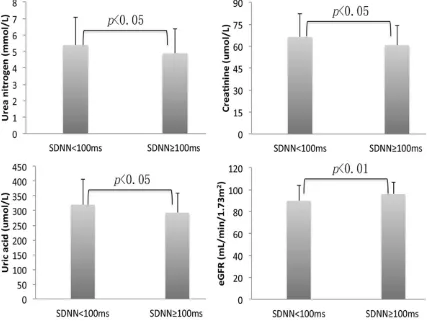
[image:3.612.89.418.97.438.2]Baseline population characteristics
Figure 1. Biomarkers of renal function of patients with low HRV (SDNN < 100 ms) versus patients with normal HRV (SDNN ≥ 100 ms). Compared by use of independent- sample Student’s T-test or the Mann-Whitney U test.
suppressed HRV were older (P < 0.05), and they had elevated SBP (P < 0.05), heart rate and serum levels of homocysteine (P < 0.01) and decreased left ventricular ejection fraction (P < 0.05).
Figure 1 shows the comparison of renal func-tion parameters between the two groups. Compared with patients with normal HRV, patients with suppressed HRV had relatively increased blood levels of urea nitrogen (5.38 ± 1.68 vs. 4.88 ± 1.48 mmol/L, P < 0.05), creati-nine (66.22 ± 16.27 vs. 60.77 ± 13.8 umol/L, P < 0.05) and uric acid (319.91 ± 86.79 vs. 292.79 ± 67.18 umol/L, P < 0.05). Moreover, those patients with suppressed HRV had lower values of eGFR than patients with normal HRV (89.87 ± 14.23 vs. 95.95 ± 11.12 mL/min/1.73 m2, P < 0.01).
Relationship between HRV parameters and biomarkers of renal function
Table 2 shows univariate analyses between HRV parameters and biomarkers of renal
func-tion. SDNN were inversely correlated with blood levels of urea nitrogen, creatinine and uric acid (P < 0.01), while positively correlated with eGFR (P < 0.01). No relationship was found between RMSSD and any the renal function parameters. In addition, Spearman’s analyses show that HRV frequency-domain parameter LF was posi -tively correlated with eGFR (P < 0.05), and HF (P < 0.01) and TP (P < 0.05) were inversely cor-related with blood levels of creatinine.
-renal injury. Together with the results of prior results, these findings make it clear that de- creased autonomic mo- dulation is indeed relat-ed to the declinrelat-ed renal function in essential-hypertension patients. Although both the renal injury and autonomic dysfunction are compli-cations of hyperten-sion, the complex inter-action and cause-and-
Table 2. Pearson’s and Spearman’s correlation analysis between HRV index and renal function pa-rameters
Urea nitrogen (mmol/L) Creatinine (umol/L) Uric acid (umol/L) eGFR (mL/min/1.73 m2)
r p r p r p r p
SDNN -0.246 0.001 -0.339 < 0.001 -0.224 0.002 0.273 < 0.001
RMSSD -0.027 0.710 -0.100 0.165 -0.066 0.363 0.065 0.366
LF -0.020 0.782 -0.128 0.079 -0.005 0.951 0.172 0.018
HF -0.043 0.560 -0.194 0.008 -0.072 0.325 0.105 0.151
TP 0.054 0.567 -0.165 0.023 -0.085 0.241 0.142 0.050
Pearson’s analyses were performed between SDNN and RMSSD and renal function parameters; Spearman’s analyses were
performed between LF, HF and TP and renal function parameters.
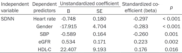
tistically significant (beta = 0.534, P < 0.01). Moreover, SDNN was also associated with heart rate, gender, SBP and blood levels of HDL-C.
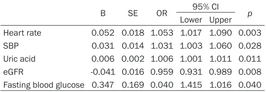
Consistent with the results of multiple linear regression analysis, binary logistic regression analysis revealed that increased blood levels of uric acid (OR = 1.006 [1.001, 1.011]) and decreased eGFR (OR = 0.959 [0.931, 0.989]) were independent variables relating the sup-pressed HRV (Table 4).
Discussion
Recently, a large cross-study comprised 9600 non-diabetic, never-treated hypertension indi-viduals has demonstrated an association be- tween the coefficient of variation of heart rate (Cv = standard deviation × 100/mean value) and declined renal function [14]. In agreement with this result, our present study showed a robust inverse association between time-domain HRV parameters SDNN and eGFR in 195 hypertensive patients without evident
[image:5.612.91.403.262.353.2]effect relationship between them is scanty and perplexing in the process of essential hyperten-sion. Theoretically, renal sympathetic nerve ter-minals directly contact with renal vasculature and renal tubules [15], and modulate renal hemodynamics, tubular transport and renin secretion [16]. Indeed, a clinical study has indi -cated that pure autonomic dysfunction contrib-uted to elevated serum levels of creatinine and urea nitrogen and decreased eGFR compared with patients without autonomic failure [17]. In the development of hypertension, it has be- en demonstrated that sympathetic activity is already increased at earlier stages when renal function is not or only slightly impaired in hyper-tension [18]. Prolonged sympathetic hyperac -tivity can induce changes in intrarenal blood vessels by releasing catecholamines, which induce proliferation of smooth muscle cells and adventitial fibroblasts in the renal vascular wall [19, 20]. Additionally, renal sympathetic over-activity leads to renal dysfunction through affecting filtration, secretion and reabsorption function [15, 21, 22]. With development of
Table 3. Multivariate analysis of factors associated with SDNN in total sample
Independent
variable Dependent predictors
Unstandardized coefficient Standardized co-efficient (beta) p
B SE
SDNN Heart rate -0.748 0.180 -0.297 < 0.001
Gender -17.915 4.704 -0.283 < 0.001
SBP -0.589 0.164 -0.260 0.001
eGFR 0.534 0.171 0.223 0.002
HDL-C 22.407 9.193 0.176 0.016
Clinical variables (age, sex, body weight, smoking, alcohol drinking), history of ischemic heart disease and stroke, heart rate, SBP and DBP, medical therapy (antiplatelet drugs,
β-blockers, ACEI/ARB, calcium channel blockers, diuretics and statins), laboratory param
-eters (urea nitrogen, uric acid, fasting blood sugar, triglycerides, total cholesterol, LDL-C,
markers of renal function, as they reflect the tubular reabsorption function and glomerular filtration function. In the present study, we found that hypertensive patients with depressed HRV had lower eGFR and higher blood levels of urea nitrogen, creatinine and uric acid when compared with patients with normal HRV. The results of univariate correlation analyses showed that SDNN was positively correlated with eGFR and inversely correlated with blood levels of urea nitrogen, creatinine, and uric acid. After adjusting the compounding factors, only eGFR remained posi -hypertension, the impaired kidney
progressive-ly exacerbates sympathetic excitation through increasing sodium retention and hypervolemia and activating the renin-angiotensin -aldoste-rone system, which in turn result in elevated BP [23-25]. Thus, autonomic dysfunction charac -terized by sympathetic over-excitation may be an important underlying mechanism of hyper-tension-induced renal dysfunction.
HRV is determined by a complex interaction of sympathetic and parasympathetic activity. Decreased HRV is associated with autonomic dysfunction and a host of adverse outcomes in cardiovascular diseases and chronic kidney disease [26-28]. SDNN is the main parameter of time-domain analysis; it reflects the total modulation of both parasympathetic and sym-pathetic nerve system, and a suppressed SDNN value (SDNN < 100 ms) usually indicates relative sympathetic dominance and inhibited parasympathetic activity [13, 29]. RMSSD and HF predominantly reflect parasympathetic modulation, while LF and LF/HF represent sym -pathetic-parasympathetic balance. Moreover, LF has been variably though to represent sym -pathetic activity by some reports [5, 30].
In clinical practice, GFR is considered the best overall index of kidney function in health and disease. In the present study, we used CKD-EPI equation to estimate the values of GFR, which is applicable to a variety of populations and clinical conditions, and improves the bias at higher levels of estimated GFR [10]. Additionally, the blood levels of urea nitrogen, creatinine and uric acid are always considered as the
tively correlated with SDNN. Moreover, eGFR is an independent variable predicting the pres-ence of depressed HRV according to the logis-tic analysis. The present study was consistent with the retrospective analysis on a cohort of 200 hypertensive patients performed by Paolo et al. [31]. In his study, it was shown that LF/HF, a marker of sympatho-vagal balance, was sig-nificantly decreased in the groups with mild and moderate decreased eGFR, suggesting the involvement of autonomic imbalance in hyper-tension-related renal dysfunction. Similarly, studies performed in patients with chronic kid-ney diseases also suggested that lower fre-quency-domain HRV parameters were associ -ated with higher risk of progression to end-stage renal diseases [26, 32]. These results power -fully supported that autonomic dysfunction is closely related to decreased renal function, or even renal damage.
High uric acid levels have been associated with decreased renal perfusion and tubular secre-tion of uric acid [33]. Another study has report -ed that increas-ed blood levels of uric acid were related with an accelerated progression of hypertension and development of end-organ injury [34]. In the present study, elevated uric acid levels were found to be associated with the presence of depressed HRV according to the results of logistic analysis. Consistently, in pre-hypertensive patients, it has been demon-strated that uric acid inversely correlated with HRV parameters LF, suggesting uric acid is associated with sympathetic activity [35]. Nevertheless, the plausible mechanisms by which uric acid correlated with HRV parameters are not clearly known, it might be partly
attrib-Table 4. Binary logistic regression analysis results of the pos-sible correlates for depressed HRV
B SE OR 95% CI p
Lower Upper Heart rate 0.052 0.018 1.053 1.017 1.090 0.003
SBP 0.031 0.014 1.031 1.003 1.060 0.028
Uric acid 0.006 0.002 1.006 1.001 1.011 0.011
eGFR -0.041 0.016 0.959 0.931 0.989 0.008
Fasting blood glucose 0.347 0.169 0.040 1.415 1.016 0.040 Clinical variables (age, sex, body weight, smoking, alcohol drinking), history of ischemic heart disease and stroke, heart rate, SBP and DBP, medical therapy
(antiplatelet drugs, β-blockers, ACEI/ARB, calcium channel blockers, diuretics
and statins), laboratory parameters (urea nitrogen, uric acid, fasting blood
[image:6.612.90.359.98.191.2]uted to the increased sympathetic activity, because study has demonstrated that sympa-thetic over-activity in hypertension decreased renal clearance of uric acid, leading to elevated plasma levels of uric acid [16].
Limitations
There are limitations that should be acknowl-edged. First, the serum levels of uric acid, cre -atinine and urea nitrogen were obtained at a single time-point. In order to reduce the fluctua -tions, further study should measure these bio-markers at several different time-point. Second, this is an observational study and any associa-tions between HRV parameters and renal func-tion parameters did not definitely prove causal -ity. Nevertheless, this study elucidated that adverse HRV parameters are associated with declined renal function. Longitudinal analysis of hypertensive patients will determine possi-ble early predictive value of HRV on hyperten-sion-induced renal dysfunction.
Conclusion
In conclusion, using a simple, cheap and non-invasive autonomic test, we have demonstrat-ed that autonomic dysfunction is associatdemonstrat-ed with declined renal function in essential hyper-tension. This finding may be helpful to early detect and diagnose hypertension nephropa-thy. Further research is needed to clarify the underlying mechanisms between autonomic dysfunction and decreased renal function in hypertension.
Acknowledgements
This study was supported in part by Chinese National Natural Science Foundation (No. 81673970) and the First-class Subject Funded Projects of Shandong Province (No. 220306). We thank all our colleagues working in the Electrocardiogram Room and Clinical Bioche- mistry Room, Affiliated Hospital of Traditional Chinese Medicine. This study was approved by the ethics committee of Shandong University of Traditional Chinese Medicine. A written infor- med consent was obtained from all the patients at the time of admission, with which the blood was authorized to scientific purpose.
Disclosure of conflict of interest
None.
Address correspondence to: Xiao Li, Department of Cardiology, Affiliated Hospital of Shandong Uni-versity of Traditional Chinese Medicine, 42 Wenhua Road, Jinan 250011, Shandong, P. R. China. Tel: +86-18753151199; E-mail: [email protected]
References
[1] Kim MJ, Lim NK, Park HY. Relationship be-tween prehypertension and chronic kidney dis-ease in middle-aged people in Korea: the Ko-rean genome and epidemiology study. BMC Public Health 2012; 12: 960.
[2] Seccia TM, Caroccia B, Calo LA. Hypertensive nephropathy. Moving from classic to emerging pathogenetic mechanisms. J Hypertension 2017; 35: 205-212.
[3] Yun JS, Ahn YB, Song KH, Yoo KD, Kim HW, Park YM, Ko SH. The association between ab-normal heart rate variability and new onset of chronic kidney disease in patients with type 2 diabetes: a ten-year follow-up study. Diabetes Res Clin Pract 2015; 108: 31-37.
[4] von Kanel R, Carney RM, Zhao S, Whooley MA. Heart rate variability and biomarkers of sys-temic inflammation in patients with stable cor -onary heart disease: findings from the Heart and Soul Study. Clin Res Cardiol 2011; 100: 241-247.
[5] Chandra P, Sands RL, Gillespie BW, Levin NW, Kotanko P, Kiser M, Finkelstein F, Hinderliter A, Pop-Busui R, Rajagopalan S, Saran R. Predic-tors of heart rate variability and its prognostic significance in chronic kidney disease. Nephrol Dial Transplant 2012; 27: 700-709.
[6] Kurata C, Uehara A, Sugi T, Ishikawa A, Fujita K, Yonemura K, Hishida A, Ishikawa K, Tawara-hara K, Shouda S, Mikami T. Cardiac autonom-ic neuropathy in patients with chronautonom-ic renal failure on hemodialysis. Nephron 2000; 84: 312-319.
[7] Tahrani AA, Dubb K, Raymond NT, Begum S, Altaf QA, Sadiqi H, Piya MK, Stevens MJ. Car -diac autonomic neuropathy predicts renal function decline in patients with type 2 diabe-tes: a cohort study. Diabetologia 2014; 57: 1249-1256.
[8] Cho YH, Craig ME, Davis EA, Cotterill AM, Couper JJ, Cameron FJ, Benitez-Aguirre PZ, Dalton RN, Dunger DB, Jones TW, Donaghue KC; Adolescent Type 1 Diabetes Cardio-Renal Intervention Trial. Cardiac autonomic dysfunc-tion is associated with high-risk albumin-to-creatinine ratio in young adolescents with type 1 diabetes in AdDIT (adolescent type 1 diabe-tes cardio-renal interventional trial). Diabediabe-tes Care 2015; 38: 676-681.
AM, Kjeldsen SE, Laurent S, Narkiewicz K, Ruilope L, Rynkiewicz A, Schmieder RE, Struijk-er BoudiStruijk-er HA, Zanchetti A; European Society of Hypertension; European Society of Cardiol-ogy. 2007 Guidelines for the management of arterial hypertension: the task force for the management of arterial hypertension of the European Society of Hypertension (ESH) and of the European Society of Cardiology (ESC). Eur Heart J 2007; 28: 1462-1536.
[10] Levey AS, Stevens LA, Schmid CH, Zhang YL, Castro AF 3rd, Feldman HI, Kusek JW, Eggers P, Van Lente F, Greene T, Coresh J; CKD-EPI (Chronic Kidney Disease Epidemiology Collab-oration). A new equation to estimate glomeru -lar filtration rate. Ann Intern Med 2009; 150: 604-612.
[11] Dufang M, Yongcheng W, Ping J, Yonghui Y, Xiao L. N-Terminal Pro-B-Type natriuretic pep-tide levels inversely correlated with heart rate variability in patients with unstable angina pectoris. Int Heart J 2016; 57: 292-298. [12] Odemuyiwa O, Malik M, Farrell T, Bashir Y, Pol
-oniecki J, Camm J. Comparison of the predic-tive characteristics of heart rate variability in-dex and left ventricular ejection fraction for all-cause mortality, arrhythmic events and sud-den death after acute myocardial infarction. Am J Cardiol 1991; 68: 434-439.
[13] Kleiger RE, Miller JP, Bigger JT Jr, Moss AJ. De-creased heart rate variability and its associa-tion with increased mortality after acute myo-cardial infarction. Am J Cardiol 1987; 59: 256-262.
[14] Liakos CI, Karpanou EA, Markou MI, Grassos CA, Vyssoulis GP. Correlation of 24-Hour blood pressure and heart rate variability to renal function parameters in hypertensive patients. The effect of smoking. J Clin Hypertens 2015; 17: 938-943.
[15] DiBona GF. Physiology in perspective: the wis -dom of the body. Neural control of the kidney. Am J Physiol Regul integr Comp Physiol 2005; 289: R633-641.
[16] Joles JA, Koomans HA. Causes and conse-quences of increased sympathetic activity in renal disease. Hypertension 2004; 43: 699-706.
[17] Garland EM, Gamboa A, Okamoto L, Raj SR, Black BK, Davis TL, Biaggioni I, Robertson D. Renal impairment of pure autonomic failure. Hypertension 2009; 54: 1057-1061.
[18] Klein IH, Ligtenberg G, Oey PL, Koomans HA, Blankestijn PJ. Sympathetic activity is in-creased in polycystic kidney disease and is as-sociated with hypertension. J Am Soc Nephrol 2001; 12: 2427-2433.
[19] Erami C, Zhang H, Ho JG, French DM, Faber JE. Alpha(1)-adrenoceptor stimulation directly
in-duces growth of vascular wall in vivo. Am J Physiol Heart Circ Physiol 2002; 283: H1577-1587.
[20] Zhang H, Faber JE. Trophic effect of norepi -nephrine on arterial intima-media and adventi-tia is augmented by injury and mediated by different alpha1-adrenoceptor subtypes. Circ Res 2001; 89: 815-822.
[21] Spallone V, Gambardella S, Maiello MR, Barini A, Frontoni S, Menzinger G. Relationship be -tween autonomic neuropathy, 24-h blood pres-sure profile, and nephropathy in normotensive IDDM patients. Diabetes Care 1994; 17: 578-584.
[22] Winocour PH, Dhar H, Anderson DC. The rela-tionship between autonomic neuropathy and urinary sodium and albumin excretion in insu-lin-treated diabetics. Diabe Med 1986; 3: 436-440.
[23] Abdulla MH, Johns EJ. The innervation of the kidney in renal injury and inflammation: a cause and consequence of deranged cardio -vascular control. Acta Physiol (Oxf) 2017; 220: 404-416.
[24] Esler M. The sympathetic nervous system in hypertension: back to the future? Curr Hyper-tens Rep 2015; 17: 11.
[25] Barrett CJ. Renal sympathetic nerves - what have they got to do with cardiovascular dis-ease? Exp Physiol 2015; 100: 359-365. [26] Brotman DJ, Bash LD, Qayyum R, Crews D,
Whitsel EA, Astor BC, Coresh J. Heart rate vari-ability predicts ESRD and CKD-related hospi-talization. J Am Soc Nephrol 2010; 21: 1560-1570.
[27] Binkley PF. Promise of a new role for heart rate variability in the clinical management of pa-tients with heart failure. JACC Heart Fail 2017; 5: 432-434.
[28] Kleiger RE, Stein PK, Bigger JT Jr. Heart rate variability: measurement and clinical utility. Ann Noninvasive Electrocardiol 2005; 10: 88-101.
[29] Heart rate variability. Standards of measure-ment, physiological interpretation and clinical use. Task force of the European society of car-diology and the North American society of pac-ing and electrophysiology. Eur Heart J 1996; 17: 354-81.
[30] Cygankiewicz I, Zareba W. Heart rate variabili-ty. Handb Clin Neurol 2013; 117: 379-393. [31] Melillo P, Izzo R, De Luca N, Pecchia L. Heart
rate variability and target organ damage in hy-pertensive patients. BMC Cardiovasc Disord 2012; 12: 105.
pa-tients with chronic kidney disease. J Bras Nefrol 2014; 36: 155-162.
[33] Perlstein TS, Gumieniak O, Williams GH, Spar -row D, Vokonas PS, Gaziano M, Weiss ST, Liton -jua AA. Uric acid and the development of hy-pertension: the normative aging study. Hypertension 2006; 48: 1031-1036.
[34] Mazzali M, Kanellis J, Han L, Feng L, Xia YY, Chen Q, Kang DH, Gordon KL, Watanabe S, Na -kagawa T, Lan HY, Johnson RJ. Hyperuricemia induces a primary renal arteriolopathy in rats by a blood pressure-independent mechanism. Am J Physiol Renal Physiol 2002; 282: F991-997.