RESEARCH ARTICLE
STUDY OF BIOCHEMICAL PARAMETERS OF FOUR DIFFERENT MEDICINAL PLANTS
*Gayathri, V. and Kiruba, D.
Department of Botany, Avinashilingam Institute for Home Science and Higher Education for Women, Coimbatore – 641043, Tamilnadu, India
ARTICLE INFO ABSTRACT
Medicinal plants are the most exclusive source of life saving drugs for majority of the world’s population. The use of medicinal plant extract increases the body’s immune system and lowers the allergies and asthma. The four medicinal plants taken for the present study possess digestive property. The experiment conducted to study the biochemical parameters of the leaves of the four medicinal plants viz., R. tomentosa, P. guajava, C, aurantium and C. limonum showed a significantly higher protein, carbohydrate and chlorophyll content in C. aurantium. The chlorophyll 'b' and total chlorophyll content was found to be significantly higher in R. tomentosa.
Copyright © 2014 Gayathri, V. and Kiruba, D. This is an open access article distributed under the Creative Commons Attribution License, which permits unrestricted use, distribution, and reproduction in any medium, provided the original work is properly cited.
INTRODUCTION
Plants have been known to relieve various diseases through traditional medicine. Medicinal plants are the richest bio-resource of drugs in traditional systems of medicine, modern medicines, nutraceuticals, food supplements, folk medicines, pharmaceutical intermediates and chemical entities for synthetic drugs (Neube et al., 2008). Medicine from plant sources have been in use in Homeopathy, Ayurveda, Allopathy and in traditional medicine since time immemorial (Gogoi and Islam, 2012). Knowledge of the chemical constituents of the plant is desirable, not only for the discovery of therapeutic drugs but also to discover the actual value of folklore remedies (Mojab et al., 2003).
Medicinal plants were used by people of ancient cultures, without the knowledge of their active ingredients. Some medicinal plants are used for remedy at household level. Medicinal plants have proved to be effective for prevention and cure of various disorders. Their use against digestive disorders is very common at household level. Usually herbal medicines are widely perceived by the public as being natural, healthful and free from side effects (Mordi and Akanji, 2012). In rural areas, people suffer from common digestive disorders such as diarrhea, dysentery and food poisoning. The extract of medicinal plants in these cases raises the body’s immune
*Corresponding author: Gayathri, V. Department of Botany,
Avinashilingam Institute for Home Science and Higher Education for Women,Coimbatore – 641043, Tamilnadu, India.
system and lowers the allergies (Sidhu, 2007). It is generally known that the consumption of a variety of local herbs and vegetables by man contributes significantly to the improvement of human health, in terms of prevention and or cure of disease because plants have long served as useful and natural source of therapeutic agents (Chevellier, 1996).
MATERIALS AND METHODS
In the present study, fresh samples of four different medicinal plant leaves were used. The medicinal plants are Rhodomyrtus
tomentosa, Psidium guajava, Citrus aurantium and Citrus
limonum. A study was carried out on various biochemical
parameters such as protein, carbohydrate and chlorophyll content of the leaves of the four plants.
COLLECTION OF PLANT SAMPLES
The fresh leaves of Rhodomyrtus tomentosa, Psidium guajava,
Citrus aurantium and Citrus limonum were obtained from
Kengarai village in Nilgiri District of Tamil Nadu, India.
MORPHOLOGY OF THE PLANTS
RHODOMYRTUS TOMENTOSA
Systematic Position Kingdom : Plantae
Class: Eudicot
ISSN: 0975-833X
International Journal of Current Research
Vol. 6, Issue, 05, pp.6490-6496, May,2014
INTERNATIONAL JOURNAL OF CURRENT RESEARCH
Article History:
Received 05th
January, 2014 Received in revised form
04th February, 2014
Accepted 15th April, 2014
Published online 20th May,2014
Key words:
Rhodomyrtus tomentosa, Psidium guajava, Citrus aurantium,
Citrus limonum,
Order: Myrtales
Family: Myrtaceae
Genus:Rhodomyrtus Species: R.tomentosa
Description- Plate- I
Ceylon hill cherry is an evergreen shrub which grows up to a height of about 2-3 m tall.
The leaves are densely white or yellow and arranged in alternative position.
Leaves are elliptic or oblong-elliptic, 5-8 cm long and 2-4 cm wide. Leaf margin is entire. The petiole is 3-5 mm long
The upper leaf surface is glossy and glabrous, with three conspicuous longitudinal veins. The lower surface is white (or) yellowish with longitudinal veins
Flowers are solitary or in three lowered dichasia in upper axils; peduncles are upto 1 cm long, pedicels 0.5-2.5 cm long; bracts elliptic, leaf-like, 6-12 mm long, bracteoles elliptic or ovate, 2-3 mm long, persistent.
Calyx is campanulate, 5-7 mm long, tomentose, five-ten
ribbed, five lobed and persistent.
Petal is broadly obovate, stamens are numerous, 10-15 mm long, filaments pink; style 13-15 mm long; ovary three-four locular.
The fruit is an oblong edible berry, purplish- black, soft, crowned by persistentcalyx lobes.
Each fruit contains 40-45 seeds in six to eight pseudo-locules, divided by thin false septa.
Plate- I. Rhodomyrtus tomentosa
PSIDIUM GUAJAVA
Systematic Position
Kingdom: Plantae
Class: Eudicot
Order: Myrtales
Family: Myrtaceae
Genus: Psidium Species: P. guajava
Description – Plate- II
Guava is a large tropical evergreen shrub (or) small shade tree, wild tree grown upto 20 m height and are well branched, twigs glabrous.
Leaves are opposite, short-petiolate, the blade oval with prominent pinnate veins, 5-15cm long.
Flower somewhat showy, 3cm across, axillary, solitary or in pairs; bracteoles 1 to 8mm; pedicel upto 1.5cm long.
Calyx tube subglobose, urceolate, 1x0.4cm, tomentose persistent; lobes 4/5, ovate-lanceolate, unequal, 1x0.7cm.
Petals whitish and 4 or more, outer once broadly ovate, 2.5x1.5cm, inner once linear-lanceolate, 1.5x0.7cm, caducous. Disc broad, thick.
Stamen is infinite in several series, exserted filaments white, 0.5-1.5cm.
Ovary globose,1cm, Ovules infinite ; style 1.5cm, berry globose, 4x3.5 cm; seeds infinite
Plate- II. Psidium guajava
CITRUS AURANTIUM
Systematic Position
Kingdom: Plantae
Class: Eudicot
Order: Sapindales
Family: Rutaceae
Genus: Citrus Species: C. aurantium
Description – Plate- III
This occurs truly wild as a shrub or tree. Young shoots glabrous, angled spines ascending straight.
Leaves are dark green, elliptic, ovate or oblong rounded or with an obtuse apex.
Flower waxy, white usually tinged with red, sweet scented.
Petals are 4-5, stamens 20-40, petiole winged, stamens are in bundle.
Fruit is rounded in shape and inside, the fruits contain fibres.
When fruit is ripe, it turns to bright orange colour and becomes juicy.
Plate- III. Citrus aurantium
CITRUS LIMONUM
Systematic Position
Kingdom: Plantae
Class: Eudicot
Order: Sapindales
Family: Rutaceae
Genus: Citrus Species: C. limonum
Description – Plate- IV
A bushy shrub, young shoots glabrous, somewhat-angled spines ascending straight; bark greenish.
The lemon grows upto 6m tall.
The leaves are dark green, leathery and evergreen, oblong, elliptic or oval and upto 14 cm long.
Flower waxy, white having violet streaks from inside.
Flower open to have 5 white petals, upto 5cm across.
Petals 4-5, stamens 20-40.
Fruits are ovoid in shape and have a pointed tip at one end. They have thick covering over it.
When fruit is unripe it is hard, dark green and non juicy but on ripening it turns to bright yellow, becomes soft and juicy.
When ripe very aromatic with thin spongy rind and coherent colorless small vesicels filled with acid aromatic juice.
Fruits are smooth to bumpy, rinds dotted with oil glands.
Plate- IV. Citrus limonum
Biochemical Parameters
The following biochemical parameters were observed in the leaves of all the four medicinal plants used for the present study.
Chlorophyll
Protein
Carbohydrate
METHODS
1. Estimation of Chlorophyll Content
Chlorophyll ‘a’, ’b’ and total chlorophyll were analyzed following the method of Arnon (1949).
Materials Required
Analytical grade acetone was diluted to 80 % acetone.
Procedure
One gram of freshly cut sample of leaf was taken into a clean mortar.
The leaf bits were ground to a fine pulp with the addition of 20ml of 80 % ( w/v) acetone.
The mixture thus obtained was centrifuged at 5000 rpm for 5 minutes.
The supernatant was transferred to 100ml volumetric flask. This procedure was repeated until the residue became colourless.
The absorbance of the solution was read in a spectrophotometer at 645 and 663 nm against the solvent blank (80% acetone).
Calculation
The amount of chlorophyll present in the extract was calculated (mg chlorophyll / gm tissue) using the formula,
mg chlorophyll ‘a’/gm tissue = 12.7 A663 - 2.69 A645 x
V/(100xW)
mg chlorophyll ‘b’/gm tissue = 22.9A645-4.68A663x
V/(1000xW)
mg total chlorophyll /gm tissue = 20.2A645+8.02A663x
V/(1000xW)
Where
A = Absorbance of specific wave length
V = Final volume of chlorophyll extract in 80% acetone W = Fresh weight of the tissue
2. Estimation of Protein (Lowry et al., 1951)
Principle
The blue colour developed by phosphomolybdic phosphotungstic components in the folin- ciocalteau reagent by the amino acids, tyrosine and tryptophan present in the protein and the colour developed by the biuret reaction of the protein with the alkaline cupric tartrate are measured by Lowry’s method.
Materials Required
Two percent sodium carbonate in 0.1N sodium hydroxide (Reagent A).
0.5 percent copper sulphate (CuSo4.5H2O) in 1 percent potassium sodium tartarate (Reagent B).
Alkaline copper solution: mix 50ml of reagent A and 1ml of reagent B prior to use (Reagent C).
Folin- Ciocalteau reagent (Reagent D).
Protein solution (stock standard): Accurately 50mg of bovine serum albumin (fraction V) was weighed and dissolved in distilled water and made up to 50ml in a standard flask.
Working standard: Ten ml of stock solution was diluted to 50ml with distilled water in a standard flask. One ml of this solution contains 200mg protein.
Procedure
Extraction of protein from samples
Extraction was carried out with buffers. About 500mg of the sample was weighed and ground well with a pestle and mortar in 5-10ml of the phosphate buffer, centrifuged and supernatant was used for protein estimation.
Estimation of Protein
About 0.2, 0.4, 0.6, 0.8 and 1ml of the working standard were pipetted out into a series of test tubes.
About 0.1 and 0.2ml of the sample was pipetted out in two other test tubes.
The volume was made up to 1ml using distilled water in all the test tubes. A tube with one ml of water served as the blank.
About 5ml of reagent C was added to each tube including the blank. Mixed well and allowed to stand for 10 min.
To this, 0.5ml of reagent D was added, mixed well and incubated in dark for 30 minutes. Blue colour developed was read at 660nm.
A standard graph was drawn and the amount of protein present in the sample was calculated.
Calculation
The amount of protein present in the sample was expressed in mg / gm = mg of protein/volume of test standard x concentration of the standard.
3. Estimation of Carbohydrate Content (Hedge and Hofreiter, 1952).
Anthrone Method
Principle
Concentrated sulphuric acid hydrolyses the glycoside bond of carbohydrate to the given monosaccharides which were then dehydrated to furfural. The furfural reacted with anthrone (10-Keto 9, 10-dihydroanthracene) to give the blue coloured complex which was measured colorimetrically at 630 nm.
Materials Required
2.5 N HCl
Anthrone reagent was prepared by dissolving 200mg anthrone in 100ml of ice cold 95% H2SO4. Prepared
freshly before use.
Stock standard: 100mg of glucose was dissolved in 100ml of water.
Working standard: 5ml of stock standard solution was diluted to 100ml using distilled water (50mg/ml).
Procedure
About 100mg of the sample was taken in a boiling tube and was hydrolysed by keeping it in boiling water bath for three hours with 5ml of 2.5 N HCl and cooled at room temperature.
Then it was neutralized with solid sodium carbonate until the effervescence ceases.
The volume was made up to 100ml and centrifuged.
The supernatant was collected and 0.5 and 1ml aliquot were taken for analysis.
The standard was prepared by taking 0, 0.2, 0.4, 0.6, 0.8 and 1ml of the working standard and a blank was maintained.
The volume was made up to 1ml in all the tube including the sample tube by adding distilled water.
Then, 4ml of anthrone reagent was added and heated for eight minutes in a boiling water bath.
Then, it was cooled rapidly and blue green colour developed was read at 630nm.
A standard graph was drawn by plotting concentration of the standard on the X-axis versus absorbance on the Y-axis.
From the graph, the amount of carbohydrate present in the sample was calculated.
Calculation
Amount of carbohydrate present in 100 mg of the sample = mg of glucose/volume of test samplex100
RESULTS AND DISCUSSION
The experiments conducted in Rhodomyrtus tomentosa,
Psidium guajava, Citrus aurantium and Citrus limonum for
analysis of biochemical parameters showed the following results.
BIOCHEMICAL ANALYSIS OF THE LEAVES OF
Rhodomyrtus tomentosa, Psidium guajava, Citrus aurantium
and Citrus limonum
ESTIMATION OF PROTEIN
[image:5.612.62.307.399.664.2]The protein content of four medicinal plants were observed and presented in Table - 1 and Figure - 1.
Table 1. Protein content of the four medicinal plants
Plant name Protein content
(mg/ 100 g)
Rhodomyrtus tomentosa Psidium guajava Citrus aurantium Citrus limonum
14.55 ± 0.027 12.69 ± 0.022 16.80 ± 0.012 4.369 ± 0.023 SEd
Cd (p<0.01)
[image:5.612.334.534.404.470.2]0.02005 0.04274 Values are mean of three triplicates
Figure 1. Protein content of the four medicinal plants
The highest protein content was estimated in C. aurantium
(16.80±0.012 mg/100g of leaf sample) followed by R.
tomentosa (14.55±0.027 mg/100g of leaf sample), P. guajava
(12.69 ± 0.022 mg /100g of leaf sample) and C. limonum (4.369 ±0.023 mg/100g of leaf sample) (Table 1). Jimoh and Oladiji (2005) screened the protein content of the plant Piliostigma
thonningii through Bovine serum reagent and estimated the
protein content to be 30.33 ± 0.31mg /100g. The protein content was found to be significantly higher in C. aurantium
(Table-1). Thomsen et al. (1991) have indicated the presence of higher protein level in the plants parts contributing to the increase in food value. According to Snehal and Madhukar (2012) leaf extracts of Stevia were evaluated for their biochemical composition. Results of their work showed higher concentration of sugars, protein, amino acid in dry leaves than in fresh leaves due to reduction in moisture content and increase in dry mass. According to Laitonjam et al. (2013), the biochemical studies of Phlogacanthus pubinerrius and
Phlogacanthus jenkinsii leaves showed relatively high levels of
antioxidant activity in the methanolic extracts of leaves of both the plants.
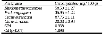
ESTIMATION OF CARBOHYDARTE
Among the medicinal plants taken for the study, the carbohydrate content was estimated to be higher in C.
aurantium (87.75 ± 1.11 mg /100g of leaf sample) and the
lower value was observed in C. limonum (20.08 ± 0.93 mg / 100g of leaf sample) (Table 2 and Figure 2). R. tomentosa and
P. guajava showed 58.50 ± 1.27 mg/100g of leaf sample and
35.95±1.22 mg/100g of leaf sample respectively (Table - 2 and Figure - 2).
Table 2. Carbohydrate content of the four medicinal plants
Plant name Carbohydrates (mg / 100 g)
Rhodomyrtus tomentosa Psidium guajava Citrus aurantium Citrus limonum
58.50 ± 1.27 35.95 ± 1.22 87.75 ± 1.11 20.08 ± 0.93 SEd
Cd (p<0.01)
0.938 1.896 Values are mean of three triplicates
Manikandan and Victor (2010) estimated the carbohydrate content of Ruellia tuberosa and Dipteracanthus patulus Jacq. to be 56.4 mg / and 62.8 mg / 100g respectively.
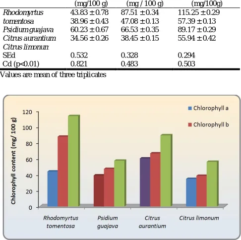
[image:5.612.82.276.408.482.2] [image:5.612.319.560.531.684.2]Estimation of chlorophyll "a", chlorophyll "b" and total chlorophyll content
Chlorophyll 'a', chlorophyll 'b' and total chlorophyll content was calculated for all the four medicinal plants. The chlorophyll 'a' content was estimated to be 43.83 ± 0.78 mg /100g leaf sample, 38.96 ± 0.43 mg /100g leaf sample, 60.23 ± 0.67mg /100g leaf sample and 34.56 ± 0.26 mg /100g leaf sample in the four medicinal plants R. tomentosa, P. guajava,
C. aurantium and C. limonum respectively (Table - 3 and
Figure - 3). Higher chlorophyll 'b' content was observed in both
R. tomentosa (87.51 ± 0.34) and C. aurantium (66.53 ± 0.35
mg/100g). P. guajava and C. limonum showed a chlorophyll 'b' content of 47.08 ± 0.13mg / 100g leaf sample and 38.45 ± 0.15mg / 100g leaf sample respectively (Table-3; Fig-3). The total chlorophyll content was maximum in R. tomentosa and the value was 115.25 ± 0.29 mg/100g leaf sample (Table-3, Fig -3). The other three medicinal plants studied showed a total chlorophyll content of 57.39 ± 0.13 (P. guajava), 89.17 ± 0.29
(C. aurantium) and 55.94 ± 0.42 mg / 100g leaf sample (C.
limonum) (Table - 3, Fig - 3). The total chlorophyll content was
[image:6.612.58.302.323.407.2]found to be significantly higher in R. tomentosa (Table - 3).
Table 3. Chlorophyll 'a', Chlorophyll 'b' and Total Chlorophyll content of the four medicinal plants
Plant names Chlorophyll a
(mg/100 g)
Chlorophyll b (mg / 100 g)
Total chlorophyll (mg/100g) Rhodomyrtus tomentosa Psidium guajava Citrus aurantium Citrus limonum
43.83 ± 0.78 38.96 ± 0.43 60.23 ± 0.67 34.56 ± 0.26
87.51 ± 0.34 47.08 ± 0.13 66.53 ± 0.35 38.45 ± 0.15
115.25 ± 0.29 57.39 ± 0.13 89.17 ± 0.29 55.94 ± 0.42
SEd Cd (p<0.01) 0.532 0.821 0.328 0.483 0.294 0.503 Values are mean of three triplicates
Figure 3. Chlorophyll a, Chlorophyll b and Total Chlorophyll content of the four medicinal plants
Among the biochemical parameters studied for the four medicinal plants viz., R. tomentosa and P. guajava belonging to the family Myrtaceae; C. aurantium and C. limonum
belonging to the family Rutaceae, the protein content was significantly higher in C.aurantium (Table-1 and Fig-2). Among the chlorophyll pigments estimated, the chlorophyll content was significantly higher in C. aurantium (Table-3). The
chlorophyll 'b' and total chlorophyll content was found to be significantly higher in R. tomentosa (Table-3).
Primary metabolites are of prime importance and essentially required for growth of plants (Jayaram et al., 1981). Murray et al. (1986) have identified chlorophyll as the most indispensable class of primary compounds as they are the only substance that capture sunlight during photosynthesis and make it available to plant system for its growth. Earlier studies by Gayathri et al.
(2013) on the effect of herbal leaf extracts on biochemical parameters of Vigna mungo L. have revealed a significant increase in chlorophyll, protein and carbohydrate content. Studies by Shad et al. (2013) on the biochemical analysis of different parts of Cichorium intybus L. have revealed a statistically significant difference among various parts of the plant regarding sugars, free amino acids and water soluble proteins.
REFERENCES
Arnon, D.E. 1949. Copper enzymes in isolated chloroplast. Pl.
Physiol., 24: 1-5.
Chevellier, A. 1996. The Encyclopedia of medical plant. London. Dorling Kindresley Ltd. (online).http : // www. Chclibrary.org / plant. html.
Gayathri, V., Prabha Sherlina, F. and Anju Singh 2013. Effect of inorganic fertilizers and herbal leaf extracts on biochemical parameters of black gram (Vigna mungo L.).
Global J. Biosci. Biotech.,2(4) : 518-521.
Gogoi, P. and Islam , M. 2012. Phytochemical screening of
Solanum nigrum L. and Solanum myriacanthus Dunal from
districts of upper Assam. India. IOSR J. pharma.,2(3) : 455 – 459.
Hedge, J.E. and Hofreiter, B.T. 1952. Determination of total carbohydrate by anthrone method.In : Carbohydrate chemistry (Eds.), Whistler, R.L. and Bo Miller, J.N., Academic Press, New York. pp. 17.
Jayaram, J., Bray, H.G. and Thorpe, W.V. 1981. Analysis of phenolic compounds of interest in metabolism.
Meth.Biochem. Anal., 1 : 27 – 52.
Jimoh, F.O. and Oladiji, A.T. 2005. Preliminary studies on
Piliostigma thonningii seeds: Proximate analysis, mineral
composition and phytochemical screening. African J.
Biotechnol. 4(12): 1439-1442.
Laitonjam, W.S., Satyavama, R.Y., Asem, D. and Wangkheirakpam, S.D. 2013. Evaluative and comparative study of biochemical, trace elements and antioxidant activity of Phlogacanthus pubinervius T. Anderson and
Phlogacanthus Jenkinsii C.B.Clarke leaves. Indian
J.Nat.prod. Res., 4(1) : 67-72.
Lowry, O.H., Rosenbrough, N.S., Farr, A.L. and Randall, R.J. 1951. Protein measurement with folin phenol reagent. J.
Biol. Chem., 193 : 267 – 2
Manikandan, A. and Victor Arokia Doss.D. 2010. Evalution of biochemicconents, nutritional value, trace elements, SDS-PAGE and HPTLC profiling in the leaves of Ruellia
tuberose L. and Dipteracanthus patulus (Jacq.) J. Chem.
Pharma. Res., 2(3) : 295-303.
Mojab, F., Kamalinijal, M. Ghaderi, N. and Vahidipour, H. 2003. Phytochemical screening of some Iranian plants.Iranian J. Pharma. Res., pp. 77-32.
[image:6.612.60.303.337.579.2]Mordi, J.C. and Akanji, M.A. 2012. Phytochemical screening of the dried leaf extract of Cnidoscolus aconitifolius and associated changes in liver enzymes induced by its administration in Wistar rats. Curr. Res. J. Bio. Sci., 4(2) : 153 – 158.
Murray, A.P., Gibbs, C.G. and Longmore, A.R. 1986. Determination of chlorophyll in marine waters : Inter comparison of a Rapid HPLC method with full HPLC, Spectrophotometric and Fluorometric methods. Mar.
Chem., 19 : 211 – 227.
Ncube, N.S., Afotayan, A.J. and Okoh, A.I. 2008. Assessment techniques of anti-microbial properties of natural compounds of plant origin current methods and future trends.Afric. J. Biotech., 7(12) : 1797-1806.
Shad, M.A., Nawaz, H., Rehma, T. and Ikaram, N. 2013. Determination of some biologicals, phytochemicals and antioxitant properties of different parts of Cichorium
intybus L. : A Comparative study. J. Anim. Plant Sci.23(4)
: 1060-1066.
Sidhu, K., Kaur, J., kaur, G. and Pannu, K. 2007. Prevention and cure of digestive disorders through the use of medicinal plants .J .Hum. Ecol., 21(2) : 113-116.
Snehal, P. and Madhukar, K. 2012) Quantitative estimation of biochemical content of various extracts of Stevia
rebaudiana leaves. Asian. J. Pharma. Clin. Res., 5(1) :
115 – 117.
Thomsen, S., Handen, H.S. and Nyman, V. 1991. Ribosome inhibiting proteins from invitro cultures of Phytolacea
dodecandra. Planta. Med., 57 : 232 – 236.