Sonographically guided lymph node biopsy: Complication
rates
Michael Mueller1, Genia Wittich1, Suemeyra Oeztuerk1, Wolfgang Kratzer1, Mark Martin Haenle1, Richard Andrew Mason2
1Department of Internal Medicine I, University Hospital Ulm, Ulm, Germany
2Department of Veterans Affairs, Louis Stokes Cleveland Medical Center, Brecksville, USA Email: [email protected]
Received 23 January 2012; revised 22 February 2012; accepted 21 March 2012
ABSTRACT
Purpose: The study investigated the rate of compli- cations associated with sonographically guided lymph node biopsies and assessed potential risk factors. Methods: A total of 536 sonographically guided punc- ture procedures (283 males, 52.8%; 253 females, 47.2%; average age 57.0 ± 16.0 years; range 14 - 87 years) were performed in 469 patients for the work-up of unclear lymphadenopathy. Events, complications and potential risk factors, were prospectively do- cumented. Results: The 469 patients underwent a total of 536 puncture procedures (PP) including 663 punc- tures and 1485 passes. Lymph node localizations were intraabdominal (55.2%, n = 296), cervical (22.4%, n = 120), inguinal (12.9%, n = 69), axillary (7.8%, n = 42) and other (1.7%, n = 9). No complications were documented during the entire study period. There was no increased risk of complications documented for the potential risk factors number of punctures, the number of passes, the localization, diameter of the lymph node (s), puncture technique, needle gauge, as well as patients’ sex, age and coagulation parameters, and the experience of the examiner. Conclusions: Our findings confirm the safety of percutaneous sono- graphically guided lymph node biopsies in different regions of the body in patients with adequate coagula- tion parameters undergoing pre-interventional color Doppler ultrasound examination.
Keywords:Lymph Nodes; Complications; Biopsy;
Ultrasonography
1. INTRODUCTION
Sonographically guided lymph node biopsy is considered a safe and effective diagnostic tool [1,2]. It served to ex- clude or confirm the diagnosis in patients with enlarged lymph nodes and is of decisive clinical value in determin- ing therapy and prognosis [3-5]. Despite real-time imag-
ing and pre-interventional assessment of the planned puncture path, complications may very infrequently oc- cur. Complication rates in the literature are in the range of 0% - 2.9% [1-3,6]. To our knowledge, however, no large studies have addressed the causes of complications in patients undergoing sonographically guided diagnostic lymph node biopsy. The present study prospectively documented complications and required treatments and assessed potential risk factors, including the number of punctures, the number of passes, the localization, diame- ter of the lymph node (s), puncture technique, needle gauge, as well as patients’ sex, age and coagulation pa- rameters, and the experience of the examiner.
2. MATERIALS AND METHODS
2.1. Patient Selection
A total 4278 diagnostic puncture biopsies (liver, 2280 punctures; lymph nodes, 536 punctures; pancreas, 351 punctures; other locations, 1111 punctures) were per- formed in 3353 patients in the department of diagnostic ultrasound of our institution between September 1999 and November 2009. The 536 biopsies of lymph nodes were performed in 469 patients aged 14 - 87 years (mean 57.0 ± 16.0 years). 52.8% (n = 283) of the puncture proce-dures were performed in male patients, 47.2% (n = 253) in female patients. Of these, 42.7% (n = 229) of biopsies were performed on an outpatient basis, while 57.3% (n = 307) were performed on inpatients.
2.2. Nomenclature
For the sake of clarity, the present report gives only the outer diameter of puncture needles in millimeters. Assess- ment considered patients, puncture procedures (PP), punc- tures and passes.
be used. A puncture was defined as a biopsy using a nee- dle of the same diameter and technique; multiple passes with the same needle were counted as a single puncture. Passes were defined as each entry of the needle into the target region. Thus, throughout the study, individual pa- tients could undergo several PP with numerous punctures, each of these potentially including several passes. Com- plications were evaluated on the basis of PP as complica- tions occurring during procedures with multiple punc- tures could not be assigned to any single puncture.
2.3. Data Collection
For data collection, a commercially available, PC-based, standardized documentation system (ViewPoint Bildver- arbeitung GmbH, Wessling, Germany) whose structured data entry met standards established by the German So- ciety for Ultrasonography in Medicine (DEGUM), was used. The following parameters were systematically re- corded in order to calculate their effect on the complica- tion risk: number of punctures, number of passes, sex, age, localization, size, puncture technique, needle di- ameter, examiner’s experience and patient’s coagulation status. During the study period, all complications and subsequent therapeutic measures, if any, were docu- mented prospectively in an Excel spread sheet (Microsoft Office Excel 2003).
2.4. Definition of Complications
Major complications were defined as post-interventional events that could clearly be attributed to the PP; were clinically relevant; and required therapeutic intervention (e.g. blood transfusion, drainage etc.). Minor complica- tions were events that were not clinically relevant and did not require specific therapeutic intervention. Ad- ministration of analgesics was not considered a therapeu- tic intervention; hence, pain requiring analgesic treat- ment was regarded as a minor complication.
2.5. Puncture Requirements
The following criteria had to be met before each punc- ture: clear indications with therapeutic consequence; ade- quate coagulation status (platelet count ≥ 70,000/µl, Quick’s test value ≥ 70%, partial thromboplastin time (PTT) < 50 seconds, no application of anticoagulants); and written consent signed by the patient one day prior to the procedure. In rare cases, especially with superficial lymph nodes, punctures were also performed in patients not meeting the required coagulation criteria. This was often indicated in the work-up of leukemic disorders in which the choice of appropriate chemotherapy depended on timely diagnosis. Every puncture performed in pa- tients not meeting coagulation criteria was based on a careful evaluation of the relative benefits and risks of the
procedure. Examiner’s experience was divided into two categories: Those who had performed a minimum of 150 PP were considered “experienced examiners”, whereas those who had performed less than 150 PP were consid- ered “inexperienced examiners”.
2.6. Execution
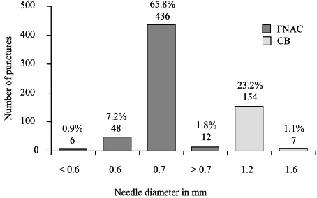
All punctures were performed under sonographic guid- ance (Philips HDI 3000/5000 Sono CT; C5-2 curved array transducers and biopsy guidance for deep lymph nodes; sonographically guided free-hand puncture using a L12-5 parallel transducer head for superficial lymph nodes). 75.7% of punctures (n = 502) were performed using fine needle aspiration cytology (FNAC), while 24.3% (n = 161) involved cutting biopsies (CB).
First, the target lesion was sonographically visualized in the epigastric region. Then, penetration length was determined and routine Doppler ultrasound examination was performed in order to minimize the risk of injury to adjacent organs or vessels. After disinfection of the skin directly above the puncture site, local anesthesia with mepihexal 1% was induced, followed by intravenous
application of piritramide (Dipidolor®; 1 - 2 ml = 10 - 20
mg) and metoclopramide (Reglan®; 2 ml = 10 mg), and
volume and pulse-oxymetric monitoring prior to the ac- tual puncture. For FNAC of deeper lymph nodes, exam- iners used biopsy guidance in order to insert a catheter (diameter: 1.3 mm) into a small skin incision through which the actual puncture needle (Chiba, diameter: 0.7 mm) was inserted and advanced into the mass lesion un-der sonographic guidance. Once the puncture needle was in place, the assistant actuated the Binder valve, thus generating a vacuum, and dislodged cells from their for- mation by means of poking and fanning movements be- fore aspirating them into the tube. After the puncture needle had been removed the aspirated cytological mate-rial was transferred onto a clean glass slide and several smears were made using the glass coverslip. The aspira- tion procedure could be repeated several times if inade- quate cytological material was collected on the first try. Puncture with FNAC (Chiba, diameter: 0.95 mm) did not requiring a leader catheter.
into 3.5% - 3.7% of formaldehyde solution and sent in for histological evaluation. In case both CB and FNAC were performed, the FNAC was always performed first. After FNAC, the patient was required to maintain four hours’ bed rest including a one hour of sandbag com- pression (three hours after CB) of the puncture site and fasting during the same period. Patients’ pulse rate and blood pressure were recorded hourly (every 30 minutes after CB). Following CB, patients underwent a final com- plete blood count, physical examination by the treating physician and sonographic examination. If all findings were within normal limits, patients were discharged.
2.7. Statistical Evaluation
Statistical analyses were performed using the SAS 9.2 statistics software package (SAS Institute Inc., Cary, North Carolina, USA). The data of the 536 PP were evaluated descriptively regarding absolute and relative frequencies, mean, standard deviation, minimum and maximum. A multivariate analysis was not performed since no major complications were recorded in our study.
3. RESULTS
No complications, either major or minor, were recorded in the present patient collective. No elevated complica- tion rate could be attributed to any of the potential risk factors such as number of punctures, number of passes, patients’ sex, age or coagulation status, tumor localiza- tion or size, puncture technique, needle diameter, ex- perience of examiner. The 469 patients recruited for the present study underwent a total 536 puncture procedures including 663 punctures and 1485 passes. The localiza- tion of all lymph nodes and the associated number of PP
is presented in Figure 1. The exact localization of in-
tra-abdominal lymph nodes is given in Figure 2.
Maximum lymph node diameter was documented in 92.4% (n = 495) PP. Mean maximum diameter was 30.9 ± 24.8 mm (range: 5.0 - 300 mm). Needles with a di-
ameter of 0.3 - 1.6 mm were used for punctures (Figure
3). In 76.5% (n = 410) of PP, only one puncture was per-
formed; of these, 88.8% (n = 364) were FNAC and 11.2% (n = 46) were CB. In 23.3% (n = 125) of PP, two punctures were performed. Of these, PP included both CB and FNAC in 91.2% (n = 114), while, in 8.8% (n = 11), patients underwent two FNAC using puncture nee- dles of different diameters. Only in one instance (0.2%, n = 1) did PP consist of three punctures, two of which were FNAC and one, CB. The average number of passes was 1.2 ± 0.5 (range: 1 - 3) for CB and 2.6 ± 0.9 (range: 1 - 6)
for FNAC (Figure 4).
During the period of the study, a total of 33 different examiners performed a mean 125 ± 254.6 PP (range 4 - 1480) on various organs. Overall, 9.1% (n = 3) of the
[image:3.595.310.539.89.249.2]intraabdominal 55.2% (n=296) inguinal 12.9% (n=69) axillary 7.8% (n=42) cervical 22.4% (n=120) other localizations 1.7% (n=9) intraabdominal 55.2% (n=296) inguinal 12.9% (n=69) axillary 7.8% (n=42) cervical 22.4% (n=120) other localizations 1.7% (n=9)
Figure 1. Lymph node localizations.
hilary (liver) 14.2%(n=42) other intraabdominal localizations 33.4% (n=99) trunculary 13.2% (n=39) parailiacal 10.1% (n=30) paraaortal 29.1% (n=86) hilary (liver) 14.2%(n=42) other intraabdominal localizations 33.4% (n=99) trunculary 13.2% (n=39) parailiacal 10.1% (n=30) paraaortal 29.1% (n=86)
Figure 2. Intraabdominal localizations.
0 100 200 300 400 500
< 0.6 0.6 0.7 > 0.7 1.2 1.6
Needle diameter in mm
Nu m ber of punc tur es FNAC CB 0.9% 6 7.2% 48 65.8% 436 1.8% 12 23.2% 154 1.1% 7 0 100 200 300 400 500
< 0.6 0.6 0.7 > 0.7 1.2 1.6
Needle diameter in mm
[image:3.595.307.535.285.667.2]Nu m ber of punc tur es FNAC CB 0.9% 6 7.2% 48 65.8% 436 1.8% 12 23.2% 154 1.1% 7
Figure 3. Biopsy type and needle gauge.
[image:3.595.307.534.526.668.2]was documented in 42.5% (n = 228) of the PP and was below the minimum limit of ≥70% in 6.1% (n = 14) of all cases; the lowest Quick’s value registered for a PP was at 32%. PTT was documented in 39.6% (n = 212) of the PP and was above the limit of <50 seconds in 1.4% (n = 3) of cases. The highest PTT registered for a PP was 105 seconds. In only one case was PP performed in the presence of abnormal findings for all three coagulation parameters.
6
3
0 1 2 3 4 5 6 7
FNAC CB
Technique
M
axi
m
um num
be
r
of
pas
se
s/
pu
nct
ur
e
M = 2.6
M = 1.2 6
3
0 1 2 3 4 5 6 7
FNAC CB
Technique
M
axi
m
um num
be
r
of
pas
se
s/
pu
nct
ur
e
M = 2.6
M = 1.2
4. DISCUSSION
Lymph node biopsies are generally considered to be very safe diagnostic procedures [1]. No complications were recorded in the present patient collective. In the literature, reported rates are low, ranging from 0 to 2.9% [1-10] (Table 1). Major complications were reported in two
[image:4.595.62.284.83.238.2]publications, both secondary to vascular injuries [3,6]. The available studies, however, concentrated on the out- come of the punctures; complications were addressed more as a secondary issue that, in some cases, was not specifically discussed [11,12]. To our knowledge, no cur- rent studies have specifically investigated the complication rate of lymph node punctures. Other studies that have ad-
Figure 4. Maximum and mean needle passes per puncture.
[image:4.595.57.536.395.724.2]Coagulation parameters (platelets, Quick’s value, PTT) were documented in 38.6% (n = 207) of all PP. Of these, 90.3% (n = 187) were performed in patients meeting minimum criteria established for coagulation status, while, in 9.7% (n = 20) criteria were not fully met. Plate- let count was documented in 41.6% (n = 223) of the PP and was below the limit of ≥70,000/µl in 4.5% (n = 10); the lowest platelet count was 11,000/µl. Quick’s value
Table 1. A review of the literature on sonographically guided lymph node biopsies.
Authors Date Complications per total punctures Complication rate in % complication Type of Imaging doppler Color Coagulation parameters Maximum needle gauge
Takashima
et al. [3] 1997 1 major/91 1.1
Puncture of
carotid artery SFHP n/a n/a 0.7 mm
Fisher
et al. [6] 1997 1 major/35 2.9
Puncture of inferior epigastric
artery
SGP Yes n/a 1.2 mm
Screaton
et al. [1] 2002 3 minor/260 1.2 3 hematomas SFHP Yes No 1.6 mm
Wittich
et al. 2010 0/663 0 -
SGP/
SFHP Yes Yes 1.6 mm
Kim
et al. [2] 2007 0/155 0 - SFHP Yes No 1.6 mm
Cheung
et al. [4] 2000 0/60 0 - SFHP n/a n/a 1.2 mm
Abe
et al. [7] 2007 0/>100 0 - SFHP Yes n/a 2.1 mm
al-Mofleh [8] 1992 0/240 0 - SPU n/a n/a 0.9 mm
Veerapand
et al. [10] 2004 0/72 0 - SFHP n/a n/a 0.9 mm
Nagano
et al. [5] 1991 0/16 0 - SGP n/a n/a 0.8 mm
Memel
et al. [9] 1996 0/26 0 - SGP Yes Yes 1.2 mm
dressed complications have reported smaller series of 16 to 260 lymph node punctures without clear description of how the data were gathered [3,6].
Because there were no complications reported in the present study, it was not possible to show an increased risk of complications for the potential risk factors of number of punctures, the number of passes, the localiza- tion, diameter of the lymph node (s), puncture technique, needle gauge, as well as patients’ sex, age and coagula- tion parameters, and the experience of the examiner. To our knowledge, no other study has included an analysis of this type, which, because of the often small number of punctures and complications, is not always feasible [1, 3,6].
We attribute our low complication rate to the fact that all punctures were performed under sonographic real- time guidance and that we routinely obtained pre-inter- ventional color Doppler ultrasound examination and co- agulation screening. To date, color Doppler ultrasound examination and routine coagulation screening have yet to be established in the standard pre-interventional work-up
[3,5,6,8,10] (Table 1). Based on the current literature,
and supported by the findings of the present study, we recommend obtaining a color Doppler ultrasound ex- amination prior to every planned lymph node biopsy: this includes cases of both superficial lymph nodes and nodes that would appear to be easily distinguished from blood vessels. Further, we recommend routine screening of patients’ coagulation status. Quick’s test of 70% and platelet count of 70,000/µl appear to be justified and con- tribute to maintaining low complication rates. In cases of superficial lymph nodes, a decision to perform a punc- ture in patients not meeting the above coagulation crite- ria might be made on the basis of a careful benefit-to- risk consideration.
In the United States, CT is considered the imaging method of choice for guided puncture of intra-abdominal structures [9]. Advantages of CT over ultrasound include the superior anatomical visualization, the capacity for detailed delineation of the target lesion from surrounding structures, uniform viewing conditions not affected by the presence of intestinal gas and more exact visualize- tion of the puncture needle [6]. With respect to the suc- cess rate for biopsies, however, ultrasound and CT are
comparable [9]. In fact, Takashima et al. [3] presents
data that suggests that ultrasound is superior to CT and MRI. Complication rates for CT-guided punctures are reported in the range of 0.05% - 2.5% [13], which is comparable to complication rates reported for sono- graphically guided punctures (0% - 2.9%) [1-10]. A
comparison study reported by Sheafor et al. [14] also
found no significant difference in the rate of complica- tions between CT- and sonographically guided punctures. Be- cause of the capacity for real-time visualization of
both needle placement and the entire puncture procedure, as well as more rapid and mobile application, absence of radiation exposure and cost efficiency, ultrasound would appear to be the imaging method of choice for guided lymph node puncture; with comparable results and a similar complication rate it is a sensible alternative to
CT-guided punctures. Our findings support Memel et al.
[9], who concluded that ultrasound is the imaging method of choice for guided puncture of intra-abdominal lymph nodes.
The findings of the present study are in accord with reports in the current literature which show conclusively that percutaneous sonographically guided lymph node puncture biopsy is a very safe procedure. Routine pre- interventional color Doppler ultrasound and coagulation screening minimizes the risk of serious bleeding compli- cations. As an imaging method for puncture guidance, CT and ultrasound exhibit comparable success and com- plication rates. Given the absence of radiation exposure, real-time visualization and cost efficiency, we conclude that ultrasound should be considered the imaging method of choice for guided puncture of lymph nodes. Further studies are needed to develop evidence-based guidelines that with further minimize the risk of complications and enhance the safety of patients undergoing sonographi- cally guided puncture.
REFERENCES
[1] Screaton, N.J., Berman, L.H. and Grant, J.W. (2002) Head and neck lymphadenopathy: Evaluation with US- guided cutting-needle biopsy. Radiology, 224, 75-81. doi:10.1148/radiol.2241010602
[2] Kim, B.M., Kim, E.K., Kim M.J., et al. (2007) Sono-graphically guided core needle biopsy of cervical lym-phadenopathy in patients without known malignancy.
Journal of Ultrasound in Medicine, 26, 585-591.
[3] Takashima, S., Sone, S., Nomura, N., et al. (1997) Non-palpable lymph nodes of the neck: Assessment with US and US-guided fine-needle aspiration biopsy. Journal of Clinical Ultrasound, 25, 283-292.
doi:10.1002/(SICI)1097-0096(199707)25:6<283::AID-JC U1>3.0.CO;2-8
[4] Cheung, Y.C., Wan, Y.L., Lui, K.W. and Lee, K.F. (2000) Sonographically guided core-needle biopsy in the diag- nosis of superficial lymphadenopathy. Journal of Clinical Ultrasound, 28, 283-289.
doi:10.1002/1097-0096(200007/08)28:6<283::AID-JCU3 >3.0.CO;2-T
[5] Nagano, T., Nakai, Y., Taniguchi, F., et al. (1991) Diag-
nosis of paraaortic and pelvic lymph node metastasis of gynecologic malignant tumors by ultrasound-guided per- cutaneous fine-needle aspiration biopsy. Cancer, 68,
2571-2574.
doi:10.1002/1097-0142(19911215)68:12<2571::AID-CN CR2820681207>3.0.CO;2-9
and Nelson, R.C. (1997) Small lymph nodes of the ab-domen, pelvis, and retroperitoneum: Usefulness of sono-graphically guided biopsy. Radiology, 205, 185-190.
[7] Abe, H., Schmidt, R.A., Sennett, C.A., Shimauchi, A. and Newstead, G.M. (2007) US-guided core needle biopsy of axillary lymph nodes in patients with breast cancer: Why and how to do it. RadioGraphics, 27, S91-S99.
doi:10.1148/rg.27si075502
[8] Al-Mofleh, I.A. (1992) Ultrasound-guided fine needle aspiration of retroperitoneal, abdominal and pelvic lymph nodes. Diagnostic reliability. Acta Cytology, 36, 413-415. [9] Memel, D.S., Dodd, G.D. Ⅲ and Esola, C.C. (1996) Ef-ficacy of sonography as a guidance technique for biopsy of abdominal, pelvic, and retroperitoneal lymph nodes.
American Journal of Roentgenology, 167, 957- 962.
[10] Veerapand, P., Chotimanvijit, R., Laohasrisakul, N. and Muennooch, W. (2004) Percutaneous ultrasound-guided fine needle aspiration of abdominal lymphadenopathy in AIDS patients. Journal of the Medical Association of Thailand, 87, 400-404.
[11] Kline, T.S., Kannan, V. and Kline, I.K. (1984) Lympha- denopathy and aspiration biopsy cytology. Review of 376 superficial nodes. Cancer, 54, 1076-1081.
doi:10.1002/1097-0142(19840915)54:6<1076::AID-CNC R2820540624>3.0.CO;2-W
[12] Lioe, T.F., Elliott, H., Allen, D.C. and Spence, R.A. (1999) The role of fine needle aspiration cytology (FNAC) in the investigation of superficial lymphadenopathy; uses and limitations of the technique. Cytopathology, 10, 291- 297. doi:10.1046/j.1365-2303.1999.00183.x
[13] Oyen, R.H., Van Poppel, H.P., Ameye, F.E., et al. (1994)
Lymph node staging of localized prostatic carcinoma with CT and CT-guided fine-needle aspiration biopsy: Prospective study of 285 patients. Radiology, 190, 315- 322.