FORMULATION DESIGN, OPTIMIZATION AND
RESPONSIVE OPHTHALMIC
IN SITU
1, 2,*
Shiva Kumar Yellanki,
1
Department of Pharmacy, Jawaharlal Nehru Technological
2
Department of Pharmaceutics, Trinity College of Pharmaceutical Sciences, Peddapalli
3
Department of Pharmaceutics, Govt. Polytechnic for Women, Hanamkonda
ARTICLE INFO ABSTRACT
The poor bioavailability of ocular solution is caused by dilution and drainage from the eye can be overcome by using
exhibit
situ gel of
and Sodium alginate respecti
as viscosity enhancing polymer. The Forskolin (FSK) solid dispersion was prepared by kneeding method in various ratios (1:1 to 1:4) with β
dispersion 1:4 was selected for further formulation of ocular pH and ion activated
factorial design was employed to optimize the formulation considering Carbopol 940, Sodium alginate and Hydroxy Propyl methyl cellulose (HPMC K4M) as independent variables,
transition time (sec) and
prepared successfully and assessed for appearance, gelling capacity, pH, drug content, viscosity,
vivo ocular irritation and acceptable range. Ba
formulation was found to be best optimized formulation from the nine formulations developed by 2 factorial design. The study revealed that the
antiglaucoma activity up to 8 h for F9 formulation.
Copyright © 2015 Shiva Kumar Yellanki et al. This
unrestricted use, distribution, and reproduction in any medium, provided the original work is properly cited.
INTRODUCTION
There is a high clinical demand for treating ocular diseases, and priority is in increasing the delivery efficiency of therapeutic drugs to the eye. To date, safe and effective treatments of most ocular diseases rely heavily on topical applications because of ease of use and low-cost manufacturing. Conventional dosage forms, including aqueous solutions, suspensions, and ointments, are administered topically and dominate the global market of ocular drug delivery, accounting for nearly 90% of
marketed formulations (Kaur and Smitha, 2
glaucomas are a group of diseases characterized by gradual visual field loss with excavation and atrophy of the optic nerve head due to the death of retinal ganglion cells. Although intraocular pressure (IOP) is often elevated, which is considered the greatest risk factor for glaucoma, vision loss and
*Corresponding author: 1,2Shiva Kumar Yellanki,
1Department of Pharmacy, Jawaharlal Nehru Technological
Kakinada, Kakinada - 533003, Andhra Pradesh, India.
2Department of Pharmaceutics, Trinity College of Pharmaceutical Sciences,
Peddapalli-505172, Karimnagar, Telangana, India.
ISSN: 0975-833X
Vol.
Article History:
Received 17th December, 2014 Received in revised form 23rd January, 2015 Accepted 17th January, 2015 Published online 28th February,2015
Key words:
Forskolin, Solid dispersion, Two-level factorial design,
In vivo intra ocular pressure reduction.
RESEARCH ARTICLE
FORMULATION DESIGN, OPTIMIZATION AND
IN VIVO
EVALUATION OF A PH AND ION
IN SITU
GEL OF FORSKOLIN- BETA CYCLODEXTRIN COMPLEX
Shiva Kumar Yellanki,
2Balaji Anna and
3Radha Kishan, M.
Department of Pharmacy, Jawaharlal Nehru Technological University Kakinada, Kakinada
Andhra Pradesh, India
Department of Pharmaceutics, Trinity College of Pharmaceutical Sciences, Peddapalli
Karimnagar, Telangana, India
Department of Pharmaceutics, Govt. Polytechnic for Women,
Hanamkonda-Telangana, India
ABSTRACT
The poor bioavailability of ocular solution is caused by dilution and drainage from the eye can be overcome by using In situ gel forming ocular drug delivery syst
exhibit sol to gel transition. The objective of the study was to develop optimized formulation of gel of Forskolin (FSK) antiglaucoma agent using pH and Ion activated polymers Carbopol 940 and Sodium alginate respectively as gelling agents, Hydroxy Propyl Methyl Cellulose (HPMC K4M) as viscosity enhancing polymer. The Forskolin (FSK) solid dispersion was prepared by kneeding method in various ratios (1:1 to 1:4) with β- cyclodextrinas solubility enhancing agent. Solid dispersion 1:4 was selected for further formulation of ocular pH and ion activated
factorial design was employed to optimize the formulation considering Carbopol 940, Sodium alginate and Hydroxy Propyl methyl cellulose (HPMC K4M) as independent variables,
transition time (sec) and In vitro percentage drug release as dependent
prepared successfully and assessed for appearance, gelling capacity, pH, drug content, viscosity, ocular irritation and in vivo intra ocular pressure (IOP) reduction studies and results observed in acceptable range. Based on sol to gel transition time (sec) and
formulation was found to be best optimized formulation from the nine formulations developed by 2 factorial design. The study revealed that the in situ gel system of Forskolin
antiglaucoma activity up to 8 h for F9 formulation.
This is an open access article distributed under the Creative Commons Att use, distribution, and reproduction in any medium, provided the original work is properly cited.
demand for treating ocular diseases, and priority is in increasing the delivery efficiency of therapeutic drugs to the eye. To date, safe and effective treatments of most ocular diseases rely heavily on topical applications because of ost manufacturing. Conventional dosage forms, including aqueous solutions, suspensions, and ointments, are administered topically and dominate the global market of ocular drug delivery, accounting for nearly 90% of Kaur and Smitha, 2002). The glaucomas are a group of diseases characterized by gradual visual field loss with excavation and atrophy of the optic nerve head due to the death of retinal ganglion cells. Although intraocular pressure (IOP) is often elevated, which is d the greatest risk factor for glaucoma, vision loss and
Department of Pharmacy, Jawaharlal Nehru Technological University
Trinity College of Pharmaceutical Sciences,
progressive neuropathy can occur without elevations in IOP. Other risk factors for the development of glaucoma include advanced age, family history, and black ra
Vitale, 1997) (Quigley, 1996) (Sommer, 1996)
chronic eye diseases such as glaucoma, considerable efforts have been devoted to development of new topical drug carriers and formulations to increase ocular residence time of drugs and increase drug adsorption. Consequently, enhanced drug delivery helps extend the duration of drug activity and reduce dosing frequency for patient compliance improvement. Nonconventional delivery systems and formulations for topical delivery of antiglaucoma drugs are under rapid development. Mucoadhesive polymers such as hyaluronic acid and chitosan are able to enhance retention time and drug penetration through the corneal barriers because of their bioadhesiveness
2007) (Lele and Hoffman, 2000)
are liquid upon instillation and form viscoelastic gels in response to environmental changes such as pH or temperature (Rozier et al., 1989). Forskolin (FSK), a labdane diterpene compound
International Journal of Current Research
Vol. 7, Issue, 02, pp.12943-12953, February, 2015
INTERNATIONAL
EVALUATION OF A PH AND ION
CYCLODEXTRIN COMPLEX
Radha Kishan, M.
University Kakinada, Kakinada - 533003,
Department of Pharmaceutics, Trinity College of Pharmaceutical Sciences, Peddapalli-505172,
-506009, Warangal,
The poor bioavailability of ocular solution is caused by dilution and drainage from the eye can be gel forming ocular drug delivery system prepared from polymers that transition. The objective of the study was to develop optimized formulation of In
Forskolin (FSK) antiglaucoma agent using pH and Ion activated polymers Carbopol 940 vely as gelling agents, Hydroxy Propyl Methyl Cellulose (HPMC K4M) as viscosity enhancing polymer. The Forskolin (FSK) solid dispersion was prepared by kneeding cyclodextrinas solubility enhancing agent. Solid dispersion 1:4 was selected for further formulation of ocular pH and ion activated In situ gels. The 23 factorial design was employed to optimize the formulation considering Carbopol 940, Sodium alginate and Hydroxy Propyl methyl cellulose (HPMC K4M) as independent variables, Sol to gel
percentage drug release as dependent variables. Formulations were prepared successfully and assessed for appearance, gelling capacity, pH, drug content, viscosity, in
intra ocular pressure (IOP) reduction studies and results observed in transition time (sec) and in vitro percentage drug release F9 formulation was found to be best optimized formulation from the nine formulations developed by 23
gel system of Forskolin (FSK) sustained the
is an open access article distributed under the Creative Commons Attribution License, which permits
progressive neuropathy can occur without elevations in IOP. Other risk factors for the development of glaucoma include
advanced age, family history, and black race (Quigley and
Vitale, 1997) (Quigley, 1996) (Sommer, 1996). To treat chronic eye diseases such as glaucoma, considerable efforts have been devoted to development of new topical drug carriers and formulations to increase ocular residence time of drugs and increase drug adsorption. Consequently, enhanced drug delivery helps extend the duration of drug activity and reduce dosing frequency for patient compliance improvement. Nonconventional delivery systems and formulations for topical a drugs are under rapid development. Mucoadhesive polymers such as hyaluronic acid and chitosan are able to enhance retention time and drug penetration through
the corneal barriers because of their bioadhesiveness (Diebold,
2007) (Lele and Hoffman, 2000). In situ–forming hydrogels are liquid upon instillation and form viscoelastic gels in response to environmental changes such as pH or temperature ). Forskolin (FSK), a labdane diterpene
isolated from the roots of Coleus forskohlii Briq. (Bhat et al.,
1977), is useful in the treatment of health disorders including
cardiovascular diseases, hypertension (Kansal et al., 1978)
(Dubey et al., 1997), asthma, glaucoma (Suryanayanan and Pai, 1998). The pharmacological activities of FSK are mainly due to its role as an activator of adenylate cyclase. FSK increases the amount of cyclic AMP (cAMP) (adenosine monophosphate) in cells by activating adenylate cyclase enzyme. cAMP is an important secondary messengers in the cell, and is considered to be an effective cell regulating
compound (Dubey et al., 1981). The aim of this work was to
formulate ocular in situ gelling systems using pH and ion
activated polymers containing Forskolin to be appliedtopically,
and to evaluate the in vitro and invivo performance of the
prepared in situ gelling systems.
MATERIALS AND METHODS
Forskolin (FSK) with purity of > 98 % was obtained from Madvik Labs, Hyderabad, India. Carbopol 940, Hydroxy Propyl methyl cellulose (HPMC K4M), Sodium Alginate with
a molecular weight (Mw) of 196 000 g. mol-1 was obtained
from SD fine chem., Mumbai, India. All the other chemicals were procured from HiMedia Lab, Mumbai, India. All the solvents were of High Performance Liquid Chromatography (HPLC) grade. Triple-distilled water was used throughout the studies.
Preparation of ocular In Situ gelling systems
The In situ gelling systems were prepared as per the procedure
reported by Srividya et al. with little modifications (Srividya
et al., 2001). Solid dispersions of Forskolin were preparedby kneeding method for improving the aqueous solubility using β- cyclodextrin as complexing agent. Drug with β- cyclodextrin ratios 1:1 to 1:4 were kneeded separately in a mortar and pestle using deionized distilled water as solvent for 30 min. The slurry is allowed to dry for 24 h in vacuum finally obtained granules were pulverized and passed through sieve no. 60 and analyzed for solubility. Accurately weighed quantity of HPMC K4M was dispersed in 50ml of purified water, HPMC K4M was added as viscosity enhancing agent, Carbopol 940 and Sodium alginate were sprinkled over this solution, stirred with an overhead stirrer and allowed to hydrate overnight at room
temperature. Forskolin (FSK) solid dispersion (FSK: β- cyclodextrin- 1:4) equivalent to 50 mg of active ingredient
was dissolved in small quantity of acidic medium (2% of HCl solution), 0.03% v/v of benzalkonium chloride (BKC) was added and pH was adjusted to 6.0 by using 0.1N sodium hydroxide solution. The drug solution was added to the polymer solution and stirred homogeneously using magnetic stirrer, Iosotonicity was adjusted by addition of 0.9% w/v sodium chloride (NaCl) solution. Purified water was then added to make up the volume to 100ml and the solution was filtered through 0.2μm membrane filter and all formulations
were sterilized in an autoclave at 121°C for 20 min (Srividya
et al., 2001).
Optimization by 23 factorial design
A three-factor, two-level factorial design (23) were employed
for optimization procedure with quantity of Carbopol 940 (A),
Sodium alginate (B), and HPMC K4M (C) as three prime selected independent variables, which were varied at two levels, low level (−1) and high level (+1). The values of two coded levels of three factors were assumed after preliminary
trials. The In vitro sol to gel transition time (sec) and In vitro %
[image:2.595.302.565.182.328.2]drug release were measured as dependent variables. Design-Expert® 9.0.3 Software was used for the generation and evaluation of the statistical experimental design. The factorial designed batches and responses are shown in (Table 1).
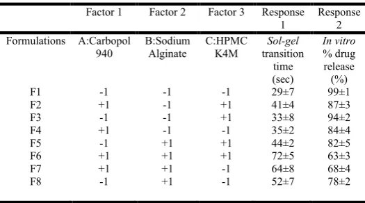
Table 1. Composition of different coded values in 23 full factorial design
Factor 1 Factor 2 Factor 3 Response 1
Response 2 Formulations A:Carbopol
940
B:Sodium Alginate
C:HPMC K4M
Sol-gel transition
time
In vitro % drug release (sec) (%)
F1 -1 -1 -1 29±7 99±1
F2 +1 -1 +1 41±4 87±3
F3 -1 -1 +1 33±8 94±2
F4 +1 -1 -1 35±2 84±4
F5 -1 +1 +1 44±2 82±5
F6 +1 +1 +1 72±5 63±3
F7 +1 +1 -1 64±8 68±4
F8 -1 +1 -1 52±7 78±2
Fourier-transformed infrared (FTIR) spectroscopy
FTIR spectroscopy was carried out to characterize the possible interaction between the drug and excipients, if any. The FTIR spectra of pure drug, FSK solid dispersion and physical mixture of drug with selected polymers were recorded using KBr disc using an FTIR spectrophotometer (Shimadzu, Tokyo, Japan).
pH
pH of the In-situ gels after addition of all ingredients was
measured using digital pH meter (Pandey et al., 2010).
Visual appearance and clarity
Visual appearance and clarity was done under fluorescent light against a white and black back ground for presence of any
particulate matter (Pandey et al., 2010).
Gelation studies
Gelation studies were carried out in test tube, The studies were carried out using simulated tear fluid (STF) of composition 1 (sodium chloride 0.670 g, sodium bicarbonate 0.200 g, calcium chloride dihydrate 0.008 g and purified water sufficient to make 100 g) and of composition 2 (bovin serum albumin 0.268
g, lysozyme 0.268 g, -globulin 0.134 g, calcium chloride
dehydrate 0.008 g, D-glucose 0.15 g, sodium chloride 0.65 g
and distilled water sufficient to make 100 g), which simulated either the divalent cation content or both the protein and
divalent cation content of the tear fluid equilibrated at 370 C.
The one drop of preparation was carefully placed into the test tube using a micropipette and 2 ml of gelation solution
(composition 1 or 2) was added slowly (Srividya et al., 2001).
Gelation was assessed by visual examination and time taken for
sol to gel formation is recorded (Nanjawade et al., 2007).
Viscosity
The viscosity of developed formulation was determined at 20°C by pouring it into the sample adaptor of Brookfield viscometer (DV-II+ Pro, Brookfield Engineering Ltd, Middleboro, MA, USA). Viscosity of sample was measured at different angular velocities. A typical run comprised changing angular velocity from 10 to 100 rpm with equal weight for each rpm. The hierarchy of angular velocity was reversed (100 to 10 rpm) with similar weight. The average of three readings was
used to calculate the viscosity. The sol was allowed to gel and
then viscosity was again measured using the same Brookfield viscometer with T spindle in conjunction with heli path stand (Nanjawade et al., 2007).
Drug content uniformity
The vials (n = 3) containing the preparation were shaken for
2–3 min manually and 100 µl of the preparation was transferred aseptically to 1ml of ethanol containing 25ml volumetric flasks with a micropipette and the final volume was
made up with simulated tear fluid solution pH 7.4 at 35± 10c
and sonicated for homogeneity. Forskolin concentration was determined at 210 nm by UV spectroscopy (Shimadzu,
UV-1601, Japan) (Srividya et al., 2001).
Sterility test
It was performed for aerobic, anaerobic and fungi microorganisms using fluid thioglycollate and soya bean casein digest medium as per IP 2007. Formulation took into laminar flow and passed through a membrane filter of 0.45μm with the help of vaccum pump. After filtration the filter paper was removed and cut into two halves. One half was dropped in fluid thioglycollate and other in soya bean casein digest. Both the media kept for incubation at 37°C for 7 days, and observed for
any microbial growth (Singh et al., 2010).
In vitro release study
Drug released study from prepared formulation was studies using Franz- diffusion cell. Cellophane membrane and artificial tear fluid (ATF) pH 7.4 was used as a diffusion membrane and medium respectively. The cellophane membrane (previously soaked overnight in the receptor medium) was tied at one end
of the glass diffusion cell. Accurately weighed 1ml of gel was
spread uniformly on a cellophane membrane, which was in contact with receptor medium. The receptor medium was stirred continuously at 20rpm to simulate blinking action of eyelids. The whole assembly was adjusted on magnetic stirrer and maintained at 35±1°C. At specify intervals (0.5 h, 1 h, 2 h up to 8 h) 1 ml of sample was withdrawn from receptor compartment, replace with 1ml of freshly ATF and analyzed by UV spectroscopy(Shimadzu, UV-1601, Japan) at 210 nm (Pandey et al., 2010) (Singh et al., 2010).
In vivo Ocular irritancy test
New Zealand White rabbits, weighing 2.5–3.0 kg, were provided by National Institute of Nutrition, Hyderabad, India. The animals, housed in standard cages in a light controlled
room at 19±1oC and 50±5% RH, were given a standard pellet
diet and water ad libitum. All the studies were conducted in
accordance with the guidelines of Committee for the Purpose of Control and Supervision of Experiments on Animals (CPCSEA). Draize technique was selected for conducting the
Ocular irritancy test on New Zealand white rabbits (Baeyens
et al., 2002). The experimental protocol dated 05/06/2014. Total sixteen animals are equally divided into four groups. Group I was treated with 0.01 % w/v Sodium lauryl sulfate (SLS) (positive control), Group II animals were treated with blank, Group III with sterile optimized formulation, and Group IV with 0.9%w/v NaCl (negative control) twice a day for a period of 1 week into the rabbit’s left eye and observed for redness, swelling, watering. The ocular condition was recorded every day. The eye irritation score was obtained by dividing the total score for all rabbits by the number of rabbits. Irritation was classified according to four grades: practically non-irritating, score 0–3; slightly non-irritating, score 4–8; moderately irritating, score 9–12; and severely irritating (or corrosive), score 13–16.
Intra ocular pressure (IOP) reduction studies
The Forskolin (FSK) In situ gel formulations were tested for
their IOP lowering effects on adult normotensive male New Zealand albino rabbits and the obtained results were compared
with that of plain In situ formulations as well as plain FSK
solution. An Increase in IOP was induced by the rapid infusion of a 5% glucose solution through the marginal ear vein. The amounts of injected were 15 ml/kg of body weight and infusion was accomplished in all animals within 30 sec. The IOP was
measured by standardized Schiotz tonometer
(Zur- Benutzungdes Schioetz, Germany) (Kaur et al., 2000)
(Monem et al., 2000). Before the measurement of the tension, the cornea was anaesthetized with 2 or 3 drops of xylocaine (1% w/v). After 2 min, the eyelids were retracted gently with one hand, without exerting pressure on the eye ball. The lower cul-de-sac of right eye of each rabbit of the group (n = 4) received 25 µl of the optimized formulation while the contra lateral eye (left) received no drug and served as a control. The IOP of both eyes of each rabbit was measured immediately before the administration of formulation (zero reading), 30 min after instillation and then at every hour for a period of 8 h. The similar procedure was adapted for the measurement of IOP
after instillation of 25 µl of plain FSK solution and plain In situ
formulations, respectively. The change in IOP (ΔIOP) was determined by following equation:
ΔIOP = IOPDosed eye− IOPControl eye
All the observations were taken in triplicate and the mean values were reported. All the measurement periods began during the same hour on each day and all the data were recorded with the same tonometer.
RESULTS AND DISCUSSION
The solid dispersions FSK with β- cyclodextrin ratios 1:1, 1:2, 1:3 and 1:4 were prepared successfully by kneeding method and produced good yield. β- cyclodextrin more than four parts formed sticky mass compared to other ratios. The drug content
of formulated solid dispersions was ranging from 94.70 ± 0.26 to 96.90 ± 0.58 %. Prepared Solid dispersions were showed solubility of 2.012±0.012, 2.241±0.018, 2.759±0.016 and 3.495±0.017 mg/ml respectively for ratios 1:1, 1:2, 1:3, and 1:4 in pH 1.2 and 2.020±0.002, 2.255±0.015, 2.768±0.011, 3.499±0.012 mg/ml respectively in pH 7.4 buffer solutions. Solid dispersion 1:4 was selected for further formulation of
ocular pH and ion activated In situ gels. The purpose of using a
full 23 factorial experimental design was to conduct a
comprehensive study of the effect of polymers and viscosity
enhancing agent like Carbopol 940(A), Sodium Alginate(B),
Hydroxy Propyl Methyl Cellulose(C) and their interactions
using a suitable statistical tool (Design-Expert® 9.0.3 Software) by applying ANOVA at 0.05 levels. The effects of
the independent variables (A, B and C) on the sol–gel transition
time (sec) and In vitro % drug release were evaluated, and the
following models were obtained:
Final Equation in Terms of Coded Factors
Sol-gel transition time (R1) =+46.25+6.75 A+11.75 B+1.25 C+3.25 AB+2.25 AC-1.25BC+ 1.75ABC
In vitro % drug release (R2)=+81.88-6.38A-9.13B-0.38C-0.87AB-0.12AC+0.13BC-2.13ABC
From the ANOVA results (Table 2 and 3) of the model relating Sol-gel transition time (sec) and In vitro % drug release as response, it can be noticed that all the coefficients of this model
equations had statistical significance (p < 0.05) with the
R2 values of 0.9994 and 0.9999 for Sol-gel transition time (sec)
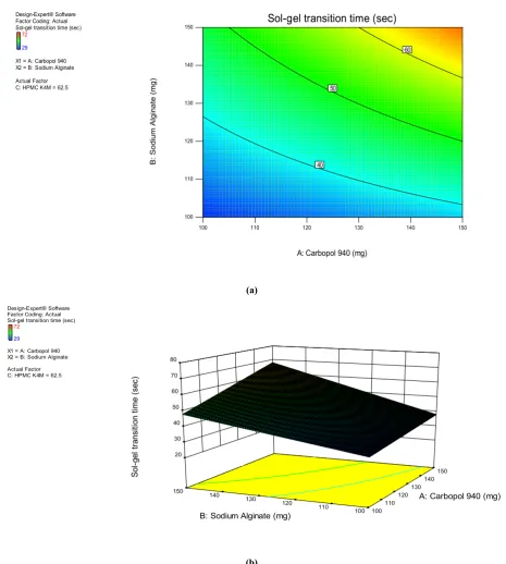
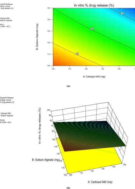
[image:4.595.145.464.298.505.2]and In vitro % drug release respectively. The three-dimensional response surface graphs (Fig. 1(b), Fig. 2 (b)) and corresponding two-dimensional contour plots (Fig. 1(a), Fig. 2 (a)) were generated by the Design-Expert z® 9.0.3 Software.
Table 2. Response 1: sol to gel transition time: ANOVA for selected factorial model Analysis of variance table [Partial sum of squares - Type III]
Table 3. Response 2: In vitro % drug release: ANOVA for selected factorial model Analysis of variance table [Partial sum of squares - Type III]
[image:4.595.122.487.549.778.2]The three-dimensional response surface graph is very useful in learning about the main and interaction effects of the independent variables (factors), whereas two-dimensional contour plot gives a visual representation of values of the response.
The three-dimensional response surface graphs relating sol to
gel transition time (sec) as response (Fig. 1 (b)) depicted the
increase in sol to gel transition time with the increasing of
polymer concentration, (Fig. 2 (b)) depicting that In vitro%
drug release was increase with decreasing of polymer concentration mainly.
The two-dimensional contour plots (Fig. 1 (a) and (Fig. 2 (a))
relating to sol to gel transition time (sec) and In vitro % drug
release indicated nonlinear relationships. The optimal value of response was obtained by numerical analysis using Design-Expert® 9.0.3 Software. In order to evaluate the optimization capability of model generated according to the results of the
full 23 factorial design, optimized In situ gel containing FSK
was prepared using the optimal process variable settings (Table 4) and the optimized formulation was evaluated to determine
the sol to gel transition time (sec) and In vitro % drug release.
The sol to gel transition time (sec) and In vitro % drug release
for the optimized formulation was measured 37±4 sec and
(a)
[image:5.595.66.532.60.579.2](b)
Fig. 1. Effect of Carbopol 940, Sodium Alginateand HPMC K4Mquantitieson sol to gel transition time (sec) presented by response surface plot (a), and contour plot (b)
Design-Expert® Software Factor Coding: Actual Sol-gel transition time (sec)
72
29
X1 = A: Carbopol 940 X2 = B: Sodium Alginate
Actual Factor C: HPMC K4M = 62.5
100 110 120 130 140 150
100 110 120 130 140
150 Sol-gel transition time (sec)
A: Carbopol 940 (mg)
B:
S
od
ium
A
lgin
a
te
(
m
g)
40 50
60
Design-Expert® Software Factor Coding: Actual Sol-gel transition time (sec)
72
29
X1 = A: Carbopol 940 X2 = B: Sodium Alginate
Actual Factor C: HPMC K4M = 62.5
100 110 120 130
140 150
100 110
120 130
140 150 20
30 40 50 60 70 80
S
o
l-g
e
l
tr
a
nsi
ti
o
n
t
im
e
(se
c
)
A: Carbopol 940 (mg)
94±3% respectively with small error value. This reveals that
mathematical models obtained from the full 23 factorial design
was well fitted.
FTIR spectra analysis revealed no significant interaction between various rational combinations containing physical mixture of drug with polymers (i.e., Carbopol 940, sodium alginate and HPMC K4M) as shown in (Fig. 3, 4, 5, Table 5). The FTIR spectra of drug-polymer mixture confirmed neither any shift in the wave numbers of the peaks nor in the intensity, construed lack of interaction. The formulations exhibited
Newtonian behavior in the sol state which is shown in the Fig.
5(A).
In the gel state the formulations exhibited pseudoplastic
rheology shown in Fig. 6(B) as evidenced by shear thinning with an increase in shear stress with increased angular velocity. Viscosity was directly dependent on the polymeric content of the formulation. The viscosity increased with increasing concentration of HPMC K4M. Maximum viscosity was observed with F9 and minimum viscosity with F8.
Table 4. Values of factors for the formulation of optimized In situ gel containing FSK (F 9)
[image:6.595.118.482.418.537.2]Table 5. Assessment of FTIR spectra
Table 6. Results of evaluation of Ocular In situgel formulations
+ Gelation after few minutes, dissolves rapidly; ++ gelation immediate, remains for few hours; +++ Gelation immediate remains for extended period.
Table 7. Scores for In VivoOcular irritation evaluation of FSK ocular In situ gel in rabbits
(a)
[image:7.595.72.517.74.698.2](b)
Fig. 2. Effect of Carbopol 940, Sodium Alginate and HPMC K4M quantities on In vitro % drug release presented by response surface plot (a), and contour plot (b)
Design-Expert® Software Factor Coding: Actual In vitro % drug release (%)
99
63
X1 = A: Carbopol 940 X2 = B: Sodium Alginate
Actual Factor C: HPMC K4M = 62.5
100 110 120 130 140 150
100 110 120 130 140 150
In vitro % drug release (%)
A: Carbopol 940 (mg)
B:
S
o
d
iu
m
Alg
in
a
te
(m
g
)
70
80
90
Design-Expert® Software Factor Coding: Actual In vitro % drug release (%)
99
63
X1 = A: Carbopol 940 X2 = B: Sodium Alginate
Actual Factor C: HPMC K4M = 62.5
100 110 120 130 140 150
100 110
120 130
140 150
60 70 80 90 100
In
v
it
ro
%
d
ru
g
re
le
a
se
(
%)
A: Carbopol 940 (mg) B: Sodium Alginate (mg)
Fig. 3. FTIR spectrum for Forskolin
[image:8.595.71.531.66.296.2]Fig. 4. FTIR spectrum for Forskolin and beta cyclodextrin Solid Dispersion
Fig. 5. FTIR spectrum of drug and other ingredients
[image:8.595.92.528.548.750.2](A)
[image:9.595.75.533.62.545.2](B)
Fig. 6. Graph representing viscosity of formulations (A) at 20°C (B) at 35°C
Fig. 7. In Vitro percentage drug release of FSK In situ gels 0
100 200 300 400 500 600
0 20 40 60 80 100 120
vi
sc
o
si
ty
(
m
p
as
)
speed (rpm)
Viscosity at 20
o
c
F1 F2 F3 F4 F5 F6 F7 F8 F9
0 200 400 600 800 1000 1200 1400 1600 1800
0 20 40 60 80 100 120
vi
sc
o
si
ty
(
m
p
as
)
speed (rpm)
Viscosity at 35
o
c
F1 F2 F3 F4 F5 F6 F7 F8 F9
0 20 40 60 80 100 120
0 1 2 3 4 5 6 7 8 9
cu
m
u
la
ti
ve
%
d
ru
g
re
le
as
e
Time (h)
In Vitro
percentage drug release
F1 F2 F3 F4 F5 F6 F7 F8 F9
[image:9.595.98.510.575.755.2]This indicates that addition HPMC K4Mlead to increase in viscosity for formulations.
All the formulations were found to be clear in appearance. The pH was within the acceptable range and hence would not cause any irritation when administered into the eye. The drug content was found to be in acceptable range and was in the range of 98.3±0.4 to 101±0.5% indicating uniform distribution of drug. All the formulations gelled instantaneously within 80 sec on contact with 7.4 pH phosphate buffer at 35 ± 1°C, formulations containing optimal quantity of HPMC K4M showed good gelling properties. By visual observations the formulations formed a translucent matrix on gelling. F9 formulation showed good gelling capacity and gelled within 37±4 sec. The pH, drug content and gelling capacity values of all the formulations were
shown in Table 6 and sol to gel transition time (sec) of all
formulations were shown in Table 1. There was no microbial growth in all formulations after 7 days of incubation period,
showing that the method used for sterilization was reliable.
In vitro release (Fig. 7) through cellophane membrane revealed that with the increase in the concentration of polymers the drug release decreased due to the formation of gel structure.
Comparing to the polymers, more concentration of carbopol 940 contain formulations showed prolonged drug release than sodium alginate with combination of HPMC K4M. These results suggested that FSK was released in a sustained manner
from formulation F9 for a period of 8 h with optimal sol to gel
transition time (37±4 sec), hence formulation F9 was selected
as optimized formulation for conducting In vivo studies. In
order to understand the drug release mechanism, the release data was tested assuming common kinetic model. The higher regression coefficient values (Higuchi model) for each formulation suggested that the formulations F1 to F9 showed matrix type of drug release.
The results of the ocular irritation studies (Table 7) indicated that the optimized formulation (F9) was non-irritant with no ocular damage or abnormal clinical signs to the cornea, iris or conjunctivae. As shown in Fig. 8 (a, b and c), no lesion
formation was observed during the test. Hence, FSK ocular In
situ Gel may be considered safe for ophthalmic drug delivery
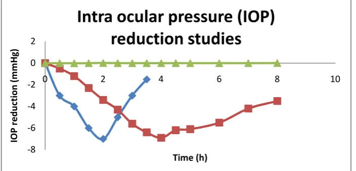
for glaucoma. The Intra ocular pressure (IOP) reduction studies
suggested that the hypotensive activity of drug loaded In situ
gel formulation (F9) was comparable to that of the plain drug solution. In the beginning, IOP decreased sharply for the first
[image:10.595.48.558.56.212.2]
(a) (b) (c)
Fig. 8. In Vivo ocular irritation studies a) 15 min before installation of In situgel (F9) b) No lesion observed for F9 on 3rd day c) No lesion observed for F9 on 7th day
Fig. 9. In Vivo IOP reduction studies for F9 -8
-6 -4 -2 0 2
0 2 4 6 8 10
IO
P
r
e
d
u
ct
io
n
(
m
m
H
g)
Time (h)
Intra ocular pressure (IOP)
reduction studies
plain FSK solution Optimized formulation Blank Formulation
[image:10.595.126.478.255.424.2]2 h in case of plain FSK solution whereas IOP was observed to
decrease slowly in case of drug loaded In situ gel formulation
(F9). The IOP was immediately and noticeably reduced up to 2 h after instillation of plain FSK solution, but increased slightly over the rest of the period of observation. This type of fluctuation was not observed in case of optimized formulation,
where the IOP continued to drop. The results suggested that In
situ gel formulation (F9) produced a significant and prolonged
reduction in IOP throughout 8 h. This overwhelming superiority over plain FSK solution was further magnified when single instillation was considered (Fig. 9). The reduction in IOP was found sustained for a period of approximately 6 h
with the In situ gel formulation (F9) as compared to the plain
drug solution.
Conclusion
The present studies, therefore, shows the successful
formulation of pH and Ion activated In situ gels of FSK using
Carbopol 940, Sodium alginate and HPMC K4M as suitable polymers for glaucoma. The application of experimental design methodology helped to prepare the optimized formulation,
which showed good sol to gel transition time (sec), In vitro
percentage drug release and In Vivo anti glaucoma activity. The
drug release was found to be diffusion controlled and followed Higuchi kinetics for all formulations. FTIR studies confirmed absence of any physiochemical interaction between drug and other ingredients. Ocular irritation studies showed that absence of ocular damage or abnormal clinical signs to the cornea, iris or conjunctivae. The Intra ocular pressure (IOP) reduction
studies suggested that In situ gel formulation (F9) produced a
significant and prolonged reduction in IOP throughout 8 h.
Conflicts of interest
The authors report that this article content does not have any conflicts of interest.
REFERENCES
Baeyens, V. Felt-Baeyens, O., Rougier, S., Pheulpin, S., Boisrame, B. and Gurny, R. 2002. Clinical evaluation of bioadhesive ophthalmic drug inserts (BODI) for the
treat-ment of external ocular infections in dogs. J. Control.
Release., 85: 163–168.
Bhat, S.V., Bajwa, B.S., Dornauer, H., De Souza, N.J. and Fehlhaber, H.W. 1977. Structures and stereochemistry of new labdane diterpenoids from Coleus forskohlii Briq. Tetrahedron Lett., 19: 1669–1672.
Coleus forskohlii on hypertension. Nagarjun., 22: 56–58.
Diebold, Y., Jarrin, M., Saez, V., Carvalho, E.L., Orea, M. and Calonge, M. 2007. Ocular drug delivery by
liposome-chitosan nanoparticle complexes (LCS-NP). Biomaterials.,
28: 1553-1564.
Dubey, C.B., Srimal, R.C. and Tandon, J.S. 1997. Clinical evaluation of ethanolic extract of Coleus forskohlii in
hypertensive patients. Sachitra Ayurveda., 49: 931–936.
Dubey, M.P., Srimal, R.C., Nityanand, S. and Dhawan, B.N. 1981. Pharmacological studies on coleonol, a hypertensive
diterpene from Coleus forskohlii. J. Ethnopharmacol.,
3: 1–13.
Kansal, C.M., Srivastava, S.P., Dube, C.B. and Tandon, J.S. 1978. Clinical evaluation of
Kaur, I.P. and Smitha, R. 2002. Penetration enhancers and ocular bioadhesives: two new avenues for ophthalmic drug
delivery. Drug Dev. Ind. Pharm., 28: 353-369.
Kaur, I.P., Singh, M. and Kanwar, M. 2000. Formulation and
evaluation of ophthalmicpreparation of acetazolamide. Int.
J. Pharm., 199: 119–127.
Lele, B.S. and Hoffman, A.S. 2000. Insoluble ionic complexes of polyacrylic acid with a cationic drug for use as a
mucoadhesive, ophthalmic drug delivery system. J.
Biomater. Sci. Polym. Edn., 11: 1319-1331.
Monem, A.S., Ali, F.M. and Ismail, M.W. 2000. Prolonged effect of liposomes encapsu-lating pilocarpine HCl in
normal and glaucomatous rabbits. Int. J. Pharm., 198:
29–38.
Nanjawade, B.K., Manvi, F.V. and Manjappa, A.S. 2007. In situ-forming hydrogels for sustained ophthalmic drug
delivery. J. Control. Release., 122:119-134.
Pandey, A,, Prashant, Y.M., Sachdeva, D., Patel, D.K. and Ramesh, R. 2010. Development and optimization of levobunolol hydrochloride in-situ gel for glaucoma
treatment. Int. J. Pharm. and Bio. Archives., 1(2): 134-139.
Quigley, H,A. and Vitale, S. 1997. Models of open-angle glaucoma prevalence and incidence in the United States. Invest. Ophthalmol. Vis. Sci., 38: 83-91.
Quigley, H.A. 1996. Number of people with glaucoma
worldwide. Br. J. Ophthalmol., 80: 389-393.
Rozier, A., Maznel, C., Grove, J. and Plazonnet, B. 1989. Gelrite: a novel, ion activated, in situ gelling polymer for ophthalmic vehicles—effect on bioavailability of timolol. Int. J. Pharm., 57: 163-168.
Singh, V., Bushetti, S.S., Appala Raju, S., Rizwan, A. and Mamata, S. 2010. In vitro and in vivo evaluation of stimuli
sensitive hydrogel for ophthalmic drug delivery. Ind. J.
Pharm. Edu. and Res., 44(4): 380- 385.
Sommer, A. 1996. Glaucoma risk factors observed in the
Baltimore Eye Survey. Curr. Op. in Ophthalmol., 7: 93-98.
Srividya, B., Cardoza, R.M. and Amin, P.D. 2001. Sustained ophthalmic delivery of ofloxacin from a pH triggered in
situ gelling system. J. Contr. Rel., 73: 205-211.
Suryanayanan, M. and Pai, J.S. 1998. Studies in
micropropagation of Coleus forskohli. J. Med. and Aro.
Plant Sciences., 20: 379–382.
![Table 2. Response 1: sol to gel transition time: ANOVA for selected factorial model Analysis of variance table [Partial sum of squares - Type III]](https://thumb-us.123doks.com/thumbv2/123dok_us/9277843.420282/4.595.122.487.549.778/response-transition-selected-factorial-analysis-variance-partial-squares.webp)