Acute headache in general neurology of China: Cause
changes and predictors of short-term outcome
*
Guangsheng Wang1, Yeting Zhou2, Xiaodong Chen1, Hongjian Wang3, Tonghui Yang1,
Chunhong Chang1, Yuanwei Wang1, Hanpei Gu1, Daoming Tong1#
1Departments of Neurology, Shuyang People’s Hospital, Shuyang, China 2Medical Evaluation Unit, Shuyang People’s Hospital, Shuyang, China 3Department of Medical Affairs, Shuyang People’s Hospital, Shuyang, China
Email: #[email protected]
Received 16 January 2012; revised 17 February 2012; accepted 21 February 2012
ABSTRACT
Background: Although patients with acute headache frequently present in emergency department, the causes and predictors of short-term outcome in pa- tients with acute headache in general neurology have not been adequately investigated. Methods: We pro- spectively reviewed the medical records of 130 hospi- talized acute headache patients in general neurology of China. Their initial CT scan was assessed, as was their lumbar puncture (LP) examination if performed. Results: The main cause of acute headache was acute secondary headache (80.8%), which was mainly at- tributable to acute cerebrovascular events (72.4%) followed by intracranial infection (19.0%). Among the 10.8% of patients who died during hospitalization most (85.7%) had subarachnoid hemorrhage (SAH). Significant predictors of survival were severe headache versus thunderclap headache, meningismus, delayed loss of consciousness, and hypertension (all p < 0.05). Multiple logistic regression analyses showed signifi-cant differences in severe headache or thunder-clap headache (OR, 0.255; 95%CI, 0.066 - 0.990; p = 0.048) and delayed loss of consciousness (OR, 0.060; 95%CI, 0.016 - 0.224, p = 0.000) between patients who died and those who survived. Conclusions: The main un-derlying cause of hospitalized acute headache was acute cerebrovascular events. Severe headache and delayed loss of consciousness are predictors for poor outcome of acute headache.
Keywords:Acute Headache; Causes; Cerebrovescular
Events; Subarachnoid Hemorrhage; Outcome
1. INTRODUCTION
Acute headache (primary and secondary) is a common
neurological emergency that may be caused by a great variety of clinical conditions. However, the underlying causes may change pattern over time particularly where there is rapid social development. Clinical study of acute headache presenting in emergency departments has been previously reported [1-5].Many acute secondary head- aches can be misdiagnosed if appropriate investigations are not made, even life-threatening subarachnoid hemor- rhage (SAH) [6-8]. However, there are few reports on patients with acute headache who are hospitalized at general department of neurology [9]. The aims of the study were to investigate underlying causes of acute headache and to determine predictors of outcome in pa- tients with acute headache.
2. METHODS
Consecutive patients with acute headache admitted either directly or through the emergency department into a general department of neurology at a public hospital in China from February 2003 to January 2006 were pros- pectively evaluated. To be eligible, patients had to have what they described as an acute onset of sudden or severe headache. Acute headache was defined as a headache of acute or sudden onset and lasting several hours, which without the history of headache, or with the history of headache but no onset of headache at least over a > 2-month period. Severe headache was defined as worst of their lives or a significant lifestyle restriction. Thunder- clap headache was defined as a sudden and severe head- ache with maximum intensity at onset. Patients with de- layed loss of consciousness were accepted if they were initially alert at the onset of headache. Exclusion criteria included age <15 years, recent head trauma or lumbar puncture (LP), initial loss of consciousness, signs of hemiplegia, and history of ≥3 recurrent headaches of the same character and intensity as the presenting headache over a >6-month period. The study was approved by the Ethical Committee on Clinical Research of the hospital;
informed consent was obtainedfrom all patients or their families.
On admission, the patients responded to an approved questionnaire providing data on sex and age, prior head- ache ictus, and family history. All patients were examin- ed by neurologist for clinical and full neurological states, especially meningismus, within 24 h from admission.
Computed tomography (CT) scan was performed by all patients within 24 h of admission to hospital. Following this procedure, patients with negative CT and suspected SAH or intracranial infection underwent LP examination. CSF was analyzed for white blood cell count and protein and glucose levels. Spectrophotometry for detection of oxyhemoglobin was also performed for these patients. Staining for acid-fast bacilli and a test for cryptococcal antigenwere investigated.
In evaluation, case histories and test data were as-sessed by two experienced neurologists; separate diag- noses were made following guidelines of the Inter- national Classification of Headache Disorders (ICHD) [10].
Statistical Methods
Data are presented as mean ± SD or n (%). Frequencies were compared by Chi-squared statisticsor Fisher’s ex- act test as appropriate. Continuous variableswere com- pared by t test.Patient survival was assessed byunivari- ate analysis.Predictors of outcome were identified with forward stepwise logistic regression. Differences with p-value <0.05 were considered significant.All statistical analyses were performed using SPSS version 10.0 soft- ware (SPSS Inc., Chicago, IL, USA).
3. RESULTS
3.1. Diagnosis of Acute Headache
A total of 130 consecutive patients (M/F, 54/64; age range, 15 - 72 years) with acute headache were enrolled, which were admitted either directly to the hospital or through the emergency department. The interval from headache onset to admission in the majority of the pa- tients was <24 h; in a few patients this was >1 week due to delayed referral from general practitioner. Shortest and longest length of hospitalization was 6 and 62 days, respectively; average stay was 18 ± 11 days.
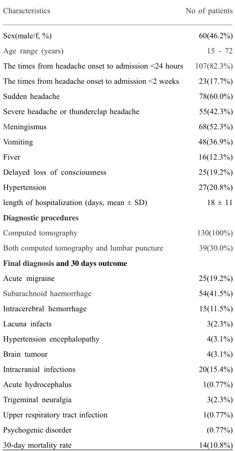
Although many patients (n= 25; 19.2%) had a primary headache (all acute migraine) most cases were acute secondary headache (n = 105; 80.8%), among whom acute cerebrovascular events were noted in the majority (72.4%). Characteristics of 130 patients with acute headacheare shown in Table 1.
Results of CT and LP evaluations are displayed in Ta- ble 2. In the positive CT group 73 patients (69.5%) were
[image:2.595.305.541.102.554.2]diagnosed, of whom 47 patients had SAH (64.4%), 15
Table 1. Characteristics of 130 patients with acute headache.
Characteristics No of patients
Sex(male/f, %) 60(46.2%)
Age range (years) 15 - 72
The times from headache onset to admission <24 hours 107(82.3%)
The times from headache onset to admission <2 weeks 23(17.7%)
Sudden headache 78(60.0%)
Severe headache or thunderclap headache 55(42.3%)
Meningismus 68(52.3%)
Vomiting 48(36.9%)
Fiver 16(12.3%)
Delayed loss of consciousness 25(19.2%)
Hypertension 27(20.8%)
length of hospitalization (days, mean ± SD) 18 ± 11
Diagnostic procedures
Computed tomography 130(100%)
Both computed tomography and lumbar puncture 39(30.0%)
Final diagnosis and 30 days outcome
Acute migraine 25(19.2%)
Subarachnoid haemorrhage 54(41.5%)
Intracerebral hemorrhage 15(11.5%)
Lacuna infacts 3(2.3%)
Hypertension encephalopathy 4(3.1%)
Brain tumour 4(3.1%)
Intracranial infections 20(15.4%)
Acute hydrocephalus 1(0.77%)
Trigeminal neuralgia 3(2.3%)
Upper respiratory tract infection 1(0.77%)
Psychogenic disorder (0.77%)
30-day mortality rate 14(10.8%)
Table 2. Diagnoses of CT and LP in patients with acute headache.
Diagnoses Positive CT group (n = 73) Negative CT group(n = 57) P Value
SAH 47(64.4%) 7(12.3%) 0.000
Intracerebral
hemorrhage 15(20.5%) 0(0) 0.000
Lacuna infacts 3(4.1%) 0(0) 0.122
Brain tumour 4(5.5%) 0(0) 0.073
Intracranial
infections 3(4.1%) 17(29.8%) 0.000
Hydrocephalus 1(1.4%) 0(0) 0.375
[image:2.595.309.538.584.735.2]intracerebral hemorrhage (20.5%; 7 cases lobar, 4 cere- bellar, 1 thalamic, 3 primary intraventricular), 3 lacunar infarction (4.1%; 2 cases internal capsule, 1 multiple la- cunar in white matter; age range, 49 - 61 years), and 3 intracranial infection (4.1%; 1 case viral encephalitis, 1 tuberculosis encephalitis, 1 unknown encephalitis; all diagnosed by LP).
The negative CT group included 57 patients in whom LP was performed in 31 cases (54.4%); in these indi- viduals 7 patients with SAH were diagnosed by oxyhemoglobin CSF (12.3%; 6 cases by LP within 1 week of onset and 1 within 13 days, 2 of whom died). We diagnosed 17 intracranial infections (29.8%; 15 pa-tients CSF positive and 2 CSF negative) including 5 cases of tuberculous meningitis, 6 viral meningitis, and 6 viral encephalitis.
3.2. Univariate and Multivariate Analysis of Outcome
Although 116 of 130 patients (89.2%) had agood out- come, 14 of 130 patients (10.8%) died during hospitali- zation, among whom the majority (12 cases; 85.7%) had SAH (of those, 75% died of rebleed); 1 patient (7%) died of fourth ventricle hemorrhage and 1 (7%) brain tumor.
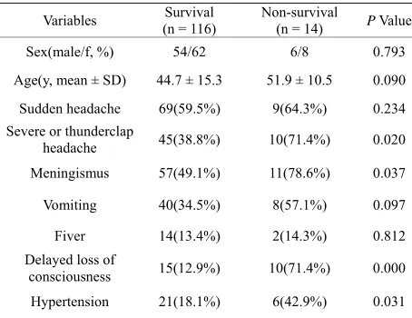
Univariate analysis of death in our patients is summa- rized in Table 3. There was no difference in sex, age,
[image:3.595.308.541.112.215.2]sudden headache, vomiting, and fever in survival patients and those who died (p > 0.05). However, there were sig-nificant differences in headache versus thunderclap headache, meningismus, delayed loss of consciousness, and hypertension in the survival and non-survival group (p < 0.05).
Table 4 summarizes the results of multiple logistic re-
[image:3.595.57.284.559.734.2]gressions based on four data of the original six variables. Significant predictors of death were severe versus thun- derclap headache (OR, 0.255; 95%CI, 0.066 - 0.990;
Table 3. The results of univariate analysis in survival and non-survival patients.
Variables (n = 116) Survival Non-survival (n = 14) P Value
Sex(male/f, %) 54/62 6/8 0.793
Age(y, mean ± SD) 44.7 ± 15.3 51.9 ± 10.5 0.090
Sudden headache 69(59.5%) 9(64.3%) 0.234 Severe or thunderclap
headache 45(38.8%) 10(71.4%) 0.020
Meningismus 57(49.1%) 11(78.6%) 0.037
Vomiting 40(34.5%) 8(57.1%) 0.097
Fiver 14(13.4%) 2(14.3%) 0.812
Delayed loss of
consciousness 15(12.9%) 10(71.4%) 0.000 Hypertension 21(18.1%) 6(42.9%) 0.031
Table 4. Results analyzed by the multiple Logistic regression for predictors of outcome in patients with acute headache.
Variables B SE Wald P Value OR 95%CI
Severe or thunderclap
headache
–1.366 0.692 17.480 0.048 0.255 0.066 ~ 0.990
Delayed loss of consciousness
–2.820 0.674 3.900 0.000 0.060 0.016 ~ 0.224
Constant 3.986 0.707 31.770 0.000 53.835
p = 0.048), and delayed loss of consciousness (OR, 0.060; 95%CI, 0.016 - 0.224, p = 0.000) between the two groups.
4. DISCUSSION
In our 130 patients diagnosed according to ICHD stan-dards secondary headache was the major cause of acute headache for hospitalization, in 80.8%, including acute cerebrovascular events in 72.4%, intracranial infection in 19.0%, and a few other causes. Acute primary headache only accounted for 19.2% of those hospitalized; all were acute migraine. This clinical study clearly indicates that in patients with acute headache referred for hospital treatment acute cerebrovascular events should be fore- most borne in mind.
On the other hand, however, in our cohort the propor- tion with secondary headache and acute cerebrovascular events was significantly higher than previously observed in emergent patients in neurology departments reported in Canada and Norway [2,3], and cerebrovascular events were significantly higher than in patients hospitalized in Minnesota [9]. The present study shows that the pattern of causes for acute headache is not the same in emer- gency room and hospitalized patients, and may differ according to region, particularly the incidence of acute cerebrovascular events were higher in China than in western countries [11-13].
group accounted for 15% - 24%. However, we have to point out that current proportion may be with potential limitations, as those SAH with initial loss of conscious- ness were not included.
Our study suggests that LP should be carried out in pa- tients with suspectedSAH and negative results on head CT [7] especially those with low Hunt and Hess grade on admission and normal diastolic blood pressure; those with suspected SAH but delayed CT scan should un- dergo LP [18] so as not to miss the most life-threatening diagnosis of SAH.
Our data show that acute lacunar infarction was also causative of acute secondary headache. Arboix et al. [19] noted that in patients with lacunar infarction, headache at stroke onset was more common in deep brain gray matter or brainstem topographies than in supratentorial white matter lesions. However, in our 3 patients with lacunar infarcts the lesions were located in the supratentorial white matter, suggesting that lacunar infarct in supraten- torial white matter may also cause acute headache.
Acute primary headache in hospitalized patients was mainly migraine headache. Because most of our patients experienced first severe sudden attack of migraine, they were diagnosed as acute migraine with or without aura. This was similar to the pattern noted of Bø, S.H. et al. [3].
In this study mortality of patients with acute headache during hospitalization was 10.8%. In these individuals SAH accounted for 85.7% of total deaths, including 2 patients who died with a negative CT scan and 1 oxyhemoglobin confirmed by LP. Our study showed that SAH is the most life-threatening acute secondary head- ache with a mortality of 75% for persons who rebleed, consistent with reported guidelines [14]. Moreover, we confirmed that sudden, severe headache rather than thun- derclap headache, meningeal irritation, delayed loss of consciousness, and hypertension in the non- survival group were significantly higher than in survivors, which are so-called “red flag” symptoms as previously reported [20,21]. On the other hand, multiple logistic regression analysis of our data revealed that severe head- ache and delayed loss of consciousness weresignificantly and in-dependently related to poor outcome in acute headache.
We observed markedly higher percent hemorrhagic stroke than that reported in Western countries [22]. The high mortality of acute headache may be not only associ- ated with higher percent hemorrhagic stroke, but also with higher mortality of hemorrhagic strokes [23], parti- cularly SAH [14].
In conclusion, the main underlying cause of hospital- ized acute headache was acute cerebrovascular events. Severe headache and delayed loss of consciousness are predictors for poor outcome of acute headache. There- fore, we believe that a better understanding of the causes
changes of acute headache and predictors of outcome for acute headache patients would facilitate design of proto- cols describing its differential diagnosis and early inter- vention.
REFERENCES
[1] Landtblom, A.M., Fridriksson, S., Boivie, J., Hillman, J., Johansson, G. and Johansson, I. (2002) Sudden onset headache: A prospective study of features, incidence and causes. Cephalalgia, 22, 354-360.
doi:10.1046/j.1468-2982.2002.00368.x
[2] Perry, J.J., Stiell, I.G., Wells, G.A., et al. (2005)Attitudes
and judgment of emergency physicians in the manage- ment of patients with acute headache. Academic Emer- gency Medicine, 12, 33-37.
doi:10.1111/j.1553-2712.2005.tb01475.x
[3] Bø, S.H., Davidsen, E.M., Gulbrandsen, P. and Dietrichs, E. (2008) Acute headache: A prospective diagnostic work-up of patients admitted to a general hospital. Euro-pean Journal of Neurology, 15, 1293-1299.
doi:10.1111/j.1468-1331.2008.02279.x
[4] Morgenstern, L.B., Luna-Gonzales, H., Huber, J.C. Jr., et al. (1998)Worst headache and subarachnoid hemorrhage:
Prospective, modern computed tomography and spinal fluid analysis. Annals of Emergency Medicine, 32, 297-
304.
[5] Grimaldi, D., Nonino, F., Cevoli, S., Vandelli, A., D’Amico, R. and Cortelli, P. (2009) Risk stratification of non-traumatic headache in the emergency department.
Journal of Neurology, 256, 51-57. doi:10.1007/s00415-009-0033-0
[6] Vermeulen, M.J. and Schull, M.J. (2007) Missed diag- nosis of subarachnoid hemorrhage in the emergency department. Stroke, 38, 1216-1221.
doi:10.1161/01.STR.0000259661.05525.9a
[7] Suarez, J.I., Tarr, R.W. and Selman, W.R. (2006) Aneu-rysmal subarachnoid hemorrhage. New England Journal of Medicine, 354, 387-396. doi:10.1056/NEJMra052732
[8] Kowalski, R.G., Claassen, J., Kreiter, K.T., Bates, J.E., Ostapkovich, N.D., Connolly, E.S. and Mayer, S.A. (2004) Initial misdiagnosis and outcome after subarach-noid hemorrhage. The Journal of the American Medical Association (JAMA), 291, 866-869.
doi:10.1001/jama.291.7.866
[9] Ramirez-Lassepas, M., Espinosa, C.E., Cicero, J.J., Johnston, K.L., Cipolle, R.J. and Barber, D.L. (1997) Predictors of intracranial pathologic findings in patients who seek emergency care because of headache. Archives of Neurology, 54, 1506-1509.
doi:10.1001/archneur.1997.00550240058013
[10] Headache Classification Subcommittee of the Inter- national Headache Society (2004) The international classification of headache disorders, 2nd edition. Cepha- lalgia, 24, 1-160.
health organization monitoring trends and determinants in cardiovascular disease. Stroke, 26, 361-367.
[12] Xu, G., Yin, Q. and Liu, X. (2009) Narrowing the gap of stroke management between developed and developing countries: Report of the fifth international stroke summit, Nanjing, China, July 17-19, 2009. Cerebrovascular Dis-eases, 29, 101-102. doi:10.1159/000259620
[13] Ferri, C.P., Schoenborn, C., Kalra, L., Acosta, D., Guerra, M., Huang, Y., et al. (2011) Prevalence of stroke and
re-lated burden among older people living in Latin America, India and China. Journal of Neurology, Neurosurgery, and Psychiatry, 82, 1074-1082.
doi:10.1136/jnnp.2010.234153
[14] Bederson, J.B., Connolly, E.S. Jr., Batjer, H.H., Dacey, R.G., Dion, J.E., Diringer, M.N., et al. (2009) Guidelines
for the management of aneurysmal subarachnoid hemor-rhage: A statement for healthcare professionals from a special writing group of the stroke council, American heart association. Stroke, 40, 994-1025.
doi:10.1161/STROKEAHA.108.191395
[15] Van der Wee, N., Rinkel, G.J.E., Hasan, D. and van Gijn, J. (1995) Detection of subarachnoid haemorrhage on early CT: Is lumbar puncture still needed after a negative scan? Journal of Neurology, Neurosurgery, and Psychia-try, 58, 357-359. doi:10.1136/jnnp.58.3.357
[16] Van Gijn, J. and van Dongen, K.J. (1982) The time course of aneurysmal haemorrhage on computed
tomo-grams. Neuroradiology,23, 153-156.
[17] O’Weill, J., Mclaggan, S. and Gibson, R. (2005) Acute headache and subarachnoid heamorrhage: A retrospective review of CT and lumbar puncture finding. Scottish Medical Journal, 50, 151-153.
[18] Tong, D.M. and Chou, Y.T. (2010) Predictors of the su-barachnoid hemorrhage of a negative CT scan. Stroke, 41,
e566-e567. doi:10.1161/STROKEAHA.109.574038
[19] Arboix, A., Grau-Olivares, M., Garcia-Eroles, L., et al.
(2006) Clinical implications of headache in lacunar stroke: Relevance of site of infarct. Headache, 46, 1172-1180. doi:10.1111/j.1526-4610.2006.00507.x
[20] Breen, D.P., Duncan, G.W., Pope, A.E., Gray, A.J. and Al-Shahi, S.R. (2008) Emergency department evaluation of sudden, severe headache. QJM: An International Journal of Medicine, 101, 435-443.
doi:10.1093/qjmed/hcn036
[21] Clinch, C.R. (2001) Evaluation of acute headaches in adults. American Family Physician, 63, 685-692.
[22] Jia, Q., Liu, L.P. and Wang, Y.J. (2010) Stroke in China. Clinical and Experimental Pharmacology and Physiology, 37, 259-264. doi:10.1111/j.1440-1681.2009.05290.x