Copyright © 1998, American Society for Microbiology. All Rights Reserved.
Sequential Evaluation of Dogs Naturally Infected with Ehrlichia canis,
Ehrlichia chaffeensis, Ehrlichia equi, Ehrlichia ewingii,
or Bartonella vinsonii
EDWARD B. BREITSCHWERDT,* BARBARA C. HEGARTY,ANDSUSAN I. HANCOCK Department of Companion Animal and Special Species Medicine, College of
Veterinary Medicine, North Carolina State University, Raleigh, North Carolina 27606
Received 2 February 1998/Returned for modification 29 April 1998/Accepted 9 June 1998
Historically, disease manifestations in dogs seroreactive to Ehrlichia canis antigens by indirect immunoflu-orescent antibody testing have been attributed to infection with either E. canis or Ehrlichia ewingii. A 1996 study by Dawson and colleagues provided PCR evidence that healthy dogs from southeastern Virginia could be naturally infected with Ehrlichia chaffeensis. This observation stimulated us to determine which Ehrlichia spp. infected sick dogs that were referred to our hospital from the same region. Based upon PCR amplification with species-specific primers, sick dogs seroreactive to E. canis antigens were determined to be infected with four
Ehrlichia species: E. canis, E. chaffeensis, E. equi, and E. ewingii. Coinfection with three Ehrlichia species (E. canis, E. ewingii, and E. equi) was documented for one dog. An additional canine pathogen presumed to be tick
transmitted, Bartonella vinsonii subsp. berkhoffii, was identified in 7 of 12 dogs. Importantly, our results indicate that in naturally infected dogs, E. chaffeensis can cause severe disease manifestations that are clinically and serologically indistinguishable from disease manifestations of E. canis or E. ewingii. In addition, our findings support the efficacy of doxycycline for treatment of E. canis, E. equi, and E. ewingii infections but indicate that, based upon the persistence of E. chaffeensis DNA for 1 year following treatment, E. chaffeensis infection in dogs may be more refractory to doxycycline treatment. Undetected coinfection with Bartonella may also complicate the evaluation of treatment efficacy while resulting in disease manifestations that mimic ehrlichiosis.
Current modalities that detect Ehrlichia canis antibodies in serum samples obtained from dogs for diagnostic purposes, such as a microimmunofluorescence assay (IFA), do not facil-itate differentiation of the infecting Ehrlichia species, particu-larly among organisms within the same genogroup. In addition to Ehrlichia muris and Cowdria ruminantium, the E. canis geno-group contains three species that are known to infect dogs:
E. canis, Ehrlichia ewingii, and Ehrlichia chaffeensis (36).
Al-though natural infection with C. ruminantium has not been reported, when dogs were experimentally infected, they devel-oped no clinical abnormalities but remained PCR positive for periods up to 3 weeks (22). As the immunodominant antigens of E. canis and C. ruminantium contain cross-reacting epitopes, serologic differentiation of these two organisms in areas in which they coexist may not be possible (28).
The pathogenicity of E. canis, E. equi, and E. ewingii in dogs has been established through the study of both natural and experimental infections (12, 17, 24, 34, 35). E. chaffeensis, which has been isolated from patients, causes monocytic ehr-lichiosis in people (7, 11, 29); however, the potential role of
E. chaffeensis as a pathogen in dogs, or the role of dogs as a
zoonotic reservoir for human infection, has not been clearly established. Although susceptible to infection with E.
chaffeen-sis, experimentally infected dogs did not develop substantial
clinical or hematologic abnormalities, despite seroconversion and reisolation of the organism (9). Recently, the detection of
E. chaffeensis DNA by PCR amplification provided the first
documentation for natural infection of dogs residing in animal shelters or in a kennel in southeastern Virginia (8). This study extends these observations by indicating that E. chaffeensis can cause severe disease manifestations in naturally infected dogs. Current evidence indicates that one or more members of the
Ehrlichia phagocytophila genogroup are responsible for causing
infection in cats, dogs, horses, human beings, and small mam-mals in the United States as well as other regions of the world (15, 20, 26, 27, 32, 37). In 1996, Greig and colleagues (15) provided clinical, serologic, and molecular evidence that dogs were infected with a granulocytic Ehrlichia species in Minne-sota and Wisconsin, the region from which the first cases of human granulocytic ehrlichiosis were identified. Subsequently, other regions where animal and human granulocytic ehrlichio-ses are endemic were identified. Dogs exposed to Ehrlichia
equi in the northeastern United States have been shown to
seroreact to E. canis antigens. In the Midwest, cross-reactivity between these two species seems less likely (15). Although
E. equi is presumably an uncommon ehrlichial pathogen in the
southeastern United States, its DNA was amplified from the blood of one dog in this study.
Previously, our laboratory isolated a novel Bartonella sub-species from a dog with endocarditis (5). Subsequently, the dog isolate was designated Bartonella vinsonii subsp. berkhoffii (American Type Culture Collection, type strain 51672) (23). A seroepidemiologic survey identified tick exposure as a risk fac-tor for the presence of B. vinsonii antibodies in dog sera (30). Antibodies to B. vinsonii were found in 3.6% of serum samples from sick dogs presented to the Veterinary Teaching Hospital, and 36% of the serum samples that were known to be reactive to E. canis antigen were also reactive to B. vinsonii. Examina-tion of sera from dogs experimentally infected with Rickettsia
rickettsii or E. canis did not indicate cross-reactivity to B.
vin-* Corresponding author. Mailing address: Department of Compan-ion Animal and Special Species Medicine, College of Veterinary Med-icine, North Carolina State University, 4700 Hillsborough St., Raleigh, NC 27606. Phone: (919) 829-4234. Fax: (919) 829-4336. E-mail: ed [email protected].
2645
on May 15, 2020 by guest
http://jcm.asm.org/
sonii antigens. This study extends our previous observations by
indicating that dogs infected with Ehrlichia species are fre-quently coinfected with B. vinsonii.
The purpose of this study was to determine which Ehrlichia species caused infection in naturally exposed dogs and to correlate this information with sequential evaluation of clini-cal, hematologic, serologic, tissue culture isolation, and PCR amplification findings. In addition, treatment outcomes were assessed on the basis of clinical response, normalization of platelet numbers, and tissue culture isolation and PCR am-plification results. Because infection with B. vinsonii, a newly recognized canine pathogen, potentially influences the clinical course of canine ehrlichiosis, the dogs in our study were eval-uated retrospectively for evidence of bartonella infection.
(Part of this research was presented as an abstract at the 13th Sesqui-annual Meeting of the American Society of Rick-ettsiology, Champion, Pa., 21 to 24 September 1997.)
MATERIALS AND METHODS
Dogs.Twelve dogs, which were presented to the Veterinary Teaching Hospi-tal, North Carolina State University (NCSU), for which seroreactivity to E. canis antigens was documented in conjunction with clinicopathologic abnormalities consistent with ehrlichiosis were chosen for follow-up examinations at three time points, approximately 2, 6, and 12 months after treatment. Diagnostic evaluations were performed and treatment regimens were defined by the attending clinician. Eleven of 12 dogs were treated with doxycycline hydrochloride at an approximate dosage of 5 mg/kg of body weight (range, 4.3 to 10.6 mg/kg; mean, 6.9 mg/kg) every 12 h for 14 to 28 days. Due to concurrent endocarditis, dog 9 was treated with a combination of amoxicillin and enrofloxacin. Medical records were re-viewed retrospectively. Complete blood counts and serum biochemical profiles were available for all 12 dogs. Following aseptic preparation, blood obtained from the jugular vein was placed in clot tubes for serum for IFA testing and Western immunoblot analysis or in EDTA anticoagulant tubes for tissue culture isolation and DNA extraction.
Serology.An IFA test was used to detect antibodies to E. canis (Florida),
E. canis (NCSU strain DJ), E. canis (NCSU strain Jake), E. chaffeensis (Ark
strain, human origin), Ehrlichia risticii, E. equi (96HE158, New York strain, human origin), and B. vinsonii subsp. berkhoffii (93-CO-1) on 30-well teflon-coated slides (16, 30). Serial twofold dilutions of sera from dogs were reacted with fluorescein isothiocyanate anti-canine immunoglobulin G conjugate (Cap-pel; Organon Teknika, West Chester, Pa.). Endpoint titers were determined as the last dilution at which brightly staining organisms could be detected on a fluorescence microscope with exciter and barrier filters.
Western immunoblotting.E. canis (Florida) antigen grown in 030 cells (25)
was purified by sucrose gradient centrifugation, and the protein concentration was determined (16). Dilutions made in final sample buffer at a protein concen-tration of 7.5 mg/ml were loaded at 20ml per well and electrophoresed on sodium dodecyl sulfate–12% polyacrylamide precast minigels (Bio-Rad Laboratories, Rockville, Centre, N.Y.). Proteins were electrotransferred to nitrocellulose pa-per (0.45-mm pore size). After being blocked with 5% milk in phosphate-buffered saline, proteins were reacted with canine serum samples at a 1:100 dilution and then by peroxidase-conjugated goat anti-canine immunoglobulin G at 1:400 in 1% milk in phosphate-buffered saline. Bands were detected with the color re-agent 4-chloro-1-napthol. Serum from a dog experimentally infected with E.
ca-nis (Florida) with a reciprocal titer of 10,240 was used as a positive control. Sera
from uninfected laboratory-raised dogs were reacted with E. canis and normal cell antigens to detect the possibility of nonspecific binding, which was not observed.
Tissue culture isolation.For each of the 12 dogs, 6 ml of EDTA-anticoagu-lated blood was collected aseptically from the jugular vein. Whole blood was spun at 1,5003g for 5 min, the erythrocyte fraction was discarded, and the
plasma was spun again for 20 min. The resulting monocyte-rich cell fractions were inoculated onto cell cultures of 030 cells (25) in 25-cm2flasks and fed with
RPMI 1640 (Gibco, Grand Island, N.Y.) containing 20% fetal bovine serum (Hyclone, Logan, Utah),L-glutamine, and sodium bicarbonate. Plates were in-cubated at 35°C with 5% CO2for 8 weeks. Cellular samples of culture
superna-tants were tested for the presence of morulae every 2 weeks by Wright stain, by indirect immunofluorescence with monoclonal antibody obtained from D. H. Walker, Galveston, Tex., and by direct immunofluorescence with direct antiehr-lichia conjugate obtained from J. E. Dawson, Centers for Disease Control and Prevention, Atlanta, Ga.
DNA extraction and nested PCR analysis.With a commercially available QIAmp blood kit (Qiagen, Chatsworth, Calif.), DNA was extracted from 600ml of stored EDTA–whole-blood samples that had been frozen at270°C. Cell-culture-grown E. canis (Jake) and E. chaffeensis (obtained from J. E. Dawson, Centers for Disease Control and Prevention) were used as positive controls. A one-tube nested-PCR method was established with primers derived from the 16S rRNA gene sequences of E. canis, E. chaffeensis, E. equi, and E. ewingii (1, 8). For
each dog and at each sampling point, PCR amplification was performed with broad-range Ehrlichia genus primers as well as E. canis, E. chaffeensis, E. ewingii, and E. equi species primers.
Ehrlichia-genus-specific amplification.Amplification by PCR was performed with a 50-ml reaction mixture containing 1mg of template DNA; 200mM (each) dATP, dTTP, dCTP, and dGTP; 0.05 pmol (each) of the outer primers desig-nated EHR-OUT1 and EHR-OUT2 (Table 1); 12.5 pmol (each) of the inner primers designated GE2f and EHRL3-IP2; 2 mM MgCl; and 2.5 U of Taq DNA polymerase (Promega, Madison, Wis.) in a 13reaction buffer (50 mM KCl, 10 mM Tris HCl [pH 8.3]). The first round of amplification included denaturation at 94°C for 45 s and annealing and chain extension at 72°C for 1.5 min. The PCR cycle was repeated 20 times. The second round of amplification included dena-turation at 94°C for 45 s, an annealing temperature of 50°C, and chain extension at 72°C for 1 min. This cycle was repeated 50 times and followed by a final extension of 5 min at 72°C.
Ehrlichia-species-specific amplification. PCR amplification was performed with a 50-ml reaction volume similar to that used in the above-described proce-dure but with the following: 0.1 pmol of the outer primers EHR-OUT1 and EHR-OUT2 and 25 pmol of the inner primer HE3-R paired with E. canis, HE3-R paired with E. chaffeensis, HE3-R paired with E. ewingii, or HE3-R paired with E. equi 3-IP2 (Table 1). The first round of amplification included denaturation at 94°C for 45 s and annealing and chain extension at 72°C for 1.5 min. This PCR cycle was repeated 20 times. The second round of amplification included denaturation at 94°C for 45 s, annealing at 55°C for 45 s, and chain extension at 72°C for 1 min. This cycle was repeated 50 times and was followed by a final chain extension of 72°C for 5 min. All PCR products were electropho-resed through 1% agarose gels in Tris-boric acid-EDTA buffer, and the DNA fragments were visualized by ethidium bromide staining under UV fluorescence. Specificity of primer sets was established by cross-testing of canine blood samples spiked with known ehrlichial DNAs representing the species E. canis, E.
chaffeen-sis, E. ewingii, and E. equi.
Bartonella amplification.PCR amplification was performed with a 50-ml reac-tion volume similar to that used in the above-described procedure but with the following: 0.2mM (each) primers Bh16SF and Bh16SR (Table 1) and 1.25 U of
Taq polymerase. Amplification cycles included denaturation at 95°C for 30 s,
annealing at 54°C for 1 min, and chain extension at 72°C for 45 s. This cycle was repeated 35 times and was followed by a final chain extension at 72°C for 5 min.
RESULTS
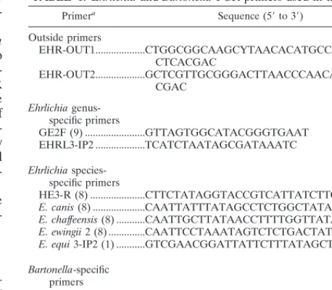
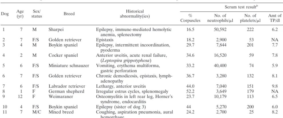
The signalments, historical abnormalities, and selected pre-treatment clinicopathologic results for 12 dogs diagnosed with canine ehrlichiosis are summarized in Table 2. Other pertinent clinical abnormalities included proteinuria in dogs 1 and 11 (urine protein/creatinine ratios, 8.7 and 5.2, respectively). Lep-TABLE 1. Ehrlichia and Bartonella PCR primers used in this study
Primera Sequence (59to 39)
Outside primers
EHR-OUT1...CTGGCGGCAAGCYTAACACATGCCAACAT CTCACGAC
EHR-OUT2...GCTCGTTGCGGGACTTAACCCAACATCTCA CGAC
Ehrlichia
genus-specific primers
GE2F (9) ...GTTAGTGGCATACGGGTGAAT EHRL3-IP2 ...TCATCTAATAGCGATAAATC
Ehrlichia
species-specific primers
HE3-R (8) ...CTTCTATAGGTACCGTCATTATCTTCCCTAT
E. canis (8) ...CAATTATTTATAGCCTCTGGCTATAGGAA E. chaffeensis (8) ...CAATTGCTTATAACCTTTTGGTTATAAATA E. ewingii 2 (8) ...CAATTCCTAAATAGTCTCTGACTATT E. equi 3-IP2 (1) ...GTCGAACGGATTATTCTTTATAGCTTG Bartonella-specific
primers
Bh16SF (3) ...AGAGTTTGATCCTGGCTCAG Bh16SR (BH1) (3) ...CCGATAAATCTTTCTCCCTAA
aReferences for the primers are given in parentheses. Primers without
listed references were established in this study (EHR-OUT1, EHR-OUT2, and EHRL3-IP2). The sizes of the products produced were 152 bp with the Ehrlichia genus-specific primers,;395 bp with the Ehrlichia species-specific primers, and 185 bp with the Bartonella-specific primers.
on May 15, 2020 by guest
http://jcm.asm.org/
[image:2.612.306.549.80.292.2]tospirosis with acute renal failure was diagnosed concurrently for dog 4. Immune-mediated hemolytic anemia in conjunction with a nonregenerative anemia (0.2% reticulocyte count) re-quiring long-term immunosuppressive drug therapy was diag-nosed for dog 5, 6 months following doxycycline treatment. Canine ehrlichiosis was diagnosed for dog 5 by the referring veterinarian approximately 1 month prior to enrollment in the study, during which time thrombocytopenia persisted despite tetracycline treatment. Dog 12 had 8,030 atypical lympho-cytes/ml and 584 immature cells of undetermined origin/ml in its peripheral blood. Lymphocyte subset analysis by flow cy-tometry identified few circulating B cells (2%) and an inversion of the CD4/CD8 ratio (ratio, 0.2; 12% CD4 cells, 60% CD8 cells). Five of 12 dogs had reciprocal IFA antibody titers of 32 or greater to B. vinsonii subsp. berkhoffii at the time of entry into the study (Table 3). Bartonella species DNA was amplified from EDTA blood samples of 7 dogs (1, 4, 7, 8, 10–12).
Prior to treatment, 9 of 12 dogs were thrombocytopenic
(platelet count,,200,000/ml) and 3 dogs had normal platelet numbers (Table 2). Seroreactivity to E. canis antigens was documented for 12 of 12 dogs (Table 3). Ehrlichiemia was documented for 4 of 10 dogs by tissue culture isolation and for 7 of 9 dogs by PCR amplification (Table 4). Because the diagnosis of ehrlichiosis was not initially suspected, pretreat-ment EDTA blood was not available for tissue culture isolation for two dogs or PCR analysis for three dogs. Cellular super-natants collected from tissue culture attempts deemed to be positive by light microscopy methods were later confirmed positive by PCR for two of four cases (dogs 2 and 3).
[image:3.612.52.547.82.288.2]Following treatment, lack of owner compliance resulted in some variation in the timing of sample collection. In addition, five dogs were not available for the entire 12-month evaluation period. Dog 9 died, dog 12 was transported to Europe, the owner of dog 8 declined blood sampling when the dog be-came pregnant, and the owners of dog 7 declined the 12-month evaluation since the dog was healthy. After treatment with TABLE 2. Signalments and historical abnormalities of and selected hematologic and biochemical values for 12 dogs with ehrlichiosis
Dog Age(yr) statusSex/ Breed abnormality(ies)Historical
Serum test resultb
%
Corpuscles neutrophils/No. of ml platelets/No. ofml Amt ofTP/dl Amt ofAlb/dl
1 7 M Sharpei Epilepsy, immune-mediated hemolytic
anemia, splenectomy 16.5 50,592 222 6.2 2.1 2 7 F/S Golden retriever Epistaxis 18.2 2,900 53 NA NA 3 4 M Boykin spaniel Epilepsy, intermittent incoordination,
pyoderma 29.7 7,844 201 7.7 2.6
4 2 M Cocker spaniel Anterior uveitis, acute renal failure,
(Leptospira grippotyphosa) 34.6 16,520 59 7.8 2.3 5 6 F/S Miniature schnauzer Vomiting, erythema multiforma,
gastric perforation 33.2 40,400 74 5.9 2.3 6 7 F/S Golden retriever Chronic demodicosis, epistaxis,
lymph-adenopathy 36.7 3,280 132 8.1 2.7
7 6 F/S Labrador retriever Lethargy, anterior uveitis 44.0 7,040 151 9.8 2.2 8 1 F German shepherd Irregular estrus cycles, splenomegaly 52.2 3,649 179 NA NA 9 12 F Weimaraner Osteomyelitis in left rear leg, Horner’s
syndrome, endocarditis 23.7 10,179 113 6.5 2.0 10 4 F/S Boykin spaniel Epilepsy (sister of dog 3) 44 5,270 200 6.0 2.7 11 7 M/C Mixed breed Coughing, aspiration pneumonia, aural
hemorrhage 24.2 2,700 25 8.2 2.1
12 5 M/C Mixed breed Splenomegaly on routine exam 37.5 4,526 87 9.6 2.4
aM, male; F, female; S, spayed; C, castrated.
bLaboratory reference ranges were as follows: 33 to 56% corpuscles, 3,000 to 11,500 neutrophils perml, 2003103to 4503103platelets perml, 4.6 to 8.2 g of total
serum protein (TP) per dl, and 2.8 to 4.4 g of serum albumin (Alb) per dl. NA, not available.
TABLE 3. Comparative seroreactivities to three E. canis isolates, E. chaffeensis, E. ristici, E. equi, and B. vinsoniia
Dog Reciprocal IFA antibody titer to:
E. canis (Florida) E. canis (DJ) E. canis (Jake) E. chaffeensis E. ristici E. equi B. vinsonii
1 .10,240 .10,240 640 1,280 ,20 20 64
2 .10,240 5,120 1,280 2,560 ,20 ,20 32
3 2,560 2,560 1,280 2,560 ,20 20 ,16
4 5,120 2,560 640 1,280 ,20 ,20 1,024
5 5,120 1,280 640 80 ,20 ,20 ,16
6 .10,240 1,280 160 .10,240 ,20 40 1,024
7 1,280 640 320 1,280 ,20 320 128
8 160 ,20 ,20 ,20 ,20 ,20 ,16
9 1,280 1,280 20 ,20 ,20 ,20 ,16
10 320 640 160 160 ,20 20 ,16
11 5,120 2,560 160 1,280 ,20 40 ,16
12 .10,240 .10,240 1,280 5,120 640 80 ,16
aBased upon DNA amplification by PCR with Ehrlichia species-specific primers, dogs 1 to 3 and 11 were determined to be infected with E. canis and dogs 4 to 6
were determined to be infected with E. chaffeensis. Based on DNA sequencing, dog 7 was determined to be infected with E. platys. The infecting Ehrlichia species were not identified for dogs 8 to 10 and 12.
on May 15, 2020 by guest
http://jcm.asm.org/
[image:3.612.51.548.555.700.2]TABLE 4. Summary of pre- and posttreatment platelet counts, reciprocal IFA titers, and tissue culture isolation and PCR amplification results
Dog Parameter Pretreatmenttest resulta
Posttreatment test resultaat mo:
Outcome
1 2 3 6 8 12
1 Platelet count 222 449 290 Healthy, chronic pyoderma Reciprocal IFA titer .10,240 .10,240 .10,240
Tissue culture isolation 1 2
PCR amplification 1/1 1/1 2/2
Species isolated Ec Ec, Eew, Eeq
2 Platelet count 53 556 539 255 Healthy
Reciprocal IFA titer .10,240 1,280 640 640
Tissue culture isolation 1 2 2 2
PCR amplification 1/1 2/2 2/2 2/2 Species isolated Ec
3 Platelet count 201 244 210 230 Healthy, epilepsy Reciprocal IFA titer 5,120 2,560 1,280 2,560
Tissue culture isolation 1 2 2 2
PCR amplification 1/1 2/2 2/2 2/2 Species isolated Ec
4 Platelet count 59 450 NA 316 Healthy
Reciprocal IFA titer 5,120 640 320 NA
Tissue culture isolation 2 2 2 NA
PCR amplification 1/1 1/1 1/1 NA Species isolated Ech Ech Ech
5 Platelet count 74 NA 125 416 Immunosuppression from IMHAb
Reciprocal IFA titer 5,120 2,560 1,280 320
Tissue culture isolation NA 2 2 2
PCR amplification NA 1/1 1/1 1/1
Species isolated Ech Ech Ech
6 Platelet count 132 244 233 NA Healthy
Reciprocal IFA titer .10,240 ND .10,240 .10,240
Tissue culture isolation 2 2 2 2
PCR amplification 1/1 1/1 2/2 1/1
Species isolated Ech Ech Ech
7 Platelet count 151 204 121 Healthy, owners declined sampling at 12 mo Reciprocal IFA titer 10,240 2,560 2,560
Tissue culture isolation 2 2 2 PCR amplification 1/2 2/2 1/2 Species isolated Ep
8 Platelet count 179 Adequate 319 Healthy at 6 mo, unavailable for follow-up (pregnant) Reciprocal IFA titer 160 20 20
Tissue culture isolation 2 2 2 PCR amplification 2/2 2/2 2/2
9 Platelet count 113 495 553 Died 10 mo posttreatment, congestive heart failure Reciprocal IFA titer 1,280 160 160
Tissue culture isolation 1 2
PCR amplification NA 2/2 2/2
10 Platelet count 200 Clumping 198 246 Healthy, epilepsy Reciprocal IFA titer 640 640 1,280 320
Tissue culture isolation 2 2 2 2
PCR amplification 2/2 2/2 2/2 2/2
11 Platelet count 25 165 453 354 Healthy
Reciprocal IFA titer 5,120 2,560 640 640
Tissue culture isolation 2 2 2 2
PCR amplification 1/1 2/2 2/2 2/2 Species isolated Ec
12 Platelet count 87 112 Clumping Healthy, owners moved to Europe 9 mo posttreatment Reciprocal IFA titer .10,240 1,280 1,280
Tissue culture isolation NA 1 2
PCR amplification NA 2/2 2/2
aPlatelet counts (103) and reciprocal IFA titers are per microliter. “Adequate” and “clumping” indicate, respectively, that platelets were of adequate numbers for
a healthy animal and that platelets clumped, making a determination of number difficult. For tissue culture isolation results, a1or2indicates that organisms were isolated or not, respectively. For PCR amplification test results, a1or2to the left of the shill indicates that Ehrlichia genus-specific DNA was or was not detected, respectively, and a1or2to the right of the shill indicates that Ehrlichia species-specific DNA was or was not detected, respectively. NA indicates that a sample was not available for testing. Species abbreviations: Ec, E. canis; Ech, E. chaffeensis; Eew, E. ewingii; Eeq, E. equi; Ep, E. platys. The species of each infecting organism was determined by PCR amplification with Ehrlichia species-specific primers for dogs 1 to 6 and 11; that of dog 7 was determined by DNA sequencing. The infecting
Ehrlichia species were not identified for dogs 8 to 10 and 12.
bIMHA, immunity-mediated hemolytic anemia.
on May 15, 2020 by guest
http://jcm.asm.org/
doxycycline, clinical abnormalities, potentially attributable to ehrlichiosis, resolved in all but dog 4, which remained PCR positive for E. chaffeensis DNA and had the highest antibody titer to B. vinsonii detected among the dogs in this study (Table 3). Posttreatment, Ehrlichia species were isolated by tissue culture only from dog 12, blood from which was PCR negative. Thrombocytopenia resolved in dogs 1 to 6 and 8 to 11, but resolution was not observed for dog 7 or 12, both of which were monitored for less than a year (Table 4). Serum IFA antibody titers decreased by more than fourfold in all dogs except dogs 1 (E. canis, E. equi, and E. ewingii infected) and 6 (E.
chaffeen-sis infected), in which stable antibody titers perchaffeen-sisted in
con-junction with positive PCR results at 6 and 12 months, respec-tively (Table 4).
With Ehrlichia genus-specific primers, amplification prod-ucts were obtained from 5 of 12 dogs (1, 4–7) at one or more times between 6 and 12 months during the posttreatment fol-low-up period (Table 4). Based upon species-specific PCR primers, dog 1 was infected with E. canis, E. equi, and E.
ew-ingii; dogs 2, 3, and 11 were infected with E. canis; and dogs 4
to 6 were infected with E. chaffeensis. With the exception of dog 7 (6-month-posttreatment sample), ehrlichial speciation was successful for each sample in which an Ehrlichia genus amplicon was obtained. In all instances, species-specific prim-ers provided reproducible results, as was best illustrated by dogs 4 to 6, from which only E. chaffeensis DNA was amplified at each datum collection point during the 12-month period of study. As the species-specific primers used in this study did not generate an amplicon, the ehrlichial-genus-positive PCR prod-uct for dog 7 was further characterized by sequencing in the laboratory of D. H. Walker, Galveston, Tex. The species was identified as Ehrlichia platys, which our primers were not ex-pected to amplify. Ehrlichia DNA could not be amplified from the available samples for four dogs (Table 4). Based upon continued amplification of ehrlichial DNA posttreatment, all three E. chaffeensis-infected dogs (Fig. 1) appeared to remain infected or were reinfected following treatment. These dogs were generally thrombocytopenic, or their platelet counts re-mained in the low reference range. The PCR results from dog 1 are notable, as only E. canis was amplified from the pretreat-ment blood sample, whereas E. canis, E. equi, and E. ewingii were amplified from two separate blood samples obtained 6 months posttreatment, approximately 1 week apart. The sec-ond of these samples was obtained because of bacterial con-tamination of the initial sample obtained for ehrlichial culture. Presumably, these results reflect continued tick exposure.
When pretreatment serum samples from these dogs were
tested against antigens derived from E. canis Florida (type strain) and two NCSU E. canis isolates (DJ and Jake) or against E. chaffeensis (type strain), the seroreactive antibody titers (Table 3) did not generally facilitate differentiation of the infecting Ehrlichia species when they were compared to the PCR results (Table 4). In addition, in some instances there was substantial variation in the IFA antibody titers among the three E. canis strains used as antigens. Except with dog 12, antibodies to E. risticii were not detected. Similarly, Western immunoblot analysis with E. canis antigens did not differenti-ate the infecting Ehrlichia species. When results were com-pared across time (periods up to 13 months), there was mini-mal or no change in the Western blot patterns regardless of the dog’s infection status, as defined by platelet count or PCR result.
DISCUSSION
Historically, infection with Ehrlichia species has generally been considered to be host specific. For example, E. canis was thought to infect only dogs and wild carnivores and E.
chaffeen-sis was thought to infect only deer and human beings. Recently,
an isolate genetically and antigenically similar to E. canis was obtained from a veterinarian in Venezuela (31). Similarly, iso-lates genetically identical to E. risticii, the cause of Potomac horse fever, have been obtained from dogs (21). Recent evi-dence indicates that a member of the E. phagocytophila group, presumably E. equi, causes disease manifestations in cats, dogs, horses, and human beings (15). In this study, E. chaffeensis, originally isolated and characterized as a cause of human dis-ease (7), was found to cause disdis-ease in dogs. Collectively, these observations suggest that several Ehrlichia species can be transmitted to a variety of hosts in nature. Therefore, addi-tional efforts to define the spectrum of host susceptibility in domestic and wild animals seem appropriate.
Previously, based upon PCR amplification and DNA se-quence analysis, we documented severe clinical and hemato-logic abnormalities due to E. ewingii infection in dogs with gran-ulocytic ehrlichiosis from North Carolina and Virginia (14). The present study indicates that E. chaffeensis, as well as E.
ca-nis, E. equi, E. ewingii, and E. platys, can cause disease
mani-festations and clinicopathologic abnormalities in dogs orig-inating from the same geographic region. Importantly, these results provide the first evidence for development of disease manifestations in dogs naturally infected with E. chaffeensis. In concert with the results derived by Dawson and Ewing (9), dogs seroreactive to E. canis antigens from this region may be infected with E. canis, E. chaffeensis, or E. ewingii. Infection with any one of these three species can cause severe disease manifestations that may be clinically, hematologically, and se-rologically indistinguishable from those of the other two spe-cies. The relative contribution of coinfection with three
Ehrli-chia species to the disease manifestations in dog 1 awaits
additional studies of coinfected dogs. Collectively, these results indicate that increased utilization of molecularly based diag-nostic modalities should enhance our understanding of poten-tially important clinical or pathologic differences associated with infection with a single Ehrlichia species or coinfection with multiple Ehrlichia species.
Serologic testing by an IFA assay was not able to consistent-ly distinguish between infection with E. canis and that with
E. chaffeensis. Similarly, when E. canis antigen was used,
[image:5.612.51.291.69.208.2]West-ern immunoblot analysis of sera from these dogs did not result in antigenic protein recognition that would facilitate diagnostic differentiation of the infecting species. Since E. ewingii has not been cultivated in an in vitro culture system, antigen from this
FIG. 1. PCR amplicons obtained with E. chaffeensis primers from sequential EDTA blood samples from dogs 4 to 6. Lane2, negative control blood sample; lane1, positive control (cultured E. chaffeensis); lane M, molecular size markers.
on May 15, 2020 by guest
http://jcm.asm.org/
organism was not available for comparative serologic testing. Although coinfection was not examined in this study, it is probable that coinfection with more than one Ehrlichia species further limits the utility of serologic testing for differentiation of the infecting Ehrlichia species. Recently, we have observed numerous examples of coinfection with E. canis, E. chaffeensis, and/or E. ewingii in a kennel of heavily tick-infested dogs (un-published data).
Since readily discernible differences in IFA or Western im-munoblot seroreactivity patterns to E. canis antigens do not appear to differentiate between the infecting species, molecu-lar detection and speciation of ehrlichial DNA is necessary to determine if predictable differences in therapeutic outcomes can be further correlated with an infecting Ehrlichia species. Several factors, including anticipated duration of infection, therapeutic responsiveness (particularly to tetracycline deriva-tives), and zoonotic potential, emphasize the importance of determining which Ehrlichia species is causing infection and antibody reactivity to E. canis antigen in a dog. For example,
E. canis causes chronic, frequently subclinical infection with
the potential for the development of severe life-threatening disease manifestations (6, 14, 24) whereas E. ewingii is consid-ered to cause polyarthritis and potentially self-limiting infec-tion (35). Ehrlichia canis and E. chaffeensis may not be elimi-nated by doxycycline therapy, whereas therapeutic elimination of E. ewingii or E. equi is an expected outcome.
PCR amplification of ehrlichial DNA is gaining acceptance as an important adjunct to serologic testing for the diagnosis of canine ehrlichiosis (18, 38). In this study, to increase the sen-sitivity of the PCR assays, two nested techniques were used in testing, first, to determine the presence of Ehrlichia DNA (genus-specific primers) and, second, to differentiate among the various Ehrlichia species. To reduce the risk of contami-nation, historically associated with nested PCR, a single-tube procedure was developed for both assays by designing the out-er primout-ers with an annealing tempout-erature substantially highout-er than that of the internal primers. The internal primers used for
Ehrlichia species differentiation were modified slightly from
the primers described by Dawson et al. (8) and Barlough et al. (1) to reduce annealing temperatures for the nested protocol. Based on the consistent amplification of only E. chaffeensis DNA at multiple time points from dogs 4 to 6 and consistently negative PCR results from dogs 8 to 10, we believe that PCR contamination and nonspecific priming are unlikely explana-tions for PCR evidence of coinfection in dog 1. When they were tested with blood samples spiked with DNAs of known ehrlichial species, these primer pairs did not amplify nonspe-cific DNA. Since E. equi is not considered to be endemic to this region, DNA was extracted from two samples on two different occasions from the dog coinfected with E. equi. Identical PCR amplicons were obtained. Since amplicons were obtained from only seven of nine pretreatment EDTA blood samples, efforts to enhance the sensitivity of detection may be warranted.
Problems recognized in dogs with increasing frequency by veterinarians are the persistence of clinical and/or hematologic abnormalities, the persistence of antibody reactivity to E. canis antigen (2), and the persistence of Ehrlichia DNA as detected by PCR following antirickettsial drug therapy (38). Documen-tation of one or more of these factors has caused some veter-inarians to treat canine ehrlichiosis with tetracycline hydro-chloride or doxycycline hydrohydro-chloride for extended periods (months to years), a less-than-optimal situation, potentially fa-cilitating the development of drug-resistant bacteria. In other instances, alternative treatment modalities such as the admin-istration of imidocarb diproprionate (33) have been used in an effort to obtain a satisfactory therapeutic response. Although
the cause of these treatment failures is most probably multi-factorial, it is of interest that all three dogs infected with
E. chaffeensis in this study remained PCR positive after
treat-ment with doxycycline at a dose and duration generally con-sidered to be efficacious for treatment of E. canis infection. Following experimental infection with E. canis, other investi-gators found that three of five dogs treated with doxycycline for only 7 days failed to clear their infection (19). However, in a study of similar design from our laboratory, using experimen-tally infected mixed-breed dogs treated for 14 days, eight of eight dogs became culture and PCR negative following treat-ment (4). In the present study, all four dogs naturally infected with E. canis appeared to clear their infection, although dog 1 remained E. canis PCR positive at 6 months following the initial treatment, which may have reflected reinfection associ-ated with continued tick exposure. In a recent study, 43 of 80
E. canis posttreatment blood samples, obtained from E. canis
seroreactive dogs from Arizona and Texas, were PCR positive (38). Collectively, these results suggest that the role of drug-resistant strains of E. canis, as well as the efficacy of doxycy-cline for the treatment of E. chaffeensis infection in naturally infected dogs, deserves additional consideration. Due to the lack of sensitivity of tissue culture isolation, the variability in posttreatment IFA antibody responses, and the possibility of DNA persistence, unassociated with viable organisms, other modalities are needed to prove therapeutic elimination of in-fection.
Although this study involved a small number of dogs,
Bar-tonella DNA was amplified from 58% of the dogs and 42%
were reactive to B. vinsonii antigen, with the two highest anti-body titers being found in dogs infected with E. chaffeensis. Since Amblyomma americanum ticks have been implicated in the transmission of E. chaffeensis, it is possible that this tick may cotransmit B. vinsonii. Since simultaneous infestation with more than one tick species is not unusual in dogs, controlled studies will be necessary to clarify the role of ticks in the transmission of Bartonella species. It is of interest that ep-istaxis, a well-recognized clinical manifestation of ehrlichiosis, has been reported in association with Bartonella henselae and
Bartonella quintana infection in humans (10, 13) and B. vinsonii
infection in a dog (5). The extent to which concurrent infection with B. vinsonii complicates the clinical course of ehrlichiosis in dogs deserves additional study.
ACKNOWLEDGMENTS
This research was supported by the State of North Carolina and through a grant from Fort Dodge Laboratories, Fort Dodge, Iowa.
We acknowledge the efforts of the house staff of the Veterinary Teaching Hospital, NCSU, for the clinical management of the patients and A. N. Billings, The University of Texas Medical Branch, Galves-ton, Texas, for performing the DNA sequencing.
REFERENCES
1. Barlough, J. E., J. E. Madigan, E. DeRock, and L. Bigornia. 1996. Nested polymerase chain reaction for detection of Ehrlichia equi genomic DNA in horses and ticks (Ixodes pacificus). Vet. Parasitol. 63:319–329.
2. Bartsch, R. C., and R. T. Greene. 1996. Post-therapy antibody titers in dogs with ehrlichiosis: follow-up study on 68 patients treated primarily with tet-racycline and/or doxycycline. J. Vet. Intern. Med. 10:271–274.
3. Bergmans, A. M., J. F. P. Schellekens, J. D. A. van Embden, and L. M. Schouls. 1996. Predominance of two Bartonella henselae variants among cat-scratch disease patients in The Netherlands. J. Clin. Microbiol. 34:254– 260.
4. Breitschwerdt, E. B., B. C. Hegarty, and S. I. Hancock. 1998. Doxycycline hyclate treatment of experimental canine ehrlichiosis followed by challenge inoculation using two Ehrlichia canis strains. Antimicrob. Agents Chemo-ther. 42:362–368.
5. Breitschwerdt, E. B., D. L. Kordick, D. E. Malarkey, B. Keene, T. L. Had-field, and K. Wilson.1995. Endocarditis in a dog due to infection with a
on May 15, 2020 by guest
http://jcm.asm.org/
novel Bartonella subspecies. J. Clin. Microbiol. 33:154–160.
6. Codner, E. C., and L. L. Farris-Smith. 1986. Characterization of the sub-clinical phase of ehrlichiosis in dogs. J. Am. Vet. Med. Assoc. 189:47–50. 7. Dawson, J. E., B. E. Anderson, J. L. Fishbein, C. S. Sanchez, C. S.
Gold-smith, K. H. Wilson, and C. W. Duntley.1991. Isolation and characterization of an Ehrlichia species from a patient diagnosed with human ehrlichiosis. J. Clin. Microbiol. 29:2741–2745.
8. Dawson, J. E., K. L. Biggie, C. K. Warner, K. Cookson, S. Jenkins, J. Levine, and J. G. Olson.1996. Polymerase chain reaction evidence of Ehrlichia
chaffeensis, an etiologic agent of human ehrlichiosis, in dogs from southeast
Virginia. Am. J. Vet. Res. 57:1175–1179.
9. Dawson, J. E., and S. A. Ewing. 1992. Susceptibility of dogs to infection with
Ehrlichia chaffeensis, causative agent of human ehrlichiosis. Am. J. Vet. Res.
53:1322–1327.
10. Drancourt, M., J. L. Mainardi, P. Brouqui, F. Bandenesch, A. Carta, F. Lehnert, J. Etienne, F. Goldstein, J. Acar, and D. Raoult.1995.
Bar-tonella (Rochalimaea) quintana endocarditis in three homeless men. N. Engl.
J. Med. 332:419–423.
11. Dumler, J. S., S. M. Chen, K. Asanovich, E. Trigiani, V. L. Popov, and D. H. Walker. 1995. Isolation and characterization of a new strain of Ehrlichia
chaffeensis from a patient with nearly fatal monocytic ehrlichiosis. J. Clin.
Microbiol. 33:1704–1711.
12. Gaunt, S. D., R. E. Corsetvet, C. M. Berry, and B. Brennan. 1996. Isolation of Ehrlichia canis from dogs following subcutaneous inoculation. J. Clin. Microbiol. 34:1429–1432.
13. Golden, S. E. 1993. Hepatosplenic cat-scratch disease associated with ele-vated anti-Rochalimaea antibody titers. Pediatr. Infect. Dis. J. 12:868–871. 14. Goldman, E. E., E. B. Breitschwerdt, C. B. Grindem, B. C. Hegarty, J. J.
Walls, and J. S. Dumler.1998. Granulocytic ehrlichiosis in dogs from North Carolina and Virginia. J. Vet. Intern. Med. 12:61–70.
15. Greig, B., K. M. Asanovich, P. J. Armstrong, and J. S. Dumler. 1996. Geographic, clinical, serologic and molecular evidence of granulocytic ehr-lichiosis, a likely zoonotic disease in Minnesota and Wisconsin dogs. J. Clin. Microbiol. 34:44–48.
16. Hegarty, B. C., M. G. Levy, R. F. Gager, and E. B. Breitschwerdt. 1997. Immunoblot analysis of the immunoglobulin G response to Ehrlichia canis in dogs: an international survey. J. Vet. Diagn. Invest. 9:32–38.
17. Iqbal, Z., and Y. Rikihisa. 1994. Reisolation of Ehrlichia canis from blood and tissues of dogs after doxycycline treatment. J. Clin. Microbiol. 32:1644– 1649.
18. Iqbal, Z., W. Chaichanasiriwithaya, and Y. Rikihisa. 1994. Comparison of PCR with other tests for early diagnosis of canine ehrlichiosis. J. Clin. Microbiol. 32:1658–1662.
19. Iqbal, Z., and Y. Rikihisa. 1994. Application of the polymerase chain reac-tion for the detecreac-tion of Ehrlichia canis in tissues of dogs. Vet. Microbiol. 42: 281–287.
20. Johansson, K. E., B. Pettersson, M. Uhlen, A. Gunnarsson, M. Malmqvist, and E. Olsson.1995. Identification of the causative agent of granulocytic ehrlichiosis in Swedish dogs and horses by direct solid phase sequencing of PCR products from 16S rRNA gene. Res. Vet. Sci. 58:109–112.
21. Kakoma, I., R. D. Hansen, B. E. Anderson, T. A. Hanley, K. G. Sims, L. Liu, C. Bellamy, M. T. Long, and B. K. Baek.1994. Cultural, molecular, and immunological characterization of the etiologic agent for atypical canine ehrlichiosis. J. Clin. Microbiol. 32:170–175.
22. Kelly, P. J., L. A. Matthewman, S. M. Mahan, S. M. Semu, T. Peter, P. R. Mason, P. R. Brouqui, and D. Raoult.1994. Serologic evidence for antigenic
relationships between Ehrlichia canis and Cowdria ruminantium. Res. Vet. Sci. 56:170–174.
23. Kordick, D. L., B. Swaminathan, C. T. Greene, K. H. Wilson, S. O’Connor, D. G. Hollis, G. M. Matar, B. G. Malcom, P. S. Hayes, T. L. Hadfield, E. B. Breitschwerdt, and D. J. Brenner.1996. Bartonella vinsonii subsp. berkhoffii subsp. nov., isolated from dogs; Bartonella vinsonii subsp. vinsonii; and emend-ed description of Bartonella vinsonii. Int. J. Syst. Bacteriol. 46:704–709. 24. Kuehn, N. F., and S. D. Gaunt. 1985. Clinical and hematologic findings in
canine ehrlichiosis. J. Am. Vet. Med. Assoc. 186:355–358.
25. Levy, M. G., D. Gebhard, R. Gager, and E. B. Breitschwerdt. 1995. A newly described canine monocytoid cell line capable of supporting growth of the rickettsial parasite Ehrlichia canis, and useful for examination of host:para-site interactions, p. 274. In Proceedings of the International Union Immu-nology Society, 4th International Veterinary ImmuImmu-nology Symposium. 26. Lewis, G. E., D. L. Huxsoll, M. Ristic, and A. J. Johnson. 1975.
Experimen-tally induced infection of dogs, cats, and nonhuman primates with Ehrlichia
equi, etiologic agent of equine ehrlichiosis. Am. J. Vet. Res. 36:85–88.
27. Madewell, B. R., and D. H. Gribble. 1982. Infection in two dogs with an agent resembling Ehrlichia equi. J. Am. Vet. Med. Assoc. 180:512–514. 28. Matthewman, L. A., P. J. Kelly, S. M. Mahan, S. M. Semu, P. R. Mason, D.
Bruce, P. Brouqui, and D. Raoult.1994. Reactivity of sera collected from dogs in Mutare, Zimbabwe, to antigens of Ehrlichia canis and Cowdria
ruminantium. Vet. Rec. 134:498–499.
29. Paddock, C. D., J. W. Sumner, G. M. Shore, D. C. Bartley, R. C. Elie, J. G. Mcquade, C. R. Martin, C. S. Goldsmith, and J. E. Childs.1997. Isolation and characterization of Ehrlichia chaffeensis strains from patients with fatal ehrlichiosis. J. Clin. Microbiol. 35:2496–2502.
30. Pappalardo, B. L., M. T. Correa, C. C. York, C. Y. Peat, and E. B. Breit-schwerdt.1997. Epidemiologic evaluation of the risk factors associated with exposure and seroreactivity to Bartonella vinsonii in dogs. Am. J. Vet. Res. 58:467–471.
31. Perez, M., Y. Rikihisa, and B. Wen. 1996. Ehrlichia canis-like agent isolated from a man in Venezuela: antigenic and genetic characterization. J. Clin. Microbiol. 34:2133–2139.
32. Petrovec, M., S. L. Furlan, T. A. Zupanc, F. Strle, P. Brouqui, V. Roux, and J. S. Dumler.1997. Human disease in Europe caused by a granulocytic
Ehrlichia species. J. Clin. Microbiol. 35:1556–1559.
33. Price, J. E., and T. T. Dolan. 1980. A comparison of the efficacy of imidocarb dipropionate and tetracycline hydrochloride in the treatment of canine ehr-lichiosis. Vet. Rec. 107:275–277.
34. Stockham, S. L., J. W. Tyler, D. A. Schmidt, and K. S. Curtis. 1990. Exper-imental transmission of granulocytic ehrlichial organisms in dogs. Vet. Clin. Pathol. 19:99–104.
35. Stockham, S. L., D. A. Schmidt, K. S. Curtis, B. G. Schauf, J. W. Tyler, and S. T. Simpson.1992. Evaluation of granulocytic ehrlichiosis in dogs of Mis-souri including serologic status to Ehrlichia canis, Ehrlichia equi, and Borrelia
burgdorferi. Am. J. Vet. Res. 53:63–68.
36. Walker, D. H., and J. S. Dumler. 1996. Emergence of the ehrlichioses as human health problems. Emerg. Infect. Dis. 2:18–29.
37. Walls, J. J., B. Greig, D. F. Neitzel, and J. S. Dumler. 1997. Natural infection of small mammal species in Minnesota with the agent of human granulocytic ehrlichiosis. J. Clin. Microbiol. 35:853–855.
38. Wen, B., Y. Rikihisa, J. M. Mott, R. Greene, H.-Y. Kim, N. Zhi, G. C. Couto, A. Unver, and R. Bartsch.1997. Comparison of nested PCR with immun-ofluorescent-antibody assay for detection of Ehrlichia canis infection in dogs treated with doxycycline. J. Clin. Microbiol. 35:1852–1855.