N A N O E X P R E S S
Open Access
Microstructure and optical properties of
Pr
3+
-doped hafnium silicate films
YongTao An, Christophe Labbé
*, Larysa Khomenkova, Magali Morales, Xavier Portier and Fabrice Gourbilleau
Abstract
In this study, we report on the evolution of the microstructure and photoluminescence properties of Pr3+-doped hafnium silicate thin films as a function of annealing temperature (TA). The composition and microstructure of the films were characterized by means of Rutherford backscattering spectrometry, spectroscopic ellipsometry, Fourier transform infrared absorption, and X-ray diffraction, while the emission properties have been studied by means of photoluminescence (PL) and PL excitation (PLE) spectroscopies. It was observed that a post-annealing treatment favors the phase separation in hafnium silicate matrix being more evident at 950°C. The HfO2phase demonstrates a pronounced crystallization in tetragonal phase upon 950°C annealing. Pr3+emission appeared atTA= 950°C, and the highest efficiency of Pr3+ion emission was detected upon a thermal treatment at 1,000°C. Analysis of the PLE spectra reveals an efficient energy transfer from matrix defects towards Pr3+ions. It is considered that oxygen vacancies act as effective Pr3+sensitizer. Finally, a PL study of undoped HfO2and HfSiOxmatrices is performed to evidence the energy transfer.
Keywords:Praseodymium, Hafnium silicate, Oxygen vacancies, Photoluminescence, Energy transfer
Background
Rare-earth elements are important optical activators for luminescent devices. Among various rare-earth lumines-cent lumines-centers, trivalent praseodymium (Pr3+) offers simul-taneously a strong emission in the blue, green, orange, and red spectral range, satisfying the complementary color relationship [1,2]. Consequently, Pr3+-doped glass/ crystals are often used as phosphor materials [2,3]. SiO2
-(Ca, Zn)TiO3:Pr3+ phosphors prepared with nanosized
silica particles exhibit an intense red photoluminescence (PL) [3]. The Pr3+ emission was achieved for Si-rich SiO2(SRSO) implanted with Pr3+ ions, but its intensity
was lower [4].
Hafnium dioxide (HfO2) and hafnium silicates (HfSiOx)
are currently considered as the predominant high-k dielec-tric candidates to replace the conventional SiO2due to the
rapid downscaling of the complementary metal-oxide semiconductor (CMOS) transistors [5,6]. It is ascribable to their good thermal stability in contact with Si, large elec-tronic bandgaps, reasonable conduction band offset in re-gard to Si, and their compatibility with the current CMOS
technology. Our group has first explored the structural and thermal stability of HfO2-based layers fabricated by
radio frequency (RF) magnetron sputtering [7,8] and their nonvolatile memory application [9,10].
It is worth to note that both HfO2and HfSiOx
matri-ces have lower phonon frequencies compared to those of SiO2, and as a consequence, both are expected to be
suitable hosts for rare-earth activators. Thus, PL proper-ties have been investigated for the HfO2 matrix doped
with Tb3+ [11], Eu3+ [11,12], or Er3+ [12,13] and have been explained by the interaction of rare-earth ions with host defects. Recently, our group has demonstrated that an enhancement of Er3+PL emission can be achieved for the Er-doped HfSiOxmatrix in comparison with that of
the Er-doped HfO2[14]. It was also observed that an
en-ergy transfer from the HfO2 host defects towards Er3+
ions, whereas the existence of Si clusters allowed an en-hancement of the Er3+ ion emission under longer-wavelength excitation. Consequently, the mechanism of the excitation process, when Si clusters and oxygen-deficient centers act as Er3+ sensitizers, has been pro-posed to explain an efficient rare-earth emission from Er-doped HfSiOxhosts [14] similar to that observed for
the Er-doped SRSO materials [15]. * Correspondence:[email protected]
CIMAP, UMR 6252 CNRS/CEA/Ensicaen/UCBN, 6 Boulevard Maréchal Juin, Caen, Cedex 4 14050, France
In this paper, we study the microstructure and optical properties of Pr-doped hafnium silicate films fabricated by magnetron sputtering versus annealing temperature. We demonstrate that an efficient Pr3+ light emission is achievable by tuning the annealing conditions. The exci-tation mechanism of Pr3+ions is also discussed.
Methods
The films were deposited onto p-type (100) 250-μm-thick Si wafers by RF magnetron sputtering of a pure HfO2 target topped by calibrated Si and Pr6O11 chips.
The growth was performed in pure argon plasma with an RF power density of 0.98 W∙cm−2; the Si substrate temperature was kept at 25°C. After deposition, a post-annealing treatment was carried out under a nitrogen flow, at temperatures (TA) varying from 800°C up to
1,100°C for 1 h.
The refractive index (n) (given always at 1.95 eV) and the film thicknesses were deduced from spectroscopic ellipsometry data. The chemical composition of the films was determined by Rutherford backscattering spectrom-etry (RBS) using a 1.5-MeV4He+ion beam with a normal incidence and a scattering angle of 165°. The infrared ab-sorption properties were investigated by means of a Nicolet Nexus (Thermo Fisher Scientific, Waltham, MA, USA) Fourier transform infrared (FTIR) spectroscopy at Brew-ster’s incidence (65°) in the range of 500 to 4,000 cm−1. X-ray diffraction (XRD) experiments were performed using a Philips Xpert MPD Pro device (PANalytical B.V., Almelo,
The Netherlands) with CuKαradiation (λ= 1.5418 Å) at a fixed grazing angle incidence of 0.5°. Cross-sectional speci-mens were prepared by standard procedure involving grinding, dimpling, and Ar+ion beam thinning until elec-tron transparency for their observation by transmission electron microscopy (TEM). The samples were observed using a FEG 2010 JEOL instrument, operated at 200 kV. The PL emission and PL excitation (PLE) measurements were carried out using a 450-W Xenon arc lamp as excita-tion source at room temperature corrected on spectral response with the help of a Jobin-Yvon Fluorolog spectrom-eter (HORIBA Jobin Yvon Inc., Edison, NJ, USA).
Results and discussion
Composition and structural characterizations
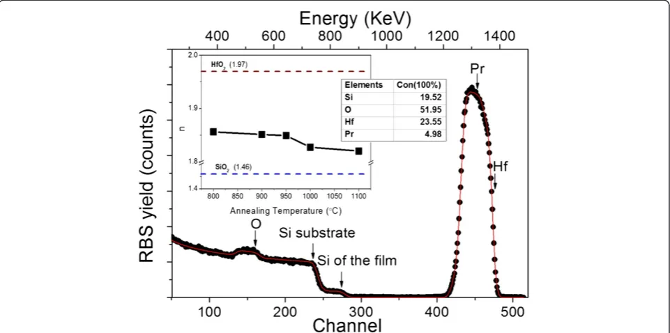
In this study, the chemical composition of the film Hf0.24Si0.20O0.52Pr0.05was determined through the
simula-tion of the corresponding RBS spectrum using the SIMNRA program (Figure 1). The RBS analysis shows that the as-deposited film cannot be considered as a matrix of SiO2 and HfO2 only, as this is usually assumed for
haf-nium silicates. In our case, we deal with a hafhaf-nium silicate matrix enriched with silicon as well as doped with Pr3+ ions.
The inset of Figure 1 displays the refractive index evo-lution upon annealing treatment between 800°C and 1,100°C. The uncertainty of the refractive index is 0.01. Nevertheless, it was notable that it decreased withTA. In
[image:2.595.58.539.454.693.2]a previous study on as-deposited film, it was found that
Figure 1Experimental RBS spectrum (points) and simulated curve using SIMNRA (solid line) for as-deposited film.Inset table is the chemical composition of the film. Inset figure is the refractive index evolution versusTA. The pure HfO2and pure SiO2indices are also shown in
the refractive index was about 2.2 [8], exceeding the value corresponding to the stoichiometric HfSiO4matrix
(1.7) due to Si enrichment [8]. However, upon annealing, the refractive index is found to be about 1.85 (TA= 800°C)
and 1.82 (TA= 1,100°C). If we exclude the decrease of
por-osity, this evolution could be explained by the increasing contribution of some phases with lower refractive index upon annealing (like SiO2(1.46)) [8].
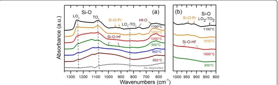
Figure 2a represents the evolution of the FTIR spectra as a function ofTA. The FTIR spectrum of as-deposited
film is represented by two broad vibration bands in the ranges of 500 to 750 and 800 to 1,200 cm−1. An anneal-ing treatment stimulates the appearance of several bands that peaked at about 827, 1,084, and 1,250 cm−1(dashed lines in Figure 2a) corresponding to the LO2-TO2, TO3,
and LO3vibration modes of the Si-O bond, respectively.
Moreover, the increase of the LO3 mode intensity is
attributed to the increase in the number of Si-O-Si bonds. This is a signature of the formation of the SiO2
phase due to a phase separation process, leading to the decrease of the refractive index forTA≥800°C.
This phase separation is confirmed also by an increase of the vibration mode intensity in the range of 600 to 780 cm−1, corresponding to Hf-O bonds for the formation of the HfO2 phase [7,14]. The appearance of well-defined
peaks at 760 and 660 cm−1 for TA ≥ 1,050°C attests the
presence of the monoclinic HfO2phase [16]. Besides, for TA≥ 1,050°C, two new absorption peaks that centered at
900 and 1,000 cm−1appeared (detailed in Figure 2b). As we showed earlier [8], at such temperature, undoped HfSiOx did not reveal the presence of Si-O-Hf bonds.
Thus, the vibration band at 900 and 1,000 cm−1 can be attributed to Si-O-Pr asymmetric mode. Similar incorpor-ation of rare-earth ions into Si-O bonds and the formincorpor-ation of rare-earth silicate phase was observed earlier for SiOx
materials doped with Er3+, Nd3+, or Pr3+and annealed at
1,100°C [17-19]. Thus, based on this comparison, one can conclude about the formation of Pr silicate revealed by FTIR spectra.
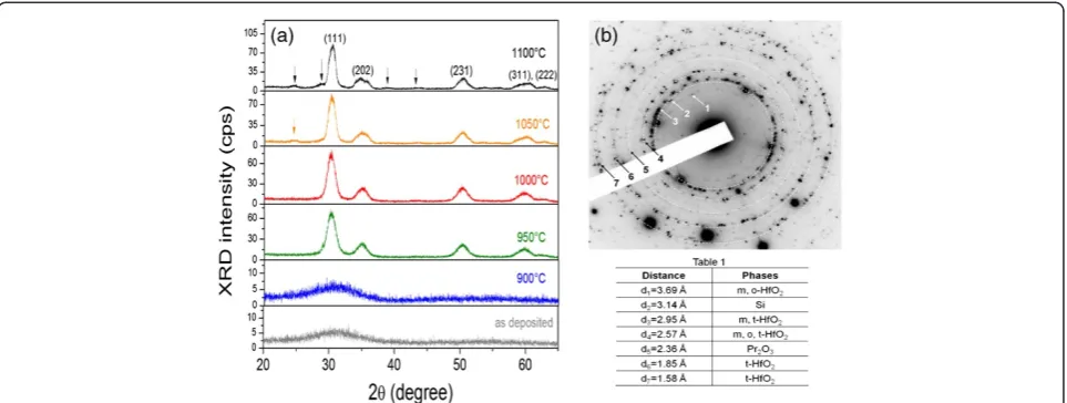
To get more information about the evolution of film structure, we performed XRD analyses. For as-deposited and 900°C annealed films, XRD spectra show a broad peak in the range of 25.0° to 35.0° with a maximum intensity located at 2θ ≈31.0° (Figure 3a). The shape of the XRD peak demonstrates the amorphous nature of both layers. WithTA increase, several defined peaks appear,
emphasizing the formation of a crystalline structure. Thus, forTA= 950°C, intense XRD peaks at 2θ≈30.3°,
35.0°, and 50.2° were detected. They correspond to the (111), (200), and (220) planes of the tetragonal HfO2
phase, respectively, confirming the FTIR analysis [8]. The peak at 2θ≈60.0° can be considered as an overlapping of the reflections from the (311) and (222) planes of the same HfO2phase. WhenTAreaches 1,050°C, the appearance of
peaks at almost 2θ≈24.6° and 28.5° occurs. The first peak is attributed to the monoclinic HfO2phase (Joint
Com-mittee on Powder Diffraction Standards (JCPDS) no. 78–0050). The second one, at 28.5°, could be ascribed to several phases such as Pr2O3 (2θ[222] ≈ 27.699°) (JCPDS
no. 78–0309), Pr6O11 (2θ[111] ≈ 28.26°) (JCPDS no. 42–
1121), Si (2θ[111] ≈ 28.44°) (JCPDS no. 89–5012), or
Pr2Si2O7(2θ[008]≈29.0°) (JCPDS no. 73–1154), due to the
overlapping of corresponding XRD peaks. This observa-tion is in agreement with the FTIR spectra (Figure 2b) showing the Hf-O vibrations and formation of Pr clusters.
[image:3.595.59.540.545.693.2]In some oxygen-deficient oxide films [20,21], the phase separation is observed with the crystallization of the stoichiometric oxide matrix in the initial step and then in metallic nanoclustering. The aforesaid results are also coherent with our previous study of nonstoichiometric Hf-silicate materials in which we have evidenced the formation of HfO2 and SiO2 phases as well as Si
Figure 2FTIR spectra of samples and detailed spectra between 800 and 1,020 cm−1.(a) FTIR spectra of samples measured at Brewster’s
angle (65°) as a function ofTAfor 1 h of nitrogen flow. Si-O bands are marked by dashed lines. (b) Detailed spectra between 800 and 1,020 cm−1
nanoclusters (Si-ncs) upon annealing treatment [14,22]. To underline this point, we performed a TEM observa-tion of 1,100°C annealed sample and observed a forma-tion of crystallized Si clusters. Figure 3b exhibits the corresponding selected area electron-diffraction (SAED) pattern. The analysis of dotted diffraction rings indicates the presence of several phases. Among them, one can see the signature of monoclinic and tetragonal HfO2
phases, Pr2O3phase, and crystallized Si phase (the Table
one found in Figure 3b). This latter confirms the pre-sence of Si-ncs but in a small amount (a few spots on the corresponding ring).
Photoluminescence properties
Figure 4a shows the PL spectra of Pr3+-doped hafnium silicate films, which were excited by a 285-nm wave-length for Pr3+ ions. Remarkable emission is observed with peaks centered at about 475, 487, 503, 533, 595, 612, 623, 640, 667, 717, and 753 nm. They are associated to the Pr3+ energy level transitions 3P1→3H4, 3P0→3H4, 3
P0→3H5, 1D2→3H4, 3P0→3H6, 3P0→3F2, 3P0→3F3, and 3
P0→3F4, respectively, as shown in Figure 4b [23]. The
maximum emission intensity corresponds to the peak centered at 487 nm due to the3P0→3H4transition.
On the first step, the effect of annealing on Pr3+ PL properties was investigated (Figure 4c). The PL intensity evolution is shown in Figure 4d for the representative peak at 487 nm. The PL intensity increases with TA
ri-sing from 800°C up to 1,000°C and then decreases with further TA increase. At the initial stage, the annealing
process is supposed to decrease the non-radiative re-combination rates [24]. Thereafter, the quenching of the Pr3+emission that occurred for TA> 1,000°C can be due
to the formation of the Pr3+silicate or Pr oxide clusters
(Figure 2) similar to the case observed in [17,18]. More-over, it is interesting to note that the position of peak (Pr3+: 3P0→3H4) redshifts from 487 nm (TA ≤ 1,000°C)
to 492 nm (TA= 1,100°C) as shown in Figure 4c. At the
same time, two split peaks contributed to the 1D2→3H4
transition that joined as one sharp peak which centered at 617 nm. All these results can be explained by the de-pendence of Pr3+ PL parameters on the crystal field associated with the type of Pr3+ environment [25]. Fur-thermore, the Pr3+ surrounding has been influenced by the crystallization of the HfO2phase for films annealed
atTA> 1,000°C.
Taking into account the formation of Si-ncs in Pr-doped HfSiOxsamples annealed at 1,100°C for 1 h, one can
ex-pect the appearance of a PL emission due to exciton re-combination inside Si-ncs, which is usually observed in the 700- to 950-nm spectral range [17,18]. However, our study of these samples did not reveal the Si-nc PL emis-sion. Two reasons can be mentioned. The first one is the low density of Si-ncs, confirmed by the SAED pattern (Figure 3b). The second one is the efficient energy transfer from the Si-ncs to Pr3+ions. However, based on the com-parison of energetic diagrams of Pr3+ions and Si-ncs, we observed that the energy levels of Si-ncs and Pr3+ ions have no overlapping. Thus, the energy transfer from Si-ncs toward Pr3+ ions should be very weak, contrary to an efficient sensitizing of other rare-earth ions such as Er3+ or Nd3+ in SiOx or HfSiOx matrices [23,24].
Thus, in the case of Pr-doped HfSiOx samples, Si-ncs
do not seem to be a major actor for the energy transfer. Nevertheless, due to the low amount of Si-ncs, their PL signal is not detectable.
[image:4.595.58.540.89.271.2]Thus, the second step of our investigation was to study the mechanism of Pr3+energy transfer under the 285-nm excitation wavelength. The energy diagram of Pr3+ ions
does not present such an absorption band wavelength at 285 nm (Figure 4b). In addition, the 4fto 5dtransition is witted in upper energy level between 250 and 220 nm [26]. This evidences the indirect excitation of Pr3+ions by the 285-nm wavelength and confirms an energy transfer behavior. To investigate this behavior in detail, we take interest in the strong background PL from 350 to 550 nm for the layers annealed at 800°C to 900°C in Figure 4c. This broad band may be ascribed to more than one kind of defect [5,6,27]. For the layers annealed at higher TA
such as 1,000°C, the intensity of this PL band drops deeply while the Pr3+ PL intensity increases notably. This sug-gests that the energy transfers from host defects to Pr3+ ions.
To understand this point, PLE spectra were recorded for the ‘optimized’sample (annealed at 1,000°C) at different detection wavelengths (400, 487, and 640 nm, correspond-ing almost to the background emission for the former and to Pr3+PL for the two latter), and they are presented in Figure 5. All the PLE spectra show a remarkable peak at about 280 nm (4.43 eV), and this peak position is in good agreement with that observed for oxygen vacancies [28]. According to some references [6,29], the O vacancies in the host matrix introduce a series of defect states (at about
1.85 to 4.45 eV) in the bandgap of HfO2, which might
pro-vide recombination centers for excited e and h pairs. These excitons can effectively transfer energy to the nearby Pr3+ions due to the overlapping with absorption levels of Pr3+and, thus, to enhance the Pr3+PL emission. Therefore, the Hf-related O vacancies in the host matrix serve as effective sensitizers to the adjacent Pr ions. An additional argument for this interaction is the increasing of Pr3+ PL intensity with TA (from 900°C to 1,000°C)
which caused the formation of HfO2 grains, providing
more Hf-related O vacancies. However, due to a decom-position process, formation of the Si-rich phase (Pr-doped SiOxand/or Pr silicate) occurs too. The decrease of the
tensity of the PL band that peaked at 400 nm and the in-crease of corresponding Pr3+emission are a signature of the contribution of these Si-rich phase to the Pr3+ion ex-citation (Figure 4c).
The excitation mechanism of Pr3+ ions was further explored by comparing two matrices. We carried out the PL experiments for three kinds of samples annealed at 1,000°C: undoped HfO2, undoped HfSiOx films, and
Pr-doped HfSiOx films excited by a 285-nm source
(Figure 6). According to [6], in HfSiOxfilms, two types of
[image:5.595.58.538.88.386.2]O vacancies coexist: one is an O vacancy surrounded by
Figure 4PL spectra, schematics, and PL behavior.(a) PL spectrum in logarithmic scale for 1,000°C annealed layer. (b) The schematics of the Pr3+intra-4f transitions. (c) PL spectra of the films annealed at different annealing temperatures (TA= 800°C to 1,100°C). The excitation wavelength
is 285 nm. (d) The behavior of the PL intensity of the3P
Si atoms (Si-related O vacancy), while the other is an O vacancy surrounded by Hf atoms (Hf-related). Since the HfO2phase is ionic, it is obvious that it forms easier in
the HfSiOx film upon annealing, and thus, Hf-related O
vacancy formation is most preferable than Si-related O vacancy [6]. Herein, a particular interest is focused on the emissions from the defects: the Pr-doped film shows a broad band peaked at 420 nm, while the peak positions redshift to about 450 and 490 nm for HfSiOxand HfO2
films, respectively. The 450-nm band can be fitted in en-ergy into four Gaussian bands centered at 3.1, 2.84, 2.66,
and 2.11 eV (table inset of Figure 6). The former two peaks are related to defects of the SiOx phase, for
in-stance, Si-related oxygen deficient centers [13,28]. The peak at 2.66 eV is ascribed to O vacancies related to the HfO2phase. The disappearance of the 2.66-eV PL
com-ponent is accompanied with the appearance of the strong 487-nm emission and series of other Pr3+ transitions in Pr-doped HfSiOx film, which implies the energy transfer
from O vacancies to the Pr sites.
As a result, the Si-rich HfO2host not only serves as a
[image:6.595.61.539.88.290.2]suitable matrix to achieve efficient Pr3+ emission, but
Figure 5PLE spectra in logarithmic scale for 1,000°C annealed layer detected for different emission peaks.
Figure 6PL spectra of Pr-doped and undoped HfSiOxand undoped pure HfO2films excited at 285 nm.The films were annealed at 1,000°C.
[image:6.595.56.539.473.703.2]also provides a sufficient amount of O vacancies acting as effective sensitizers of rare-earth ions.
Conclusions
In summary, we have fabricated the Pr3+-doped hafnium silicate layers by RF magnetron sputtering. The effect of the annealing temperature on the film properties has been investigated by means of ellipsometry, XRD, and FTIR spectroscopies. We showed that the highest Pr3+PL inten-sity is obtained for 1,000°C annealing. The PL and PLE measurements demonstrate that the Pr3+ ions were effi-ciently excited by oxygen vacancies in the films, and thus, remarkable Pr3+PL can be obtained by a non-resonant ex-citation process. The present results show the promising application of Pr-doped films for future optoelectronic devices.
Competing interests
The authors declare that they have no competing interests.
Authors’contributions
YTA fabricated the Pr-doped layers, carried out the characterization studies, as well as wrote the draft of manuscript. LK fabricated the undoped layers. MM performed the RBS measurements and refinements. XP performed the TEM study. CL and FG coordinated the study. All authors discussed and commented on the manuscript. All authors read and approved the final manuscript.
Acknowledgments
The authors would like to thank Dr. Ian Vickridge from SAFIR, Institut des NanoSciences de Paris for the RBS data as well as Dr. Sophie Boudin from CRISMAT Lab for the measurement of PL and PLE spectra. This work is supported by the CEA/DSM/ENERGY contract (Project HOFELI) and the Chinese Scholarship Council (CSC) program.
Received: 9 October 2012 Accepted: 23 December 2012 Published: 21 January 2013
References
1. Birkhahn R, Garter M, Steckl AJ:Red light emission by photoluminescence and electroluminescence from Pr-doped GaN on Si substrates.Appl Phys Lett1999,74:2161.
2. Wang W, Jiang C, Shen MR, Fang L, Zheng FG, Wu XL, Shen JC:Effect of oxygen vacancies on the red emission of SrTiO3:Pr3+phosphor films.
Appl Phys Lett2009,94:081904.
3. Haranath D, Khan AF, Chander H:Luminescence enhancement of (Ca, Zn) TiO3: Pr3+phosphor using nanosized silica powder.Appl Phys Lett2006,
89:091903.
4. Zhu F, Xiao ZS, Yan L, Zhang F, Zhong K, Cheng GA:Photoluminescence and radiation effect of Er and Pr implanted silicon-rich silicon oxide thin films.Nucl Instr Meth Phys RES, Sect B2009,267:3100.
5. Choi JH, Mao Y, Chang JP:Development of hafnium based high-k materials-a review.Mater Sci Eng, R2011,72:97.
6. He G, Zhu LQ, Sun ZQ, Wan Q, Zhang LD:Integrations and challenges of novel high-kgate stacks in advanced CMOS technology.Prog Mater Sci
2011,56:475.
7. Khomenkova L, Dufour C, Coulon PE, Bonafos C, Gourbilleau F:High-k Hf-based layers grown by RF magnetron sputtering.Nanotechnology
2010,21:095704.
8. Khomenkova L, Portier X, Cardin J, Gourbilleau F:Thermal stability of high-kSi-rich HfO2layers grown by RF magnetron sputtering.
Nanotechnology2010,21:285707.
9. Khomenkova L, Portier X, Sahu BS, Slaoui A, Bonafos C, Schamm-Chardon S, Carrada M, Gourbilleau F:Silicon nanoclusters embedded into oxide host for non-volatile memory applications.ECS Trans2011,35:37.
10. Khomenkova L, Sahu BS, Slaoui A, Gourbilleau F:Hf-based high-kmaterials for Si nanocrystal floating gate memories.Nanoscale Res Lett2011,6:172. 11. Liu LX, Ma ZW, Xie YZ, Su YR, Zhao HT, Zhou M, Zhou JY, Li J, Xie EQ:
Photoluminescence of rare earth3+doped uniaxially aligned HfO2
nanotubes prepared by sputtering with electrospun polyvinylpyrolidone nanofibers as templates.J Appl Phys2010,107:024309.
12. Lange S, Kiisk V, Aarik J, Kirm M, Sildos I:Luminescence of ZrO2and HfO2
thin films implanted with Eu and Er ions.Phys Stat sol (c)2007,4:938. 13. Wang JZ, Xia Y, Shi Y, Shi ZQ, Pu L, Zhang R, Zheng YD, Tao ZS, Lu F:1.54
μm photoluminescence emission and oxygen vacancy as sensitizer in Er-doped HfO2 films.Appl Phys Lett2007,91:191115.
14. Khomenkova L, An YT, Labbé C, Portier X, Gourbilleau F:Hafnia-based luminescent insulator for phosphor applications.ECS Trans2012,
45(5):119.
15. Cueff S, Labbé C, Dierre B, Cardin J, Khomenkova L, Fabbri F, Sekiguchi T, Rizk R:Cathodoluminescence and photoluminescence comparative study of Er-doped Si-rich silicon oxide.J Nanophotonics2011,5:051504. 16. Nguyen NV, Davydov AV, Chandler-Horowitz D, Frank MM:Sub-bandgap
defect states in polycrystalline hafnium oxide and their suppression by admixture of silicon.Appl Phys Lett2005,87:192903.
17. Talbot E, Lardé R, Pareige P, Khomenkova L, Hijazi K, Gourbilleau F:
Nanoscale evidence of erbium clustering in Er doped silicon rich silica.
Nanoscale Res Lett, . in press.
18. Debieu O, Bréard D, Podhorodecki A, Zatryb G, Misiewicz J, Labbé C, Cardin J, Gourbilleau F:Effect of annealing and Nd concentration on the
photoluminescence of Nd3+ions coupled with silicon nanoparticles.
J Appl Phys2010,108:113114.
19. Kukli K, Ritala M, Pilvi T, Sajavaara T, Leskela M, Jones AC, Aspinall HC, Gilmer DC, Tobin PJ:Evaluation of a praseodymium precursor for atomic layer deposition of oxide dielectric films.Chem Mater2004,16:5162.
20. Perrière J, Hebert C, Petitmangin A, Portier X, Seiler W, Nistor M:Formation of metallic nanoclusters in oxygen deficient indium tin oxide films.
J Appl Phys2011,109:123704.
21. Millon E, Nistor M, Hebert C, Davila Y, Perrière J:Phase separation in nanocomposite indium oxide thin films grown at room temperature: on the role of oxygen deficiency.J Mater Chem2012,22:12179.
22. Talbot E, Roussel M, Genevois C, Pareige P, Khomenkova L, Portier X, Gourbilleau F:Atomic scale observation of phase separation and formation of silicon clusters in Hf high-ksilicates.J Appl Phys2012,
111:103519.
23. Maqbool M, Richardson HH, Kordesch ME:Luminescence from praseodymium doped AlN thin films deposited by RF magnetron sputtering and the effect of material structure and thermal annealing on the luminescence.J Mater Sci2007,42:5657.
24. Polman A, Jacobson DC, Eaglesham DJ, Kistler RC, Poate JM:Optical doping of waveguide materials by MeV Er implantation.J Appl Phys1991,
70:3778.
25. Ramos-Brito F, Alejo-Armenta C, Garcia-Hipolito M, Camarillo E, Hernandez AJ, Murrieta SH, Falcony C:Photoluminescence emission of Pr3+ions in different
zirconia crystalline forms.Opt Mater1840,2008:30.
26. van der Kolk E, Dorenbos P, van Eijk CWE:Vacuum ultraviolet excitation and quantum splitting of Pr3+in LaZrF7andα-LaZr3F15.Opt Commun
2001,197:317.
27. Chen TJ, Kuo CL:First principles study of the oxygen vacancy formation and the induced defect states in hafnium silicates.J Appl Phys2012,
111:074106.
28. Wang JZ, Shi ZQ, Shi Y, Pu L, Pan LJ, Zhang R, Zheng YD, Tao ZS, Lu F:
Broad excitation of Er luminescence in Er-doped HfO2films.Appl Phys A
2009,94:399.
29. Xiong K, Du Y, Tse K, Robertson J:Defect states in the high-dielectric-constant gate oxide HfSiO4.J Appl Phys2007,101:024101.
doi:10.1186/1556-276X-8-43