ScholarWorks @ Georgia State University
ScholarWorks @ Georgia State University
Biology Dissertations Department of Biology
Spring 5-7-2011
The 26S Proteasome and Histone Modifying Enzymes Regulate
The 26S Proteasome and Histone Modifying Enzymes Regulate
Agnieszka D. Truax
Follow this and additional works at: https://scholarworks.gsu.edu/biology_diss Part of the Biology Commons
Recommended Citation Recommended Citation
Truax, Agnieszka D., "The 26S Proteasome and Histone Modifying Enzymes Regulate." Dissertation, Georgia State University, 2011.
https://scholarworks.gsu.edu/biology_diss/91
THE 26S PROTEASOME AND HISTONE MODIFYING ENZYMES REGULATE TRANSCRIPTION OF THE CLASS II TRANSACTIVATOR, CIITA
by
AGNIESZKA D. TRUAX
Under the Direction of Susanna Fletcher Greer, PhD
ABSTRACT
Major Histocompatibility Complex Class-II (MHC-II) molecules are critical regulators of
adaptive immunity that present extracellular antigens required to activate CD4+ T cells. MHC-II
are regulated at the level of transcription by master regulator, the Class II Transactivator
(CIITA), whose association with the MHC-II promoter is necessary to initiate transcription.
Re-cently, much research focused on novel mechanisms of transcriptional regulation of critical
genes like MHC-II and CIITA; findings that the macromolecular complex of the 26S-proteasome
is involved in transcription have been perhaps the most exciting as they impart novel functions to
capped by a 19S-regulatory particle. The 19S contains six ATPases which are required for
tran-scription initiation and elongation. We demonstrate that 19S ATPase-S6a inducibly associates
with CIITA promoters. Decreased expression of S6a negatively impacts recruitment of the
tran-scription factors STAT-1 and IRF-1 to the CIITA due to significant loss in histone H3 and H4
acetylation. S6a is robustly recruited to CIITA coding regions, where S6a binding coordinates
with that of RNA polymerase II. RNAi mediated S6a knockdown significantly diminishes
re-cruitment of Pol II and P-TEF-b components to CIITA coding regions, indicating S6a plays
im-portant roles in transcriptional elongation.
Our research is focused on the ways in which accessibility to and transcription of DNA is
regulated. While cancers are frequently linked to dysregulated gene expression, contribution of
epigenetics to cancers remains unknown. To achieve metastatic ability, tumors alter gene
expres-sion to escape host immunosurveilance. MHC-II and CIITA expresexpres-sion are significantly
down-regulated in highly metastatic MDA-MB-435 breast cancer cells. This suppression correlates
with elevated levels of the silencing modification H3K27me3 at CIITA and a significant
reduc-tion in Pol II recruitment. We observe elevated binding of the histone methyltransferase to
CII-TApIV and demonstrate this enzyme is a master regulator of CIITA gene expression. EZH2
knockdown results in significant increases in CIITA and MHC-II transcript levels in metastatic
cells. In sum, transcriptional regulation by the 19S-proteasome and histone modifying enzymes
represents novel mechanisms of control of mammalian gene expression and present novel
thera-peutic targets for manipulating MHC expression in disease.
THE 26S PROTEASOME AND HISTONE MODIFYING ENZYMES REGULATE
TRANSCRIPTION OF THE CLASS II TRANSACTIVATOR, CIITA
by
AGNIESZKA D. TRUAX
A Thesis Submitted in Partial Fulfillment of the Requirements for the Degree of
Doctor of Philosophy
in the College of Arts and Sciences
Georgia State University
Copyright by Agnieszka D. Truax
THE 26S PROTEASOME AND HISTONE MODIFYING ENZYMES REGULATE
TRANSCRIPTION OF THE CLASS II TRANSACTIVATOR, CIITA
by
AGNIESZKA D. TRUAX
Committee Chair: Susanna F. Greer, PhD
Committee: Richard D. Dix, PhD
Zhi-Ren Liu, PhD
Electronic Version Approved:
Office of Graduate Studies
College of Arts and Sciences
Georgia State University
DEDICATION
Today‟s media often describes the life of a scientist as a person that makes a great
dis-covery with relatively no effort. In reality, science is a profession filled with many more failures
than successes. Most of time life of scientist is filled with lots of reading, followed by some well
educated guesses and never ending experiments that provide you no results or the results that are
completely opposite of what you have predicted. In short science requires lots of time, effort and
sacrifice and would not be possible without the support of friends around you.
Therefore, I would like to dedicate this documents to all folks around me that have
sup-ported my journey throughout that degree; my husband Jon Truax whom I thank from the bottom
of my heart, his parents Rita and Ric Truax who has been an excellent support throughout my
studies, my brother Jacek and my dad, Jozef Groniecki who has always been there for me.
This thesis is dedicated to the memory of my mother, Grazyna, who never got to see me
achieving my Doctoral Degree as she always dreamed. She has provided me guidance, patience
and support of my learning and development throughout my childhood. Without the support of
ACKNOWLEDGEMENTS
It is a pleasure to have this opportunity to acknowledge some of the people that have
con-tributed to my degree. First and foremost I would like to thank Dr. Susanna Fletcher Greer for
providing an excellent laboratory atmosphere that is positive and very supportive, where an
in-dependent thought is encouraged but guidance is always available. Her enthusiasm for science is
definitely contagious. Greer lab has been definitely a place that I have matured as a scientist and
had a chance to interact with a wonderful group of people.
My thesis committee has been very helpful and always accessible. I would like to thank
Dr. Richard Dix for his encouragement and Dr. Zhi-Ren Liu for providing me new perspective
on my research.
The idea of working in isolation is absolutely absurd. Therefore I would like to extent my
thanks to all my labmates for their continuous support and encouragement. You are all so willing
to help, and have helped me really grow during our time together. I look forward to our future
not as fellow students, but as colleagues and friends.
TABLE OF CONTENTS
ACKNOWLEDGEMENTS ... v
ABBREVIATIONS: ... x
LIST OF FIGURES ... xiii
1 CHAPTER I: INTRODUCTION ... 1
1.1 MHC MOLECULES AND THE ADAPTIVE IMMUNE SYSTEM: ... 1
1.2 Innate immunity: ... 1
1.3 Adaptive immunity: ... 2
1.4 Major Histocompatibility Complex:... 4
1.5 Regulation of MHC II Transcription: ... 7
1.6 CIITA, the Master Regulator of MHC II: ... 10
1.7 The 26S Proteasome ... 14
1.8 Non-proteolytic roles of the proteasome: ... 20
1.9 ROLES FOR THE 26S PROTEASOME IN TRANSCRIPTION: ... 22
1.10 Roles in transcription initiation: ... 23
1.11 Regulation of transcriptional elongation: ... 25
1.12 THE ACCESSIBILITY OF CHROMATIN: ... 29
1.13 Histone acetylation: ... 33
1.15 Additional Histone Modifications: ... 36
1.16 Histone modifications and DNA accessibility: ... 37
1.17 Chromatin structure of MHC II and CIITA promoters: ... 38
1.18 DYSREGULATION OF MHC II MOLECULES IN CANCER: ... 40
1.19 SUMMARY: ... 42
2 CHAPTER II: THE 19S ATPASE S6a (S6’/TBP1) REGULATES TRANSCRIPTION INITIATION OF THE CLASS II TRANSACTIVATOR ... 46
2.1 ABSTRACT: ... 47
2.2 BACKGROUND... 48
2.3 RESULTS ... 51
2.4 DISCUSSION ... 80
2.5 MATERIAL AND METHODS ... 85
2.6 ACKNOWLEDGMENTS ... 91
3 CHAPTER III: THE 19S ATPase S6a PLAYS A ROLE IN RECRUITMENT OF RNA POL II AND CYCLIN T1 TO CLASS II TRANSACTIVATOR, CIITA CODING REGION ... 92
3.1 ABSTRACT ... 93
3.2 BACKGROUND... 94
3.3 RESULTS ... 97
3.5 MATERIAL AND METHODS ... 117
3.6 ACKNOWLEDGEMENTS: ... 122
4 CHAPTER IV: EARLY EPIGENETIC EVENTS REGULATE THE ADAPTIVE IMMUNE RESPONSE GENE, CIITA ... 123
ABSTRACT ... 124
4.1 BACKGROUND... 125
4.2 RESULTS ... 128
4.3 DISCUSSION ... 145
4.4 MATERIALS AND METHODS ... 150
4.5 ACKNOWLEDGMENTS ... 155
5 CHAPTER V: THE HISTONE METHYLTRANSFERASE EZH2 IS A CRITICAL REGULATOR OF INDUCIBLE EXPRESSION OF CLASS II TRANSACTIVATOR, CIITA ………..156
5.1 ABSTRACT ... 157
5.2 BACKGROUND... 158
5.3 RESULTS: ... 162
5.4 DISCUSSION ... 196
5.5 MATERIALS AND METHODS ... 200
5.6 ACKNOWLEDGMENTS ... 205
ABBREVIATIONS:
Acetylation (Ac)
Acetylated histone H3 (acH3)
Adenosine diphosphate (ADP)
Adenosine triphosphate (ATP)
Antigen Presenting Cells (APCs)
Arginine (R)
Bare Lymphocyte Syndrome (BLS)
Brahma-related gene (BRG-1)
Coactivator-associated arginine methyltransferase 1 (CARM1)
Camp Responsive Element Binding Protein (CREB)
Chromatin Immunoprecipitation (ChIP)
Class II Transactivator (CIITA)
Class II Transactivator promoter IV (CIITApIV)
CREB binding protein (CBP)
Co-Immunoprecipitation (Co-IP)
C-Terminal Domain (CTD)
Cyclin Dependent Kinase (CDK)
DRB-Sensitive Inducing Factor (DSIF)
Enhancer of Zeste homolog 2 (EZH2)
Gamma Activated Sequence (GAS)
Heterochromatin Protein 1 (HP1)
Histone 2A (H2A)
Histone 3 (H3)
Histone 4 (H4)
Histone H3 trimethylated at lysine 4 (H3K4me3)
Histone H3 acetylated at lysine 9 (H3K9ac)
Histone H3 acetylated at lysine 18 (H3K18ac)
Histone H3 trimethylated at lysine 27 (H3K27me3)
Histone H3 trimethylated at lysine 36 (H3K36me3)
Histone Acetyltransferase (HAT)
Histone Deacetylase (HDAC)
Human Leukocyte Antigen (HLA)
Histone Methyltransferase (HMT)
Immunoblot (IB)
Interferon-γ (IFN-γ)
Immunoprecipitation (IP)
Interferon Response Element (IRE)
Interferon regulatory Factor (IRF)
Janus Activated Kinase (JAK)
Lysine (K)
Major Histocompatibility Complex (MHC)
Messenger RNA (mRNA)
Methylation (Me)
Negative Transcription Elongation Factor (N-TEF)
Polymerase Chain Reaction (PCR)
Phosphorylation (P)
Positive Transcription Elongation Factor (P-TEFb)
Regulatory Factor X Complex (RFX)
RNA Polymerase II (RNA Pol II)
Reverse Transcription Polymerase Chain Reaction (RT-PCR)
Sodium dodecyl sulfate polyacrylamide gel electrophoresis (SDS-PAGE)
Serine 5 (Ser5)
Serine 2 (Ser2)
Severe Combined Immunodeficiency (SCID)
Short Interfering RNA (siRNA)
Signal Transducer and Activator of Transcription (STAT)
Transcriptional Activation Domain (TAD)
TATA Binding Protein (TBP)
Transcription Factor (TF)
Transcription Factor II H (TFII H)
Tumor Necrosis Factor-α (TNF-α)
Ubiquitin (Ub)
Untreated (NT)
LIST OF FIGURES
Figure 1.1 MHC II molecules are an indispensable arm of the adaptive immune response as they
present processed extracellular antigens to CD4+T cells which in turn initiate adaptive immune
responses. ... 6
Figure 1.2 MHC II promoter region. ... 9
Figure 1.3 CIITApIV proximal region... 11
Figure 1.4 Activation of a JAK –STAT signal transduction pathway. ... 12
Figure 1.5 Enzymatic cascade of ubiquitination. ... 15
Figure 1.6 Ubiquitination regulates numerous cellular processes. ... 16
Figure 1.7 The 26S Proteasome. ... 19
Figure 1.8 Dynamics at the transcriptional initiation and elongation of CIITA. ... 28
Figure 1.9 Structure of nucleosomes. ... 30
Figure 1.10 Histone code. ... 31
Figure 2.1 The 19S ATPase S6a associates with MHC-II and CIITApIV proximal promoters. 52 Figure 2.2 The 19S ATPase S6a is required for optimal CIITA activity and CIITA gene expression. ... 56
Figure 2.3 19S ATPase S6a associates with STAT-1 ... 59
Figure 2.4 STAT-1 and IRF-1 recruitment to the CIITApIV proximal promoter is dramatically decreased in the absence of S6a. ... 61
Figure 2.5 S6a knockdown decreases CIITApIV histone H3 and histone H4 acetylation. ... 64
Figure 2.7 S6a knockdown decreases histone H3 and H4 acetylation in a promoter specific
manner... 70
Figure 2.8 S6a dependent regulation of CIITApIV histone acetylation is independent of the
proteolytic activity of 26S proteasome. ... 72
Figure 2.9 S6b dependent regulation of CIITApIV histone acetylation. ... 76
Figure 2.10 (Supplementary figure 1) The 19S ATPase S6a associates with MHC-II exon III,
exon V and CIITA coding regions upon IFN-γ stimulation. ... 77
Figure 2.11 (Supplementary figure 2) The 19S ATPase S6b siRNA specifically and efficiently
decreases S6b protein expression and does not influence levels of histone H3 and histone H4 at
CIITApIV. ... 79
Figure 3.1 The 19S ATPase S6a binds to both promoter and coding region of CIITA. ... 100
Figure 3.2 S6a knockdown reduces RNA pol II occupancy on CIITA promoters and coding
regions. ... 102
Figure 3.3 Proteasome inhibition and its effect RNA pol II occupancy at either the proximal
promoter (A) or exon IV (B) of CIITA. ... 104
Figure 3.4 The 19S ATPase S6a associates with Hexim-1 and CDK9. ... 109
Figure 3.5 Decreased expression of S6a decreases occupancy of cyclin T1 at CIITA proximal
promoter and coding region ... 111
Figure 3.6 (Supplementary Figure 1) The 19S ATPase Sug1 (A) and S7 (B) associates with
Hexim-1 and CDK9. ... 113
Figure 4.1 Rapid increases in histone 3 lysine 18 (H3K18ac) acetylation and in histone 3 lysine
Figure 4.2 Rapid decreases in histone 3 lysine 9 (H3K9me3) and histone 3 lysine 27
(H3K27me3) trimethylation occur at CIITA promoter IV upon IFN-γ stimulation. ... 131
Figure 4.3 Figure 3. Transcriptional activation of CIITApIV occurs within 20‟ of IFN-γ
stimulation... 134
Figure 4.4 EZH2 binding to CIITApIV decreases significantly following IFN-γ stimulation. 135
Figure 4.5 Figure 5. The EZH2 knockdown is specific. ... 138
Figure 4.6 EZH2 knockdown significantly increases CIITA mRNA in the presence or absence
of cytokine stimulation. ... 140
Figure 4.7 (Supplementary Figure 1) EZH2 half life is unchanged during a 120 minute IFN-γ
time course. ... 142
Figure 4.8 (Supplementary Figure 2) BRG-1 binding to CIITAPIV is not required for removal of
EZH2 from CIITAPIV. ... 144
Figure 5.1 MHC II cell surface expression and mRNA expression is decreased in MDA MB 435
cells. ... 163
Figure 5.2 Despite expected patterns of epigenetic modifications and enzyme recruitment at
MHC II HLA-DRA, RNA pol II fails to inducibly bind the proximal promoter region. ... 167
Figure 5.3 Significantly decreased levels of CIITA mRNA in MDA MB 435 breast cancer cells
in the presence of lower levels of promoter acetylation (A). ... 169
Figure 5.4 Decreased CIITApIV levels of H3K18 acetylation and decreased CPB and RNA Pol
II recruitment correlates with significantly elevated levels of H3K27me3 in metastatic cells. . 173
Figure 5.5 Although expressed, recruitment of STAT-1 and IRF-1 to CIITApIV decreases in
Figure 5.6 In MDA MB cells,
IFN-binding of EZH2 at CIITApIV. ... 178
Figure 5.7 EZH2 knockdown decreases CIITApIV histone H3K27 trimethylation. ... 182
Figure 5.8 EZH2 knockdown significantly increases mRNA levels of CIITA and MHC II in
MDA MB 435 (A), MDA MB 435 Br-1 (B), and MDA MB 435 Lu 2 cells (C). ... 184
Figure 5.9 (Supplementary Figure 1) Control cell surface staining for variants of MDA MB 435
breast cancer cells (A) HeLa (B) and MCF 10A (C). ... 187
Figure 5.10 (Supplementary Figure 2)
IFN-H3K9me3, H3K27me3 and RNA pol II at MHC II HLA-DRA in HeLa cells... 188
Figure 5.11 (Supplementary Figure 3) Levels of acetylated H3 and H4 increase in HeLa cells
stimulated with IFN-γ and levels of histone H3 and H4 are unchanged in MDA MB 435 cells.
... 190
Figure 5.12 (Supplementary Figure 4) Levels of H3K18 acetylation, CBP, H3K9me3,
H3K27me3, and RNA pol II at CIITApIV in HeLa cells. ... 191
Figure 5.13 (Supplementary Figure 5) Levels of H3K27me3, H3K9 me3, and H3K18
acetylation at the GAPDH proximal promoter are unaffected by cytokine in MDA MB 435 (top),
MDA MB 435 Brain 1 (middle), and MDA MB 435 Lung 2 cells (bottom). ... 194
Figure 5.14 (Supplementary Figure 6) Levels of EZH2 decrease at MHC II and CIITApIV post
cytokine stimulation. ... 195
Figure 6.1 Transcription regulation of CIITA genes. ... 209
Figure 6.2 Transcriptional regulation of CIITA genes by the histone methyltransferase EZH2.
1 CHAPTER I: INTRODUCTION
1.1 MHC MOLECULES AND THE ADAPTIVE IMMUNE SYSTEM:
Human beings are constantly exposed to pathogens; thus the immune system is essential
for survival as it functions to protect against invading pathogens. The immune system of higher
vertebrates is composed of two responses, termed innate and adaptive, which collaborate to
pro-vide efficient and effective protection. Innate immunity is non-specific and propro-vides the first line
of defense against pathogens (Janeway and Medzhitov 2002). Adaptive immunity has evolved
only in higher vertebrates and provides specific, although delayed, responses against pathogens.
Cells of the adaptive immune response, unlike those of the innate response, have the capacity to
develop memory against invading pathogens they have previously contacted and can provide
long term protection against pathogens. Specificity and long term memory are therefore critical
characteristics of the adaptive immune response (Janeway 2001) .
1.2 Innate immunity:
Innate immunity broadly includes molecular and cellular mechanisms present prior to
in-fection capable of quickly developing a localized non-specific response against invading
patho-gens. Invading microorganisms initially encounter the host‟s physical barriers of defense
the acidic stomach and perspiration. Beyond these initial barriers, the innate response includes
groups of effector cells and antimicrobial compounds capable of neutralizing the invading
patho-gen based on common molecular markers present on pathopatho-gen surfaces. If the pathopatho-gen
over-comes initial barriers, reactions of the innate system are activated based on recognition of the
presence of the pathogen and recruitment of effector cells capable of killing and eliminating the
pathogen (Janeway and Medzhitov 2002). Effector cells of the innate response include dendritic
cells, natural killer cells, mast cells, basophils, and eosinophils which function together to
recog-nize the invading pathogen and to prevent the infection from spreading.
1.3 Adaptive immunity:
A second form of immunity, the adaptive immune response develops in response to
infec-tion. Adaptive immunity differs from innate in that the adaptive response is highly specific and
has the ability to develop immunological memory. Adaptive immunity is slower to develop in a
primary response, but is able to recognize a much wider repertoire of foreign substances.
An effective adaptive immune response involves lymphocytes and antigen presenting
cells. Lymphocytes are produced in primary lymphoid organs and, following development,
circu-late through the blood to various secondary lymphoid organs. Two major populations of
lympho-cytes are B cells and T cells. Both B and T lympholympho-cytes have membrane bound receptors, the B
cell receptor (BCR) and T cell receptor (TCR), respectively, which recognize specific antigens
plas-ma cells which produce antibody to aid in clearance of extracellular pathogens. T cells also
par-ticipate in pathogen clearance, but unlike B cells, are able to clear infections of both intracellular
and extracellular origin. There are three major types of T cells: cytotoxic CD8+ T cells (Tc),
CD4+ T helper T cells (Th), and regulatory T cells (Treg). T lymphocytes are defined as CD4 or
CD8 based on the presence of a CD4 or CD8 glycoprotein which is bound on the cell surface
(Farber, Acuto et al. 1997). CD4 T cells are further subdivided into T helper 1 (Th1) and T
hel-per 2 (Th2) cells, based upon cytokine secretion profiles (Gerloni and Zanetti 2005). Broadly
speaking, cytotoxic T cells defend against intracellular infections and helper T cells fight
extra-cellular infections. Both CD4+ and CD8+ T cells can, following activation, transition to a subset
of memory cells which persist after an infection has been resolved and allow for quick expansion
following re-exposure to the same pathogen (Jelley-Gibbs, Lepak et al. 2000; Seder and Ahmed
2003). CD4+ T cells also play roles in establishing immunological memory by promoting the
generation of CD8+ memory T cells (Harbertson, Biederman et al. 2002).
In contrast to antibodies or B cell receptors which recognize free antigens, a majority of T
cell receptors recognize antigen only when bound to cell surface Major Histocompatibility
com-plex (MHC) molecules (Gerloni and Zanetti 2005). Recognition of antigens by a BCR or TCR
complex together with MHC presentation and additional co-stimulatory signals is required to
ac-tivate both B and T lymphocytes (Janeway 2001). Acac-tivated T and B cells induce multiple
sig-naling cascades which activate transcription of genes and result in efficient proliferation of
acti-vated, antigen specific B and T cells, and initiation of the adaptive immune response (Janeway
1.4 Major Histocompatibility Complex:
The Major Histocompatibility complex (MHC) was initially studied as a genetic
compo-nent that determines the ability to accept or reject transplanted tissue from individuals of the
same species (Kuo, Maruyama et al. 2005). All mammals have a tightly packed cluster of genes
located on a chromosome 6 which determines the structure of the MHC. MHC molecules play
important roles in the ability of the immune system to differentiate between „self‟ and „non-self,‟
and are thus crucial in the activation of immune responses against both pathogens and tumors.
There are two major types of MHC molecules: MHC class I which present peptides derived from
intracellular pathogens and MHC class II which present peptides derived from extracellular
pa-thogens (Drozina, Kohoutek et al. 2005). MHC I are expressed on all nucleated cells with the
level of expression differing between cell type, while MHC II are constitutively expressed on
antigen presenting cells (APCs) and are inducible by inflammatory cytokine on all nucleated
cells. MHC I present intracellular antigens to cytotoxic T cells via the CD8 receptor while MHC
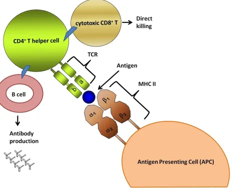
II molecules present extracellular peptides to CD4+ T helper cells (Figure 1.1) (Rojo, Saizawa et
al. 1989; Gerloni and Zanetti 2005).
MHC II molecules play important roles in activating adaptive immune responses and
MHC II deficiencies lead to the development of bare lymphocyte syndrome, (BLS), which
re-sults in death in infancy (Reith and Mach 2001). Severe combined immunodeficiency (SCID) is
also developed due to lack of MHC II expression. Patients with SCID are extremely vulnerable
to infection because their lack of CD4+ T cells also results in an inability to activate B cells, nor
expression of MHC II molecules is also associated with disease development as all autoimmune
diseases correlate with over expression of MHC II in the targeted tissues (Swanberg, Lidman et
al. 2005). MHC II molecules play important roles in anti-tumor immunity. The presentation of
tumor cell antigens by MHC class II molecules is particularly critical in the detection of a newly
formed tumor as MHC class II proteins are capable of activating multiple arms of the anti-tumor
immune response (Guy, Krajewski et al. 1986; Garrido and Ruiz-Cabello 1991). As MHC II
mo-lecules play critical roles in the activation of adaptive immune responses, and as deregulation of
MHC II expression has such dire consequences, MHC II expression is tightly regulated,
primari-ly at the level of transcription (Benoist and Mathis 1990).
Figure 1.1 MHC II molecules are an indispensable arm of the adaptive immune response as they present processed extracellular antigens to CD4+T cells which in turn initiate adaptive immune responses.
Antigen presenting cells (APC) play an essential role in recognizing, processing, and presenting proteins. Processed antigens are presented by APCs and, by all interferon gamma (IFN-) stimu-lated cells, on MHC II molecules. MHC II are cell surface glycoproteins that present antigens to CD4+ T cells resulting in T cell activation. Activated CD4T cells can activate B cells to pro-duce specific antibodies and elicit adaptive immune responses. Activated CD4+ T cells can also activate cytotoxic CD8+T cells to mediate direct killing (Kindt Thomas J 2007; Parham 2009).
1.5 Regulation of MHC II Transcription:
MHC II molecules are glycoproteins which are constitutively expressed on the surface of
antigen presenting cells such as activated macrophages, B cells, dendritic cells, and activated T
cells (Cresswell and Howard 1997; Morris, Beresford et al. 2002). Expression of MHC II can be
induced in all nucleated cells by inflammatory cytokines, primarily the cytokine interferon
gam-ma (IFN-γ) (Benoist and Mathis 1990; Kaufgam-man, Salomonsen et al. 1994). IFN-γ is a type II
in-terferon which, through binding to the type II IFN receptor, activates transcription of target genes
including MHC II. In contrast to IFN-α and IFN-β, which can be expressed by all cells, IFN-γ
secretion is restricted to T lymphocytes, dendritic cells, and NK cells (Mach, Steimle et al. 1996;
Boss and Jensen 2003; Platanias 2005). Inducible expression of MHC II allows for enhanced
antigen presentation and induction of localized immune responses. The expression of MHC II
genes is tightly regulated by multiple transcription factors and by chromatin remodeling enzymes
that bind to a conserved regulatory region in the MHC II gene (Ting and Trowsdale 2002; Boss
and Jensen 2003; Drozina, Kohoutek et al. 2005). This regulatory region consists of conserved
sequences designated as the X1 box, X2 box, and Y box (Benoist and Mathis 1990; Ting and
Trowsdale 2002). These sequences are respectively recognized by ubiquitously expressed DNA
binding factors. The X1 box is bound by the regulatory factor X (RFX) which is a trimer
con-sisting of RFX5, RFXANK, and RFXAP (Masternak, Barras et al. 1998; Nagarajan,
Louis-Plence et al. 1999). The c-AMP responsive element-binding protein (CREB) binds to the X2
box (Moreno, Beresford et al. 1999). The Y box is bound by the nuclear factor Y (NF-Y)
trimer-ic complex whtrimer-ich consists of NF-YA, NF-AB, and NF-YC (Mantovani 1999; Drozina, Kohoutek
not sufficient for the initiation of MHC II transcription. Instead, the enhanceosome serves as
platform for binding the master regulator of MHC II transcription, the class II transactivator,
CIITA (Masternak, Muhlethaler-Mottet et al. 2000) (Figure 1.2).
CIITA is a co-activator as it is not a DNA binding protein, but instead binds to the
enhan-ceosome complex through interactions with RFX, NFY, and CREB (Masternak,
Muhlethaler-Mottet et al. 2000; Ting and Trowsdale 2002). As seen in Figure 1.2, CIITA initiates
transcrip-tion of MHC II genes by recruiting the basal transcriptranscrip-tional machinery including the TATA
binding protein (TBP), TATA associated factors (TAFs) (Mahanta, Scholl et al. 1997), histone
acetyltransferases (HATs) (Spilianakis, Papamatheakis et al. 2000; Wright and Ting 2006;
Koues, Dudley et al. 2008), and histone methyltransferases (HMTs) (Koues, Mehta et al. ; Zika,
Fauquier et al. 2005), which act together to change MHC II promoter accessibility. CIITA
re-cruitment leads to enhanced levels of acetylation at the proximal promoter of MHC II genes that
in turn promotes efficient assembly of transcription machinery and rapid activation of MHC II
transcription (Koues, Mehta et al. ; Sisk, Gourley et al. 2000; Wang, Huang et al. 2001).
There-fore, interactions of CIITA with the MHC II proximal promoter are crucial for the activation of
Figure 1.2 MHC II promoter region.
(Top) MHC II proximal promoter contains X1, X2 and Y boxes. The X1 box is bound by RFX which is a trimer consisting of RFX-5, RFX-ANK and RFX-AB. CREB binds X2 box. The Y box is bound by the NF-Y trimeric protein consisting of NF-YA, NF-YB and NF-YC. The bind-ing of each of these constitutively expressed transcription factors forms the MHC II enhanceo-some complex, which is necessary but insufficient to mediate cytokine inducible MHC II tran-scription (Kretsovali, Agalioti et al. 1998; Fontes, Kanazawa et al. 1999; Zika, Greer et al. 2003; Zika, Fauquier et al. 2005).
1.6 CIITA, the Master Regulator of MHC II:
CIITA is the master regulator for MHC II gene expression (Mach, Steimle et al. 1996).
Its activity is absolutely required for the expression of the MHC class II genes, however the
regulation of CIITA itself is less understood (Chang, Fontes et al. 1994). In order to maintain
tight regulation of MHC II, CIITA expression is also tightly regulated at the level of transcription
(Ting and Trowsdale 2002). Transcription of CIITA is regulated in a cell specific manner by
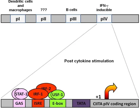
four different promoters: pI, pII, pIII and pIV (Muhlethaler-Mottet, Otten et al. 1997) (Figure
1.3). By virtue of different transcriptional start sites illustrated in the top part of Figure 1.3, each
promoter yields a different CIITA isoform that will have a unique first exon. The promoter I (pI)
isotype is expressed primarily in dendritic cells and macrophages. Promoter II (pII) is not well
conserved amongst different species and often considered to be inactive (Baton, Deruyffelaere et
al. 2004). Promoter III generates the CIITA isotype (pIII) in constitutively expressed in B cells,
Figure 1.3 CIITApIV proximal region.
(TOP) CIITA is transcribed from 4 different promoter regions depending on a cell type. Promo-ter I drives expression of CIITA in dendritic cells. The function of promoPromo-ter II is not conserved between species and the promoter is believed to be inactive. Promoter III primarily functions in B cells and promoter IV is responsible for the IFN-γ induced expression of CIITA.
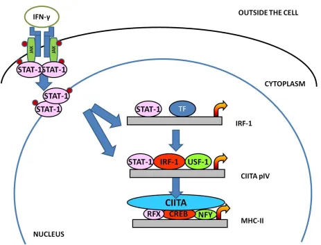
Figure 1.4 Activation of a JAK –STAT signal transduction pathway.
Promoter IV CIITA isotype (pIV) is the IFN-γ responsive form of CIITA which is
pre-dominantly expressed in IFN-γ stimulated cells (Piskurich, Gilbert et al. 2006) resulting in
paral-lel INF-γ induction of MHC II expression (Mottet, Otten et al. 1997;
Muhlethaler-Mottet, Di Berardino et al. 1998; Morris, Beresford et al. 2002). It is critical that CIITA
expression is tightly regulated because abbearant expression is associated with multiple diseases
including autoimmune conditions and cancer (van den Elsen, Gobin et al. 2001; Satoh, Toyota et
al. 2004).
Cytokine dependent MHC II transcription allows for enhanced antigen presentation by all
nucleated cells and is achieved by binding of the transcription factors signal transducer and
acti-vator of transcription (STAT-1) and interferon regulatory factor 1 (IRF-1) to CIITApIV
(Piskurich, France et al. 1993; Piskurich, Blanchard et al. 1995; Piskurich, Youngman et al.
1997; Piskurich, Wang et al. 1998; Piskurich, Linhoff et al. 1999). Cytokine activated CIITApIV
is also occupied by the constitutively expressed transcription factor upstream stimulating factor 1
(USF-1) (Figure 1.3) (Muhlethaler-Mottet, Di Berardino et al. 1998; Piskurich, Linhoff et al.
1999; Pattenden, Klose et al. 2002). Activation of inducible CIITApIV is initiated when IFN-γ
binds to its surface receptor and occurs via the Janus kinase signal transducer and activator of
transcription (JAK-STAT) signal transduction pathway (Figure 1.4) (Morris, Beresford et al.
2002; Pattenden, Klose et al. 2002; Piskurich, Gilbert et al. 2006). Activated JAK1 and JAK2
phosphorylate STAT-1, causing STAT-1 to translocate to the nucleus. In addition to binding to
the gamma activating sequence (GAS) box of CIITApIV, STAT-1 binds the IRF-1 gene STAT-1
binding site (Muhlethaler-Mottet, Di Berardino et al. 1998). CIITApIV also has conserved GAS
interferon regulatory factors (IRFs) that are globally involved in IFN responses (Piskurich,
Linhoff et al. 1999; Piskurich, Gilbert et al. 2006; Wright and Ting 2006). In addition to
promot-ing bindpromot-ing of transcription factors to pIV, IFN-γ induces acetylation of histones which loosens
the chromatin structure and increases the accessibility of CIITApIV (Sterner and Berger 2000;
Wright and Ting 2006). Because MHC II plays important roles in initiating, maintaining, and
eventually terminating adaptive immune responses, the unique ability of CIITA to regulate the
expression of MHC II genes makes CIITA a major player in the regulation of antigen
presenta-tion and adaptive immune responses.
1.7 The 26S Proteasome
The ubiquitin proteasome system (UPS) is well known for its role in the degradation of
polyubiquitinated proteins, including transcription factors and chromatin remodeling proteins
(Ciechanover 1994). However, recent investigations indicate subunits of the 26S proteasome are
essential for transcriptional regulation of various genes independent of their roles in protein
de-gradation (Conaway, Brower et al. 2002; Ferdous, Kodadek et al. 2002; Greer, Zika et al. 2003).
Research in the Greer lab has shown that subunits of the UPS are necessary for transcriptional
regulation of MHC II and CIITA genes (Koues, Mehta et al. ; Truax, Koues et al. ; Bhat, Turner
et al. 2008; Koues, Dudley et al. 2008; Koues, Dudley et al. 2009). The role of the proteasomal
Figure 1.5 Enzymatic cascade of ubiquitination.
Figure 1.6 Ubiquitination regulates numerous cellular processes.
It is well established that polyubiquitination targets proteins for degradation by the 26S
proteasome (Pickart 2004; Haglund and Dikic 2005). The 8.5 Kd, 76 amino acid protein
ubiqui-tin was initially described as a protein modification which is covalently attached to lysine
resi-dues of target molecules (Ciechanover 1994; Adams 2003; Shmueli and Oren 2005). Chains of
four ubiquitin molecules are necessary for recognition and degradation by the 26S proteasome
(Figure 1.5) (Ciechanover 1998; Conaway, Brower et al. 2002). Ubiquitin is attached to proteins
via an isopeptide bond between the C terminus of the ubiquitin molecule and a lysine (K) side
chain on the target protein. A protein targeted for degradation is tagged with ubiquitin molecules
via a three step enzymatic cascade. Polyubiquitination occurs when these steps are repeated,
re-sulting in the formation of a chain of four ubiquitin molecules on the target protein (Figure 1.5)
(Thrower, Hoffman et al. 2000).
Ubiquitin has seven lysine residues (K-6, K-11, K-27, K-29, K-33, K-48 and K-63) and,
while a majority of proteins are marked for proteasomal degradation by polyubiquitination via
K-48, recent studies have shown that K-63 linked polyubiquitination also marks proteins for
de-gradation (Chau, Tobias et al. 1989; Finley, Sadis et al. 1994) (Kim, Kim et al. 2007). Proteins
polyubiquitinated via K-63 play roles in the activation of several cellular pathways and are
even-tually degraded in lysosomes (Tan, Wong et al. 2007). Alternatively, monoubiquitination
regu-lates many cellular processes including nuclear export, DNA repair, protein interactions, and
his-tone modifications (Figure 1.6) (Terrell, Shih et al. 1998; Lucero, Penalver et al. 2000; Lotocki,
Alonso et al. 2003; Gupta-Rossi, Six et al. 2004; Sigismund, Polo et al. 2004; Haglund and Dikic
the outcomes of alternative, ie, non-lysine 48 or 63 linked, ubiquitination; these alternative
ubi-quitination reactions will likely be a primary focus of ubiquitin research for years to come.
Once a protein is tagged via lysine 48 polyubiquitination with 4 or more molecules of ubiquitin,
the protein is targeted to the 26S proteasome for degradation (Ciechanover 1998; Thrower,
Hoffman et al. 2000; Finley, Ciechanover et al. 2004).
The 26S proteasome in mammalian cells is a 2.5 MDa multi-protein complex made of a
19S regulatory particle (RP) and a 20S proteolytic core (Baumeister, Walz et al. 1998) which can
exist independently in both the nucleus and cytoplasm (Peters, Franke et al. 1994). The 19S can
be further divided into two parts: a lid and a base. The lid is composed of eight non-ATPase
sub-units which are required for protein degradation (Coux, Tanaka et al. 1996; Baumeister, Walz et
al. 1998; Gorbea, Taillandier et al. 1999). The base of the 19S contains six ATPases belonging to
the ATPases associated with a variety of cellular activities (AAA) family: S4, S6a, S6b, S7,
Sug1 (S8) and Sug2 (S10b) which correspond, respectively, to the yeast homologous proteins
Rpt 1-6, and four non-ATPase subunits, Rpn1, Rpn2, Rpn10, and Rpn13 (Figure 1.7) (Gorbea,
Taillandier et al. 1999; Adams 2003; Bhat, Turner et al. 2008). The 20S catalytic core is a 700
kDa cylinder which consists of four stacked rings, with each ring containing seven α and β
Figure 1.7 The 26S Proteasome.
The base ATPases contain a C terminal hydrophobic tyrosine X motif that docks into the
pockets of the α rings of the 20S (Smith, Chang et al. 2007). In the presence of ATP, the 20S
catalytic core associates with the 19S regulatory particle on both sides to form the 26S
protea-some, allowing for the recognition of the ubiquitinated substrates which are marked for
degrada-tion (Coux, Tanaka et al. 1996; Gonzalez, Delahodde et al. 2002). The 19S regulatory particle
recognizes the ubiquitin chains on targeted proteins, cleaves the chains, unfolds the protein, and
directs the unfolded protein to the 20S core for degradation (Figure 1.5) (Coux, Tanaka et al.
1996; Strickland, Hakala et al. 2000). The result of proteasomal degradation is cleaved peptides
with an average length of 8-12 amino acids and free ubiquitin, which is reactivated and recycled
(Jung, Catalgol et al. 2009). Through these mechanisms the 26S proteasome eliminates a variety
of unwanted and/or misfolded proteins, (Hilt and Wolf 1996; He, Qi et al. 1998) and thus
regulates many important cellular processes.
1.8 Non-proteolytic roles of the proteasome:
Growing studies indicate the 26S proteasome plays critical regulatory, but degradation
independent, roles in gene expression. While it is well accepted that 19S ATPases recognize and
unfold ubiquitinated proteins for degradation, these ATPases have RNA/DNA helicase activity,
indicating their potential involvement in cellular processes independent of proteolysis (Coux,
Tanaka et al. 1996; Baumeister, Walz et al. 1998; Adams 2003). Studies in yeast demonstrate
the 19S ATPases associate with transcription factors, with RNA polymerase II, with activated
promoters, and with transcriptional elongation complexes (Ferdous, Kodadek et al. 2002;
in mammalian cells have shown that the 19S base is essential for initiation and elongation by
RNA pol II, but that proteolytic activity of the 26S is not required (Ferdous, Kodadek et al.
2002). Work in the Greer has demonstrated the 19S ATPases associate with the MHC II
prox-imal promoter, with CIITA, and with the enhanceosome complex where they play important role
in regulating CIITA activity and MHC II expression (Truax, Koues et al. ; Bhat, Turner et al.
2008; Koues, Dudley et al. 2008; Koues, Dudley et al. 2009). Evidence also indicates inhibition
of the 19S decreases elongation, while inhibiting the catalytic activity of the 20S increases
elon-gation, thus the balance between the 19S/20S subunits is crucial for transcriptional regulation
(Gillette, Gonzalez et al. 2004). These observations indicate the ubiquitin/proteasome pathway
plays multiple roles in transcription and imply the non-degradative roles of the proteasome may
1.9 ROLES FOR THE 26S PROTEASOME COMPONENTS IN TRANSCRIPTION:
Transcription is a first step in gene expression and is divided into 5 stages: pre-initiation,
initiation, promoter clearance, elongation, and termination. During pre-initiation, general
tran-scription factors guide RNA pol II to proximal promoter regions. In eukaryotes trantran-scription
re-quires special sequences, known as TATA boxes, located around 30 base pairs upstream of start
sites. The TATA box is a binding site for the TATA binding protein (TBP) which is itself a part
of Transcription Factor II B (TFIIB), here illustrated on the MHC II proximal promoter (Figure
1.2). The core domain of TFIIB associates with TBP at the TATA box and interacts with the
DNA both upstream and downstream of the TATA box, thus recruiting RNA pol II to the correct
site (Calvo and Manley 2003; Deng and Roberts 2007). Eukaryotic RNA pol II is not directly
recruited to the DNA region of interest. Instead, groups of transcription factors create an
initia-tion complex that recruits RNA pol II and initiates transcripinitia-tion. Although extensively
investi-gated, the full mechanisms that control transcriptional initiation, elongation, and termination
re-main unknown.
In 1992, Swaffield and colleagues first indentified roles for proteasome subunits in
tran-scriptional regulation. These researchers demonstrated 19S ATPase subunits Rpt6 (Sug1) and
Rpt4 (Sug2) rescued a mutant transactivator Gal4 in Saccharomyces cerevisae (Swaffield,
Bromberg et al. 1992; Rubin, Coux et al. 1996). Soon thereafter, the intact 26S proteasome, as
well as the 19S regulatory particle and the 20S core, were demonstrated to localize in both the
nucleus and the cytoplasm (Peters, Franke et al. 1994). These early experiments were the first to
ob-served that the 19S component of the proteasome also plays important roles in regulating
tran-scription at a variety of different genes (Koues, Mehta et al. ; Kinyamu, Chen et al. 2005; Lassot,
Latreille et al. 2007; Zhu, Wani et al. 2007; Bhat, Turner et al. 2008; Koues, Dudley et al. 2008;
Koues, Dudley et al. 2009). Subunits of the proteasome are required in the formation of initiation
complexes, in maintaining elongation, and in executing termination. Roles for the 19S ATPases
in transcriptional initiation at CIITApIV will be described in this dissertation in Chapter 2 and in
Chapter 5, we demonstrate how 19S subunits maintain transcriptional elongation of CIITA
genes.
1.10 Roles in transcription initiation:
Recent publications have demonstrated the 19S ATPases play important roles in initiating
transcription of various yeast and mammalian genes. In yeast, the 19S proteasome has been
shown to be responsible for the recruitment of the histone acetyltransferase
(Spt-Ada-Gcn5-acetyl-transferase) SAGA to promoter regions. Lack of the 19S ATPase Rpt6, the yeast homolog
of mammalian Sug1, results in reduced recruitment of the HAT SAGA and in lower levels of
histone H3 acetylation, indicating that 19S ATPases are important for recruiting HATs to
promo-ter regions (Lee, Ezhkova et al. 2005). Furthermore, as mentioned above, alleles of Rpt6 rescue
mutations in the Gal4 activation domain (Rubin, Coux et al. 1996). Rpt6 also mediates the
re-cruitment of transcription factors to TBP (Swaffield, Melcher et al. 1995) and binds to various
actively transcribing genes (Gonzalez, Delahodde et al. 2002). Overall, these observations
dem-onstrate the 19S proteasome plays important roles in transcriptional initiation of yeast promoters.
2006, Rasti and colleagues demonstrated the 19S ATPase Sug1 plays positive roles in adenovirus
E1A dependent transcription (Rasti, Grand et al. 2006). In 2007, Zhu determined the 26S
protea-some enhances recruitment of p53 to p21 wafl responsive promoters (Zhu, Wani et al. 2007).
Concurrent observations from Lassot and colleagues demonstrated 19S ATPases play important
roles in regulating Tat dependent transcription of HIV-1 genes (Lassot, Latreille et al. 2007). The
19S ATPase Sug-1 also plays an important proteolytic and non proteolytic role in regulating
transcription mediated by retinoic acid (Ferry, Gianni et al. 2009).
The 19S ATPases play critical roles in regulating transcription initiation of MHC II and
CIITA genes. The ATPase Sug1 is recruited to the MHC II proximal promoter following IFN-γ
stimulation. Decreased expression of Sug1 results in decreased recruitment of CIITA to the
MHC II promoter and in reduced expression of MHC II genes (Bhat, Turner et al. 2008). Sug1
exerts control over CIITA promoter binding by regulating histone H3 acetylation at the MHC II
proximal promoter through interactions with acetylated histone H3. In cells treated with Sug1
specific siRNA, MHC II histone acetylation is decreased with a preferential impact on
acetyla-tion at histone H3 lysine 18. Sug1 also recruits the histone acetyltransferase CREB binding
pro-tein (CBP) to MHC II promoters, further implicating Sug1 as an important regulator of histone
modifications at the MHC II proximal promoter (Koues, Dudley et al. 2008).
Sug1 is also important for regulating H3 lysine 4 trimethylation and H3 arginine 17
dime-thylation at both MHC II and CIITA genes. However, Sug1 does not impact H3 lysine 36
4 trimethylation are important activating modifications, these observations further implicate
Sug1 as being involved in activating transcription initiation (Koues, Dudley et al. 2009).
Sup-porting a role for Sug1 in initiation are observations that the absence of Sug1 enhances levels of
H3 lysine 27 trimethylation, resulting in repression of transcription initiation (Koues, Mehta et
al.). Chapter 2 of this dissertation details observations that an additional 19S ATPase, S6a, plays
critical roles in initiating transcription of CIITApIV. Decreased expression of S6a significantly
impacts the recruitment of transcription factors STAT-1 and IRF-1 to the CIITApIV proximal
promoter and diminishes levels of activating acetylation on histone H3 and histone H4 with a
preferential loss of histone H3 lysine 18 acetylation and H4 lysine 8 acetylation (Truax, Koues et
al.) Together, these observations provide evidence that the19S proteasome plays regulatory roles
in transcriptional initiation of the cytokine inducible genes MHC II and CIITA.
1.11 Regulation of transcriptional elongation:
Following transcription initiation, the multi-component transcriptional machinery falls
under the control of positive and negative transcription factors which regulate gene expression.
During the early steps of elongation, RNA polymerase II is paused by negative transcription
elongation factors (N-TEFs) (Sims, Belotserkovskaya et al. 2004). As illustrated in Figure 1.8,
positive transcription elongation factor (P-TEF-b) is recruited to the N-TEF complex where it
phosphorylates the carboxy terminal domain (CTD) of RNA pol II and N-TEF, thus permitting
elongation (Sims, Belotserkovskaya et al. 2004). P-TEF-b exists in two distinct molecular forms
as an active and inactive complex (Nguyen, Kiss et al. 2001; Yang, Zhu et al. 2001). P-TEF-b
1 (Hexim-1) or 2 (Hexim-2) that is bound to 7SK small nuclear RNA (snRNA) (Figure 1.8)
(Barboric, Kohoutek et al. 2005; Blazek, Barboric et al. 2005; Dulac, Michels et al. 2005;
Schulte, Czudnochowski et al. 2005). The CTD domain of RNA pol II is composed of 52
re-peats of the heptapeptide sequence Tyr-Ser-Pro-Thr-Ser-Pro-Ser. During transcriptional
initia-tion, the CTD domain remains hypophosphorylated and becomes hyperphosphorylated during
elongation (Corden, Cadena et al. 1985). The switch from transcriptional initiation to elongation
is dynamic and is controlled by interactions between positive and negative transcription factors.
Additional regulation through pausing of RNA pol II allows the replacement of initiation factors
with molecules required for transcriptional elongation and RNA processivity.
In addition to the above described roles in transcription initiation, the 26S proteasome
al-so regulates transcription elongation. Initial observations that inhibition of proteaal-some activity
reduces recruitment of RNA pol II to various yeast promoters first implicated the proteasome as
involved in of transcriptional elongation (Lipford, Smith et al. 2005). Evidence has since
accu-mulated that 19S ATPases play important roles in pol II dependent elongation that are
indepen-dent of the presence of the 20S core (Ferdous, Gonzalez et al. 2001). Rpt 6, the yeast homolog of
mammalian Sug1, has been demonstrated to link histone ubiquitination and methylation, which
are both important steps in elongation (Sun and Allis 2002; Ezhkova and Tansey 2004;
Pokholok, Harbison et al. 2005). 19S components also interact with the yeast elongation factor
CDc8 (Ferdous, Gonzalez et al. 2001). Studies in mammalian systems are less extensive, but also
support roles for proteasomal subunits in transcriptional elongation. 19S ATPases are recruited
to coding regions of HIV-1 genes with a preferential abundance of the 19S ATPase S6a (Lassot,
receptor responsive pS2 genes a complex with elongin and RNA pol II (Zhang, Sun et al. 2006),
suggesting proteasomal subunit cooperation with transcriptional factors to mediate elongation.
Observations in the Greer lab indicate robust occupancy of 19S ATPases in coding regions of
CIITA and MHC II genes (Truax, Koues et al.), mechanistic details of these associations are
de-scribed in detail in Chapter 5 of this dissertation.
More than a decade has passed since scientists first explored non-degradative roles for
the proteasome. The discovery of nuclear proteasome, followed by preliminary research in yeast
first indicated the proteasome is „not just degrading‟ anymore. Recent studies in both mammalian
and yeast systems indicates 19S ATPase components of the ubiquitin proteasome system play
important roles in regulating transcription of various genes via both proteolytic and non
proteo-lytic roles. Data described in detail in Chapters 2 and 5 of this dissertation adds to this growing
body of evidence by demonstrating that the 19S ATPase S6a is a crucial regulator of both
tran-scriptional initiation and elongation of CIITA genes. As MHC II and its master regulator CIITA
are major players in adaptive immunity, tight transcriptional regulation of these genes is essential
to a properly functioning immune system. The contributions of my data increase our
understand-ing of the transcriptional regulation of not only these genes, but of all inducible genes where a
Figure 1.8 Dynamics at the transcriptional initiation and elongation of CIITA.
1.12 THE ACCESSIBILITY OF CHROMATIN:
DNA exists in a highly organized, tightly packed form termed chromatin (Berger 2007).
Differential packaging of DNA with histone and non-histone proteins into chromatin determines
DNA accessibility during transcription. Chromatin is dynamic and can be found in multiple
forms, from condensed, tightly packed heterochromatin that is inactive to loosely organized, and
transcriptionally accessible, euchromatin (Berger, Kouzarides et al. 2009). The fundamental unit
of chromatin is the nucleosome (Mandel and Fasman 1976; Berger 2007). Histones (H) are
integral components of nucleosome structures as they provide a scaffold for double stranded
DNA to wrap around. There are four histone proteins: H2A, H2B, H3, and H4, two copies of
which form the octameric nucleosome structure along with a fifth linker histone, H1 (Figure 1.9)
(Kornberg and Thomas 1974; Luger, Mader et al. 1997). Histones are positively charged, while
DNA is negatively charged due to phosphate groups in its sugar backbone. Because of this
charge differential, DNA is wrapped tightly around the histone octamers (Ward, Bowman et al.
2009). As such, the base unit of chromatin are nucleosomes, consisting of histone proteins
around which DNA is wrapped 1.67 times (Luger, Mader et al. 1997). Nucleosome cores consist
of two H2A-H2B dimers and H3-H4 tetramers. Histone H1 stabilizes nucleosomes by interacting
with double stranded DNA as it enters and exits the nucleosome (Kouzarides 2007) and aids
formation of dinucleosomes (Widom 1998). Dinucleosomes fold over to form 30nm fibers that
Figure 1.9 Structure of nucleosomes.
Histones are small proteins that provide a scaffold for the DNA to wrap around. There are four histone proteins which form an octomer structure. The nuclesome histone core consists of central H3/H4 tetramer that is surrounded on either side by a H2A/H2B dimer. DNA wraps tightly around the histone proteins to form the intact nucleosome structure. Chromatin is very dynamic and can be found in multiple forms depending on how tightly DNA wraps around those histone proteins (Kornberg and Thomas 1974; Luger, Mader et al. 1997; Clapier and Cairns 2009).
Figure 1.10 Histone code.
In inactive heterochromatin, histone proteins bind tightly to DNA and regulate access to
DNA of transcription factors and the general transcription machinery. Structural changes to
his-tones are facilitated by posttranslational modifications that render chromatin more or less
availa-ble for transcription. The N- and C-terminal tails of histones are regions availaavaila-ble for
posttran-slational modifications (Felsenfeld and Groudine 2003). Modifications to histone tails include
acetylation (Morris, Rao et al. 2007), phosphorylation (Ivaldi, Karam et al. 2007), methylation
(Li, Lin et al. 2002), sumoylation (Nathan, Sterner et al. 2003), ADP ribosylation (Boulikas
1990) and ubiquitination (Davies and Lindsey 1994; Ng, Xu et al. 2002; Ting and Trowsdale
2002; Shukla, Stanojevic et al. 2006; Shukla and Bhaumik 2007). These modifications have
been linked to gene expression through a histone code hypothesis which proposes that the
com-binatory effects of these modifications determines the “open” and “closed” state of chromatin
and thereby aids in determining the expressivity of genes (Wray, Hahn et al. 2003). Histone
modifications create a variety of modifications that alter N terminal tails of histones and create
the epigenetic code that, in part, determines the accessibility of chromatin.
Contributions from histone modifications are just one element of regulation that
deter-mines the overall structure of chromatin. The state of chromatin packaging is determined by a
combination of factors in addition to histone modifications including DNA methylation and
nuc-leosome remodeling, which together define gene expression patterns and regulate transcription.
In eukaryotes, DNA methylation occurs by covalent modification of cytosines to CpG
dinucleo-tides (Illingworth and Bird 2009). Promoter regions in the human genome are embedded with
CpG islands, where methylation occurs and provides an additional mechanism of transcription
multiple mechanisms contribute to gene expression, histone modifications are the most studied
of these mechanisms. Due to the complex relationship between DNA and the histones around
which it is wound, modifications to histones are a key component of transcriptional regulation.
Of histone modifications, acetylation and methylation are the most critical as they impart
signifi-cant structural alterations in histones and dramatically change interactions with DNA.
1.13 Histone acetylation:
One of the most studied histone modifications is acetylation of lysine residues on
his-tones which loosens interactions between histone proteins and DNA, and contributes to an
“open” state of chromatin (Cosgrove 2007). Acetylation primarily occurs on lysine residues
lo-cated on histones H3 and H4 (Figure 1.10). A deacetylated tail of histones H3 and H4 are
posi-tively charged and, as DNA is negaposi-tively charged; deacetylated histones tightly bind DNA and
render it inaccessible to transcriptional activation. The addition of acetyl groups to targeted
ly-sine residues neutralizes the positive charge on histone tails and loosens interactions with DNA
(Struhl 1998). Alterations in histone acetylation result from changes in the balance between the
enzymes which catalyze histone acetylation: histone acetyltransferases (HATs) and histone
dea-cetyltransferases (HDACs), which remove acetyl groups (Utley, Ikeda et al. 1998; Yang 2004).
HATs and HDACs are recruited to chromatin as transcriptional coactivators by transcription
fac-tors and are generally found in large remodeling complexes. There are a variety of known HATs,
including several which are discussed in this dissertation: general control of amino acid synthesis
5 (GCN5), CREB binding protein (CBP), p300 and p300/CBP associated factor (PCAF),
Legube and Trouche 2003). Acetylation is reversed by histone deacetylases (HDACs) which
re-move acetyl groups from lysine residues (de Ruijter, van Gennip et al. 2003). Both HATs and
HDACs influence a myriad of cellular processes including signal transduction, apoptosis, cell
cycle regulation, cell growth and transcription (Utley, Ikeda et al. 1998; Yang 2004).
1.14 Histone methylation:
Methylation plays dual roles in regulating histone accessibility as it has been linked to
both activation and silencing of transcription (Daujat, Bauer et al. 2002; Naeem, Cheng et al.
2007). Methylation confers additional levels of complexity as methylation can be mono, di- or
tri- methylation and occurs at lysine (K) and arginine residues (R) (Figure 1.10). Methylation of
arginine residues has been linked to gene activation (Bauer, Daujat et al. 2002) while lysine
me-thylation has been linked with gene silencing and activation (Jenuwein and Allis 2001;
Nakayama, Rice et al. 2001; Berger 2002; Lehnertz, Ueda et al. 2003; Shilatifard 2006).
Argi-nine is a positively charged amino acid and the nitrogen of argiArgi-nine can be modified by the
addi-tion of one or two methyl groups (Gary and Clarke 1998). Currently eight mammalian protein
arginine methyltransferases (PRMTs) have been identified (Bedford and Richard 2005). While
the mechanism through which arginine methylation enhances transcripition remains unknown,
evidence exists that methylated arginines collaborate with other transcriptional activators and
enhance their activity to promote transcriptional activation (Stallcup 2001). Methylation on
ly-sine residues generally correlates with gene suppression but can also contribute to gene
activa-tion. One, two or three methyl groups can be added by histone methyltransferases (HMTases)
is associated with gene silencing, while H3K4 di- and trimethylation, H3K36 trimethylation, and
H3K79 di- and trimethylation methylation contributes to gene activation (Varier and Timmers
2011).
The histone methyltransferase Enhancer of Zeste Homolog 2 (EZH2) belongs to
Poly-comb Group (PcG) and is involved in gene repression (Laible, Wolf et al. 1997; Satijn and Otte
1999). EZH2 catalyzes trimethylation of H3K9 and K27 with a strong preference for K27 (Cao,
Wang et al. 2002; Czermin, Melfi et al. 2002; Muller, Hart et al. 2002). EZH2 has a cysteine rich
SET domain which is involved in binding to the Polycomb repressive complex (Cao, Wang et al.
2002). Data in Chapter 3 of this dissertation demonstrates EZH2 binds to the CIITA proximal
promoter and is a “master regulator” of the silencing histone modifications at CIITApIV, thus
implicating roles for EZH2 in regulating other silenced, but inducible genes (Mehta, Truax et al.
2011). Of note are recent studies indicating expression of EZH2 is elevated in multiple human
cancers, including breast cancer, which will be described in detail in this dissertation in Chapter
4.
While the mechanisms by which histone methylation contributes to gene silencing are not
known, cross talk between various modifications has been observed (Muller and Verrijzer 2009).
H3K4 methylation blocks methylation of H3K9 and induces the dimethylation of H3K27,
result-ing in an openresult-ing of chromatin structure (Wang, Huang et al. 2001; Agger, Cloos et al. 2007;
Lee, Villa et al. 2007). H3K36 methylation recruits histone deacetylases and reestablishes closed
2007). Similar to reversible acetylation, histone methylation can also be reversed by histone
de-methylases (HDMTs). One of first identified HDMT is LSD1 which demethylates mono- and
di- H3K4in a flavin adenine dinucleotide (FAD) dependent oxidative reaction (Wysocka, Milne
et al. 2005). This observation led to discovery of highly conserved Jumonji C containing proteins
which remove methyl groups from lysine residues (Fodor, Kubicek et al. 2006; Klose, Yamane
et al. 2006; Tsukada, Fang et al. 2006; Whetstine, Nottke et al. 2006; Yamane, Toumazou et al.
2006). This recent observation that methylation is reversible by histone demethylases provides
new levels of regulation for gene expression. There are also additional interactions between
me-thylated histones and the DNA methylation machinery which further determines the availability
of DNA for gene expression. While histone methylation and the impact on DNA structures is
reversible, DNA methylation is permanent and leads to stable repression of gene expression. The
interactions between methylated histones and methylated DNA are complex as histone
methyla-tion mediates DNA methylamethyla-tion and DNA methylamethyla-tion also serves as a template for
modifica-tions to histones (Ooi, Qiu et al. 2007; Mohn, Weber et al. 2008). Greater understanding of the
crosstalk between modifications to histones and modifications to DNA provide new, and largely
unexplored, levels of regulation of gene expression (Cedar and Bergman 2009).
1.15 Additional Histone Modifications:
Although histone acetylation and methylation are the most studied, there are a variety of
additional histone modifications including phosphorylation, ubiquitination, sumoylation, and
poly ADP ribosylation which contribute to the status of chromatin. While phosphorylation of
gene expression is unknown (Nowak and Corces 2000). Phosphorylation of histone H3 at serine
10 (Figure 1.10) plays an important role in the very early events of transcriptional elongation
that occur prior to P-TEF-b recruitment (Ivaldi, Karam et al. 2007). Furthermore, several histone
acetyltransferases have enhanced activity towards phosphorylated histone substrates (Cheung,
Tanner et al. 2000; Lo, Trievel et al. 2000). Histone sumoylation is generally associated with
transcriptionally silenced genes (Nathan, Sterner et al. 2003) (Shiio and Eisenman 2003) and
his-tone ubiquitination, similarly to hishis-tone methylation, plays dual roles in transcription activation
and silencing, depending on the residue targeted. For example, histone H2A ubiquitination
(ubH2A) plays roles in both gene activation and in H3K27me3 mediated silencing (Cao,
Tsukada et al. 2005; Wei, Zhai et al. 2006; Lee, Norman et al. 2007). Finally, poly (ADP
ribosy-lated) histones are important in the assembly of histone complexes during DNA replication
(Boulikas 1990). Thus, the structure of chromatin and the availability of DNA for transcription is
determined by various posttranslational modifications.
1.16 Histone modifications and DNA accessibility:
Posttranslational modifications to N terminal tails of histones affect chromatin structure
and availability for gene expression (Figure 1.10). The state of chromatin is determined by a
combination of modifications on single histones which orchestrates a histone code that regulates
transcriptional activation. While this field remains in its infancy, a histone code has begun to
emerge. Methylation at histone H3K9 and H3K27 are considered to be silencing modifications,
trimethylation at H3K4. Poised chromatin is identified by the activating modification H3K4me2
and the silencing modification H3K27me3 (Jenuwein and Allis 2001; Lachner, O'Carroll et al.
2001; Zhang and Reinberg 2001; Bernstein, Humphrey et al. 2002; Santos-Rosa, Schneider et al.
2002; Shilatifard 2006). The full mechanisms which determine interactions between various
his-tone modifications and regulate their effects on chromatin remain unknown and are areas of
in-tense investigation. Thus, the interplay between those various modifications creates a so-called
“epigenetic landscape” which determines the expressivity of genes.
1.17 Chromatin structure of MHC II and CIITA promoters:
Extensive work has established the epigenetic map of MHC II and CIITA genes as
know-ledge of the epigenetic modifications to histones of these genes is critical to understanding their
transcriptional regulation. Among the most well documented modifications associated with
ac-tive transcription of MHC II genes is acetylation of lysine residues on histone H3 (primarily K9
and K18) and histone H4 (primarily K5 and K8) (Beresford and Boss 2001; Masternak, Peyraud
et al. 2003; Spilianakis, Kretsovali et al. 2003; Zika, Fauquier et al. 2005; Rybtsova, Leimgruber
et al. 2007). Basal levels of acetylation are constitutively observed on MHC II histones H3 and
H4 which dramatically increases upon recruitment of CIITA (Beresford and Boss 2001; Zika,
Fauquier et al. 2005). Multiple histone modifying enzymes are recruited to MHC II with CIITA
including histone acetyltransferases CBP/p300, pCAF, and Src-1, to establish an open state of
chromatin. In the absence of CIITA, basal levels of acetylation are observed, but the lack of
re-cruitment of HATs diminishes the availability of MHC II promoter DNA, indicating HATs are