The talented interferon-gamma
Fan-Ching Lin, Howard A. Young
Laboratory of Experimental Immunology, Cancer and Inflammation Program, Center for Cancer Research, National Cancer Institute, Frederick, USA
Email: [email protected], [email protected]
Received 12 April 2013; revised 13 May 2013; accepted 26 May 2013
Copyright © 2013 Fan-Ching Lin, Howard A. Young. This is an open access article distributed under the Creative Commons Attribu- tion License, which permits unrestricted use, distribution, and reproduction in any medium, provided the original work is properly cited.
ABSTRACT
IFN-γ is an extraordinarily pleotropic cytokine. It can not only heighten both the innate and adaptive im- mune response against pathogens and tumors, but also has the ability to maintain immune homeostasis. Since the effects of IFN-γ are cell and tissue specific, it is important to consider the recent advances in IFN-γ signaling in the context of different diseases. To this end, we review the involvement of IFN-γ in the patho- genesis of several inflammatory diseases, its thera- peutic potential as an anti-tumor agent and its effects upon stem cells.
Keywords:IFN-γ; Inflammation; Autoimmune; Cancer;
Inflammatory Bowel Diseases; Neurodegenerative Diseases; Stem Cells
1. INTRODUCTION
Interferons (IFNs) were first described in 1957 by Issacs and Lindenmann as substances that restrict viral repli- cation. In the 1970s, these substances were further cha- racterized based on the inducing properties and cell type
expression patterns. Interferon-γ (IFN-γ) was first named
Immune IFN, then later Type II IFN. The major sources
of IFN-γ are natural killer (NK) cells, T cells and NKT
cells, and its receptor is expressed ubiquitously on almost all cell types. Binding of IFN-γ to its receptor triggers the Janus kinase (JAK)/Signal Transducer and Activator of Transcription (STAT) pathway. STAT1 is phosphory- lated, subsequently dimerizes and then translocates into the nucleus to initiate the transcription of target genes.
IFN-γ can induce both pro- and anti-inflammatory res-
ponses, and its ability to induce these two responses is critical for a balanced immune response. In addition to
its function in activating innate immune cells, IFN-γ
signaling also plays a role in T cell development. IFN-γ
signaling facilitates Th1 development by inducing T-bet
expression and suppressing the expression of GATA3, a
protein that drives Th2 differentiation. In addition, IFN-γ
inhibits development of Th17 cells by inhibiting the ef- fects of cytokines that promote Th17 cell development. This complex yet delicate signaling network allows
IFN-γ to tailor the immune response either for defense
against infection or towards maintaining the homeostasis of the host [1]. In this review, we will recapitulate the
recent advances in the study of IFN-γ signaling in the
context of disease and therapeutic potential.
2. ATHEROSCLEROSIS
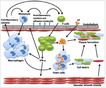
Atherosclerosis is a chronic inflammatory condition that contributes to the development of cardiovascular diseases (CAD). The process is initiated by retention of oxidized lipoproteins (LPs) in pre-lesional endothelium of the artery wall. Endothelial cells (EC) sense LPs through receptors include scavenger receptors and pattern re- cognition receptors. Triggered by LPs, ECs produce cell surface adhesion molecules, chemokines and inflamma- tory cytokines that recruit macrophages to extravasate into the arterial wall and phagocytose oxidized LPs. Lipid engorged macrophages evolve into foam cells, many of which become activated via scavenger or toll- like receptors and produce inflammatory chemokines and cytokines. The increased production of inflammatory chemokines and cytokines attracts other immune cells to the blood vessel wall, such as T cells, resulting in a ca- scading inflammatory response. As the condition pro- gresses, the oxidized LPs and cell debris together with smooth muscle cells develop into atheroma, an advance lesion. This atheroma can further develop to form a
fibrous plaque which is subject to rupture [2] (Figure 1).
IFN-γ expressed by the incoming T cells activates macro-
Figure 1. In response to lipoprotein retention in pre-lesional artery wall, endothelial cell permit monocyte infiltration. This infiltrated monocyte derived macrophages ingest lipid and transform into foam cells. This foam cell eventually died. Dying cells and other cellular debris along with choles-terol crystals form a necrotic core that eventually progress into atherosclerotic plaques.
ATP-binding cassette transporter A1 (ABCA1) is a key regulator that facilitates cholesterol efflux from ma- crophages and prevents the formation of pathological
foam cells. IFN-γ has been shown to down regulate
ABCA1 expression in Human THP-1 cells, a macro- phage cell line, by suppressing ABCA1 expression regu- lation factor through the JAK/STAT pathway [3]. In addi-
tion, IFN-γ activates the ERK pathway that is required
for the uptake of modified lipoprotein by theTHP-1 cells
[4]. These data indicate that IFN-γ plays a very important
role in the lipid accumulation in the artery wall by increasing lipoprotein uptake and inhibiting cholesterol efflux from marcophages. In addition to macrophages,
IFN-γ also has effects on nonimmune cells, the coronary
arteries and smooth muscle cells. IFN-γ sensitizes intact
arteries and cultured vascular smooth muscle cells (VSMCs) to exogenous dsRNA as well as self-RNA.
These IFN-γ primed VSMCs express higher levels of
TNF-α and IP-10 when stimulated compared to non-
treated samples. This study implies in the presence of
viral infection and cell death, IFN-γ would further ac-
celerate atherosclerosis by augmenting inflammatory responses in the microenviorment [5]. A recent report
also showed that peripheral IFN-γ may contribute to
coronary artery disease (CAD). In the peripheral blood of
CAD patients, CD8+CD56+ T cells constitute 30% of the
total CD8+ T cell population compared to 21% in normal
subjects. The presence of these cells has been linked to several autoimmune diseases. They produce higher levels
of IFN-γ, and are resistant to apoptosis. The capacity of
CD8+CD56+ T cells to express higher level IFN-γ may
drive the disease progression by maintaining an inflam- matory response in the periphery [6].
3. AUTOIMMUNE DISEASE
IFN-γ has been associated with promoting autoimmune
diseases due to its proinflammatory properties. The most
notable diseases associated with IFN-γ are systemic
lupus erythematous (SLE), multiple sclerosis (MS) and Rheumatoid Arthritis (RA). The main characteristic of the complex autoimmune disease SLE is the generation of autoantibodies by activated B cells. The antibody- complement complex causes local and systemic inflam- mation that can lead to kidney failure. Although previous
studies identified the importance of IFN-α in lupus
pathogenesis, recent studies have implied IFN-γ as the
the single-nucleotide polymorphisms (SNP) of IFN-γ
genes to lupus susceptibility. The allele with the greatest
risk, SNP (rs2430561), is located in an NF-κB binding
site and has elevated IFN-γ expression compared to a
low-risk allele [7]. Also, peripheral blood T cells from SLE patients expressed significantly higher levels of
IFN-γ when stimulated with anti-CD3/CD28 antibodies
compared to those from normal controls [8]. Increased level of STAT1/pSTAT1 can be detected in both lym- phocytes and monocytes from SLE patients, and ex- pression levels of STAT1 correlate with disease activity [9]. These results align with the findings in our labora- tory. We have observed rapid appearance of SLE-like symptoms in our AU-rich element deletion mice whose deletion results in a constant expression of low levels of
IFN-γ (Hodge et al. manuscript in preparation). Further-
more, a recent report by Lee and colleges provides a
probable mechanism of the IFN-γ contribution to lupus
pathogenicity. At 7 weeks of age, IFN-γ can be detected
in the serum in lupus-prone Roquinsan/san mice used in
this study. The detection of IFN-γ coincided with the
onset of disease. They found excessive IFN-γ expres-
sion contributed to lupus pathology by promoting accu- mulation of T follicular helper cells, leading to abnor- mal germinal center formation and autoantibody produc- tion [10].
MS is an autoimmune disease caused by infiltration of autoreactive T lymphocytes into the central nervous sys- tem and result in damages to neurons and axons. Al-
though MS pathogenesis involves IFN-γ secreting T cells,
IFN-γ has been shown to be neuron protective and able
to mitigate the severity of disease. In the MS animal model, experimental autoimmune encephalomyelitis (EAE), IFN-γ receptor knockout (IFN-γR−/−) in the CNS
resulted in more severe neurological deficits as compared
to IFN-γR−/− in the periphery [11]. In another similar
study, EAE was induced in transgenic mice expressing
signaling defective dominant-negative IFN-γ receptors
(GFAPγ R1 mice) on astrocytes. Inhibition of IFN-γ
signaling did not prevent disease onset. In contrast,
GFAPγR1 mice exhibited extensive demyelination at
peak acute disease with increased mortality [12]. These data could explain the findings that treatment in MS
patients with anti-IFN-γ antibodies did not ameliorate the
symptoms, but actually aggravated the disease. Without
the inhibition of IFN-γ, Th1 cells will commit to the
Th17 lineage whose contributory role in EAE has been
widely reported. Nevertheless, administration of IFN-γ is
not an appropriate therapy for MS, since only CNS-re-
stricted IFN-γ is neural protective. Exogenous IFN-γ in
the periphery would only aggravate the inflammatory responses and lead to undesirable outcomes. In fact early
clinical trials with IFN-γ in MS patients did show a
worsening of the disease [13].
The role of IFN-γ in the pathogenesis of RA still re-
mains controversial. RA is characterized by the accumu- lation of effector T cells that target joints resulting in da-
mage to the cartilage and bone. In several studies, IFN-γ
has been shown to be a disease limiting factor in an RA
animal model, collagen-induced arthritis (CIA), IFN-γ
administration ameliorated disease severity, while CIA
disease progression was worse in IFN-γR−/−[14]. IFN-γ
was also found to inhibit IL-1β-induced cartilage-de-
grading matrix metalloproteinase production when cul- tured with RA synovial tissue specimens [15]. However,
other reports implied the involvement of IFN-γ in RA
pathogenesis. In RA patients, higher level of STAT1 can be found in the peripheral blood and there was a corre- lation of STAT1 levels to disease activity [16]. A study
by Doodes et al. found neutralization of IFN-γ inhibits
arthritis. IFN-γ−/− mice developed less severe proteogly-
can-induced arthritis with delayed onset. The IL-17 level in these mice contributed to the disease development, since IFN-γ/IL-17−/− mice have disease in less severity in
comparison. The results suggested that both IFN-γ and
IL-17 have the potential to induce arthritis, though the
strength of IFN-γ signaling dictates IL-17 contribution to
disease onset [17]. While this study clearly addresses the
importance of IFN-γ in RA pathogenesis, it also displays
the discrepancy among disease models. In the CIA model, IL-17 expression is more detrimental with respect to disease progression; hence, disease severity is worse in
IFN-γR−/− mice. However in the Doodes’ report [17],
arthritis induced by proteoglycan was more severe in IL-17−/− mice. Also, the Saha et al. have shown that, in
the CIA model, the symptoms improved when animals
treated with anti-IFN-γ antibodies in the early phase of
disease induction while symptoms worsen when the anti- bodies were given during the later stage of the disease [18]. These results indicate that even under the same di- sease context, any change in disease factors, such as anti- gen, gender, timing of treatment and age of subjects, would result in different immune environments, leading to differences in outcome.
4. CANCER
It is well-documented that IFN-γ can contribute to the
containment of tumor progression and growth by in- creasing tumor antigen presentation to tumor specific T cells and increased susceptibility to NK cytotoxicity. In
addition to promote immune response to the tumor, IFN-γ
also can induce the expression of tumor suppressing fac- tors. For example, Mig-1, the monokine attracting acti-
vated T and NK cells, is induced by IFN-γ and was re-
cently shown to limit metastasis in a mammary tumor
model. Furthermore, GBP-1, a major product of IFN-γ
of highly malignant TS/A mammary carcinoma cells in
immune-competent Balb/c mice [19]. IFN-γ was also
shown to inhibit growth and promote cell death in human hepatocyte carcinoma cells by inducing autophagy— through IRF-1 signaling pathway [20]. In the study by
Schmitt et al., IFN-γ induced the expression of miR-29,
an anti-tumor factor. MiR-29 family members target the expression of proteins involved in invasion, migration or proliferation of cells and silencing of these target pro- teins would significantly inhibit tumor growth. MiR-29 expression levels were inversely correlated with the pro- liferation rate of various melanoma cell lines [21]. In murine renal cell carcinoma cell line model (RCC), cul-
turing the cells with IFN-γ induced expression of nitric
oxide synthase (iNOS). The resulting high elevation of nitric oxide (NO) and citrulline, and a decrease in argi- nase activity lead to cell cycle arrest, and significantly inhibit RCC proliferation [22].
Despite the antiangiogenesis property of IFN-γ, there
is ample evidence to indicate that it has a protumorigenic effect as well. A meta-analysis genetic study with over
1900 cancer cases indicated a correlation of IFN-γ + 874
T/A with a significantly increased risk of cervical can- cer in the comparison of the AT versus TT genotype [23].
Transgenic expression of IFN-γ in the mouse stomach
induced an extensive inflammatory response accom- panied with increased cell proliferation. These data imply
that IFN-γ may contribute to inflammation-associated
gastric neoplasia [24]. Furthermore in an animal model
of UVB-induced melanoma, IFN-γ was indicated as dri-
ving a protumorigenic microenvironment through the ac- tivation of melanocytes, an effect that was abolished by
systemic administration of anti-IFN-γ antibodies [25]. In
the tumor microenvironment, IFN-γcan also function to
protect tumor cells from immune destruction. It has been
shown that IFN-γ induced PD-L1 expression on acute
myeloid leukemia and human oral squamous carcinoma. The interaction of PD-L1 expressed on the cancer cells and PD-1 expressed on T cells suppresses T cell activation and induces T cell apoptosis. As a result, the antitumor immunity of T cells is inhibited in the micro-environ- ment, thus promoting tumor survival [26,27].
These studies suggest that anti- and protumorigenic-
functions of IFN-γ are cell and tumor specific. Therefore,
to be effective, the therapies and vaccines incorporating
IFN-γ may need to be tailored in the context of the spe-
cific tumor type.
5. INFLAMMATORY BOWEL
DISEASE (IBD)
IBD, including Crohn’s disease (CD) and ulcerative colitis (UC), is triggered by abnormal immune responses toward common gut microbiota. During the course of
disease, epithelial cells are exposed to an array of proin- flammatory cytokines which results in a disruption of epithelium homeostasis and compromises the mucosal barrier. Subsequently, the penetration of gut bacteria across the corrupted mucosal epithelium leads to infiltra-
tion of T cells [28]. Vigorous IFN-γ production by T
cells can be detected in colonic mucosal tissue cultures and intestinal lamina propria mononuclear cells from IBD
patients. Ample evidence has implicated IFN-γ signaling
to the pathogenesis of this disease by both augmenting the inflammatory response and compromising the mucosal barrier.
Wnt-β-catenin signaling is one of the signaling path-
ways that maintain epithelium homeostasis by regulating intestinal epithelial cell (IECs) proliferation and survival.
There is a strong association between aberrant Wnt-β-
catenin signaling and IBD as well as intestinal cancer.
Treatment of IECs with IFN-γ leads to activation of β-
catenin signaling through phosphoinositide-3 kinase
(PI3K) and AKT. IFN-γ induced AKT-β-catenin activa-
tion promotes IEC proliferation. However, this activation can also induce the expression of a Wnt inhibitor, Dkk. As a result, increased incidence of apoptosis and reduced IEC proliferation can be seen in a mouse colitis model
and IEC cultured with extended IFN-γ treatment, as
Dkk1 suppresses Wnt-β-catenin signaling [29]. In addi-
tion, IFN-γinduces GBP-1 expression which suppresses
the expression of β-catenin, and in turn disrupts Wnt-
β-catenin signaling [30]. The mucosal barrier in the
intestine provides a physical hurdle that limits access of toxins and microbes to underlying tissues. During inflam- mation, epithelial cells express hypoxia-inducible factor (HIF)-1,resulting in induction of several genes that streng-
then the mucosal barrier [31]. IFN-γ was shown to inhibit
the expression of HIF-1β which in turns suppresses HIF-1
activity and expression in IECs both in vitro and in vivo
[32]. This weakening of the mucosa enhances bacterial content leakage, thus attracting immune cells to the in- flamed gut and hence further promoting the developing of disease.
Contrary to its role in promoting IBD, several reports
have shown IFN-γ could be a negative regulator of
disease severity by inhibiting Th17 generation. IL-23 has been shown to be colitogenic, as it promotes the genera- tion of Th17 cells and suppresses Treg differentiation in the intestinal microenvironment [33]. Sheikh and collea-
gues used an IL-10−/− mice experimental colitis model
and showed that IFN-γ inhibits IL-23 expression in
macrophages isolated from lamina propria of IL-10−/−
mice. Furthermore, IFN-γR1/IL-10−/− mice have severe
colonic inflammation with increased IL-23 expression
[34]. A study by Jin et al. also showed that IL-17 plays a
critical role in IBD pathogenesis. IFN-γ−/− mice develop
treatment , while IL-17−/− mice develop colitis to a lesser
degree [35]. However, one needs to be cautious because
these reports do not suggest IFN-γ as a treatment for IBD.
While blocking the tissue destructive effects of IL-17
may ameliorate the severity of IBD, the presence of IFN-γ
would induce profound inflammatory responses and re- sults in IBD pathology as shown in numerous reports [35,36].
6. NEURODEGENERATIVE DISEASES
Injury or infection in the brain activates microglia and glial cells function as the first line of defense as well as astrocytes, another type of glial cell that plays a role in the repair process. When activated, these cells release an
array of proinflammatory cytokines, including IL-1β,
TNF-α, and IL-6, as well as ROS and NO, which are
toxic to neurons and result in cell death [37]. When in- flammation resolves, astrocytes start the repair process. However, in neurodegenerative diseases, such as Parkin- son’s disease (PD), Alzheimer’s disease (AD) and MS, sustained inflammation is observed that ultimately causes the pathology associated with these diseases. The charac- teristic of PD is gradual and progressive loss of dopami-
nergic neurons in the substantia nigra. Microglial ac-
tivation and T cell infiltration are commonly observed in postmortem PD brain tissue. Accumulation of proinflam- matory cytokines can be detected in the brain and cere- brospinal fluid of PD patients. These observations suggest the involvement of inflammation in disease development, although the pathogenic mechanisms are still unclear. In AD, the two hallmark features of this disease are the
extracellular Aβ plaques, a result of the cleavage of the
amyloid precursor protein (APP), and intracellular neuro- fibrillary tangles (NFTs). NFTs are composed of the tau protein which is abnormally hyperphosphorylated and forms insoluble fibrils in AD patient that initiate the deposition within the cell. It has been widely believed
that Aβ deposition play a major role in the pathogenesis
of AD by activating microglia in an effort to clear Aβ.
However, microglia in AD patients fail to phagocytose
Aβ that results in Aβ plaque formation and profound
inflammatory response.
IFN-γ has been known to activate microglia and astro-
cytes and induces iNOS expression in CNS [38]. Injec- tion in the mouse cerebral ventricles with recombinant
adeno-associated viruses (rAAV) expressing mIFN-γ re-
sults in accumulation of activated microglia and astro- cytes in the CNS. Basophilic lesions in the basal ganglia caused by calcified deposits can be found in the brain of these mice. This pathology is described in PD as well as idiopathic basal ganglia calcification, also a neurode- generative disease [39]. In chronic Parkinsonian mon-
keys, increased IFN-γ was found in the serum and CNS
years after disease initiation. Also, constant IFN-γR sig-
naling was detected in both microglial and astroglial cells in these animals. Moreover, there was a positive
correlation between the levels of IFN-γ in CNS and the
degree of dopaminergic neuronal degeneration in the di- seased monkeys. In PD-induced mice, microglia and astroglia are activated before the death of dopaminergic cells, and the activation level of these cells was severely
dampened in IFN-γ−/− mice [40]. These data imply that
IFN-γ not only is critical in the initial glial cell activation in PD, but also is responsible for maintaining the per- petual activation status throughout the course of disease. In the AD mouse model, where APP is overexpressed, infiltration of IFN-γ expressing T cells can be detected in
the CNS. Adoptively transferring Aβ specific Th1 cells
into these mice increased Aβ deposition and microglial
activation. Treatment with anti-IFN-γ antibodies amelio-
rated AD-like symptoms [41]. This study clearly shows
the association of IFN-γ in accelerating the pathology of
AD.
Despite the proinflammatory properties, IFN-γ has
been shown to be also neuroprotective. Studies have
shown that IFN-γ plays a major part in CNS reparation
after injury. In the rat hippocampus following status epi- lepticus, neutralizing IFN-γ or its receptor aggravated the neuronal injury, while intracerebroventricular injection
of IFN-γ attenuates the damage [42]. IFN-γ might also
play a role in neuron injury repair, since more extensive
neurodegeneration can be observed in IFN-γ−/− mice
compared to WT mice after injury induction in the ventral horn of the spinal cord [43]. In the case of AD,
IFN-γ has been shown to decrease Aβ plaque burden in
the beginning stage of the disease. Microglial cells of APP transgenic mice upregulate MHCII and CD11c as well as a component of complement system after injec-
tion of rAAV expressing mIFN-γ in the brain. These
IFN-γ primed mouse microglia cells are able to decrease
Aβ aggregation through phagocytosis. However, the im-
mune response against the Aβ aggregate is clearly ineffi-
cient to clear Aβ deposition since the Aβ deposit con-
tinues to accumulate with age in these animals. [44].
7. STEM CELL RESEARCH
Hematopoietic stem cells (HSCs) are in a constant state of differentiation and proliferation to maintain sufficient blood cells in the periphery. Recently, more and more research is focused on the impact of proinflammatory cytokines on hematopoiesis, since HSC output is altered in response to these signals during infection, injury or radiation/chemotherapy. In line with previous reports
that IFN-γ suppresses hematopoiesis, a recent report by
de Bruin et al. showed that IFN-γ inhibited STAT5
for HSC self-renewal. When infected with lymphocytic choriomeningitis virus, HSC recovery was more efficient
in IFN-γ−/− mice than WT [45]. However, new studies
have emerged that suggest the effects of IFN-γ on HSCs
may not be all negative. IFN-γ has been shown to be able
to increase proliferation and mobilize HSCs during chronic
infection by Mycobacterium avium [46]. In a study by
MacNamara and colleges, IFN-γ signaling altered myeloid
progenitor function and phenotype in order to augment the production of granulocytes and monocytes during
Ehrlichia muris infection [47]. These results indicate that
IFN-γ regulates HSC in the states of homeostasis as well
as infection. The discrepancy among reports could be a result of different microenvironments used for experi- ments, as the BM niche is proven to be important for
HSC maintenance. Thus, the system used for HSC gen-
eration or maintenance may affect the results. Also,
IFN-γ may only affect certain HSC/progenitors at specific
stages of differentiation. Thus, different experimental set- tings may give rise to different results.
IFN-γ also has been indicated in inducing tissue re-
generation and differentiation in stem cell transplantation studies. Stem cell transplantation provides a cell source that could replace dead or injured tissue. Factors that control stem cell proliferation and tissue differentiation would be beneficial for optimizing the efficacy of therapy.
Results from in vitro screening of different neural stem/
progenitor cells (NSPCs) factors found that IFN-γ had
the best capacity among tested treatments in promoting neuronal differentiation based on neural morphology and
β-III tubulin expression [48]. However, not all neural
stem cell/progenitors respond to IFN-γ in the same man-
ner and can be used for transplantation therapy. In the
report by Duque et al., treatment of IFN-γ on oligo-
dendrocyte-type 2 astrocyte progenitor cells (O-2A/ OPC) caused cell arrest and inhibited the generation of oligo- dendrocytes [49]. In addition to neural stem cell research,
IFN-γ also has been showed to drive the differentiation
of human mesenchymal stem cells (hMSC) into osteo- blasts in an autocrine manner. Additional of exogenous
IFN-γ in hMSC cell culture accelerated osteoblast dif-
ferentiation and induced higher levels of Runx2 ex- pression, a gene essential for osteoblast differentiation and function, [50]. As the interest in stem cell transplant- ation therapy continues to increase, we can expect to see
an influx of studies on the impact of IFN-γ on stem cell
development and maturation in the very near future.
8. CONCLUSION
Nearly five decades after the first discovery of IFN-γ, we
are yet to fully understand the complexity of its function.
What we can be sure about is that the effects of IFN-γ
are cell and tissue specific. Researchers should pay addi-
tional attention to the samples and conditions used when
studying IFN-γ. Different cells, tissues and mouse genetic
backgrounds will give rise to different outcomes. In the content of disease, the stage of disease progression, age and gender during sampling will impact on the study readout. All these factors make it even more difficult to decipher the biological function of this complex gene and thus there will be a continuing effort to understand the role of IFN-γ on host cell biology.
9. DISCLAIMER
The publisher or recipient acknowledges right of the U.S. Government to retain a nonexclusive, royalty-free license in and to any copyright covering the article.
The contents of this publication do not necessarily reflect the views or policies of the Department of Health and Human Services, nor does mention of trade names, commercial products, or organizations imply endorse- ment by the U.S. Government.
Conflict of interest disclosure: The authors declare no competing financial interests
REFERENCES
[1] Lin, F.-C. and Young, H. (2012) Interferon-gamma, in encyclopedia of signaling molecules. Springer, New York, 966-972.
[2] Tabas, I., Williams, K.J. and Boren, J. (2007) Subendo- thelial lipoprotein retention as the initiating process in athe- rosclerosis: Update and therapeutic implications. Circula- tion, 116, 1832-1844.
doi:10.1161/CIRCULATIONAHA.106.676890
[3] Hao, X.R., et al. (2009) IFN-gamma down-regulates ABCA1 expression by inhibiting LXRalpha in a JAK/ STAT sig- naling pathway-dependent manner. Atherosclerosis, 203, 417-428.doi:10.1016/j.atherosclerosis.2008.07.029 [4] Li, N., et al. (2010) ERK is integral to the IFN-gamma-
mediated activation of STAT1, the expression of key genes implicated in atherosclerosis, and the uptake of mo- dified lipoproteins by human macrophages. The Journal of Immunology, 185, 3041-3048.
doi:10.4049/jimmunol.1000993
[5] Ahmad, U., et al. (2010) IFN-gamma primes intact hu- man coronary arteries and cultured coronary smooth mu- scle cells to double-stranded RNA- and self-RNA-induced inflammatory responses by upregulating TLR3 and mela- noma differentiation-associated gene 5. The Journal of Immunology, 185, 1283-1294.
doi:10.4049/jimmunol.0902283
[6] Bergstrom, I., et al. (2012) Persistent accumulation of interferon-gamma-producing CD8+CD56+ T cells in blood from patients with coronary artery disease. Atherosclero- sis, 224, 515-520.
doi:10.1016/j.atherosclerosis.2012.07.033
lupus susceptibility. Annals of the Rheumatic Diseases, 70, 1878-1879.doi:10.1136/ard.2010.147249
[8] Harigai, M., et al. (2008) Excessive production of IFN- gamma in patients with systemic lupus erythematosus and its contribution to induction of B lymphocyte stimulator/B cell-activating factor/TNF ligand super-family-13B. The Journal of Immunology, 181, 2211-2219.
[9] Karonitsch, T., et al. (2009) Activation of the interferon- gamma signaling pathway in systemic lupus erythemato- sus peripheral blood mononuclear cells. Arthritis & Rheu- matism, 60, 1463-1471.doi:10.1002/art.24449
[10] Lee, S.K., et al. (2012) Interferon-gamma excess leads to pathogenic accumulation of follicular helper T cells and germinal centers. Immunity, 37, 880-892.
doi:10.1016/j.immuni.2012.10.010
[11] Lee, E., et al. (2012) IFN-gamma signaling in the central nervous system controls the course of experimental auto- immune encephalomyelitis independently of the localiza- tion and composition of inflammatory foci. Journal of Neuroinflammation, 9, 7.
[12] Hindinger, C., et al. (2012) IFN-gamma signaling to as- trocytes protects from autoimmune mediated neurological disability. PLoS One, 7, e42088.
doi:10.1371/journal.pone.0042088
[13] Lees, J.R. and Cross, A.H. (2007) A little stress is good: IFN-gamma, demyelination, and multiple sclerosis. Jour- nal of Clinical Investigation, 117, 297-299.
doi:10.1172/JCI31254
[14] Schurgers, E., Billiau, A. and Matthys, P. (2011) Colla- gen-induced arthritis as an animal model for rheumatoid arthritis: Focus on interferon-gamma. Journal of Inter- feron & Cytokine Research, 31, 917-926.
doi:10.1089/jir.2011.0056
[15] Page, C.E., et al. (2010) Interferon-gamma inhibits inter- leukin-1beta-induced matrix metalloproteinase produc- tion by synovial fibroblasts and protects articular carti- lage in early arthritis. Arthritis Research & Therapy, 12, R49.
[16] Karonitsch, T., et al. (2012) Interferon signals and mono- cytic sensitization of the interferon-gamma signaling path-way in the peripheral blood of patients with rheuma- toid arthritis. Arthritis & Rheumatism, 64, 400-408. doi:10.1002/art.33347
[17] Doodes, P.D., et al. (2010) IFN-gamma regulates the requirement for IL-17 in proteoglycan-induced arthritis. The Journal of Immunology, 184, 1552-1559.
doi:10.4049/jimmunol.0902907
[18] Saha, B., et al. (2009) Gene modulation and immunore- gulatory roles of interferon gamma. Cytokine, 50, 1-14. doi:10.1016/j.cyto.2009.11.021
[19] Lipnik, K., et al. (2010) Interferon gamma-induced hu- man guanylate binding protein 1 inhibits mammary tumor growth in mice. Molecular Medicine, 16, 177-187. [20] Li, P., et al. (2012) Interferon-gamma induces autophagy
with growth inhibition and cell death in human hepato- cellular carcinoma (HCC) cells through interferon-regu- latory factor-1 (IRF-1). Cancer Letters, 314, 213-222. doi:10.1016/j.canlet.2011.09.031
[21] Schmitt, M.J., et al. (2012) Interferon-gamma-induced activation of Signal Transducer and Activator of Tran- scription 1 (STAT1) up-regulates the tumor suppressing microRNA-29 family in melanoma cells. Cell Communi- cation and Signaling, 10, 41.
[22] Tate, D.J., et al. (2012) Interferon-gamma-induced nitric oxide inhibits the proliferation of murine renal cell carci- noma cells. International Journal of Biological Sciences, 8, 1109-1120.doi:10.7150/ijbs.4694
[23] Mi, Y.Y., et al. (2011) Interferon gamma +874 T/A poly- morphism contributes to cancer susceptibility: A meta- analysis based on 17 case-control studies. Molecular Bi- ology Reports, 38, 4461-4467.
doi:10.1007/s11033-010-0575-3
[24] Syu, L.J., et al. (2012) Transgenic expression of inter- feron-gamma in mouse stomach leads to inflammation, metaplasia, and dysplasia. American Journal of Pathol- ogy, 181, 2114-2125.doi:10.1016/j.ajpath.2012.08.017 [25] Zaidi, M.R., et al. (2011) Interferon-gamma links ultra-
violet radiation to melanomagenesis in mice. Nature, 469, 548-553.doi:10.1038/nature09666
[26] Berthon, C., et al. (2010) In acute myeloid leukemia, B7-H1 (PD-L1) protection of blasts from cytotoxic T cells is induced by TLR ligands and interferon-gamma and can be reversed using MEK inhibitors. Cancer Im- munology, Immunotherapy, 59, 1839-1849.
doi:10.1007/s00262-010-0909-y
[27] Chen, J., et al. (2012) Interferon-gamma-induced PD-L1 surface expression on human oral squamous carcinoma via PKD2 signal pathway. Immunobiology, 217, 385-393.
doi:10.1016/j.imbio.2011.10.016
[28] Marsal, J. and Agace, W.W. (2012) Targeting T-cell mi- gration in inflammatory bowel disease. Journal of Inter- nal Medicine, 272, 411-429.
doi:10.1111/j.1365-2796.2012.02588.x
[29] Nava, P., et al. (2010) Interferon-gamma regulates intes- tinal epithelial homeostasis through converging beta- catenin signaling pathways. Immunity, 32, 392-402. doi:10.1016/j.immuni.2010.03.001
[30] Capaldo, C.T., et al. (2012) IFN-gamma and TNF-alpha- induced GBP-1 inhibits epithelial cell proliferation through suppression of beta-catenin/TCF signaling. Mucosal Im- munology, 5, 681-690.
doi:10.1038/mi.2012.41
[31] Colgan, S.P. and Taylor, C.T. (2010) Hypoxia: An alarm signal during intestinal inflammation. Nature Reviews Gastroenterology & Hepatology, 7, 281-287.
doi:10.1038/nrgastro.2010.39
[32] Glover, L.E., et al. (2011) IFN-gamma attenuates hy- poxia-inducible factor (HIF) activity in intestinal epithet- lial cells through transcriptional repression of HIF-1beta. The Journal of Immunology, 186, 1790-1798.
doi:10.4049/jimmunol.1001442
[33] Ahern, P.P., et al. (2010) Interleukin-23 drives intestinal inflammation through direct activity on T cells. Immunity, 33, 279-288.doi:10.1016/j.immuni.2010.08.010
experimental colitis. The Journal of Immunology, 184, 4069-4073.doi:10.4049/jimmunol.0903600
[35] Jin, Y., et al. (2012) IL-17/IFN-gamma interactions re- gulate intestinal inflammation in TNBS-induced acute colitis. Journal of Interferon & Cytokine Research, 32, 548-556.doi:10.1089/jir.2012.0030
[36] Ito, R., et al. (2006) Interferon-gamma is causatively involved in experimental inflammatory bowel disease in mice. Clinical & Experimental Immunology, 146, 330- 338. doi:10.1111/j.1365-2249.2006.03214.x
[37] Glass, C.K., et al. (2010) Mechanisms underlying in- flammation in neurodegeneration. Cell, 140, 918-934. doi:10.1016/j.cell.2010.02.016
[38] Jung, J.S., Kim, D.H. and Kim, H.S. (2010) Ginsenoside Rh1 suppressesinducible nitric oxide synthase gene ex- pression in IFN-gamma-stimulated microglia via modula- tion of JAK/STAT and ERK signaling pathways. Bio- chemical and Biophysical Research Communications, 397, 323-328. doi:10.1016/j.bbrc.2010.05.117
[39] Chakrabarty, P., et al. (2011) Interferon-gamma induces progressive nigrostriatal degeneration and basal ganglia calcification.Nature Neuroscience, 14, 694-696.
[40] Barcia, C., et al. (2012) IFN-gamma signaling, with the synergistic contribution of TNF-alpha, mediates cell spe- cific microglial and astroglial activation in experimental models of Parkinson’s disease. Cell Death & Disease, 2, e142.
[41] Browne, T.C., et al. (2013) IFN-gamma production by amyloid beta-specific Th1 cells promotes microglial ac- tivation and increases plaque burden in a mouse model of Alzheimer’s disease. The Journal of Immunology, 190, 2241-2251. doi:10.4049/jimmunol.1200947
[42] Ryu, H.J., et al. (2010) The protective effects of inter- leukin-18 and interferon-gamma on neuronal damages in the rat hippocampus following status epilepticus. Neuro- science, 170, 711-721.
doi:10.1016/j.neuroscience.2010.07.048
[43] Victorio, S.C., Havton, L.A. and Oliveira, A.L. (2010) Absence of IFNgamma expression induces neuronal de- generation in the spinal cord of adult mice. Journal of Neuroinflammation, 7, 77.
[44] Chakrabarty, P., et al. (2010) IFN-gamma promotes com- plement expression and attenuates amyloid plaque depo- sition in amyloid beta precursor protein transgenic mice. The Journal of Immunology, 184, 5333-5343.
doi:10.4049/jimmunol.0903382
[45] De Bruin, A.M., et al. (2013) Interferon-gamma impairs proliferation of hematopoietic stem cells in mice. Blood, 121, 3578-3585. doi:10.1182/blood-2012-05-432906 [46] Baldridge, M.T., et al. (2010) Quiescent haematopoietic
stem cells are activated by IFN-gamma in response to chronic infection. Nature, 465, 793-797.
[47] MacNamara, K.C., et al. (2011) Infection-induced mye- lopoiesis during intracellular bacterial infection is criti- cally dependent upon IFN-gamma signaling. The Journal of Immunology, 186, 1032-1043.
doi:10.4049/jimmunol.1001893
[48] Zahir, T., et al. (2009) Neural stem/progenitor cells dif- ferentiate in vitro to neurons by the combined action of dibutyryl cAMP and interferon-gamma. Stem Cells and Development, 18, 1423-1432.doi:10.1089/scd.2008.0412 [49] Tanner, D.C., Cherry, J.D. and Mayer-Proschel, M. (2011)
Oligodendrocyte progenitors reversibly exit the cell cycle and give rise to astrocytes in response to interferon-gam- ma. The Journal of Neuroscience, 31, 6235-6246. doi:10.1523/JNEUROSCI.5905-10.2011