0095-1137/00/$04.00⫹0
Copyright © 2000, American Society for Microbiology. All Rights Reserved.
Community Acquisition of Gentamicin-Sensitive Methicillin-Resistant
Staphylococcus aureus
in Southeast Queensland, Australia
GRAEME R. NIMMO,1* JACQUELINE SCHOONEVELDT,1GABRIELLE O’KANE,1† BRAD MCCALL,2ANDALISON VICKERY3
Microbiology Department, Queensland Health Pathology Service, Princess Alexandra Hospital, Woolloongabba 4102,1 Brisbane Southside Public Health Unit, Coopers Plains 4108,2and Microbiology Department,
Royal Prince Alfred Hospital, Camperdown 2050,3Australia
Received 12 April 2000/Returned for modification 28 June 2000/Accepted 18 August 2000
Community-acquired methicillin-resistantStaphylococcus aureus(MRSA) susceptible to gentamicin has been reported in a number of countries in the 1990s. To study the acquisition of gentamicin-sensitive MRSA (GS-MRSA) in southeast Queensland and the relatedness of GS-MRSA to other strains of MRSA, 35 cases of infection due to GS-MRSA from October 1997 through September 1998 were examined retrospectively to determine the mode of acquisition and risk factors for MRSA acquisition. Thirty-one isolates from the cases were examined using a variety of methods (antibiotyping, phage typing, pulsed-field gel electrophoresis [PFGE] fingerprinting, and coagulase typing by restriction analysis of PCR products) and were compared with strains of local hospital-acquired gentamicin-resistant MRSA (GR-MRSA) and of Western Australian MRSA (WA-MRSA). Only 6 of 23 cases of community-acquired GS-MRSA had risk factors for MRSA acquisition. Twenty of 21 isolates from cases of community-acquired infection were found to be related by PFGE and coagulase typing and had similar phage typing patterns. Hospital- and nursing home-acquired GS-MRSA strains were genetically and phenotypically diverse. Community-acquired GS-MRSA strains were not related to nosocomial GR-MRSA or WA-MRSA, but phage typing results suggest that they are related to GS-MRSA previously reported in New Zealand.
Methicillin (oxacillin)-resistantStaphylococcus aureus(MRSA) has proven to be one of the more widespread and durable nosocomial pathogens of the late 20th century (1, 34). In east-ern Australia the appearance of MRSA was documented as early as 1965 (26) and was followed by an epidemic of gen-tamicin-resistant MRSA (GR-MRSA) in the late 1970s and early 1980s (22). MRSA has remained endemic in eastern Australian states in the 1980s and 1990s, and the majority of isolates have been resistant to gentamicin and multiple other non-beta-lactam antimicrobials (30, 31). Throughout this pe-riod GR-MRSA did not become established as an endemic problem in the state of Western Australia (25). However, in the late 1980s strains of gentamicin-sensitive MRSA (GS-MRSA) began to cause community-acquired infections in re-mote Aboriginal communities in northern Western Australia and subsequently spread to the Perth metropolitan area in the south, causing both community-acquired and nosocomial in-fection (20, 24). These strains have been referred to as WA-MRSA. Further spread of WA-MRSA to the Northern Terri-tory has since been documented (16).
The emergence of GS-MRSA, as either a nosocomial or community-acquired infection phenomenon, is now worldwide. GS-MRSA with increased susceptibility to other antimicrobials has recently been reported in six widely dispersed hospitals in France and one in the West Indies (15). In the United States an increase in the incidence of community-acquired MRSA infections in children in Chicago has been observed (12). Many children had no identified risk factors for MRSA infection, and
14 of 15 isolates from such children were gentamicin suscep-tible and were more likely to be suscepsuscep-tible to other antimi-crobials than nosocomially acquired isolates. In the southwest Pacific region, community-acquired infections due to GS-MRSA have been reported in the mid-1990s in Auckland, New Zealand. The majority of strains involved belong to the West-ern Samoan phage pattWest-erns (WSPP), and infections are par-ticularly common among the Polynesian population (17, 24). The emergence of community-acquired GS-MRSA infections has also been observed in Brisbane, Sydney, Canberra, and Melbourne in eastern Australia in the late 1990s (5).
The observation in our laboratory that GS-MRSA was being isolated de novo from patients attending hospital emergency departments and outpatient clinics prompted a prospective collection of all GS-MRSA isolates from clinical specimens from October 1997 through September 1998 and a subsequent retrospective survey of associated clinical and epidemiological data. We wished to determine the mode of acquisition associ-ated with GS-MRSA, the spectrum of infection associassoci-ated with it, genetic relationships within GS-MRSA strains, and related-ness to local strains of GR-MRSA. We also hoped to deter-mine the relationship of these isolates to WA-MRSA and to the MRSA reported in the southwest Pacific region (SWP-MRSA).
MATERIALS AND METHODS
Setting.The study was performed at the microbiology laboratory at Princess
Alexandra Hospital. This laboratory serves a 900-bed university hospital and three community hospitals (400 beds in all) within the cities of Brisbane and Logan and the shire of Redland, all of which fall within the Brisbane metropol-itan area in southeast Queensland. A total of 820,000 people live within the area served by these institutions, although another three laboratories also provide services within the same area.
Study design.We conducted a retrospective analysis of all new unique clinical
isolates of GS-MRSA and cases associated therewith from October 1997 through September 1998. Medical records of all cases were reviewed and patients were interviewed by phone where possible to determine type of infection, acquisition * Corresponding author. Mailing address: QHPS Microbiology
De-partment, Princess Alexandra Hospital, Woolloongabba, Queensland 4102, Australia. Phone: 61 7 3240 2389. Fax: 61 7 3240 5786. E-mail: [email protected].
† Present address: Microbiology Department, The Prince Charles Hospital, Chermside 4032, Australia.
3926
on May 15, 2020 by guest
http://jcm.asm.org/
status (community, hospital, or nursing home), ethnicity, and outcome of infec-tion. Classification of infections as community acquired or nosocomial (hospital or nursing home) was in accordance with Centers for Disease Control and Prevention definitions (9). In addition, ascertainment of acquisition status in-cluded searching the medical record and questioning during the interview for evidence of contact with health care institutions (including nursing homes) within the preceding 12 months, previous surgery, underlying chronic disorder, or a household member with contact with health care institutions; cases of community-acquired infection were subclassified as either having or not having risk factors for prior MRSA acquisition.
Identification.S. aureuswas identified by the presence of clumping factor and
detection of thenucgene, and oxacillin (methicillin) resistance was confirmed by detection of themecgene. The multiplex PCR procedure used was based on a modification of the method by Unal et al. (32); themecAprimers were described by Murakami and Minamide (19), and thenucprimers were described by Brak-stad et al. (4). The 25-l reaction mixture consisted of 10l of lysate, 100M (each) deoxynucleoside triphosphate, 0.2 M (each) primer, 0.5 U of Dy-NAzyme II DNA polymerase (Finnzymes Oy, Espoo, Finland) in 10⫻PCR buffer (1⫻is 10 mM Tris-HCL [pH 8.8], 50 mM KCl, 1.5 mM MgCl2, 0.1%
[wt/vol] Triton X-100). DNA amplification consisted of an initial cycle of 94°C for 5 min, 55°C for 30 s, and 72°C for 2 min; this was followed by 29 cycles of 94°C for 30 s, 55°C for 30 s, and 72°C for 2 min. PCR products were visualized on 2% agarose gels stained with ethidium bromide.
Control and comparator strains.Control strains used for coagulase typing
were Staphylococcus epidermidisATCC 12228, S. aureus ATCC 29213, and
MRSA ATCC 49476. Control strains used formecandnucgene detection were
S. aureusATCC 29213, MRSA ATCC 49476,S. epidermidisATCC 14990, andS.
epidermidis(wild,mecApositive) PA2E16433. For the purpose of comparison,
WA-MRSA B8-10 and B8-31 (Pathcentre, Perth, Western Australia, Australia) and six local isolates of hospital-acquired GR-MRSA selected to represent prev-alent antibiograms (results not shown) and phage types were also tested. All isolates were stored on Protect bacterial preservers (Technical Service Consult-ants Ltd., Lancashire, United Kingdom) at⫺80°C.
Antimicrobial susceptibility tests.Tests for susceptibility to fusidic acid,
ri-fampin, tetracycline, erythromycin, and ciprofloxacin were performed by the Vitek IMS using the GPS-IX card (bioMerieux Vitek, Hazelwood, Mo.). The production of-lactamase was determined by the use of a Cefinase disc (Becton Dickinson, Cockeysville, Md.). Heteroresistance to oxacillin was detected by a disc method (7). Briefly, a 5-g oxacillin disc was added to a Mueller-Hinton agar plate containing no NaCl using an inoculum of 108CFU/ml. The plate was
incubated for 48 h at 30°C. A zone diameter of⬍20 mm indicated a resistant isolate. Heterogeneously resistant isolates exhibited partial growth or microcolo-nies within the inhibition zone.
Coagulase typing by PCR.Molecular typing on the basis of coagulase gene
polymorphisms was performed by a modification of the method of Goh et al. (10). Overnight broth cultures (1 ml of tryptic soy; 35°C) were washed by cen-trifugation (1,000⫻g) in 1 ml of 50 mM Tris-EDTA (TE) buffer (Sigma, St. Louis, Mo.). Pellets were resuspended in 500l of TE containing 15 U of lysostaphin (Sigma)/ml. Suspensions were heated to 37°C for 1 h. One milliliter of lysing buffer (0.45l of Igepal CA-630 [Sigma], 0.45l of Tween 20 [Sigma], 6l of proteinase K [Sigma], and 1 ml of PCR buffer [50 mM KCL, 10 mM Tris-HCL, 1.5 mM MgCl]) was added before the samples were heated for 1 h at 56°C. Samples were heated at 95°C for 10 min and centrifuged, and the super-natant was frozen at⫺80°C for subsequent use. The 3⬘-end region of the coag-ulase gene was amplified using primers COAG2, 5⬘CGAGACCAAGATTCAA CAAG3⬘, and COAG3, 5⬘AAAGAAAACCACTCACATCA3⬘, which hybridize to sites 1632 to 1651 and 2589 to 2608, respectively. PCR amplifications were performed by adding the cell lysate (10l) to a 40-l PCR mixture with the addition of 1.5 mM MgCl2, as described in detail by Goh et al. (10). Seventeen
microliters of the PCR product was digested for 15 min at 37°C with 6 U of the restriction enzymeHaeIII (Sigma) in 1.9l of buffer provided as described by Lawrence et al. (14). TheHaeIII digests were visualized on 2% agarose gels stained with ethidium bromide. Isolates were allocated to types based on the sizes of their PCR products and to subtypes based on restriction fragment length polymorphisms (RFLPs) of the digested product. Subtypes were determined by the number of bands present and their sizes.
Fingerprinting by PFGE.Pulsed-field gel electrophoresis (PFGE) of
chromo-somal DNA was performed using the enzymeSmaI. DNA was separated on a GenePath system (Bio-Rad, Hercules, Calif.) using the GenePath group 1 re-agent kit (Bio-Rad) with initial pulse times of 5.3 and 34.9 s at the end of the 20-h run. Gels were stained with ethidium bromide and were photographed under UV illumination. The patterns were compared visually using the criteria of Tenover et al. (28) and were analyzed with GelCompar software (Applied Maths, Kor-trijk, Belgium). Results were analyzed using the unweighted pair group method for arithmetic averages and the Dice coefficient (6) with 1.2% band tolerance.
Phage typing.Phage typing was performed using the method of Blair and
Williams (2). The 23 phages of the Basic International Set of Typing Phages were supplemented by 10 phages of the International Set of Experimental Phages for MRSA (23), by two experimental phages issued by the International Centre at Colindale, London, United Kingdom, and by eight experimental phages isolated at the Royal Prince Alfred Hospital, Sydney, Australia (33). All phages were used at 100⫻routine test dilution.
Statistical analysis.Categorical data were analyzed by comparing differences
in proportions. Medians were compared using the Mann-Whitney rank sum test. The significance level was set at 0.05. Rank sum and confidence interval calcu-lations were performed using Graphpad Prism, version 3.00 (GraphPad Software Inc., San Diego, Calif.) and C.I.A., version 1, 1989 (BMA Publishing, London, United Kingdom), respectively.
RESULTS
Thirty-five cases of infection due to GS-MRSA were iden-tified in 35 patients. The majority of cases were community-acquired infections, and most of these occurred in Polynesians (Table 1). Six (26%) of 23 cases of community-acquired infec-tion had risk factors for MRSA: one Polynesian patient worked in a hospital in a non-patient contact position, and two of her family household members were also patients in the study; another Polynesian patient was a domestic worker in a nursing home; the remaining two had previous hospital contact as patients. All resided in the laboratory’s service area with the exception of one who lived in Sydney. The predominant types of infection were soft-tissue abscesses in community-acquired infections and surgical wound infection in hospital-acquired infections (Table 1). Three Caucasian patients died following hospital-acquired infection. One 87-year-old patient died of GS-MRSA septicemia with no primary focus identified. Two other patients (aged 86 and 56) died of other causes. Five cases of community-acquired infection occurred in two Polynesian families (two in one and three in the other).
[image:2.612.309.551.100.345.2]Isolates were available for study in 21 of 23 community-acquired infections, 8 of 10 hospital-community-acquired infections, and 2 of 2 nursing home-acquired infections (P, 0.6). All isolates were positive for nuc and mecAgene products, and all pro-duced-lactamase. Resistance to other antimicrobials was rare TABLE 1. Epidemiological and clinical characteristics of 35 cases
of GS-MRSA infection analyzed according to mode of acquisitionb
Characteristic
No. of cases of infections that were:
Pa
Community acquired
Health care facility
acquired No risk
factors With riskfactors Hos-pital Nursinghome
Total no. of cases 17 6 10 2
Sex 0.19
Male 11 2 4
Female 6 4 6 2
Ethnicity
Aboriginal 1
Caucasian 5 1 8 2 0.001
Polynesian 11 5 2
Nature of infection
Abscess 13 3 ⬍0.0001
Cellulitis 3 1 2 1 0.58
Surgical wound 5
Decubitis ulcer 1
Hydradenitis suppurativa 1
Impetigo 1
Pneumonia 1
Septicemia 1
Septicemia and
osteo-myelitis 1
Urinary catheter 1
aStatistical tests compare combined results for community-acquired infections with those for health care facility-acquired infections.
bMedian ages (confidence intervals) of patients with community- and health care facility-acquired infections, 29 (24 to 45 years) and 67 years (43 to 28 years), respectively (P⫽0.0002).
on May 15, 2020 by guest
http://jcm.asm.org/
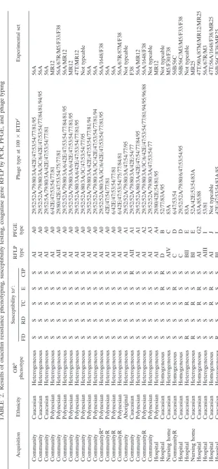
TABLE 2. Results of oxacillin resistance phenotyping, susceptibility testing, coagulase gene RFLP by PCR, PFGE, and phage typing Isolate Acquisition Ethnicity OR b phenotype Susceptibility to c: RFLP type PFGE type Phage type at 100 ⫻ RTD d Experimental set FD RD TC E CIP F810539 Community Caucasian Heterogeneous S S S S S AI A0 29/52/52A/79/80/3A/42E/47/53/54/77/81/95 56A D817541 Community Caucasian Heterogeneous S S S S S AI A0 29/52/52A/79/80/3A/3C/6/42E/47/53/54/77/84/81/94/95 56A A830538 Community Caucasian Heterogeneous S S S S S AI A0 29/52/5A/79/80/3A/42E/47/53/54/77/81 56A I825560 Community Polynesian Heterogeneous S S S S S AI A0 6/42E/47/53/54/77/81 MR12 B826559 Community Polynesian Heterogeneous S S S S S AII A0 29/80/42E/47/53/54/75/77/81 56A/67R/M5/F33/F38 I816601 Community Polynesian Heterogeneous S S S S S AI A0 29/52/52A/79/80/3A/6/42E/47/53/54/77/84/81/95 56A/MR12 H823537 Community Polynesian Heterogeneous S S S S S AI A0 29/52/52A/79/80/3A/6/42E/47/53/54/77/81/95 MR12 G823530 Community Polynesian Heterogeneous S S S S S AI A0 29/52/52A/80/3A/6/42E/47/53/54/77/81/95 47T/MR12 D828570 Community Polynesian Heterogeneous S S S S S AI A0 29/52/52A/80/3A/3C/47/53/54/77/95 Not typeable A803355 Community Polynesian Heterogeneous S S S S S AI A0 29/52/52A/79/80/3A/42E/47/53/54/77/81/94 56A A806533 Community Polynesian Heterogeneous S S S S S AI A0 29/52/52A/79/80/3A/3C/42E/47/53/54/77/81/94 56A E803543 CommunityR a Polynesian Heterogeneous S S S S S AI A0 29/52/52A/80/3A/3C/6/42E/47/53/54/77/81/95 56A/1648/F38 E822547 CommunityR Polynesian Heterogeneous S S S S S AI A0 42E/47/54/77/81 56A F809718 CommunityR Polynesian Heterogeneous S S S S S AI A0 6/42E/47/53/54/77/81 56A F809715 CommunityR Polynesian Heterogeneous S S S S S AI A0 6/42E/47/53/54/75/77/84/81 56A/67R/87M/F38 A823547 Community Aboriginal Heterogeneous S S S S S AI A1 29/52/52A/79/80/3A/47/54/77/95 Not typeable C810534 Community Caucasian Heterogeneous S S S S R AII A1 29/52/52A/79/80/3A/42E/54/77 F38 G821561 Community Polynesian Heterogeneous S S S S S AI A1 29/52/52A/80/3A/42E/47/54/77/84/95 56A/MR12 D821552 CommunityR Polynesian Heterogeneous S S S S S AI A2 29/52/52A/79/80/3A/3C/6/42E/47/53/54/77/81/94/95/96/88 56A/1648/F38 E802537 Community Polynesian Heterogeneous S S S S S AI A3 29/52/52A/79/80/3A/47/53/54/77 Not typeable I802552 Hospital Polynesian Heterogeneous S S S S S AI A4 29/80/42E/54/81/95 MR12 E822485 Hospital Caucasian Homogeneous S S S R R D B 52/77/83A/95 Not typeable J710566 Nursing home Caucasian Heterogeneous S S S S S AIV C 83A M5/F30/F38 F829549 CommunityR Caucasian Homogeneous S S S R S C D 6/47/53/54 56B/56C C801535 Hospital Caucasian Homogeneous S S S S S C D 29/52/52A/79/80/6/47/53/54/95 56B/56C/M3/M5/F33/F38 D828354 Hospital Caucasian Heterogeneous R S R S S BII E 83A Not typeable B827549 Nursing home Caucasian Heterogeneous R S R S S BI E 52A/42E/53/54/83A MR25 I823541 Hospital Caucasian Homogeneous S S R R S AI G2 83A/85/88 47T/90A/87M/MR12/MR25 E804531 Hospital Caucasian Heterogeneous S S S S S AIII I 53/81 56A/67R/M3 E812560 Hospital Caucasian Homogeneous S S R R S AI J Not typeable 47T/56A/1648/F38/MR25 D808118 Hospital Caucasian Heterogeneous R S S S S BI L 42E/47/53/54/83A/85 56B/56C/F38/MR25 aCommunityR, community acquired with risk factors. bOR, oxacillin resistance. cS, susceptible; R, resistant. FD, fusidic acid; RD, rifampin; TC, tetracycline; E, erythromycin; CIP, ciprofloxacin. dRTD, routine test dilution.
on May 15, 2020 by guest
http://jcm.asm.org/
[image:3.612.166.477.60.729.2]in community-acquired isolates but was common in isolates acquired in a hospital or nursing home: two community-ac-quired isolates had single-agent resistance, while five hospital-or nursing home-acquired isolates had two-agent resistance and one had single-agent resistance (Table 2). Expression of oxacillin resistance was homogeneous in only one community-acquired isolate and in four hospital-community-acquired isolates (P, 0.01). All of these isolates were from Caucasian patients, and the patient with the community-acquired isolate had a risk factor for MRSA acquisition (Table 2). All local GR-MRSA strains tested expressed homogeneous resistance (Table 3).
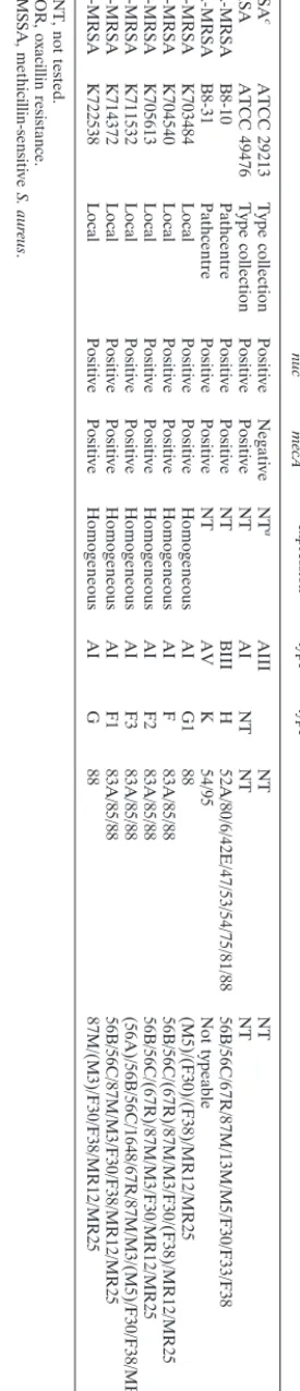
Coagulase gene RFLP patterns of the 31 GS-MRSA isolates were divided into four types (A to D), with types A and B being further divided into four (I to IV) and two (I and II) closely related subtypes, respectively (Fig. 1 and Table 2). All but one of the community-acquired isolates fell into subtypes AI and AII, and conversely only three of the health care facility-ac-quired isolates belonged to subtype AI. Three additional sub-types for the control and WA-MRSA strains were described (Table 3).
The 31 study isolates were divided into nine pulsotypes (A to E, G, I, J, L) by PFGE (Fig. 2, Table 2). There were five closely related subtypes (A0 to A4) within type A. Isolates from all 16 Polynesian cases, the 1 aboriginal case, and 4 of the 14 Cau-casian cases fell within type A. Pulsotype A subtypes accounted for all community-acquired isolates but one. The pulsotypes of all but one of the GS-MRSA isolates tested differed from those of the GR-MRSA isolates (Fig. 2, Table 3). GS-MRSA isolate I823541 belonged to pulsotype G2, which was related to two GR-MRSA isolates (Fig. 2).
Twenty of the community-acquired GS-MRSA isolates ap-peared to be closely related to the isolates of the WSPP as described by Heffernan et al. (11) (Table 2). Fifteen of these isolates were related to WSPP1, and five were related to WSPP2, the latter isolates showing lysis with phage 81 but none with phages 52, 52A, 3A, or 95. However, within the isolates related to WSPP1 and WSPP2 there were several distinct phage typing patterns. The only one of the community-acquired isolates which did appear not to be related to the WSPP strains of MRSA was F829549; this isolate also differed from other community-acquired isolates in PFGE pulsotype and coagulase RFLP. The hospital- and nursing home-ac-quired isolates are a varied group, all having different phage typing patterns. The GR-MRSA isolates with PFGE pulso-types G and F had closely related or identical phage pulso-types (Table 3).
DISCUSSION
The classification of acquisition status in the study of com-munity-acquired MRSA remains controversial. Previous stud-ies have shown that contact with a health care institution in the 12 months prior to admission is the most common risk factor for MRSA carriage (21, 27). The need to document risk factors for MRSA infection and especially contact with health care institutions and not to rely on an arbitrary time-related defi-nition when determining acquisition has been canvassed pre-viously (3). One study in southern Texas dealt with this issue by performing a case control study comparing community-acquired MRSA and community-community-acquired methicillin-sensitive S. aureusinfections (18). They found no significant difference when risk factors for MRSA within the preceding 6 months were compared. We have endeavored to overcome this diffi-culty by subdividing apparently community-acquired cases into those with and those without risk factors for MRSA acquisi-tion. The presence of risk factors for MRSA in only 6 of 23
TABLE
3.
Results
of
nuc
and
mecA
PCR,
oxacillin
resistance
expression,
coagulase
gene
RFLP
by
PCR,
PFGE,
and
phage
typing
for
control
and
comparator
strains
Description
Identification
Origin
Presence
of
:
OR
b
expression
RFLP
type
PFGE
type
Phage
type
Experimental
set
nuc
mecA
MSSA
c
ATCC
29213
Type
collection
Positive
Negative
NT
a
AIII
NT
NT
MRSA
ATCC
49476
Type
collection
Positive
Positive
NT
AI
NT
NT
NT
WA-MRSA
B8-10
Pathcentre
Positive
Positive
NT
BIII
H
52A/80/6/42E/47/53/54/75/81/88
56B/56C/67R/87M/13M/M5/F30/F33/F38
WA-MRSA
B8-31
Pathcentre
Positive
Positive
NT
AV
K
54/95
Not
typeable
GR-MRSA
K703484
Local
Positive
Positive
Homogeneous
AI
G1
88
(M5)/(F30)/(F38)/MR12/MR25
GR-MRSA
K704540
Local
Positive
Positive
Homogeneous
AI
F
83A/85/88
56B/56C/(67R)/87M/M3/F30/(F38)/MR12/MR25
GR-MRSA
K705613
Local
Positive
Positive
Homogeneous
AI
F2
83A/85/88
56B/56C/(67R)/87M/M3/F30/MR12/MR25
GR-MRSA
K711532
Local
Positive
Positive
Homogeneous
AI
F3
83A/85/88
(56A)/56B/56C/1648/67R/87M/M3/(M5)/F30/F38/MR12/MR25
GR-MRSA
K714372
Local
Positive
Positive
Homogeneous
AI
F1
83A/85/88
56B/56C/87M/M3/F30/F38/MR12/MR25
GR-MRSA
K722538
Local
Positive
Positive
Homogeneous
AI
G
88
87M/(M3)/F30/F38/MR12/MR25
a
NT,
not
tested.
b
OR,
oxacillin
resistance.
c
MSSA,
methicillin-sensitive
S.
aureus
.
on May 15, 2020 by guest
http://jcm.asm.org/
[image:4.612.361.499.88.727.2]cases of apparently community-acquired infection suggests that the majority were truly community acquired.
The PFGE results demonstrate that all of the isolates from Polynesians and all except one (F829549) of the other com-munity-acquired isolates were closely or possibly related (pul-sotype A). Both coagulase RFLP and phage typing results also support this conclusion. It is noteworthy that the one exception was isolated from a Caucasian patient with previous hospital contact. In addition, the only hospital-acquired Polynesian iso-late was recovered from a postappendectomy wound. As the procedure was performed on the day of admission, it is likely that infection was caused by the patient’s endogenous flora. WA-MRSA isolates have been shown to be distinct from GR-MRSA isolates endemic in eastern Australia (20). Results for the two WA-MRSA strains examined confirm this finding and demonstrate that they are unrelated to any of the other GS-MRSA strains studied. The hospital-acquired GR-GS-MRSA iso-lates examined fell into two related groups, one of which ap-peared to be related to a hospital-acquired GS-MRSA isolate (I823541). Members of the other hospital-acquired GS-MRSA group were genotypically and phenotypically quite diverse, with the exception that the discrepant community-acquired isolate (F828549) appeared closely related to hospital-acquired isolate C801535.
Phage typing results suggest that community-acquired GS-MRSA strains being isolated in southeast Queensland are related to SWP-MRSA strains reported in Auckland, New Zealand, where infections with these organisms are also pre-dominantly community acquired and mainly seen in the Pacific Island patients (17, 24). There was substantial migration of New Zealanders (including Polynesians) to Australia in the 1980s and 1990s (Australia Now—A Statistical Profile, Austra-lian Bureau of Statistics, Commonwealth of Australia, 2000 [http//www.abs.gov.au]). The predominantly Polynesian eth-nicity of cases in southeast Queensland and the earlier appear-ance of these strains in Auckland supports the view that their introduction to Australia was from Polynesia via New Zealand. Confirmation by direct comparison of these geographically di-verse strains is awaited.
The range and severity of infections caused by these GS-MRSA strains are in keeping with those reported previously (5). The appearance of these strains in the community and their potential for further spread are of public health impor-tance. The prevalence of methicillin resistance in community-acquired S. aureusshould be monitored, as a significant in-crease would necessitate changes to prescribing guidelines for community-acquired staphylococcal infections. The currently recommended first-line agents for common staphylococcal in-fections, isoxazolyl penicillins and cephalosporins (D. N. Gil-bert, R. C. Moellering, and M. A. Sande (ed.), The Sanford guide to antimicrobial therapy, 30th ed., Antimicrobial Ther-apy Inc., Hyde Park, Vt.), will not be effective, and selection of alternative agents will be dependent on local susceptibility patterns.
Lack of resistance to the other antimicrobials tested was also quite uniform in the community-acquired isolates, with only one expressing resistance to ciprofloxacin. The phenotypic ex-pression of resistance to oxacillin in pulsotype A was uniformly heterogeneous. Furthermore, the six hospital-acquired GR-MRSA isolates expressed resistance homogeneously, as did F829549, the genotypically unrelated community-acquired iso-late, and four of eight hospital-acquired GS-MRSA isolates. Phenotypic expression of methicillin resistance inS. aureushas been shown to be stable (29), and reemergence of heteroge-neous expression has also been noted in France with the re-appearance of GS-MRSA since 1993 (8). The relationship of the heterogeneous phenotype to expression of gentamicin re-sistance is uncertain but may be related to genes other than mecAsuch as the regulatory genes mecIand mecRIand the mecpromoter region (13, 35). Sequence analysis of themec regulatory and promoter regions of GR-MRSA and GS-MRSA may provide an explanation.
ACKNOWLEDGMENTS
We thank the staffs of the Departments of Microbiology and Infec-tion Control, Princess Alexandra Hospital, and of the Brisbane South-side Public Health Unit for their assistance in isolate and data collec-tion and A. Morton for assistance in statistical analysis.
REFERENCES
1.Ayliffe, G. A.1997. The progressive intercontinental spread of
methicillin-resistantStaphylococcus aureus. Clin. Infect. Dis.25(Suppl.):74–79.
2.Blair, J. E., and R. E. O. Williams.1961. Phage typing of staphylococci. Bull.
W. H. O.24:771–784.
3.Boyce, J. M.1998. Are the epidemiology and microbiology of
methicillin-resistantStaphylococcus aureuschanging? JAMA279:623–624.
4.Brakstad, O. G., K. Aasbakk, and J. A. Maeland.1992. Detection of
Staph-ylococcus aureusby polymerase chain reaction amplification of thenucgene.
J. Clin. Microbiol.30:1654–1660.
5.Collignon, P., I. Gosbell, A. Vickery, G. Nimmo, T. Stylianopoulos, and T.
Gottlieb.1998. Community-acquired methicillin-resistantStaphylococcus
[image:5.612.53.292.70.141.2]au-reusin Australia. Lancet352:146–147. FIG. 1. Electrophoresis of PCR coagulase gene products andHae
III-di-gested products of representative strains. (A) Lanes 1 and 6, size markers; lanes 2 to 5, products A to D, respectively. (B) Lanes 1 and 5, size markers; lanes 2 to 4, RFLP patterns BIII, BII, and AI, respectively. (C) Lanes 1 and 15, size markers, lanes 2 to 14, RFLP patterns AIII, AI, AV, AI, BIV, AIV, AII, C, BI, AII, BI, AI, and AI, respectively.
FIG. 2. Schematic representation of PFGE pulsotypes of 31 study isolates (lanes 1 to 31), 6 nosocomial GR MRSA isolates (lanes 31 to 37), and 2 WA-MRSA isolates (lanes 38 and 39), together with a dendrogram showing percent similarities of patterns and nomenclature of pulsotypes. Letters, pulso-types (seven or greater band differences); numerals, subpulso-types (one to six band differences).
on May 15, 2020 by guest
http://jcm.asm.org/
[image:5.612.53.289.485.674.2]6.Dice, L. R.1945. Measures of the amount of ecological association between species. Ecology26:297–302.
7.Frebourg, N. B., D. Nouet, L. Lemee, E. Martin, and J. F. Lemeland.1998.
Comparison of ATB Staph, Rapid ATB Staph, Vitek, and E-Test methods for detection of oxacillin heteroresistance in staphylococci possessingmecA. J. Clin. Microbiol.36:52–57.
8.Galdbart, J.-O., A. Morvan, and N. E. Solh.2000. Phenotypic and molecular
typing of nosocomial methicillin-resistantStaphylococcus aureusstrains sus-ceptible to gentamicin isolated in France from 1995 to 1997. J. Clin. Micro-biol.38:185–190.
9.Garner, J. S., W. R. Jarvis, T. G. Emori, T. C. Horan, and J. M. Hughes.
1988. CDC definitions for nosocomial infections, 1988. Am. J. Infect. Con-trol16:128–140.
10.Goh, S.-H., S. B. Byrne, J. L. Zhang, and A. W. Chow.1992. Molecular typing
ofStaphylococcus aureuson the basis of coagulase gene polymorphisms.
J. Clin. Microbiol.30:1642–1645.
11.Heffernan, H., H. Davies, and M. Brett.1995. MRSA increasing in New
Zealand. N. Z. Public Health Rep.2:97–98.
12.Herold, B. C., L. C. Immergluck, M. C. Maranan, D. S. Lauderdale, R. E.
Gaskin, S. Boyle-Vavra, C. D. Leitch, and R. S. Daum.1998.
Community-acquired methicillin-resistantStaphylococcus aureusin children with no iden-tified predisposing risk. JAMA279:593–598.
13.Hiramatsu, K.1995. Molecular evolution of MRSA. Microbiol. Immunol.
39:531–543.
14.Lawrence, C., M. Cosseron, O. Mimoz, C. Brun-Buisson, Y. Costa, K. Samii,
J. Duval, and R. Leclercq.1996. Use of the coagulase gene typing method for
the detection of carriers of methicillin-resistantStaphylococcus aureus. J. Antimicrob. Chemother.37:687–696.
15.Lelievre, H., G. Lina, M. E. Jones, C. Olive, F. Forey, M. Roussel-Delvallez,
M.-H. Nicolas-Chanoine, C. M. Bebear, V. Jarlier, A. Andremont, F.
Vandenesch, and J. Etienne.1999. Emergence and spread in French
hospi-tals of methicillin-resistantStaphylococcus aureuswith increasing suscepti-bility to gentamicin and other antibiotics. J. Clin. Microbiol.37:3452–3457.
16. Maguire, G. P., A. D. Arthur, P. J. Boustead, B. Dwyer, and B. J. Currie.
1996. Emerging epidemic of community-acquired methicillin-resistant
Staph-ylococcus aureusinfection in the Northern Territory. Med. J. Aust.164:721–
723.
17. Mitchell, J. M., D. MacCulloch, and A. J. Morris.1996. MRSA in the
community. N. Z. Med. J.110:411.
18. Moreno, F., C. Crisp, J. H. Jorgensen, and J. E. Patterson.1995.
Methicillin-resistantStaphylococcus aureusas a community organism. Clin. Infect. Dis. 21:1308–1312.
19. Murakami, K., and W. Minamide.1993. PCR identification of
methicillin-resistantStaphylococcus aureus, p. 539–542.InD. H. Persing, T. F. Smith, F. C. Tenover, and T. J. White (ed.), Diagnostic molecular microbiology: principles and applications. American Society for Microbiology, Washington, D.C.
20. O’Brien, F. G., J. W. Pearman, M. Gracey, T. V. Riley, and W. B. Grubb.
1999. Community strain of methicillin-resistantStaphylococcus aureus in-volved in a hospital outbreak. J. Clin. Microbiol.37:2858–2862.
21. Palmer, B., R. Dula, W. Zakaria, and D. Reagan.1994. Factors associated
with outpatient acquisition of methicillin-resistant Staphylococcus aureus
(MRSA). Infect. Control Hosp. Epidemiol.15:S22.
22. Pavillard, R., K. Harvey, D. Douglas, A. Hewstone, J. Andrew, B. Collopy, V.
Ashe, P. Carson, A. Davidson, G. Gilbert, J. Spicer, and F. Tosolini.1982.
Epidemic of hospital-acquired infection due to methicillin-resistant
Staphy-lococcus aureusin major Victorian hospitals. Med. J. Aust.1:451–454.
23. Richardson, J. F., V. T. Rosdahl, W. J. van Leeuwen, A. M. Vickery, A.
Vindel, and W. Witte.1999. Phages for methicillin-resistant Staphylococcus
aureus: an international trial. Epidemiol. Infect.122:227–233.
24. Riley, D., D. MacCulloch, and A. J. Morris.1998. Methicillin-resistant
Staph-ylococcus aureusin the suburbs. N. Z. Med. J.111:59.
25. Riley, T. V., J. W. Pearman, and I. L. Rouse.1995. Changing epidemiology
of methicillin-resistantStaphylococcus aureusin Western Australia. Med. J. Aust.163:412–414.
26. Rountree, P. M., and M. A. Beard.1968. Hospital strains ofStaphylococcus
aureuswith particular reference to methicillin-resistant strains. Med. J. Aust.
2:1163–1168.
27. Sumrall, B., and R. Nolan.1996. Retrospective study of
‘community-ac-quired’ (CA) methicillin-resistantStaphylococcus aureus(MRSA) occurring during an epidemic of MRSA at a Veterans Affairs hospital. Infect. Control Hosp. Epidemiol.17:28.
28. Tenover, F. C., R. D. Arbeit, R. V. Goering, P. A. Mickelsen, B. E. Murray,
D. H. Persing, and B. Swaminathan.1995. Interpreting chromosomal DNA
restriction patterns produced by pulsed-field gel electrophoresis: criteria for bacterial strain typing. J. Clin. Microbiol.33:2233–2239.
29. Tomasz, A., S. Nachman, and H. Leaf.1991. Stable classes of phenotypic
expression in methicillin-resistant clinical isolates of staphylococci. Antimi-crob. Agents Chemother.35:124–129.
30. Turnidge, J., P. Lawson, R. Munro, and R. Benn.1989. A national survey of
antimicrobial resistance inStaphylococcus aureusin Australian teaching hos-pitals. Med. J. Aust.150:65–72.
31. Turnidge, J. D., G. R. Nimmo, and G. Francis.1996. Evolution of resistance
inStaphylococcus aureusin Australian teaching hospitals. Med. J. Aust.
164:68–71.
32. Unal, S., J. Hoskins, J. E. Flokowotsch, C. Y. E. Wu, D. A. Preston, and P. L.
Skatrud.1992. Detection of methicillin-resistant staphylococci by using the
polymerase chain reaction. J. Clin. Microbiol.30:1685–1691.
33. Vickery, A. M., M. A. Beard-Pegler, and E. Stubbs.1986. Phage typing
patterns and lysogenicity of methicillin-resistantStaphylococcus aureusfrom Sydney, Australia. J. Med. Microbiol.22:209–216.
34. Voss, A., and B. N. Doebbeling.1995. The worldwide prevalence of
methi-cillin-resistantStaphylococcus aureus. Int. J. Antimicrob. Agents5:101–106.
35. Weller, T. M. A.1999. The distribution of mecA,mecR1andmecIand
sequence analysis ofmecIand themecpromoter region in staphylococci expressing resistance to methicillin. J. Antimicrob. Chemother.43:15–22.