0095-1137/05/$08.00⫹0 doi:10.1128/JCM.43.2.539–545.2005
Copyright © 2005, American Society for Microbiology. All Rights Reserved.
Identification of Variable-Number Tandem-Repeat Loci
in
Leptospira interrogans
Sensu Stricto
Z. Majed,
1E. Bellenger,
1D. Postic,
1C. Pourcel,
2G. Baranton,
1and M. Picardeau
1*
Laboratoire des Spiroche`tes, Institut Pasteur, Paris,1and Laboratoire GPMS, Institut de Ge´ne´tique et Microbiologie,
Universite´ Paris-Sud, Orsay,2France
Received 24 June 2004/Returned for modification 6 September 2004/Accepted 17 October 2004
Leptospira interroganssensu stricto is responsible for the most frequent and severe cases of human lepto-spirosis. The epidemiology and clinical features of leptospirosis are usually associated with the serovars and
serogroups of Leptospira. Because of the difficulties associated with serological identification of Leptospira
strains, we evaluated a novel PCR-based method for typingL. interrogansserovars. Based upon the genome
sequence ofL. interrogansserovar Lai type strain 5660, 44 loci were analyzed by PCR for their variability in size
due to the presence of variable-number tandem repeats (VNTR). Seven VNTR loci were found to be powerful
markers for serovar identification, epidemiology, and phylogenetic studies ofL. interrogans. This rapid and
easy method should greatly contribute to a better knowledge of the epidemiology ofLeptospira.
The genusLeptospiraconsists of a heterogeneous group of pathogenic and saprophytic species belonging to the order
Spirochaetales. PathogenicLeptospiraspecies, currently
classi-fied in seven species based on DNA relatedness (2, 25), are the agents of leptospirosis. Transmission to humans occurs through direct or indirect contacts with urine of infected ani-mals.Leptospira interroganssensu stricto (25) is the main spe-cies associated with human leptospirosis. In France,L.
inter-rogans sensu stricto is responsible for about 60% of human
cases and for the most severe ones. The intraspecies taxonomy of leptospires is well established and based on antigenic deter-minants. Since the description of serovars in 1915, about 80 serovars have been identified inL. interroganssensu stricto (2); among them, 60 serovars are validly described (12). Since each serovar is usually associated with a particular host, identifica-tion of serovars is essential to epidemiological studies and strategies for prevention (5). The reference method for sero-logical identification is the microagglutination test, which is a complex and fastidious test since it requires cross adsorption of many rabbit hyperimmune sera (24).
Antigenically related serovars are grouped into serogroups. However, a given serogroup is often found in several
Lepto-spiraspecies. For instance, the nine validly described serovars
from serogroup Bataviae are distributed amongL. interrogans
sensu stricto species (two serovars),L. santarosai(four sero-vars),L. kirschneri (one serovar), L. noguchii (one serovar),
andL. borgpetersenii(one serovar). Several studies have thus
shown that the system of serogroups is not related to molecular classifications. In contrast, serovars can be characterized by different molecular methods, such as restriction fragment length polymorphism-based methods (15, 22), arbitrarily primed PCR (19), and pulsed-field gel electrophoresis (PFGE) (8, 9). How-ever, these techniques are not widely applied, because PFGE and restriction fragment length polymorphism are laborious and
re-quire significant volumes of culture and arbitrarily primed PCR results in poor reproducibility and interpretation of results. In addition, lateral genetic transfer among leptospires (18) and large chromosomal rearrangements between serovars (26) prevent the construction of species phylogenetic trees by gene sequencing (7) or discrete whole-genomic data (19).
Analysis of variable-number tandem repeats (VNTR), also called multiple-locus VNTR analysis, has proven to be a highly powerful and discriminant method to study the population structure of bacteria (17) and to characterize isolates even from monomorphic bacterial populations (6, 11, 13). The ge-nome ofL. interrogansserovar Lai has recently been sequenced (20), and this allowed us to define pairs of primers flanking some VNTR-like loci. Our goal is to determine whether VNTR analysis will be able to differentiate most of the serovar reference strains fromL. interroganssensu stricto, providing a practical and simple PCR-based method for the identification
ofL. interrogansserovars.
MATERIALS AND METHODS
Bacterial strains and culture conditions.L. interrogansserovars (Table 1),L. borgpeterseniiserovar Castellonis strain Castellon 3,L. kirschneriserovar Grip-potyphosa strain Moskva V,L. kirschneriserovar Cynopteri strain 3522C,L. biflexaserovar Patoc strain Patoc1, and L. meyeriserovar Semaranga strain Veldrat were obtained from the strain collection at the National Reference Laboratory forLeptospiraat the Institut Pasteur, Paris, France. Leptospiral strains used in this study were also isolated from patients (one strain ofL. interrogansserovar Canicola, six strains ofL. interrogansserovar Icterohaemor-rhagiae, three strains ofL. interrogansserovar Pomona, and one strain ofL. interrogansserovar Hardjo), dogs (seven strains ofL. interrogansserovar Cani-cola), bovines (two strains ofL. interrogansserovar Hardjo), horses (one strain of
L. interrogansserovar Hardjo), and sheep (two strains ofL. interrogansserovar Hardjo) in the last 20 years.Leptospirastrains were grown at 30°C in EMJH (4, 10) liquid medium.
DNA manipulations.Genomic DNA ofLeptospirawas isolated as previously described (16). Amplification was achieved withTaqpolymerase (Amersham), using one cycle of denaturation (94°C for 5 min) followed by 35 cycles of amplification consisting of denaturation (94°C for 30 s), annealing (55°C for 30 s), and primer extension (72°C for 1 min 30 s) and a final extension of 10 min at 72°C. The amplified products were analyzed by 1.5% agarose gel electrophore-sis. The sizes of the amplified products were estimated by comparison with a 100-bp ladder (Invitrogen). Some of the amplified products were sequenced at the Genomic Platform (Institut Pasteur).
* Corresponding author. Mailing address: Laboratoire des Spiro-che`tes, Institut Pasteur, 28 rue du docteur Roux, 75724 Paris Cedex 15, France. Phone: 33 (1) 45 68 83 68. Fax: 33 (1) 40 61 30 01. E-mail: [email protected].
539
on May 16, 2020 by guest
http://jcm.asm.org/
Sequence analysis. The large chromosome CI sequence (4,332 kb) ofL. interrogansserovar Lai (20) was analyzed by using the Repeat Finder software (1) and the Tandem Repeats Database (http://iech5.igmors.u-psud.fr/GPMS/) (3). The copy number of repeats of each VNTR locus was deduced from sequencing data and sizes of the amplified products. The data were then imported into the Bionumerics software package (Applied Maths, Kortrijk, Belgium), and a phy-logenetic tree was constructed by using the neighbor-joining method. The mul-tiple phylogenetic methods showed similar tree topology. The ClustalX program (23) was used to generate nucleotide sequence alignments.
Nucleotide sequence accession numbers.The sequences of the VNTR loci described in this report can be found in GenBank under accession numbers AY766398, AY766399, AY766400, AY766401, AY766402, and AY766403.
RESULTS
Computer-assisted analysis of VNTR-like regions in theL.
[image:2.585.49.544.87.581.2]interrogansgenome. At the beginning of our work, only one
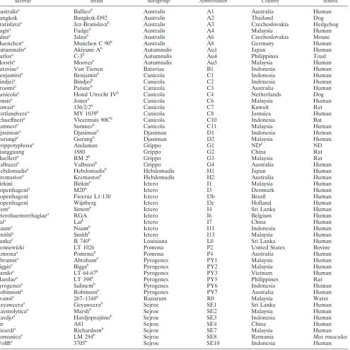
TABLE 1. Strains used in this study
Serovar Strain Serogroupc Abbreviation Country Source
Australisa Ballicob Australis A1 Australia Human
Bangkok Bangkok-D92 Australis A2 Thailand Dog
Bratislavaa Jez-Bratislavab Australis A3 Czechoslovakia Hedgehog
Fugisa Fudgeb Australis A4 Malaysia Human
Jalnaa Jalnab Australis A6 Czechoslovakia Mouse
Muenchena Munchen C 90b Australis A8 Germany Human
Autumnalisa Akiyami Ab Autumnalis Au1 Japan Human
Carlosa C-3b Autumnalis Au4 Philippines Toad
Moorisa Mooresb Autumnalis Au5 Malaysia Human
Bataviaea Van Tienen Bataviae B1 Indonesia Human
Benjaminia Benjaminb Canicola C1 Indonesia Human
Bindjeia Bindjeib Canicola C2 Indonesia Human
Broomia Pataneb Canicola C3 Australia Human
Canicolaa Hond Utrecht IVb Canicola C4 Netherlands Dog
Jonsisa Jonesb Canicola C6 Malaysia Human
Kuwaita 136/2/2b Canicola C7 Kuwait Rat
Portlandverea MY 1039b Canicola C8 Jamaica Human
Schueffneria Vleermuis 90Cb Canicola C10 Indonesia Bat
Sumneria Sumnerb Canicola C11 Malaysia Human
Djasimana Djasimanb Djasiman D1 Indonesia Human
Gurungia Gurungb Djasiman D2 Malaysia Human
Grippotyphosaa Andaman Grippo G1 NDd ND
Liangguang 1880 Grippo G2 China Rat
Muelleria RM 2b Grippo G3 Malaysia Rat
Valbuzzia Valbuzzib Grippo G4 Australia Human
Hebdomadisa Hebdomadisb Hebdomadis H1 Japan Human
Kremastosa Kremastosb Hebdomadis H2 Australia Human
Birkini Birkinb Ictero I1 Malaysia Human
Copenhagenia M20b Ictero I3 Denmark Human
Copenhageni Fiocruz L1-130 Ictero I3b Brazil Human
Copenhageni Wijnberg Ictero I3c Holland Human
Gema Simonb Ictero I4 Sri Lanka Human
Icterohaemorrhagiaea RGA Ictero I6 Belgium Human
Laia Laib Ictero I7 China Human
Naama Naamb Ictero I11 Indonesia Human
Smithia Smithb Ictero I13 Malaysia Human
Lankaa R 740b Louisiana L0 Sri Lanka Human
Kennewicki LT 1026 Pomona P2 United States Bovine
Pomonaa Pomonab Pomona P4 Australia Human
Abramisa Abrahamb Pyrogenes PY1 Malaysia Human
Biggisa Biggsb Pyrogenes PY2 Malaysia Human
Camloa LT 64-67b Pyrogenes PY3 Vietnam Human
Manilaea LT 398b Pyrogenes PY5 Philippines Rat
Pyrogenesa Salinemb Pyrogenes PY6 Indonesia Human
Robinsonia Robinsonb Pyrogenes PY7 Australia Human
Evansia 267–1348b Ranarum R0 Malaysia Water
Geyaweeraa Geyaweerab Sejroe SE1 Sri Lanka Human
Haemolyticaa Marshb Sejroe SE2 Malaysia Human
Hardjoa Hardjoprajitnob Sejroe SE3 Indonesia Human
Jin A81 Sejroe SE4 China Human
Ricardia Richardsonb Sejroe SE7 Malaysia Human
Romanicaa LM 294b Sejroe SE8 Romania Mus musculus
Wolffia 3705b Sejroe SE10 Indonesia Human
a
Serovar present in the serovar list of 1992 (12). Other serovars are mentioned in reference 2 but are not yet validated. b
Serovar reference strain. c
Ictero, Icterohaemorrhagiae; Grippo, Grippotyphosa. d
ND, not determined.
on May 16, 2020 by guest
http://jcm.asm.org/
Leptospiragenome sequence was available (at the time of this writing, another genome, that ofL. interrogansserogroup se-rovar Copenhageni, has been sequenced [14]), preventing strain comparison and prediction of polymorphic loci. Analysis of the genome sequence of L. interrogans serovar Lai (20) enabled the rapid identification of loci containing repetitions of short sequences. Among more than 1,000 VNTR-like re-gions, an initial selection of 44 loci was chosen after comparing the lengths of repeats (repeats of 30 to 75 bp), sequence iden-tities (nucleotide sequence identity between repeats of⬎85%), and copy numbers (between two and eight copies of the unit repeat).
PCR analysis of VNTR-like regions inL. interroganssenso
stricto.Forty-four primer pairs (sequences of the primers are
available on request) were tested for their usefulness with a set of six well-characterizedL. interrogansstrains (strains A1, I3, I7, C4, PY6, SE3) (Table 1). The primers used for the PCR correspond to the VNTR flanking regions identified in theL.
interrogansserovar Lai genome. Analysis of the amplified
prod-ucts by agarose gel electrophoresis revealed size variations in most of the loci. However, either no amplification or
amplifi-cation of several faint bands was obtained for several loci, which were therefore excluded from this study (data not shown). This could be due to low conservation of the VNTR flanking regions among serovars. The seven most discrimina-tive VNTR loci (i.e., VNTR4, VNTR7, VNTR9, VNTR10, VNTR11, VNTR19, and VNTR23) that exhibited a single PCR product whose size could be easily determined in 1.5% agarose gel electrophoresis for the six reference strains were further evaluated with a large collection of strains.
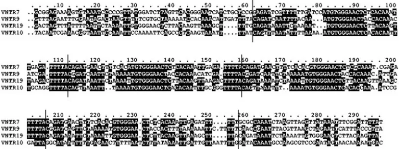
Sequence features of selected VNTR loci.The positions of
[image:3.585.44.544.82.263.2]these VNTR loci were scattered in different locations in chro-mosomeCIofL. interrogans(Table 2). Further sequence anal-ysis indicated that none of the selected VNTR loci were lo-cated in open reading frames. However, some of the VNTR loci could contain small open reading frames. In addition, despite obvious sequence similarities between unit repeats, a closer look at the selected loci revealed sequence similarities among VNTR7, VNTR9, VNTR10, and VNTR19 (Fig. 1). These four loci share a 47-bp consensus sequence which is repeated in tandem (Fig. 1). Sequences of the repeats from the VNTR4 and VNTR23 loci also display significant similarities
[image:3.585.96.487.554.700.2]FIG. 1. Nucleotide sequence alignment of the VNTR7, VNTR9, VNTR10, and VNTR19 loci fromL. interrogansserovar Lai. The 47-bp repeated units are delineated by vertical bars.
TABLE 2. VNTR loci from theL. interrogansserovar Lai genome used in this study
VNTR
locus Primers (5⬘33⬘) Position inCI(bp)
Unit length (bp)
Copy no.
Total length of PCR product
(bp)
No. of alleles/51 serovars
Copy no. range in
L. interrogans
serovars
VNTR4 4a (CAAAATCAGTCACTACCCTG) 1122221–1122580 34 5 362 10 0–23
4b (CTTTGTTGGAGCGCAATCTC)
VNTR7 7a (TCATCTGCTCCGGAGATTCG) 3312338–3312035 46 3 304 15 0–14
7b (TCCCTCCACAGGTTGTCTTG)
VNTR9 9a (TCGCTCTACAGGTCGGTGTT) 2652531–2652151 46 4 381 13 1–13
9b (GGTGAAGAGCAAACCTTTGG)
VNTR10 10a (TCCAAAATTCAGCCCTCAAG) 1666395–1666157 45 2 239 15 1–18
10b (GACGCTTGGCATTTGTATCC)
VNTR11 11a (ACAGAAGCCGTCTCATTTTG) 167476–167184 45 4 293 7 1–11
11b (CACAGGTCGGAATTTGTCA)
VNTR19 19a (CAGAAACAAGAGGGAAGATTC) 2877449–2877029 47 6 421 15 1–18
19b (ACTCTCATTTAAGAGTGGCTG)
VNTR23 23a (TTTCCAAATATACTTACTCGG) 2179070–2178732 46 5 339 13 0–14
23b (GCAAGAGAATTATTGGGATGG)
VNTR31 31a (TTCATGAAGGTCCCGAAAAC) 2729699–2730370 77 4 671 NDa ND
31b (ACGTGAGTTCGACCATGATTC)
aND, not determined (VNTR31 was used to differentiate betweenL. interrogansserovars Canicola and Portlandvere from serogroup Canicola).
on May 16, 2020 by guest
http://jcm.asm.org/
with repeats in other locations of theL. interrogans genome (data not shown). These results suggest that VNTR loci could be grouped in distinct families of tandem-repeat-containing loci.
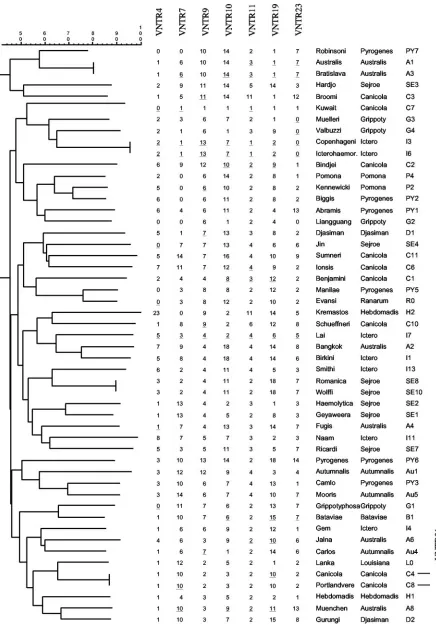
Characterization ofL. interrogansserovars by VNTR
poly-morphism analysis. PCR was performed with the seven
se-lected VNTR loci and a total of 51 serovars, clustered in 13 distinct serogroups (Table 1). The sizes of the amplified prod-ucts displayed a wide range of polymorphism, suggesting vari-ation in tandem-repeat copy numbers in the seven VNTR loci (Fig. 2). This was confirmed by sequencing of 60 amplified products from the seven loci (Fig. 3). Figure 4 shows sequences of VNTR19, which contains variable number of perfectly iden-tical repeats. For each VNTR locus, sequence analysis of am-plified products indicated a high conservation of repeat units and flanking regions amongL. interrogansserovars (data not shown). Although the presence of multiples of a full-length repeat was the general rule, an absence of the unit repeat, insertions, and/or deletions (4 out of the 60 sequenced ampli-cons) could result in a size of the amplified product which is not compatible with the variation in copy number of a full-length repeat.
For each locus, the number of tandem repeats was calcu-lated by measuring the sizes of the amplified products. The strains were typed by the numbers of variable tandem repeats in each of the seven VNTR loci. It should be noted that the value of 0 was used for amplified fragments shorter than a one-copy VNTR locus (this is the case in VNTR4, VNTR7, and VNTR23). These data could then be easily stored in da-tabases and imported in Bionumerics for analysis.
The seven markers were able to differentiate 43 of 51 L.
interrogansserovars (Fig. 3). An identical level of
discrimina-tion was obtained with only three markers, i.e., VNTR7, VNTR10, and VNTR19, that displayed the widest range of polymorphism with 15 distinct alleles among the 51 serovars (Table 2). Only four strain pairs were not differentiated what-ever VNTR locus was used. The two strains within each pair belong to the same serogroup (L. interrogansserovars Copen-hageni and Icterohaemorrhagiae from serogroup
Icterohaem-orrhagiae,L. interrogansserovars Australis and Bratislava from serogroup Australis, L. interrogans serovars Romanica and Wolffi from serogroup Sejroe, andL. interrogansserovars Ca-nicola and Portlandvere from serogroup CaCa-nicola).L. interro-gansserovars Copenhageni and Icterohaemorrhagiae were not differentiated with the 44 VNTR loci, as they were initially used as reference strains for the screening of the markers. We have undertaken PCR with VNTR loci that were previously excluded from the study to differentiate serovars that gave identical results with the seven selected markers. VNTR31 (four copies of a 77-bp repeat inL. interrogansserovar Lai) was able to differentiate betweenL. interrogansserovars Canicola (three copies) and Portlandvere (four copies) from serogroup Canicola. The three other pairs of strains were not differenti-ated whatever other VNTR was used.
Interestingly, three strains belonging to a same serovar (se-rovar Copenhageni strain M20 from Denmark, se(se-rovar Copen-hageni strain Wijnberg from Holland, and serovar Copenhag-eni strain Fiocruz L1-130 from Brazil) exhibited identical results with the seven VNTR loci (data not shown). SinceL.
interrogansis phylogenetically related to other pathogenic
spe-cies, we performed the VNTR assay with a few strains fromL.
kirschneriandL. borgpetersenii. Analysis of the PCR products
of two of the seven markers, i.e., VNTR11 and VNTR19, exhibited a single band, variable in size, with the four strains of
L. kirschneri and L. borgpetersenii. The size variation
corre-sponded to multiples of the unit repeat identified inL.
inter-rogansserovar Lai. These results suggest thatL. interrogans,L.
kirschneri, andL. borgpeterseniishared similar VNTR loci. In
contrast, no amplification was obtained with DNAs from the saprophytic species L. biflexa and L. meyeri with the seven markers.
Application of our VNTR-based method to clinical strains.
To assess our PCR-based method for genotyping, we analyzed 23 clinical strains (including 11 strains isolated from humans) with the seven most discriminative VNTR loci (Table 1). The serovars of these isolates (L. interrogansserovars Icterohaem-orrhagiae, Pomona, Hardjo, and Canicola) were previously identified by NotI restriction and PFGE (data not shown). All
FIG. 2. PCR analysis of the polymorphism of two representative VNTR loci. Amplification was performed on the VNTR10 and VNTR19 loci ofL. interrogansstrains. Lanes indicateLeptospiraserovars (Table 1).
on May 16, 2020 by guest
http://jcm.asm.org/
FIG. 3. Dendrogram of the VNTR-typed serovars ofL. interrogans. The copy number of each VNTR locus is indicated. The serovars and serogroups of reference strains are also indicated. PCR products that were sequenced are underlined. VNTR31was used to differentiate between
L. interrogansserovars Canicola and Portlandvere from serogroup Canicola.
on May 16, 2020 by guest
http://jcm.asm.org/
isolates displayed a pattern identical to that of the correspond-ing reference strain, suggestcorrespond-ing the stability of PCR patterns in strains belonging to the same serovar. Again, the use of only three markers, i.e., VNTR7, VNTR10, and VNTR19, was enough to identify these strains at the serovar level.
DISCUSSION
Recently, a giant leap forward has been achieved with the completion of the genome sequence ofL. interrogansserovar Lai (20).L. interroganscontains a 4.33-Mb large circular chro-mosome, a 359-kb small circular chrochro-mosome, and no extra-chromosomal element (14, 20). Numerous repeated sequences have been found in its genome. For example, about 50 inser-tion sequences have been identified. The L. interrogans ge-nome also contains abundant small repetitive DNA sequences. Among these DNA repeats, the structure of short sequence repeats is typical of tandem repeats. Tandem repeats consist of head-to-tail repetitions of short sequence motifs of about 10 to 100 bp. Polymorphic tandem repeats, also called VNTR, have been extensively used for fingerprinting in higher eukaryotes, including humans. Recently, the use of VNTR has also been described for phylogenetic and epidemiological studies of pathogenic bacteria.
A database of tandem repeats in more than 140 completely sequenced genomes is available (3). By using this database (http://iech5.igmors.u-psud.fr/GPMS/), genome analysis indi-cates that most of the tandem repeats (901 of 1,100; 82%) inL.
interrogans strain Lai are between 15 and 50 bp, which is
convenient for observation of polymorphism by analyzing PCR
products of polymorphic loci on agarose gels. Compared to those of other bacteria, theL. interrogans genome exhibits a high number of tandem repeats with a total length greater than 100 bp (27 and 29 per Mb for strains Fiocruz and Lai, respec-tively). Comparative genomics shows that 131 VNTR loci (length of unit repeat, between 5 and 500 bp) were shared between the two availableL. interrogansgenomes (3, 14, 20). For strains within a species or genus, this number varies from 4 (strains fromChlamydia pneumoniae) up to 163 (strains from
Yersinia pestis). Tandem repeats predicted to be polymorphic
by genome comparison between the two serovars from sero-group Icterohaemorrhagiae indeed exhibit size variations by PCR typing ofLeptospirastrains.
The function of repetitive elements in bacteria is not fully understood. Similar to theMycobacterium tuberculosis myco-bacterial interspersed repetitive units (21), tandem-repeat-containing loci inL. interrogans are located in intergenic re-gions, are dispersed throughout the genome, and constitute subfamilies based on sequence similarities. The dissemination of homologous VNTR loci in the genome may suggest fre-quent intragenomic rearrangement or that these elements are (or have been) mobile elements. Studies on the structural and functional properties of these families of repetitive DNA should improve our knowledge of the role of these abundant repeat sequences inLeptospiraspp.
All molecular tools described so far for the diagnosis of
Leptospira suffer from significant drawbacks, such as a low
discrimination level, lack of reproducibility, and requirement for large quantities of purified DNA. We therefore took ad-vantage of the presence of VNTR-like regions to design a PCR-based test. Our data showed that VNTR typing was able to differentiate 45 out of 51 serovars of L. interrogans. Our VNTR assay shows that serovars from either the same sero-group or the same geographical area are not sero-grouped together (Table 1; Fig. 3). This was true when the seven markers were used in combination but also when only three markers (VNTR7, -10, and -19) were used. This result confirms the heterogeneity among serovars of a given serogroup previously found by DNA relatedness (2) and PFGE (8, 9). Most serovar reference strains fromL. interroganshave been isolated in Asia (2), mainly in southeast Asia (Table 1). It is noteworthy to find that strains originating from distant continents can be grouped together in the dendrogram. We also show that VNTR analysis was able to differentiate serovars amongL. interrogansclinical strains, therefore demonstrating the usefulness of this PCR-based method for the identification ofL. interrogansserovars. A current method for typing Leptospira strains is macror-estriction by PFGE. This method is labor-intensive and not accessible to most laboratories in tropical and subtropical countries, where the incidence of the disease is the highest. Similar to PFGE, VNTR typing was not able to discriminate between L. interrogans serovars Icterohaemorrhagiae and Copenhageni. However, L. interrogans serovars Muenchen, Jalna, and Bratislava from serogroup Australis, which gave identical NotI macrorestriction patterns by PFGE (9), were differentiated by VNTR analysis. The use of only one VNTR locus, VNTR19 or VNTR23, was enough to discriminate se-rovars Muenchen, Jalna, and Bratislava (Fig. 3). In contrast, VNTR analysis ofL. interrogansserovars Bratislava and
Aus-FIG. 4. Sequence alignment of VNTR19 loci. Brackets on the left indicate the repeat units. Cani,L. interrogansserovar Canicola; Lai,L. interrogansserovar Lai; Grip,L. interrogansserovar Grippotyphosa; Autu,L. interrogansserovar Autumnalis; Cope,L. interrogansserovar Copenhageni; Aust,L. interrogansserovar Australis.
on May 16, 2020 by guest
http://jcm.asm.org/
tralis from serogroup Australis gave identical results (Fig. 3), but the two serovars were differentiated by PFGE (9). Distinct macrorestriction profiles of closely related strains could be due to large genomic rearrangements. For example, comparative genomics between twoL. interrogansserovars from serogroup Icterohaemorrhagiae revealed a one-Mb chromosomal inver-sion (14).
In conclusion, this method based on VNTR polymorphism provides rapid typing as well as a highly discriminant assay to identify L. interrogans serovars. In addition, VNTR typing could be widely accessible for research and public health lab-oratories, particularly in developing countries. This method should also be suitable for sharing results and for the genera-tion of databases. Further studies should include the develop-ment of a VNTR typing test with biological materials. The genome sequences of otherLeptospirapathogenic strains are at different stages of completion. These sequences would greatly facilitate the development of multiple-locus VNTR as-says for pathogenicLeptospiraspp.
ACKNOWLEDGMENTS
This work was supported by the Programme Transversal de Recher-che (PTR no. 139), Institut Pasteur.
We thank A. Ko for the generous gift of DNA from strain Fiocruz L1-130. We are grateful to G. Vergnaud and F. Denoeud for the strain comparison database. We thank C. Gutierrez Perez for help in using Bionumerics and I. Saint Girons for her support.
REFERENCES
1.Benson, G.1999. Tandem repeats Finder: a program to analyze DNA se-quences. Nucleic Acids Res.27:573–580.
2.Brenner, D. J., A. F. Kaufmann, K. R. Sulzer, A. G. Steigerwalt, F. C. Rogers, and R. S. Weyant.1999. Further determination of DNA relatedness between serogroups and serovars in the familyLeptospiraceaewith a proposal for
Leptospira alexanderisp. nov. and four newLeptospiragenomospecies. Int. J. Syst. Bacteriol.49:839–858.
3.Denoeud, F., and G. Vergnaud.2004. Identification of polymorphic tandem repeats by direct comparison of genome sequence from different bacterial strains: a web-based resource. BMC Bioinformatics12:4.
4.Ellinghausen, H. C., and W. G. McCullough.1965. Nutrition ofLeptospira pomonaand growth of 13 other serotypes: fractionation of oleic albumin complex and a medium of bovine albumin and polysorbate 80. Am. J. Vet. Res.26:45–51.
5.Faine, S., B. Adler, C. Bolin, and P. Perolat.1999.Leptospiraand leptospi-rosis. MedScience, Melbourne, Australia.
6.Farlow, J., D. Postic, K. L. Smith, Z. Jay, G. Baranton, and P. Keim.2002. Strain typing ofBorrelia burgdorferi,Borrelia afzelii, andBorrelia gariniiby using multiple-locus variable-number tandem repeat analysis. J. Clin. Micro-biol.40:4612–4618.
7.Haake, D. A., M. A. Suchard, M. M. Kelley, M. Dundoo, D. P. Alt, and R. L. Zuerner. 2004. Molecular evolution and mosaicism of leptospiral outer membrane proteins involves horizontal DNA transfer. J. Bacteriol.186:
2818–2828.
8.Herrmann, J. L., C. Baril, E. Bellenger, P. Perolat, G. Baranton, and I. Saint Girons.1991. Genome conservation in isolates ofLeptospira interrogans. J. Bacteriol.173:7582–7588.
9.Herrmann, J. L., E. Bellenger, P. Perolat, G. Baranton, and I. Saint Girons.
1992. Pulsed-field gel electrophoresis ofNotI digests of leptospiral DNA: a new rapid method of serovar identification. J. Clin. Microbiol.30:1696–1702. 10.Johnson, R. C., and V. G. Harris.1967. Differentiation of pathogenic and
saprophytic leptospires. J. Bacteriol.94:27–31.
11.Keim, P., L. B. Price, A. M. Klevytska, K. L. Smith, J. M. Schupp, R. Okinaka, P. J. Jackson, and M. E. Hugh-Jones.2000. Multiple-locus vari-able-number tandem repeat analysis reveals genetic relationships within
Bacillus anthracis. J. Bacteriol.182:2928–2936.
12.Kmety, E., and H. Dikken.1993. Classification of the species Leptospira interrogansand history of its serovars. University Press, Groningen, The Netherlands.
13.Le Fleche, P., M. Fabre, F. Denoeud, J. L. Koeck, and G. Vergnaud.2002. High resolution, on-line identification of strains from theMycobacterium tuberculosiscomplex based on tandem repeat typing. BMC Microbiol.2:37. 14.Nascimento, A. L., A. I. Ko, E. A. Martins, C. B. Monteiro-Vitorello, P. L. Ho, D. A. Haake, S. Verjovski-Almeida, R. A. Hartskeerl, M. V. Marques, M. C. Oliveira, C. F. Menck, L. C. Leite, H. Carrer, L. L. Coutinho, W. M. Degrave, O. A. Dellagostin, H. El-Dorry, E. S. Ferro, M. I. Ferro, L. R. Furlan, M. Gamberini, E. A. Giglioti, A. Goes-Neto, G. H. Goldman, M. H. Goldman, R. Harakava, S. M. Jeronimo, I. L. Junqueira-de-Azevedo, E. T. Kimura, E. E. Kuramae, E. G. Lemos, M. V. Lemos, C. L. Marino, L. R. Nunes, R. C. de Oliveira, G. G. Pereira, M. S. Reis, A. Schriefer, W. J. Siqueira, P. Sommer, S. M. Tsai, A. J. Simpson, J. A. Ferro, L. E. Camargo, J. P. Kitajima, J. C. Setubal, and M. A. Van Sluys.2004. Comparative genomics of twoLeptospira interrogansserovars reveals novel insights into physiology and pathogenesis. J. Bacteriol.186:2164–2172.
15.Perolat, P., I. Lecuyer, D. Postic, and G. Baranton.1993. Diversity of ribo-somal DNA fingerprints ofLeptospiraserovars provides a database for sub-typing and species assignation. Res. Microbiol.144:5–15.
16.Picardeau, M., A. Brenot, and I. Saint Girons.2001. First evidence for gene replacement inLeptospiraspp. Inactivation ofL. biflexa flaBresults in non-motile mutants deficient in endoflagella. Mol. Microbiol.40:189–199. 17.Pourcel, C., Y. Vidgop, F. Ramisse, G. Vergnaud, and C. Tram.2003.
Char-acterization of a tandem repeat polymorphism inLegionella pneumophila
and its use for genotyping. J. Clin. Microbiol.41:1819–1826.
18.Ralph, D., and M. McClelland.1994. Phylogenetic evidence for horizontal transfer of an intervening sequence between species in a spirochete genus. J. Bacteriol.176:5982–5987.
19.Ralph, D., M. McClelland, J. Welsh, G. Baranton, and P. Perolat.1993.
Leptospiraspecies categorized by arbitrarily primed polymerase chain reac-tion (PCR) and by mapped restricreac-tion polymorphisms in PCR-amplified rRNA genes. J. Bacteriol.175:973–981.
20.Ren, S. X., G. Fu, X. G. Jiang, R. Zeng, Y. G. Miao, H. Xu, Y. X. Zhang, H. Xiong, G. Lu, L. F. Lu, H. Q. Jiang, J. Jia, Y. F. Tu, J. X. Jiang, W. Y. Gu, Y. Q. Zhang, Z. Cai, H. H. Sheng, H. F. Yin, Y. Zhang, G. F. Zhu, M. Wan, H. L. Huang, Z. Qian, S. Y. Wang, W. Ma, Z. J. Yao, Y. Shen, B. Q. Qiang, Q. C. Xia, X. K. Guo, A. Danchin, I. Saint Girons, R. L. Somerville, Y. M. Weng, M. H. Shi, Z. Chen, J. G. Xu, and G. P. Zhao.2003. Unique physi-ological and pathogenic features ofLeptospira interrogansrevealed by whole genome sequencing. Nature422:888–893.
21.Supply, P., J. Magdalena, S. Himpens, and C. Locht.1997. Identification of novel intergenic repetitive units in a mycobacterial two-component system operon. Mol. Microbiol.26:991–1003.
22.Thiermann, A. B., A. L. Handsaker, S. L. Moseley, and B. Kingscote.1985. New method for classification of leptospiral isolates belonging to serogroup pomona by restriction endonuclease analysis: serovar kennewicki. J. Clin. Microbiol.21:585–587.
23.Thompson, J. D., T. J. Gibson, F. Plewniak, F. Jeanmougin, and D. G. Higgins.1997. The ClustalX Windows interface: flexible strategies for mul-tiple sequence alignment aided by quality analysis tools. Nucleic Acids Res.
25:4876–4882.
24.World Health Organization.2003. Human leptospirosis: guidance for diag-nosis, surveillance and control. World Health Organization, Geneva, Swit-zerland.
25.Yasuda, P. H., A. G. Steigerwalt, K. R. Sulzer, A. F. Kaufmann, F. C. Rogers, and D. J. Brenner.1987. Deoxyribonucleic acid relatedness between sero-groups and serovars in the familyLeptospiraceaewith proposals for seven newLeptospiraspecies. Int. J. Syst. Bacteriol.37:407–415.
26.Zuerner, R. L., J. L. Herrmann, and I. Saint Girons.1993. Comparison of genetic maps for twoLeptospira interrogansserovars provides evidence for two chromosomes and intraspecies heterogeneity. J. Bacteriol.175:5445– 5451.