Comparative Evaluation of the Diagnostic Performance of the
Prototype Cepheid GeneXpert Ebola Assay
Petrus Jansen van Vuren,a,bAntoinette Grobbelaar,aNadia Storm,aOusman Conteh,cKelfala Konneh,cAbdul Kamara,cIan Sanne,d,e Janusz T. Paweskaa,b,f
Centre for Emerging and Zoonotic Diseases, National Institute for Communicable Diseases, National Health Laboratory Service, Sandringham, South Africaa
; Department of Microbiology and Plant Pathology, Faculty of Natural and Agricultural Science, University of Pretoria, Pretoria, South Africab
; Ministry of Health and Sanitation, Freetown, Sierra Leonec
; Clinical HIV Research Unit, Wits Health Consortium, Johannesburg, South Africad
; Right-to-Care, Johannesburg, South Africae
; Faculty of Medical Sciences, University of the Witwatersrand, Johannesburg, South Africaf
The Ebola virus disease (EVD) outbreak in West Africa has highlighted an urgent need for point-of-care (POC) assays for the diagnosis of this devastating disease in resource-limited African countries. The diagnostic performance characteristics of a pro-totype Cepheid GeneXpert Ebola POC used to detect Ebola virus (EBOV) in stored serum and plasma samples collected from suspected EVD cases in Sierra Leone in 2014 and 2015 was evaluated. The GeneXpert Ebola POC is a self-contained single-car-tridge automated system that targets the glycoprotein (GP) and nucleoprotein (NP) genes of EBOV and yields results within 90 min. Results from 281 patient samples were compared to the results of a TaqMan real-time reverse transcription-PCR (RT-PCR) targeting the polymerase gene and performed on two real-time PCR machines. Agreement between the three platforms was
100% at cycle threshold (CT) values of<34.99, but discordant results were noted betweenCTvalues of 35 and 45.The diagnostic
sensitivity of the three platforms was 100% in 91 patient samples that were confirmed to be infectious by virus isolation. All three molecular platforms detected viral EBOV RNA in additional samples that did not contain viable EBOV. The analytical sen-sitivity of the GeneXpert Ebola POC for the detection of NP was higher, and comparable to that of polymerase gene detection, than that for the detection of GP when using a titrated laboratory stock of EBOV. There was no detectable cross-reactivity with other hemorrhagic fever viruses or arboviruses. The GeneXpert Ebola POC offers an easy to operate and sensitive diagnostic tool that can be used for the rapid screening of suspected EVD cases in treatment or in holding centers during EVD outbreaks.
T
he unprecedented scale of Ebola virus disease (EVD)out-breaks in West Africa from 2013 to 2015 caused by the Ebola virus (EBOV) represents a dramatic expansion of case numbers and the introduction of this highly lethal disease into new
geo-graphic areas (1,2). As of 7 October 2015, the World Health
Or-ganization reported a total of 28,421 EVD cases (confirmed, prob-able, and suspected) of which 11,297 (39.7%) were fatal, including 881 confirmed cases among health care workers of which 513
(58.2%) were fatal (3). The diagnostic burden of the largest EVD
outbreak in the recorded history of the disease has been mostly borne by mobile laboratories, deployed throughout the affected countries by international agencies and institutes. Delays in the diagnosis of suspected EVD cases due to sample transport from remote areas to these laboratories have put additional pressure on outbreak control efforts. The use of a wide range of assays, often not clinically validated, has complicated the interpretation and consolidation of results from different laboratories. Rapid and accurate diagnostic results have a great impact on the manage-ment of suspected cases and on the tracing of contacts. The extent
to which qualitative cycle threshold (CT) values from real-time
reverse transcription-PCR (RT-PCR) assays correlate with the in-fectious states of patients is not well understood. Patients are often kept in isolation until three consecutive blood samples, collected
days apart, are found negative by RT-PCR. HighCTvalue results
are often recorded in the blood of recovering patients for several days after clinical recovery (National Institute for Communicable Diseases [NICD], unpublished data). It is unclear whether these
high CT values infer that the patient still represents a risk for
spreading infection.
The 2013 to 2015 West African EVD outbreak is caused by the
Ebola virus (speciesZaire ebolavirus), one of five species in the
Ebolavirusgenus, which is known to cause a high fatality rate in
infected humans (60% to 90%) (4). The EBOV genome is a
neg-ative-sense, single-strand RNA consisting of 18,959 nucleotides encoding seven structural proteins and one nonstructural protein. After 2 to 21 days of incubation, the disease presents initially with flu-like symptoms such as fever, malaise, and myalgia followed by vomiting, diarrhea, abdominal pain, edema, neurological signs, and hemorrhagic manifestations such as rash, petechiae, and
bleeding from puncture sites (4).The nonspecific clinical
presen-tation of viral hemorrhagic fevers (VHFs) and their high risk for nosocomial spread highlight the importance of accurate and rapid laboratory diagnosis. Diagnosis of infection by a filovirus can be achieved by the detection of antigen, the detection of virus nucleic acid, the isolation of virus, or by the detection of a virus-specific antibody response. Antigen can be detected in serum and other body fluid samples by antigen detection enzyme-linked
immu-nosorbent assay (ELISA) (5) and indirect immunoelectron
mi-Received8 October 2015Returned for modification23 October 2015 Accepted20 November 2015
Accepted manuscript posted online4 December 2015
CitationJansen van Vuren P, Grobbelaar A, Storm N, Conteh O, Konneh K, Kamara A, Sanne I, Paweska JT. 2016. Comparative evaluation of the diagnostic performance of the prototype Cepheid GeneXpert Ebola assay. J Clin Microbiol 54:359 –367.doi:10.1128/JCM.02724-15.
Editor:A. J. McAdam
Address correspondence to Janusz T. Paweska, [email protected].
Copyright © 2016, American Society for Microbiology. All Rights Reserved.
on May 16, 2020 by guest
http://jcm.asm.org/
croscopy (6) and in skin biopsy specimens by
immunohistochem-istry (7). Detection of immunoglobulin (Ig) M and G specific to
filoviruses can indicate a recent or past infection (8).
Tradition-ally, filoviruses have been isolated successfullyin vitroin African
green monkey cell cultures (Vero) (9) and in suckling mice (10).
In recent years, filovirus diagnostics have relied mostly on
real-time RT-PCR assays using fluorogenic probes (11–19). Two
pro-totype point-of-care assays that detect viral antigen by using lat-eral flow technology were recently evaluated using clinical
specimens from suspected EVD cases in Sierra Leon (20,21).
In this study, we evaluated the diagnostic performance of the prototype GeneXpert (Cepheid, Sunnyvale, CA, USA) Ebola assay in serum and plasma samples from suspected EVD cases in Sierra Leone. The results of this assay were directly compared to real-time RT-PCR targeting of the polymerase (L) gene run on two field deployable real-time PCR platforms, SmartCycler (Cepheid, Sunnyvale, CA, USA) and LightCycler Nano (Roche, Basel, Swit-zerland). The sensitivity of the three molecular diagnostic plat-forms was compared to that of virus isolation in Vero E6 cell culture.
MATERIALS AND METHODS
Analytical sensitivity.To determine and compare the analytical
sensitiv-ity of the GeneXpert Ebola assay to that of the polymerase gene-based TaqMan RT-PCR run on two real-time platforms, a log dilution series of stock Ebola virus (SPU220/96; passage 4 Vero; 1⫻105.050% tissue cul-ture infective dose [TCID50]/ml) was prepared in culture medium (Ea-gle’s minimal essential medium [EMEM]) and tested in quadruplicate.
Analytical specificity.To evaluate the cross-reactivity of the
Gene-Xpert Ebola assay with selected hemorrhagic fever and arthropod borne viruses (arboviruses), stocks of the following viruses were tested: Sudan and Marburg viruses (Filoviridae); Lassa and Lujo viruses (Arenaviridae); Rift Valley fever and Crimean-Congo hemorrhagic fever viruses ( Bunya-viridae); West Nile, Yellow fever, and Dengue type 1 to 4 viruses ( Flavi-viridae); and Chikungunya and Sindbis viruses (Alphaviridae). More de-tailed individual isolate information can be found inTable 1.
Diagnostic sensitivity.Diagnostic sensitivity was evaluated using 281
blood specimens from suspected EVD cases submitted from August 2014
through March 2015 to the field Ebola Molecular Laboratory of the Centre for Emerging and Zoonotic Diseases of the National Institute for Com-municable Diseases (NICD) in Freetown, Sierra Leone. This field labora-tory was established as a part of the WHO Global Outbreak Alert and Response Network international outbreak response. Serum was separated from clotted blood and plasma from EDTA, and the resulting samples were stored at⫺70°C before shipment on dry ice to the NICD biosafety level 4 facility (BSL-4) in Johannesburg, South Africa for further analysis and long-term storage.
Extraction of viral RNA.RNA was extracted from clinical and
labo-ratory generated samples (input, 140l) using the QIAamp viral RNA kit (Qiagen, Hilden, Germany) per the manufacturer’s instructions and as previously described (22). The final elution volume was 60l.
Polymerase (L) gene TaqMan real-time RT-PCR.The assay was
per-formed as previously described (14,22) using the primers and probes targeting the Ebola virus L gene and 5l RNA as the template. Anin vitro
transcribed RNA copy of the L gene was used as a positive control at a known copy number. The RNA standard was prepared as previously de-scribed (23) using L-gene-specific primers (14). All runs included two negative controls as per standard PCR practice, a no-template control and an extraction negative control. Two field-deployable real-time PCR plat-forms were used, the SmartCycler (Cepheid) using propriety single-reac-tion Smart Tubes and the LightCycler Nano (Roche) using 8-well strip tubes. The SmartCycler is operated with Cepheid SmartCycler Version 2.0d software, and the LightCycler Nano is operated with software version 1.0.7. Analysis on the two platforms’ software was run with default anal-ysis settings. Cycles were as follows: reverse transcription (50°C for 30 min), denaturation (95°C for 15 min), and amplification/detection (45 cycles of 95°C for 15 s, 52°C for 25 s plus acquisition, and 72°C for 20 s). Any fluorescence detected above the threshold before 45 cycles is regarded as positive by the software and assigned aCTvalue. The two platforms
were deployed in the NICD Ebola Mobile Laboratory Unit (EMLU) in Sierra Leone from August 2014 to run the polymerase gene TaqMan real-time RT-PCR. The SmartCycler results were used to determine RNA copy numbers in patient samples.
GeneXpert Ebola assay.The assay was performed on the GeneXpert
[image:2.585.40.545.80.292.2]IV system with the Cepheid GeneXpert Dx software package using dispos-able prototype GeneXpert Ebola cartridges. The assay integrates sample purification, nucleic acid amplification, and the detection of target a se-quence in a single automated process. In addition to targeting EBOV TABLE 1Cross-reactivity of the GeneXpert Ebola assay with other hemorrhagic fever viruses and arboviruses
Family Virus Isolate (cell line)
Virus concn per milliliter
GeneXpert result (GP and NP target)
Filoviridae Sudan 276/00/6 (Passage 2 Vero) 2.2⫻108.0RNA copies Negative
Marburg Watsa/DRC 148/99/1 (passage 2 Vero) 1⫻105.75TCID
50 Negative
Arenaviridae Lassa Luga L319 (passage 5 Vero) 1⫻106.7FFUa Negative
Lujo GM serum (passage 5 Vero) 1⫻108FFU Negative
Bunyaviridae Rift Valley fever 1981 V20368 (passage 1 BHK) 1⫻106.75TCID
50 Negative
Crimean-Congo hemorrhagic fever SPU 4/81 (passage 21 Vero) 1⫻107.6TCID
50 Negative
Flaviviridae West Nile SPU 116/89 (passage 5 Vero) 1⫻108.25TCID
50 Negative
Yellow fever A9/86 (passage 2 Vero) 1⫻105.25TCID
50 Negative
Dengue serotype 1 Prototype TVP 2172 3/22/89 (passage 2 Vero) 5.4⫻106.0RNA copies Negative
Dengue serotype 2 NGC TVP 10863 7/2/2011 (passage 2 Vero) 1.6⫻106.0RNA copies Negative
Dengue serotype 3 H87 TVP 17541 8/10/2012 (passage 2 Vero) 4.7⫻106.0RNA copies Negative
Dengue serotype 4 SA216/15 (passage 1 Vero) RNA copies unknown
(CT, 17.22)
Negative
Alphaviridae Chikungunya H817 (passage 11 C6-36) 1⫻107.5TCID
50 Negative
Sindbis AR86 (passage 10 C6-36) 1⫻108.5TCID
50 Negative
aFFU, fluorescence focus units.
on May 16, 2020 by guest
http://jcm.asm.org/
nucleoprotein (NP) and glycoprotein (GP) genes, the assay includes a sample adequacy control (SAC; human housekeeping gene hydroxymeth-ylbilane synthase [HMBS]) and an internal control (IC) to ensure the adequate addition of sample and to control for PCR inhibitors. A volume of 100l serum or plasma (or laboratory-generated samples) was added to the lysis reagent bottle (containing guanidinium thiocyanate) in the BSL-4 laboratory. Outside biocontainment, a volume of 1 ml of the sam-ple/lysis mix was added directly to the GeneXpert Ebola cartridge sample well and was processed on the GeneXpert IV system. The cycle threshold (CT) values for NP and GP were obtained through the GeneXpert Dx
software package along with a decision on the validity of a specific test based on the two internal controls (SAC and IC). Any fluorescence de-tected above the threshold with either target (NP or GP) before 45 cycles is regarded as positive by the software and assigned aCTvalue.
Virus isolation.Serum or plasma samples that tested positive either by
one or all of the above-mentioned molecular assays were subjected to virus isolation. Samples were diluted 1:5 in tissue culture medium (EMEM) prior to inoculation. Vero E6 cells at 80% to 90% confluence in 25 cm2flasks were overlaid with 1 ml diluted sample and were incubated for 1 h at 37°C. After removal of the inoculum, fresh EMEM containing antibiotics (penicillin/streptomycin/amphotericin B) was added (10 ml), and the flasks were incubated at 37°C for 14 days or until cytopathic effects (CPE) were observed. After incubation (or at early signs of CPE), flasks were frozen at⫺70°C and were subsequently thawed at room tempera-ture. The presence or absence of replicating virus in the culture superna-tants was confirmed by real-time RT-PCR. Cultures in which the virus did not replicate in the first passage were subjected to a second passage by inoculating 1 ml of undiluted supernatant from passage one followed by incubation and testing as described above.
Statistics.The percentage of agreement was calculated between
differ-ent assays and virus isolation. Diagnostic accuracy parameters for the assays at a 95% confidence interval (CI) were calculated using MedCalc version 15.8 (www.medcalc.org). The following estimates were calculat-ed: sensitivity, specificity, positive predictive value (PPV), and negative predictive value (NPV). Basic calculations of means and standard devia-tions were done in Microsoft Excel 2007. Cutoff values for determining theCTvalue at which a patient sample is likely to yield an isolate on Vero
E6 cells at the 95% accuracy level, using different gene targets or assay platforms, were optimized using the two-graph receiver operating char-acteristics (TG-ROC) analysis (24–26).
Ethics statement.Approval to conduct this study was obtained from
the Office of Sierra Leone Ethics and Scientific Review Committee (ver-sion 24/03/2015) and from the Human Research Ethics Committee of the University of the Witwatersrand, South Africa (clearance certificate num-ber M150157). Clearance for the export of samples from Sierra Leone to
South Africa was granted under export permit numbers PBSL/061/02/ 2015 and PBSL/063/02/2015 by the Pharmacy Board of Sierra Leone. Xpert Ebola assay (Cepheid) has WHO authorization for emergency use.
RESULTS
Analytical sensitivity and specificity.Tenfold serial dilutions of
live EBOV in culture medium from 10,000 to 0.01 TCID50/ml
were tested in quadruplicate using the GeneXpert Ebola assay and the TaqMan RT-PCR targeting the virus polymerase (L) gene on
the SmartCycler and LightCycler Nano platforms (Table 2). The
L-gene-based RT-PCR on the two platforms detected virus RNA
in all four replicates at 1.0 TCID50/ml. TheCTvalue obtained with
the LightCycler Nano was consistently lower than the value ob-tained with the SmartCycler at the same dilution (the smallest and
largest differences were 3.4 and 6.9CTvalues, respectively). At 0.1
TCID50/ml the LightCycler Nano yielded fluorescence in two of
the four replicates compared to one replicate with the Smart-Cycler. Although the GeneXpert also detected the NP target in all
four replicates at 1.0 TCID50/ml, the GP target could only be
de-tected in all replicates down to 10 TCID50/ml. An additional single
replicate yielded a detectable NP target at 0.1 TCID50/ml and a GP
target at 1.0 TCID50/ml, respectively. No cross-reaction was
de-tected using the GeneXpert Ebola assay with other hemorrhagic
fever viruses and arboviruses tested (Table 1).
Diagnostic accuracy.A direct comparison was done between
the GeneXpert Ebola assay and the L gene TaqMan assay on two
platforms using two differentCTcutoff values. Using the L gene
assay run on the SmartCycler platform as a comparator, the
per-centages of agreement were calculated (Table 3). A total of 122
samples were regarded as positive and 159 were regarded as
neg-ative when using aCTcutoff value of 45; when using aCTof 40, 112
samples were regarded as positive and 169 were regarded as neg-ative. The agreement between the assays was the highest when
using aCTof 45 as the cutoff. When analyzing the data separately
for the two targets in the GeneXpert assay, the agreement of the GP target was lower than that with NP regardless of the cutoff value used. The agreement was highest between the L gene
Taq-Man assay run on the two different platforms at a 45CTcutoff but
not at a 40CTcutoff. Using the lower cutoff of 40CTdecreased the
[image:3.585.40.544.87.199.2]sensitivity (from 99.18% to 97.32%) and increased the specificity (from 97.48% to 98.22%) of the GeneXpert (one or both targets)
TABLE 2Analytical sensitivity of GeneXpert Ebola assay versus that of the L gene qRT-PCR on the SmartCycler and the LightCycler Nano using serial dilutions of Ebola virus at known titers in tissue culture medium
Ebola virus 220/96 passage 4 Veroa
(TCID50/ml)
Stock virus titration Ebola GeneXpert assay SmartCycler LightCycler Nano
RNA copies per milliliterb
Ratio RNA copies to TCID50
Positive GP replicatesc C
T(⫾SD)d
Positive NP replicatesc C
T(⫾SD)d
Positive replicatesc C
T(⫾SD)d
Positive replicatesc C
T(⫾SD)d
10,000 7,020,300 702 4/4 29.03⫾0.22 4/4 24.4⫾0.22 4/4 25.3⫾0.19 4/4 21.9⫾0.13
1,000 624,120 624 4/4 32.68⫾0.21 4/4 28.25⫾0.1 4/4 28.9⫾0.12 4/4 25.5⫾0.55
100 31,773 318 4/4 36.03⫾0.37 4/4 31.68⫾0.21 4/4 33.4⫾0.36 4/4 28.9⫾0.33
10 3,660.3 366 4/4 39.83⫾0.95 4/4 35.33⫾0.1 4/4 36.7⫾0.51 4/4 32.7⫾0.09
1 163.52 163 1/4 41.50 4/4 38.2⫾0.08 4/4 42.1⫾1.75 4/4 35.2⫾0.26
0.1 73.92 739 0/4 1/4 41.3 1/4 41.1 2/4 41.2; 39.5
0.01 0 n/ae 0/4 0/4 0/4 0/4
aEbola virus isolate 220/96, passage four on Vero cells, at a known titer of 1⫻105TCID
50/ml was used to prepare 10-fold serial dilutions in tissue culture medium.
b
RNA copies calculated using the data obtained on the SmartCycler.
cEach dilution of the stock virus was tested in quadruplicate. Number positive out of number tested is shown.
d
The averageCTvalue is indicated at each dilution. Standard deviation is shown where 4 replicates yielded aCTvalue.
en/a, not available.
on May 16, 2020 by guest
http://jcm.asm.org/
compared to that of analysis with a 45CTcutoff but had the in-verse effect when analyzing the LightCycler Nano platform results (sensitivity increased from 98.36% to 99.11% and specificity de-creased from 99.37% to 94.08%).
Samples were further categorized according to the range ofCT
values obtained by the L gene TaqMan assay on the SmartCycler.
AtCTvalues ofⱕ34.99, there was a 100% agreement between all
assays and platforms (Table 4). AtCTvalues between 35 and 39.99,
the agreement decreased slightly (93.75% for all assays), but it was much lower when analyzing specifically the GP target of the
GeneXpert Ebola assay (56.25%). BetweenCTvalues of 40 and 45,
the agreement was 90% between the L gene TaqMan run on the SmartCycler and on the LightCycler, 100% between the Gene-Xpert Ebola assay and the L gene SmartCycler, and only 20% and 90% when analyzing the two targets GP and NP, respectively.
Of a total of 125 serum samples subjected to virus isolation, Ebola virus could be recovered from only 91 samples after a max-imum of two passages on Vero E6. The agreement of the RT-PCR results on all three platforms with virus isolation as the standard
reference was analyzed (Table 5). Agreement in samples from
which EBOV was isolated was 100% with all assays when usingCT
45 as the cutoff. At aCTcutoff of 40, only the agreement with the
GP target in the GeneXpert assay decreased to 98.9%, while the others remained 100% in samples from which EBOV was isolated. Agreement in virus-isolation-negative samples was poor and var-ied between 2.9% and 82.35% depending on the assay and the cutoff used.
Infectivity versusCTvalues.TheCTvalues obtained by the
different assays were compared to those of the virus isolation re-sults. Two samples yielded clearly outlying rere-sults. Virus could not be isolated from one sample (date of collection post onset is
un-known) yielding the followingCTvalues on the different assays:
26.92 on SmartCycler, 25.64 on LightCycler Nano, and 28.5 and 25.1 on GeneXpert for GP and NP, respectively. Another sample (collected on day 12 post onset) yielded live virus but the following
CTvalues: 38.62 on SmartCycler, 34.76 on LightCycler Nano, and
40.5 and 38.2 on GeneXpert for GP and NP, respectively. When
discarding these outliers, a relatively narrow intermediate CT
range was established for each assay and target wherein positive
and negative virus isolation results would overlap (Fig. 1). For the
L gene TaqMan assay, the ranges wereCT31.28 to 33.7 andCT
28.08 to 31.5 on the SmartCycler and LightCycler Nano platforms,
respectively. On the GeneXpert, the ranges wereCT34.0 to 36.8
[image:4.585.39.545.89.216.2]andCT 31.2 to 32.2 with the GP and NP targets, respectively.
TABLE 3Comparison of the GeneXpert Ebola assay and the L gene qRT-PCR on the LightCycler Nano and the SmartCycler using clinical samples collected from suspected EVD cases in Freetown, Sierra Leone from 2014 to 2105
Positive or negative qRT-PCR result (no. of samples)
GeneXpert Ebola assaya
LightCycler Nano L gene qRT-PCRa
GP target NP target
One or both targets (GP and NP)
⫹ ⫺ ⫹ ⫺ ⫹ ⫺ ⫹ ⫺
SmartCycler L gene qRT-PCR, positive (n⫽122band
n⫽112c)
107band 97c 15b,c 120band 109c 2band 3c 121band 109c 1band 3c 120band 111c 2band 1c
SmartCycler L gene qRT-PCR, negative (n⫽159band
n⫽169c)
2band 0c 157band 169c 3b,c 156band 166c 4band 3c 155band 166c 1band 10c 158band 159c
Agreement (%) 93.95b 94.66c 98.22b 97.86c 98.22b 97.86c 98.93b 96.09%c
a⫹, positive;⫺, negative.
b
CTcutoff value ofⱕ45 used.
cC
Tcutoff value ofⱕ40 used.
TABLE 4Agreement between the GeneXpert Ebola assay and the L gene qRT-PCR on the LightCycler Nano and the SmartCycler at differentCT
value ranges, using clinical samples collected from suspected EVD cases in Freetown, Sierra Leone from 2014 to 2015
SmartCycler L gene qRT-PCR
CTvalue range (no. of samples)
GeneXpertEbola assaya
LightCycler Nano L gene qRT-PCRa
GP target NP target
One or both targets (GP and NP)
⫹ ⫺ ⫹ ⫺ ⫹ ⫺ ⫹ ⫺
CT⬍30 (n⫽81) 81 0 81 0 81 0 81 0
100%b 100% 100% 100%
CT30–34.99 (n⫽15) 15 0 15 0 15 0 15 0
100% 100% 100% 100%
CT35–39.99 (n⫽16) 9 7 15 1 15 1 15 1
56.25% 93.75% 93.75% 93.75%
CT40–45 (n⫽10) 2 8 9 1 10 0 9 1
20.0% 90.0% 100% 90.0%
No fluorescence signal above the threshold (n⫽159)
2 157 3 156 4 155 1 158
98.74% 98.11% 97.48% 99.37%
a⫹
, Positive;⫺, negative.
bPercentage agreement between applicable assay and SmartCycler L gene qRT-PCR using a cutoffC
Tof 45.
on May 16, 2020 by guest
http://jcm.asm.org/
[image:4.585.40.547.548.707.2]Excluding the outliers, values outside these ranges corresponded 100% to the infectivity or noninfectivity of the samples. The range of RNA copies per milliliter that corresponded consistently to
suc-cessful virus isolation was from 9.12⫻109to 1.33⫻105copies/ml
serum (excluding the outlier) while, within the range of 1.31⫻105
to 2.42⫻104copies/ml, virus could not be isolated from all of the
samples. CutoffCTvalues for determining the infectivity of
pa-tient samples with the different assays were determined by two-graph receiver operating characteristic (TG-ROC) analysis. The
cutoff on the SmartCycler platform was determined as 31.06CT
(yielding a sensitivity of 95.45% and a specificity of 97.06%). The LightCycler Nano cutoff was 28.56 (yielding a sensitivity of 93.18% and a specificity of 94.12%). The GeneXpert cutoff values were 34.21 (yielding a sensitivity of 92.05% and a specificity of 94.12%) for the GP and 31.09 (yielding a sensitivity of 96.59% and a specificity of 97.06%) for the NP targets.
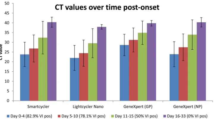
The day of sample collection post disease onset was known for 98 of the RT-PCR-positive samples (actual range, day 0 to 33). Samples were arranged according to day of collection post onset in
the following four groups: day 0 to 4 (n⫽41), 5 to 10 (n⫽42), 11
to 15 (n⫽8), and 16 to 33 (n⫽7). MeanCTvalues and standard
deviations on each assay and platform were calculated for the four
time groups (Fig. 2). Sample groups at earlier time points after
disease onset yielded the lowest averageCTvalues, with an increase
in time leading to higherCTvalues. The percentage of samples
from which Ebola virus could be recovered was highest early after onset, after which the number decreased with time, and none of the samples in the day 16 to 33 time group containing live virus.
DISCUSSION
The magnitude of the 2013 to 2015 Ebola virus disease epidemic in West Africa has highlighted the unpreparedness of the world to respond to massive transmission of this highly dangerous patho-gen. An important aspect of the control of such outbreaks is access to rapid and reliable diagnostic capacity, often required in remote and resource-constrained areas. Management of suspected cases in Ebola treatment or holding centers is heavily dependent on laboratory testing to ensure that infected patients are isolated in a timely manner and that noninfected patients are released. The availability of accurate, reliable point-of-care diagnostics would contribute greatly to the better management of infected and non-infected patients and thereby decrease the risk of unnecessary ex-posure of noninfected patients. Point-of-care diagnostic capacity was lacking during most of the West African outbreak, leading to complete dependence on mobile laboratories. The countrywide transmission in each of the most affected countries and the low number of laboratories performing Ebola diagnostics resulted in delays from patient submission to laboratory confirmation due to long distances, poor road infrastructure, and the lack of reliable sample transport.
Although real-time RT-PCR assays are currently widely used for Ebola virus diagnostics by reference laboratories worldwide and by all mobile laboratories deployed in West Africa, they were not intensively validated in the field against the gold standard, the virus isolation, mostly due to limited availability of clinical speci-mens in the past. Thus, there is no standardized molecular refer-ence test to compare and validate new prototype assays. To our knowledge, this is the first evaluation of a POC molecular assay in direct comparison to virus isolation in clinical specimens and the largest clinical evaluation of currently in-use real-time RT-PCR
TABLE 5 Agreement between qRT-PCR based assays and virus isolation from suspected EVD cases in Freetown, Sierra Leone, 2014 –2015. Positive or negative virus isolation No. with GeneXpert Ebola assay a ( C T value) No. with LightCycler Nano L-gene qRT-PCR a ( C T value) No. with SmartCycler L-gene qRT-PCR a ( C T value) GP target NP target One or both targets (GP and NP) ⫹⫺ ⫹⫺ ⫹⫺ ⫹ ⫺ ⫹⫺ Virus isolation, positive ( n ⫽ 91) 91 b (22.2–40.5) and 90 c (22.2–36.8) 0 b and 1 c (40.5) 91 b ,c (15.8–38.2) 0 b ,c 91 b ,c (15.8–40.5) 0 b ,c 91 b ,c (13.2–34.76) 0 b ,c 91 b ,c (14.77–38.62) 0 b ,c Virus isolation, negative ( n ⫽ 34) 17 b (28.5–44.6) and 6 c range (28.5–38.8) 17 b ( ⬎ 45) and 28 c ( ⬎ 40) 31 b (25.1–43.8) and 20 c (25.1–39.8) 3 b ( ⬎ 45) and 14 c ( ⬎ 40) 33 b (25.1–44.6) and 20 c (25.1–39.8) 1 b ( ⬎ 45) and 14 c ( ⬎ 40) 19 b ,c (25.6–39.8) 5 b ,c ( ⬎ 40) 30 b (26.9–42.9) and 20 c (26.9–39.96) 4 b ( ⬎ 45) and 14 c ( ⬎ 40) Agreement Virus isolation, positive (%) 100 b and 98.9 c 100 b ,c 100 b ,c 100 b ,c 100 b ,c Agreement Virus isolation, negative (%) 50.0 b and 82.35 c 8.8 b and 41.2 c 2.9 b and 41.2 c 14.3 b ,c 11.8 b and 41.2 c a ⫹ ,positive; ⫺ ,negative. b C T cutoff value of ⱕ 45 used. c C T cutoff value of ⱕ 40 used.
on May 16, 2020 by guest
http://jcm.asm.org/
[image:5.585.65.258.75.712.2]targeting the L gene. To illustrate the effect of PCR equipment choice, we also evaluated the performance of the L gene real-time RT-PCR run on the Roche LightCycler Nano platform.
The analytical sensitivity and specificity of the GeneXpert Ebola assay, which targets GP and NP genes, were compared to those of a TaqMan-based reverse transcription-quantitative PCR (qRT-PCR) targeting the polymerase gene and run on the Ceph-eid SmartCycler and Roche LightCycler Nano platforms using a laboratory-generated virus dilution series. The GeneXpert assay did not yield any cross-reaction to the hemorrhagic fever viruses or arboviruses tested in this study. The limit of EBOV detection
for all of the assays was 1.0 TCID50/ml (corresponding to 163 L
gene RNA copies per milliliter or 1.94 copies per reaction), where
all four replicates at this dilution were detected. At 0.1 TCID50/ml,
detection was intermittent by all assays (74 L gene RNA copies per milliliter or 0.88 copies per reaction). It is important to note that detection of the GP target gene was less sensitive than detection of the NP target gene in the GeneXpert assay, with a detection limit
of 1 log10less. A similar trend has been observed previously, albeit
in conventional PCR format, where the amount of DNA amplified
by an RT-PCR targeting the L gene was greater than that by a
GP-targeting assay on the same samples (12). Interestingly, in the
same study, it was found that the detection of NP was 125-fold more sensitive than the detection of the L gene, an observation that we could not reproduce in this study with the lab-generated virus titration series. The inherent multiplex characteristic of the GeneXpert assay may explain the difference. RT-PCR protocols following the one-step principle allow detection of genomic- and
antigenomic-sense (messenger) RNA (18). Although Ebola virus
has a linear, nonsegmented negative-sense RNA genome, infer-ring an equal number of copies of each of the virus’ genes per particle, it is possible that some genes are transcribed in higher numbers than others, leading to a higher number of detectable copies (including mRNA) relative to other genes during infection. The number of RNA copies is not a direct indication of the num-ber of virus particles, as RNA copies are consistently between 3
and 4 log10higher than PFU (18) or between 2 and 3 log10higher
than TCID50in our study.
The GeneXpert system is designed to use whole blood as the sample input. It has been shown that there is earlier clearance of
FIG 1The range ofCTvalues obtained by the different assays (top left, SmartCycler; top right, LightCycler Nano; bottom left, GeneXpert GP target; bottom right,
GeneXpert NP target) and the correspondence to sample infectivity is shown.CTvalues are arranged in increasing value. The dotted vertical lines indicate the
intermediate range where there is an overlap ofCTvalues corresponding to successful and unsuccessful virus isolation. Red dots indicate samples from which
virus can be isolated, and black crosses indicate unsuccessful virus isolation attempt. The two outliers are also included in the figures for reference.
on May 16, 2020 by guest
http://jcm.asm.org/
virus from serum and plasma than from whole blood (27). Testing of whole blood has an important practical and safety advantage since it does not require specimen processing. In our study, we could only evaluate and directly compare the different assays us-ing stored serum and plasma samples from suspected EVD cases. All assays yielded high estimates of diagnostic accuracy. As
ex-pected, when using a lowerCTcutoff value to characterize a
sam-ple as positive or negative, the sensitivity decreased slightly but the specificity increased. The lack of agreement between the
L-gene-based assay and the GeneXpert assay atCTvalues of⬎40 may be
due to the fact that the latter is more sensitive in detecting border-line concentrations of RNA. This emphasizes the importance of
the careful interpretation of results yielding highCTvalues, even
aboveCTvalues of 35, together with clinical data and the exposure
history of the patient. It appears that the performance of the Gen-eXpert assay is highly dependent on the detection of the NP gene rather than on the GP gene. There were only two samples where
the GP gene target was detected (CT,⬎40) but where NP was
not detected, compared to 16 samples where the NP gene target
was detected (actualCTrange, 38.4 to 43.8) but where GP was
not detected.
Arguably, a more appropriate way to directly compare perfor-mance is to look at samples grouped according to different ranges
ofCT values. All samples yieldingCT values ofⱕ34.99 (on the
SmartCycler) were detected by all of the assays (100% agreement), with agreement decreasing in lower positive samples. The value of detecting NP and GP in the GeneXpert assay is illustrated when
analyzing the results at aCTrange of 40 to 45. When detection of
GP and NP is analyzed separately at this range, the agreement to the L gene assay is 20% and 90%, respectively. However, when following the principle of regarding a sample as positive with the detection of either GP or NP or both, the agreement becomes 100%. Another important analysis was to demonstrate that the assays are reliable for the detection of RNA in samples that contain live virus. All of the assays were able to detect RNA in samples that tested positive by virus isolation. As expected, all of the assays
detected RNA in a number of samples that did not contain live virus. Similarly to what was reported for Marburg virus in bats
(28), we found a clear correlation betweenCTvalues (RNA copies)
and the ability to isolate virus from the clinical specimens.
Mar-burg virus could only be isolated from bat tissues that yieldedCT
values of⬍35 (28). In our own study, we were also unable to
isolate EBOV from the blood and tissues of experimentally
in-fected bats withCTvalues of⬍35 (NICD, unpublished data).
A direct correlation between viral RNA levels determined by qRT-PCR and the ability to isolate EBOV suggests potentially im-portant practical aspects in terms of identifying infectious or non-infectious samples. In our study, we identified an intermediate
range where there was an overlap ofCTvalues yielding positive
virus isolation and not, and thisCTrange was different for the
different assays. Excluding the limited outliers, all of the assays
perform 100% atCTvalues of⬍35, and all are able to detect RNA
in samples from which virus was isolated.
Although we do not have matching serology data available, another explanation for the detection of RNA in virus-isolation-negative samples may be that the RNA detected in the virus-isolation-negative isolation samples represents virus that is in the process of being cleared in the form of immunocomplexes. This may be supported by the fact that the percentage of successful virus isolation
de-creased over time post onset (Fig. 2). Considering that, there are
numerous factors that can have an effect on the outcome of a diagnostic test on a patient’s clinical sample. Sample quality is an obvious factor that includes various aspects such as volume, he-molysis, cold-chain transport, and storage. Probably a more im-portant factor is the timing of sample collection post disease onset. A sample collected too early or too late after disease onset can yield a false-negative result depending on which analyte is targeted. The duration of viremia caused by infection with different viruses dif-fers as does the time needed to develop a detectable antibody re-sponse. For this reason, the diagnosis of a viral hemorrhagic fever should ideally not depend on a single test result (single analyte), especially in the case of excluding VHF as a diagnosis.
FIG 2The averageCTvalues obtained by the different assays arranged according to time grouping post disease onset (blue bars, day 0 to 4; red bars, day 5 to 10;
green bars, day 11 to 15; purple bars, day 16 to 33) are shown. Error bars indicate standard deviations for each time grouping. The values in the brackets below the plots indicate the percentage of samples in the time group from which Ebola virus could be isolated (VI pos). The sample numbers per time grouping are as follows: day 0 to 4 (n⫽41); day 5 to 10 (n⫽42); day 11 to 15 (n⫽8); day 16 to 33 (n⫽7).
on May 16, 2020 by guest
http://jcm.asm.org/
[image:7.585.114.479.65.265.2]In this study, we showed that the prototype GeneXpert Ebola assay was highly accurate in the detection of RNA in serum and plasma samples containing live Ebola virus. The agreement
be-tween the different assays we compared was very high at lowCT
values and decreased with lower positive samples. Despite the
sample input volume of the GeneXpert assay being only 100l
compared to the 140l used for manual RNA extraction, it did
not noticeably affect assay sensitivity. The prototype GeneXpert Ebola assay presents several advantages over currently available qRT-PCR protocols. The assay incorporates an automated extrac-tion and sample addiextrac-tion process, making it possible to be run by minimally trained technicians within 90 min. If placed within Ebola treatment centers, the GeneXpert system would negate the need for additional biocontainment devices where patient sam-ples first have to be inactivated, processed, and RNA extracted. This technology minimizes the possibility of human error, for example, during the extraction process, RT-PCR master mix preparation, and sample addition, by being automated. One other advantage of the assay is the usage of stable reagents. The ongoing Cepheid shelf-life testing demonstrates that cartridges used for Xpert Ebola POC are stable under room temperature for at least 6 months (Cepheid, personal communication).
Hemoglobin and lactoferrin have been identified as major
in-hibitors of diagnostic PCR in human blood cells (29). However, an
internal control included in the Ebola Xpert POC ensures the detection of inhibitory effects from factors possibly present in patient samples.
False-negative results in patients with severe hemorrhagic fever
have been noted before (30). Therefore, analytical results obtained
by this and any other test should be interpreted by trained and experienced diagnosticians together with all available laboratory results and clinical, pathological, and epidemiological data to en-sure an accurate diagnosis. In conclusion, the prototype Gene-Xpert Ebola assay represents a promising point-of-care screening tool to make rapid presumptive decisions about patient manage-ment and infection control measures.
ACKNOWLEDGMENTS
The authors would like to dedicate this work to and acknowledge all individuals who contributed and who are still contributing to the fight against the Ebola epidemic in West Africa. In particular, we thank the staff of the Ministry of Health and Sanitation of Sierra Leone, the staff of the National Institute for Communicable Diseases, the National Department of Health of South Africa, the World Health Organization, and the Global Outbreak Alert and Response Network. We also thank Lynsey Isherwood for helping in preparation of ethics applications and related project logis-tics.
J.T.P., P.J.V.V., I.S., and A.K. conceived and designed the study. P.J.V.V., N.S., O.C., K.K., and J.T.P. were responsible for sample process-ing, long-term storage, and database management in Sierra Leone. P.J.V.V., N.S., and A.G. executed the laboratory work. P.J.V.V., A.G., and J.T.P. interpreted and analyzed the data. P.J.V.V. drafted the manuscript after which it was critically reviewed by all coauthors.
FUNDING INFORMATION
Cepheid provided funding to Janusz T. Paweska.
The NICD was supported by Cepheid by provision of the GeneXpert Ebola POC IV system, the GeneXpert Dx software package, disposable prototype GeneXpert Ebola cartridges, and funds for covering the costs of running polymerase (L) gene TaqMan real-time RT-PCR assays.
REFERENCES
1.WHO Ebola Response Team.2014. Ebola virus disease in West Africa– the first 9 months of the epidemic and forward projections. N Engl J Med 371:1481–1495.http://dx.doi.org/10.1056/NEJMoa1411100.
2.Carroll MW, Matthews DA, Hiscox JA, Elmore MJ, Pollakis G, Ram-baut A, Hewson R, García-Dorival I, Bore JA, Koundouno R, Abdellati S, Afrough B, Aiyepada J, Akhilomen P, Asogun D, Atkinson B, Ba-dusche M, Bah A, Bate S, Baumann J, Becker D, Becker-Ziaja B, Bocquin A, Borremans B, Bosworth A, Boettcher JP, Cannas A, Carletti F, Castilletti C, Clark S, Colavita F, Diederich S, Donatus A, Duraffour S, Ehichioya D, Ellerbrok H, Fernandez-Garcia MD, Fizet A, Fleis-chmann E, Gryseels S, Hermelink A, Hinzmann J, Hopf-Guevara U, Ighodalo Y, Jameson L, Kelterbaum A, Kis Z, Kloth S, Kohl C, Korva M, et al.2015. Temporal and spatial analysis of the 2014-2015 Ebola virus outbreak in West Africa. Nature524:97–101.http://dx.doi.org/10.1038 /nature14594.
3.World Health Organization.2015. Ebola situation report—7 October 2015. World Health Organization, Geneva, Switzerland.
4.Feldmann H, Geisbert TW. 2011. Ebola haemorrhagic fever. Lancet 377:849 – 862.http://dx.doi.org/10.1016/S0140-6736(10)60667-8. 5.Ksiazek TG, Rollin PE, Jahrling PB, Johnson E, Dalgard DW, Peters CJ.
1992. Enzyme immunosorbent assay for Ebola virus antigens in tissues of infected primates. J Clin Microbiol30:947–950.
6.Geisbert TW, Rhoderick JB, Jahrling PB.1991. Rapid identification of Ebola virus and related filoviruses in fluid specimens using indirect im-munoelectron microscopy. J Clin Pathol44:521–522.http://dx.doi.org/10 .1136/jcp.44.6.521.
7.Zaki SR, Shieh W-J, Greer PW, Goldsmith CS, Ferebee T, Katshitshi J, Tshioko FK, Bwaka MA, Swanepoel R, Calain P, Khan AS, Lloyd E, Rollin PE, Ksiazek TG, Peters CJ.1999. A novel immunohistochemical assay for the detection of Ebola virus in skin: implications for diagnosis, spread and surveillance of Ebola haemorrhagic fever. J Infect Dis 179(Suppl):S36 –S47.http://dx.doi.org/10.1086/514319.
8.Ksiazek TG, West CP, Rollin PE, Jahrling PB, Peters CJ.1999. ELISA for the detection of antibodies to Ebola viruses. J Infect Dis179(Suppl):S192– S198.http://dx.doi.org/10.1086/514313.
9.Moe JB, Lambert RD, Lupton HW.1981. Plaque assay for Ebola virus. J Clin Microbiol13:791–793.
10. van der Groen G, Jacob W, Pattyn SR.1979. Ebola virus virulence for newborn mice. J Med Virol4:239 –240.http://dx.doi.org/10.1002/jmv .1890040309.
11. Weidmann M, Mühlberger E, Hufert FT.2004. Rapid detection protocol for filoviruses. J Clin Virol30:94 –99.http://dx.doi.org/10.1016/j.jcv.2003 .09.004.
12. Sanchez A, Ksiazek TG, Rollin PE, Miranda MEG, Trappier SG, Khan AS, Peters CJ, Nichol ST.1999. Detection and molecular characterization of Ebola viruses causing disease in human and nonhuman primates. J Infect Dis179(Suppl):S164 –S169.http://dx.doi.org/10.1086/514282. 13. Onyango CO, Opoka ML, Ksiazek TG, Formenty P, Ahmed A, Tukei
PM, Sang RC, Ofula VO, Konongoi SL, Coldren RL, Grein T, Legros D, Bell M, De Cock KM, Bellini WJ, Towner JS, Nichol ST, Rollin PE. 2007. Laboratory diagnosis of Ebola hemorrhagic fever during an out-break in Yambio, Sudan, 2004. J Infect Dis196:S193–S198.http://dx.doi .org/10.1086/520609.
14. Panning M, Laue T, Olschlager S, Eickmann M, Becker S, Raith S, Courbot MC, Nilsson M, Gopal R, Lundkvist A, di Caro A, Brown D, Meyer H, Lloyd G, Kummerer BM, Gunther S, Drosten C. 2007. Diagnostic reverse-transcription polymerase chain reaction kit for filovi-ruses based on the strain collections of all European biosafety level 4 lab-oratories. J Infect Dis196:S199 –S204.http://dx.doi.org/10.1086/520600. 15. Leroy EM, Baize S, Lu CY, McCormick JB, Georges AJ, Georges-Courbot MC, Lansoud-Soukate J, Fisher-Hoch SP.2000. Diagnosis of Ebola haem-orrhagic fever by RT-PCR in an epidemic setting. J Med Virol60:463– 467.
http://dx.doi.org/10.1002/(SICI)1096-9071(200004)60:4⬍463::AID-JMV1 5⬎3.0.CO;2-M.
16. Gibb TR, Norwood DA, Jr, Woollen N, Henchal EA.2001. Development and evaluation of a fluorogenic 5=nuclease assay to detect and differentiate between Ebola virus subtypes Zaire and Sudan. J Clin Microbiol39:4125– 4130.http://dx.doi.org/10.1128/JCM.39.11.4125-4130.2001.
17. Trombley AR, Wachter L, Garrison J, Buckley-Beason VA, Jahrling J, Hensley LE, Schoepp RJ, Norwood DA, Goba A, Fair JN, Kulesh DA. 2010. Comprehensive panel of real-time TaqMan polymerase chain
on May 16, 2020 by guest
http://jcm.asm.org/
tion assays for detection and absolute quantification of filoviruses, arena-viruses and new world hantaarena-viruses. Am J Trop Med Hyg82:954 –960.
http://dx.doi.org/10.4269/ajtmh.2010.09-0636.
18. Towner JS, Rollin PE, Bausch DG, Sanchez A, Crary SM, Vincent M, Lee WF, Spiropoulou CF, Ksiazek TG, Lukwiya M, Kaducu F, Downing R, Nichol ST.2004. Rapid diagnosis of Ebola hemorrhagic fever by reverse transcription-PCR in an outbreak setting and assessment of patient viral load as a predictor of outcome. J Virol78:4330 – 4341.http://dx.doi.org /10.1128/JVI.78.8.4330-4341.2004.
19. Zhai J, Palacios G, Towner JS, Jabado O, Kapoor V, Venter M, Grolla A, Briese T, Paweska J, Swanepoel R, Feldmann H, Nichol ST, Lipkin WI.2007. Rapid molecular strategy for filovirus detection and character-ization. J Clin Microbiol45:224 –226.
20. Broadhurst MJ, Kelly JD, Miller A, Semper A, Bailey D, Groppelli E, Simpson A, Brooks T, Hula S, Nyoni W, Sankoh AB, Kanu S, Jalloh A, Ton Q, Sarchet N, George P, Perkins MD, Wonderly B, Murray M, Pollock NR.2015. ReEBOV antigen rapid test kit for point-of-care and laboratory-based testing for Ebola virus disease: a field validation study. Lancet386:867– 874.http://dx.doi.org/10.1016/S0140-6736(15)61042-X. 21. Walker NF, Brown CS, Youkee D, Baker P, Williams N, Kalawa A, Russell K, Samba AF, Bentley N, Koroma F, King MB, Parker BE, Thompson M, Boyles T, Healey B, Kargbo B, Bash-Taqi D, Simpson AJ, Kamara A, Kamara TB, Lado M, Johnson O, Brooks T.2015. Evaluation of a point-of-care blood test for identification of Ebola virus disease at Ebola holding units, Western Area, Sierra Leone, January to February 2015. Euro Surveill20(12):pii⫽21073.http://dx.doi.org/10.2807/1560-79 17.ES2015.20.12.21073.
22. Paweska JT, Jansen van Vuren P, Masumu J, Leman PA, Grobbelaar AA, Birkhead M, Clift S, Swanepoel R, Kemp A.2012. Virological and serological findings inRousettus aegyptiacusexperimentally inoculated with Vero cells-adapted Hogan strain of Marburg virus. PLoS One7(9): e45479.http://dx.doi.org/10.1371/journal.pone.0045479.
23. Le Roux CA, Kubo T, Grobbelaar AA, van Vuren PJ, Weyer J, Nel LH, Swanepoel R, Morita K, Paweska JT.2009. Development and evalua-tion of a real-time reverse transcripevalua-tion-loop-mediated isothermal ampli-fication assay for rapid detection of Rift Valley fever virus in clinical
spec-imens. J Clin Microbiol 47:645– 651. http://dx.doi.org/10.1128/JCM .01412-08.
24.Greiner M. 1995. Two-graph receiver operating characteristics (TG-ROC): a Microsoft-Excel template for the selection of cut-off values in diagnostic tests. J Immunol Methods185:145–146.http://dx.doi.org/10 .1016/0022-1759(95)00078-O.
25.Greiner M. 1996. Two-graph receiver operating characteristics (TG-ROC): update version supports optimisation of cut-off values that mini-mize overall misclassification costs. J Immunol Methods191:93–94.http: //dx.doi.org/10.1016/0022-1759(96)00013-0.
26. Greiner M, Sohr D, Göbel P.1995. A modified ROC analysis for the selection of cut-off values and the definition of intermediate results of serodiagnostic tests. J Immunol Methods185:123–132.http://dx.doi.org /10.1016/0022-1759(95)00121-P.
27. Southern TR, Rasca LD, Albarino CG, Fey PD, Hinrichs SH, Mur-phy CN, Herrera VL, Sambol AR, Hill CE, Ryan EL, Kraft CS, Campbell S, Sealy TK, Schuh A, Ritchie JC, Lyon GM, III, Mehta AK, Varkey JB, Ribner BS, Brantly KP, Ströher U, Iwen PC, Burd EM.2015. Comparison of FilmArray and qRT-PCR for the detection of
Zaire ebolavirusfrom contrived and clinical specimens. J Clin Microbiol 53:2956 –2960.http://dx.doi.org/10.1128/JCM.01317-15.
28. Towner JS, Amman BR, Sealy TK, Carroll SAR, Comer JA, Kemp A, Swanepoel R, Paddock CD, Balinandi S, Khristova ML, Formenty PB, Albarino CG, Miller DM, Reed ZD, Kayiwa JT, Mills JN, Cannon DL, Greer PW, Byaruhanga E, Farnon EC, Atimnedi P, Okware S, Katon-gole-Mbidde E, Downing R, Tappero JW, Zaki SR, Ksiazek TG, Nichol ST, Rollin PE.2009. Isolation of genetically diverse Marburg viruses from Egyptian fruit bats. PLoS Pathog5:e1000536.http://dx.doi.org/10.1371 /journal.ppat.1000536.
29. Al-Soud WA, Rådström P.2001. Purification and characterization of PCR-inhibitory components in blood cells. J Clin Microbiol39:485– 493.
http://dx.doi.org/10.1128/JCM.39.2.485-493.2001.
30. Drosten C, Panning M, Guenther S, Schmitz H.2002. False-negative results of PCR assay with plasma of patients with severe viral hemorrhagic fever. J Clin Microbiol40:4394 – 4395.http://dx.doi.org/10.1128/JCM.40 .11.4394-4395.2002.