A Survey on Medical Image Compression Algorithms and Its
Performance
Samjaikumar.S
1Marypraveena.S
2Prabakaran.S
31,3
Assistant Professor
2Associate Professor
1,2,3
Department of Electronics and Communication Engineering
1,2
Sri Ramakrishna Institute of Technology, Coimbatore, India
3
CMS College of Engineering and Technology, Coimbatore, India
Abstract— Rapid advancements in medical imaging requires an efficient methodologies for storing and transmission. A huge amount of medical images is generated through advanced medical imaging in hospitals and a large database is required to keep and maintain the diagnostic information in the form of images and it requires more space and transmission bandwidth. Medical image compression techniques help to store the medical images with relevant diagnostic information. This paper discuss about the performance analysis of different image compression algorithms and their performance variations can be determined using parameters such as MSE, PSNR and SNR. Key words: Image Compression, Compression Ratio; Mean square error (MSE), PSNR (Peak Signal to Noise Ratio), SNR Signal to Noise Ratio
I. INTRODUCTION
In general the term compression is used for compressing big data’s so that they could be stored in much less memory space rather than in their original form. Image compression is the process of reducing the size of any image files without degrading the quality of the images. The main aim of the Image compression is to reduce the correlation between one pixel and its neighbouring pixel. In recent years, digitized images are used rather than analog images. The volumes of data needed for symbolizing such images are high. Hence the transmission rate is slow and the cost of operation is high. Therefore the information contained in images should be compressed by extracting only visible elements, which has to be encoded. To increase the compression rate of the data to represent an image, the quantity of data has to be reduced. Due to this distinctive property, Image compression techniques are widely used in many applications, especially in telemedicine field in order to minimize the cost of storage [1].It is also used to increase the transmission speed of medical images and data at available bandwidth. Now a days most hospitals store medical image data in digital form. The efficient image compression techniques help the doctors to maintaining the quality of the image even after compression and it also extract the significant information with the minimum storage space. Due to digitization of data, medical image compression is necessary in all the related fields including medical image database, medical image communications etc.
II. MEDICAL IMAGE COMPRESSION
In general an image is a matrix which consists of rectangular array of dots called pixels in different rows and columns. Different types of images are used in various fields like remote sensing, video processing, medical fields, biometric, satellite imaging techniques which require efficient
compression methodologies for transmission across or within networks and storage. Medical image is the visual representation of organs or body parts. Nowadays the information about the human body is used for many diagnostic and practical purposes in clinical applications. The different types of medical images are X-ray, Magnetic Resonance Imaging (MRI), Ultrasound scans etc. The uncompressed images require large amount of RAM storage. Medical Image compression helps to reduce storage space, transmission requirements and it preserves all the relevant information received after the diagnosis. DICOM is the international standard developed for medical imaging by the National Electrical Manufacturers Association for retaining and transferring information in imaging. It also enables integration of various medical imaging devices like scanners, printers, network hardware etc. from several manufacturers. Almost all the neighbouring pixels in an image have a correlation and these results in generating redundant information. The task of compression is to modify the images in such a way that there is less correlation with the neighbouring pixels and hence the redundant information will be reduced. The two basic fundamental elements of image compression are redundancy reduction and irrelevant data reduction.
A. Compression Techniques
In general the image compression techniques are broadly categorized into two categories.
1) Lossy image compression 2) Lossless image compression
For lossless compression an exact replica of the original image can be recreated or reconstructed from the compressed image and for lossy compression an exact replica of the original image cannot be recreated from the compressed image.
1) Lossy Image Compression Technique
There is a finite amount of loss in the lossy compression and the compressed image is different from the input image. It provides higher compression ratio in comparison with lossless compression. The significant loss of information in the compressed image results in is difference between compressed image and original image [2].
The compressed images contain the degradation level relative to the original. It provides high compression rates of 30:1 or higher. Lossy compression techniques includes following schemes:
1) Transformation coding 2) Vector quantization 3) Fractal coding
4) Block Truncation Coding 5) Sub-band coding
2) Lossless Image Compression Technique
In the lossless compression the compressed image is totally replica of the original input image, there is loss of data [3]. The reconstructed image is same as that of the input image in lossless compression scheme. Here the compression rate is approximately 3:1. Lossless compression techniques includes following schemes:
1) Run length encoding 2) Huffman encoding 3) LZW coding 4) Area coding
5) DPCM
B. Performance Parameters of Image Compression
The performance of the image compression algorithms can be measured by using the parameters such as.
1) Mean square error (MSE) 2) PSNR (peak signal to noise ratio) 3) Compression Ratio (CR)
The peak error between the compressed image and original image is measured in terms of PSNR [4]. The higher value of PSNR indicates higher quality of image [5]. To calculate PSNR, MSE is first computed. Cumulative difference between the compressed image and original image is MSE.
1) Mean Square Error (MSE)
Mean Squared Error (MSE) is defined as the square of differences in the pixel values between the corresponding pixels of the two images. The mean square error (MSE) of N * M size image is given by,
MSE = ∑ [𝐼1(𝑚,𝑛)−𝐼2(𝑚,𝑛)]2 𝑀∗𝑁
𝑀,𝑁 --- (1)
M &N - number of rows and columns in the input images. 2) PSNR (Peak Signal to Noise Ratio)
PSNR Peak signal-to-noise ratio often abbreviated PSNR, is an engineering name, for the ratio between the maximum possible power of a signal and the power of corrupting noise that affects the fidelity. The peak error between the compressed image and original image is measured in terms of PSNR [6, 7]. The higher value of PSNR indicates higher quality of image. To calculate PSNR, MSE is first computed. Cumulative difference between the compressed image and original image is MSE. Small value of MSE improves image quality and reduces the error.
PSNR = 10 𝑙𝑜𝑔10 𝑅2
𝑀𝑆𝐸 --- (2) 3) Compression Ratio (CR)
Data redundancy is the centric release in digital image compression [8]. If N1 and N2 denote the number of
respectively, then the compression ratio, CR can be specify as
𝐶𝑅 =𝑁1
--- (3)
And data redundancy of the original image can be specify as 𝑅𝐷 = 1 − 1
𝐶𝑅 --- (4)
III. IMAGE COMPRESSION MODEL
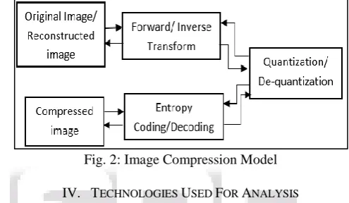
[image:2.595.300.548.265.409.2]Image compression is used to reduce the correlation between pixels. Here the input image is fed into the encoder. The first stage is a transform to eliminate the redundancy and to pack the information efficiently. Then quantization is applied to represent the packed information with as few bits as possible. Quantization is a many-to-one correspondence mapping that replaces a set of values with only one representative value [9], [10]. The output is a compressed representation of input, which can be stored for future use or can be transmitted. At the decoding side, the reverse operation is performed to generate the reconstructed image. The block diagram of an image compression model is shown below.
Fig. 2: Image Compression Model
IV. TECHNOLOGIES USED FOR ANALYSIS
The impact on storage and network bandwidth requirements is predicted to increase sharply when departments adopt digital technology in all modalities, especially digital mammography , CT and PET scanners that are generating increasingly large numbers of images per study; and archiving of 3D volume renderings and other complex advanced image reformats of the original images. The results obtained by compressing scan images using different image compression techniques are shown below.
A. Picture Archiving and Communication Systems Using ROI
Compressing a digital image can facilitate its transmission, storage and processing. As medical imaging become increasingly digital, the quantities of imaging data are forcing consideration of compression in picture archiving and communication systems (PACS) and evolving telemedicine systems. The way to store medical images are HIS (Hospital Information System) and PACS based on DICOM standard pave which in turn makes ease remote medical treatments possible. It has been reinforced through extensive research that the diagnostically important regions of medical images – the region of interest (ROI) - must be compressed by a lossless or a near lossless algorithm in order to preventing from a wrong diagnosis due to a poor image quality. The major drawback of the FR quality metrics for huge databases is that a large amount of reference information has to be provided at the final comparison point.
detecting and estimating the strength of commonly found artifact signals. A spread spectrum technique is used for estimating the quality of image in order to data hiding(watermarking) and producing a reliable metric [1].Although there are medical oriented watermarking studies in the literature and it's a valuable tool for copyright protection, patient and examination-related information hiding, data integrity control and source identification, in this work we use watermarking in order to data hiding into non-ROI part of the image while preserving the quality of non-ROI part and in a manner which can estimate the quality of original image.
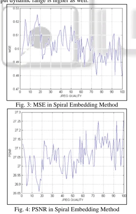
[image:3.595.312.544.69.571.2]Watermarking may be done in the spatial domain or in the transform domains such as DCT or wavelet .We choose to insert the mark in DCT domain because this is the domain still used by many compression algorithms such as JPEG standard. The performance of the proposed metric with measuring correlation of whole image degradation and MSE and PSNR of the extracted mark. We considered 100 different images which were compressed by JPEG qualities from 1 to 100 and 100 other images which were degraded by Gaussian noise with zero mean and 0.01 to 0.1 standard deviation. This metric also outperforms the algorithm of spiral embedding in spatial domain [11].The results for spiral embedding is illustrated in figures 5 and 6. As shown in figures 7 and 8, for the proposed metric the results are strongly correlated and the output dynamic range is higher as well.
Fig. 3: MSE in Spiral Embedding Method
Fig. 4: PSNR in Spiral Embedding Method
Apart from that, MSE and PSNR of the extracted mark are a good approximation of degradation of complete image which is done once by JPEG compression
Fig. 5: MSE in Spread Spectrum Embedding Method
Fig. 6: PSNR in Spread Spectrum Embedding Method
B. 3D SDB-SPIHT
A fast coding scheme is proposed for the 3D SDB-SPIHT algorithm based on an efficient spatial–temporal tree structure (STTS), which is designed for the wavelet transformed coefficients, For any coefficient, two sets are defined, the offspring set (the direct descendants of the current coefficient) and the leaves set (all the indirect descendants of the current coefficient). These two sets are processed separately according to different rules. Based on the separate descendant, 3D SDB-SPIHT has more selectivity in deciding the scanning and coding and hence achieves faster speed.
The input medical image sequences are divided into many groups of contiguous slices (GOS). All the experiments are performed in the unit of GOS. Compared with conventional 3D SPIHT, 3D SDB-SPIHT excels in the short coding time and ensures a nearly equivalent image quality.
C. Fast Coding Scheme
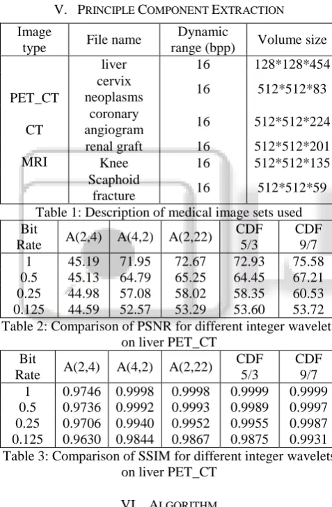
[image:3.595.57.280.372.721.2]The performance of the proposed 3D SDB-SPIHT algorithm is presented. Six typical high-resolution medical image sets from the DICOM database [20] were tested in this paper. All were stored in the standard DICOM format with 16-bit grey-scale pixel values. Table 3 provides a description of these sets. Each image set was divided into a series of GOS and each GOS was encoded and decoded as a separate unit. A GOS size of 16 was selected for all the experiments because it provides an acceptable compression performance and the required memory and computational time are reasonable for the chosen sized images.
To measure the performance of the proposed SDB-SPIHT algorithm, two objective image quality measures were utilized, peak signal-to-noise ratio (PSNR) and structural similarity index (SSIM).
V. PRINCIPLE COMPONENT EXTRACTION
Image
type File name
Dynamic
range (bpp) Volume size
PET_CT
CT
MRI
liver 16 128*128*454
cervix
neoplasms 16 512*512*83
coronary
angiogram 16 512*512*224
renal graft 16 512*512*201
Knee 16 512*512*135
Scaphoid
[image:4.595.313.539.128.641.2]fracture 16 512*512*59
Table 1: Description of medical image sets used Bit
Rate A(2,4) A(4,2) A(2,22)
CDF 5/3
CDF 9/7
1 45.19 71.95 72.67 72.93 75.58
0.5 45.13 64.79 65.25 64.45 67.21
0.25 44.98 57.08 58.02 58.35 60.53
0.125 44.59 52.57 53.29 53.60 53.72
Table 2: Comparison of PSNR for different integer wavelets on liver PET_CT
Bit
Rate A(2,4) A(4,2) A(2,22)
CDF 5/3
CDF 9/7 1 0.9746 0.9998 0.9998 0.9999 0.9999 0.5 0.9736 0.9992 0.9993 0.9989 0.9997 0.25 0.9706 0.9940 0.9952 0.9955 0.9987 0.125 0.9630 0.9844 0.9867 0.9875 0.9931 Table 3: Comparison of SSIM for different integer wavelets
on liver PET_CT
VI. ALGORITHM
Every image can be considered as non-stationary with respect to x-axis & y-axis by considering variations in pixel values. The concept provides attempt to analyze frequency information evolving with time and identifying the amount of variation due to oscillation at different scales and locations with respect to time in a target biomedical image.
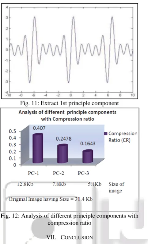
By focusing on principle oscillations in an input image we can achieve principle part of the same target image. Doing number of iterations over extracted part, methodology can extract more principle part from image. The process of oscillation detection and extraction should be continued till image gives good quality of principle component [7].
The principle component extraction methodology proves that it gives quality extraction with extraction of principle component. It provides better level of compression and it can be illustrated by considering image as a mono-dimensional signal.
Fig. 7: Identify all local maxima of x (t)
Fig. 9: Interpolate between maxima ending up with some Envelope call it e max (t)
[image:4.595.45.284.251.616.2]Fig. 11: Extract 1st principle component
Fig. 12: Analysis of different principle components with compression ratio
VII. CONCLUSION
The main objective of the image compression algorithm is to reduce the storage space and the compressed data should be feasible for transmission. The data integrity should be maintained during this process. Several techniques achieve efficient compression results. In this survey paper several algorithms involved in the image compression techniques were discussed. The performance parameters in the algorithms were estimated by two parameters such as PSNR, and MSE.
The quality of ROI part can be well-maintained and there is a strong correlation between MSE and PSNR of extracted mark and altered compression qualities. The fast algorithm based on 3D SPIHT reduces the coding time when compared with conventional 3D SPIHT. The excellent comprehensive performance confirmed this approach as an efficient solution for fast compression of medical images, especially for high resolution image sequences. Oscillation based Principle component extraction for biomedical image compression can achieve maximum compression ratio with good quality. It provides flexibility to end user to use any principle component according to his/her requirement and application. Increasing count of principle component with iteration gives more compression ratio.
REFERENCES
[1] Ansari M.A., R.S. Anand "Comparative Analysis of Medical Image Compression Techniques and their Performance Evaluation for Telemedicine", Proceedings of the International Conference on Cognition and Recognition, pp. 670-677, 2014.
[2] Sanjith S, Ganesan R, "Evaluating the Quality of Compression in Very High Resolution Satellite Images Using Different Compression Methods",International Journal of Engineering Research in Africa, Vol. 19, pp. 91-102,Mar. 2016.
[3] Sanjith S, Ganesan R. "A Review on Hyperspectral Image Compression", Control, instrumentation, communication and computational technologies (ICCICCT). International Conference, Kanyakumari, India. , pp. 10-11, Jul 2014.
[4] Sanjith S, Ganesan R, Fusion of DWT-DCT Algorithm for Satellite Image Compression", International Journal of Applied Engineering Research, ISSN 0973-4562, Vol. 10,pp. 130-137, Jul 2015.
[5] Sanjith S, Ganesan R, Rimal Isaac R. S. "Experimental Analysis of Compacted Satellite Image Quality Using Different Compression Methods", Adv Sci Eng Med Vol. 7, pp. 17, Mar 2015.
[6] M.A. Ansari , R.S. Anand "Performance Analysis of Medical Image Compression Techniques with respect to the quality of compression” IET-UK Internaional Conference on Information and Communication Technology in Electrical Sciences (ICTES 2007) Chennai tamil nadu, india , pp. 743-750. Dec.2007 [7] Smitha Joyce Pinto, Prof. Jayanand P.Gawande
"Performance analysis of medical image compression techniques”, Proceedings of the Institute of Electrical and Electronics Engineers IEEE, 2012.
[8] Rafael C. Gonzalez and Richard E. Woods. "Digital Image Processing" 3rd edition copyright 2008.
[9] Majid Rabbani, Paul W.Jones, "Digital Image Compression Techniques", 4th Edition, pp. 51, 1991. [10]Said A., Pearlman WA. An image multiresolution
representation for lossless and lossy compression. IEEE Transactions on Image Processing. 1996 Sep; 5(9):1303– 1310.
[11]H.K. Lee, H.J. Kim, K.R. Kwon, and J.K. Lee, “ROI Medical Image Watermarking using DWT and
Bit-plane,” asia –pacific conference on
communication,perth,western Australia,pp. 512-16,October 2005.
[12]Arash Ashtari Nakhaie, Shahriar B. Shokouhi, No reference medical image quality measurement based on spread spectrum and discrete wavelet transform using ROI processing, IEEE- CCECE 2011 – 000121
[13]Xiaoying Song, Qijun Huang , Sheng Chang, Jin He, Hao Wang, Three-dimensional separate descendant based SPIHT algorithm for fast compression of high-resolution medical image sequences, IET Image Process., 2017, Vol. 11 Iss. 1, pp. 80-87