Practice parameter
Practice parameter for the diagnosis and
management of primary immunodeficiency
Francisco A. Bonilla, MD, PhD*; I. Leonard Bernstein, MD†; David A. Khan, MD‡;
Zuhair K. Ballas, MD§; Javier Chinen, MD, PhD¶; Michael M. Frank, MD
储
; Lisa J. Kobrynski, MD**;
Arnold I. Levinson, MD††; Bruce Mazer, MD‡‡; Robert P. Nelson, Jr, MD§§;
Jordan S. Orange, MD, PhD¶¶; John M. Routes, MD
储储
; William T. Shearer, MD, PhD***; and
Ricardo U. Sorensen, MD†††
TABLE OF CONTENTS
I. Preface...S1 II. Executive Summary...S2 III. Algorithms ...S7 IV. Summary Statements ...S14 V. General Considerations...S20 VI. Humoral Immunodeficiencies ...S24 VII. Cellular Immunodeficiencies...S30 VIII. Combined Immunodeficiencies ...S33 IX. Phagocytic Cell Disorders ...S40 X. Complement Deficiencies...S43 XI. Acknowledgments...S45 XII. References...S45 XIII. Appendix ...S61 PREFACE
The purpose of this Practice Parameter for the Diagnosis and Management of Primary Immunodeficiency is to provide the
consultant allergist/immunologist with a practical guide for the clinical recognition and diagnosis of immunodeficiency, along with the general principles that guide management of
* Department of Medicine, Children’s Hospital Boston, and Department of Pediatrics, Harvard Medical School, Boston, Massachusetts.
† Department of Medicine and Environmental Health, University of Cincin-nati College of Medicine, CincinCincin-nati, Ohio.
‡ Department of Internal Medicine, University of Texas Southwestern Med-ical Center, Dallas, Texas.
§ Division of Allergy/Immunology, Department of Internal Medicine, Uni-versity of Iowa and the Iowa City Veteran’s Administration Medical Center, Iowa City, Iowa.
¶ National Human Genome Research Institute, National Institutes of Health, Bethesda, Maryland.
储 Department of Pediatrics, Children’s Health Center, Duke University Medical Center, Durham, North Carolina.
** Department of Pediatrics, Emory University School of Medicine, Atlanta, Georgia.
†† Section of Allergy and Immunology, Department of Medicine and Neu-rology, University of Pennsylvania School of Medicine, Philadelphia, Penn-sylvania.
‡‡ Division of Allergy and Immunology, Montreal Children’s Hospital, and Department of Pediatrics, McGill University, Montreal, Quebec.
§§ Department of Medicine, Division of Hematology/Oncology Hematolog-ical Malignancy Program/Immunology, Indianapolis, Indiana.
¶¶ Children’s Hospital of Philadelphia, Division of Immunology, and De-partment of Pediatrics, University of Pennsylvania School of Medicine, Philadelphia, Pennsylvania.
储储National Jewish Medical Research Center, Associate Professor of Medi-cine and Immunology, University of Colorado Health Sciences Center, Denver, Colorado.
*** Allergy and Immunology Service, Texas Children’s Hospital, Professor of Pediatrics and Immunology, Baylor College of Medicine, Houston, Texas. ††† Department of Pediatrics, Louisiana State University Health Science Center, New Orleans, Louisiana.
The American Academy of Allergy, Asthma and Immunology (AAAAI) and the American College of Allergy, Asthma and Immunology (ACAAI) have jointly accepted responsibility for establishing thePractice Parameter for the Diagnosis and Management of Primary Immunodeficiency.This is a complete and com-prehensive document at the current time. The medical environment is a changing environment and not all recommendations will be appropriate for all patients. Because this document incorporated the efforts of many participants, no single individual, including those who served on the Joint Task Force, is authorized to provide an official AAAAI or ACAAI interpretation of these practice parame-ters. Any request for information about or an interpretation of these practice parameters by the AAAAI or ACAAI should be direct to the Executive Offices of the AAAAI, the ACAAI, and the Joint Council of Allergy, Asthma and Immunology. These parameters are not designed for use by pharmaceutical companies in drug promotion. This parameter was edited by Dr Nicklas in his private capacity and not in his capacity as a medical officer with the Food and Drug Administration. No official support or endorsement by the Food and Drug Administration is intended or should be inferred.
these disorders. This document was developed by a Working Group under the aegis of the Joint Task Force on Practice Parameters, which has published 12 practice parameters for the field of allergy/immunology. (These can be found online at http://www.jcaai.org/Param/index.htm.) The 3 national al-lergy and immunology societies—the American Academy of Allergy, Asthma, and Immunology (AAAAI), the American College of Allergy, Asthma and Immunology (ACAAI), and the Joint Council of Allergy, Asthma and Immunology (JCAAI)— have given the Joint Task Force the responsibility for both creating new parameters and updating existing pa-rameters. The first Parameter for Primary Immunodeficiency was published in 1995. This document represents the first major revision since its original publication; the entire Prac-tice Parameter has been rewritten. The PracPrac-tice Parameter was developed by a Working Group made up of clinical immunologists who specialize in immunodeficiency. A work-ing group chaired by Dr Francisco A. Bonilla prepared the initial draft, which was subsequently reviewed by the Joint Task Force. The working draft of the Practice Parameter for the Diagnosis and Management of Primary Immunodefi-ciency was reviewed by several experts in allergy and immu-nology. These experts included reviewers appointed by the ACAAI and AAAAI. The revised final document presented herein was approved by the sponsoring organizations and represents an evidence-based consensus parameter. The project was exclusively funded by the 3 allergy and immu-nology societies noted above.
A principal aim of this Practice Parameter is to organize current knowledge and practice in the diagnosis and manage-ment of primary immunodeficiency diseases. Preparation of this Practice Parameter included a review of the medical
literature, mainly via the PubMed database. Published clinical studies or reports were rated by category of evidence and used to establish the strength of a clinical recommendation (Table 1). There are few randomized trials in the diagnosis and management of primary immunodeficiency. Thus, most of these recommendations represent evidence from published case series or reports or the opinions of experts in the field. The pathophysiology of these disorders will not be dis-cussed in detail; ample material can be found in the literature cited. The Practice Parameter consists of 224 summary state-ments, each intended to convey an important concept or point of information related to immunodeficiency in general, a specific disorder, or group of disorders. The summary state-ments are annotated to give a rationale or further elaboration along with literature references. The summary statements and references are also graded according to the Classification of Recommendations and Evidence (Table 1). The Practice Pa-rameter is divided into 6 sections. The first section contains general principles of diagnosis and management of primary immunodeficiency diseases. The remaining 5 sections pro-vide more detail regarding specific diseases or groups of diseases. Within each of these sections, the summary state-ments describe the principal clinical and laboratory features of each disorder or group of disorders, as well as principles of management that apply to that specific disease or group.
In addition to the annotated summary statements, the Prac-tice Parameter contains 6 annotated algorithms that display decision trees regarding the diagnosis and general principles of therapy of the primary immunodeficiencies. There is also an Appendix with prescribing guidelines for gammaglobulin replacement therapy.
Although developed principally with the consultant aller-gist/immunologist as the target audience, it is hoped that the Practice Parameter will also serve as a useful reference tool for physicians at all levels of training and in other disciplines as well. Other health care professionals and administrators in managed care or insurance fields may also find useful infor-mation here. The developers of this Practice Parameter hope to encourage wider recognition of primary immunodefi-ciency, increase uniformity and efficiency in evaluation, and enhance consistent application of specific diagnoses. Further-more, it is hoped that improved understanding of the princi-ples of management of these diseases will lead to better outcomes for these patients and their families.
EXECUTIVE SUMMARY
Primary immunodeficiencies are inherited disorders of im-mune system function that predispose affected individuals to increased rate and severity of infection, immune dysregula-tion with autoimmune disease, and malignancy. Primary im-munodeficiencies have many clinical similarities with, but are distinct from, secondary immunodeficiencies that may occur during certain viral infections, after immunosuppres-sion to prevent graft rejection after transplantation, during treatment of systemic autoimmune disease, or in association with cancer chemotherapy. More than 100 distinct genetic
Table 1. Classification of Evidence and Recommendations* Category of evidence
Ia Evidence from meta-analysis of randomized controlled trials Ib Evidence from at least 1 randomized controlled trial
IIa Evidence from at least 1 controlled study without randomization IIb Evidence from at least 1 other type of quasi-experimental study III Evidence from nonexperimental descriptive studies, such as
comparative studies, correlation studies, and case-control studies
IV Evidence from expert committee reports or opinions or clinical experience of respected authorities or both
LB Evidence from laboratory-based studies Strength of recommendation
A Directly based on category I evidence
B Directly based on category II evidence or extrapolated from category I evidence
C Directly based on category III evidence or extrapolated from category I or II evidence
D Directly based on category IV evidence or extrapolated from category I, II, or III evidence
E Directly based on category LB evidence
F Based on consensus of the Joint Task Force on Practice Parameters
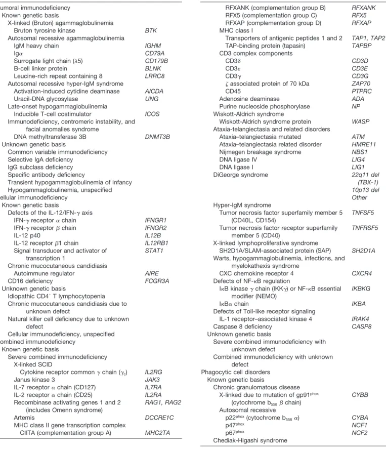
Table 2. Classification of Primary Immunodeficiencies
Disease Gene
Humoral immunodeficiency Known genetic basis
X-linked (Bruton) agammaglobulinemia
Bruton tyrosine kinase BTK
Autosomal recessive agammaglobulinemia
IgM heavy chain IGHM
Ig␣ CD79A
Surrogate light chain (5) CD179B
B-cell linker protein BLNK
Leucine-rich repeat containing 8 LRRC8
Autosomal recessive hyper-IgM syndrome
Activation-induced cytidine deaminase AICDA
Uracil-DNA glycosylase UNG
Late-onset hypogammaglobulinemia
Inducible T-cell costimulator ICOS
Immunodeficiency, centromeric instability, and facial anomalies syndrome
DNA methyltransferase 3B DNMT3B
Unknown genetic basis
Common variable immunodeficiency Selective IgA deficiency
IgG subclass deficiency Specific antibody deficiency
Transient hypogammaglobulinemia of infancy Hypogammaglobulinemia, unspecified Cellular immunodeficiency
Known genetic basis
Defects of the IL-12/IFN-␥axis
IFN-␥receptor␣chain IFNGR1
IFN-␥receptorchain IFNGR2
IL-12 p40 IL12B
IL-12 receptor1 chain IL12RB1
Signal transducer and activator of transcription 1
STAT1
Chronic mucocutaneous candidiasis
Autoimmune regulator AIRE
CD16 deficiency FCGR3A
Unknown genetic basis
Idiopathic CD4⫹T lymphocytopenia Chronic mucocutaneous candidiasis due to
unknown defect
Natural killer cell deficiency due to unknown defect
Cellular immunodeficiency, unspecified Combined immunodeficiency
Known genetic basis
Severe combined immunodeficiency X-linked SCID
Cytokine receptor common␥chain (␥c) IL2RG
Janus kinase 3 JAK3
IL-7 receptor␣chain (CD127) IL7RA
IL-2 receptor␣chain (CD25) IL2RA
Recombinase activating genes 1 and 2 (includes Omenn syndrome)
RAG1, RAG2
Artemis DCCRE1C
MHC class II gene transcription complex
CIITA (complementation group A) MHC2TA
Table 2. Continued
Disease Gene
RFXANK (complementation group B) RFXANK
RFX5 (complementation group C) RFX5
RFXAP (complementation group D) RFXAP
MHC class I
Transporters of antigenic peptides 1 and 2 TAP1, TAP2
TAP-binding protein (tapasin) TAPBP
CD3 complex components
CD3␦ CD3D
CD3 CD3E
CD3␥ CD3G
associated protein of 70 kDa ZAP70
CD45 PTPRC
Adenosine deaminase ADA
Purine nucleoside phosphorylase NP
Wiskott-Aldrich syndrome
Wiskott-Aldrich syndrome protein WASP
Ataxia-telangiectasia and related disorders
Ataxia–telangiectasia mutated ATM
Ataxia–telangiectasia related disorder HMRE11
Nijmegen breakage syndrome NBS1
DNA ligase IV LIG4
DNA ligase I LIG1
DiGeorge syndrome 22q11 del (TBX-1) 10p13 del Other
Hyper-IgM syndrome
Tumor necrosis factor superfamily member 5 (CD40L, CD154)
TNFSF5
Tumor necrosis factor receptor superfamily member 5 (CD40)
TNFRSF5
X-linked lymphoproliferative syndrome
SH2D1A/SLAM-associated protein (SAP) SH2D1A
Warts, hypogammaglobulinemia, infections, and myelokathexis syndrome
CXC chemokine receptor 4 CXCR4
Defects of NF-B regulation
IB kinase␥chain (IKK␥) or NF-B essential modifier (NEMO)
IKBKG
IB␣chain IKBA
Defects of Toll-like receptor signaling
IL-1 receptor–associated kinase 4 IRAK4
Caspase 8 deficiency CASP8
Unknown genetic basis
Severe combined immunodeficiency with unknown defect
Combined immunodeficiency with unknown defect
Phagocytic cell disorders Known genetic basis
Chronic granulomatous disease X-linked due to mutation of gp91phox
(cytochrome b558chain) CYBB Autosomal recessive p22phox(cytochrome b 558␣) CYBA p47phox NCF1 p67phox NCF2 Chediak-Higashi syndrome
disorders that affect immune system function have been iden-tified to date (a selection is listed in Table 2).
Primary immunodeficiencies occur in as many as 1 in 2,000 live births. They are most often categorized according to the immune mechanisms that are disrupted. These catego-ries include the defects of specific immunity that are subdi-vided into humoral or antibody deficiencies, cellular deficien-cies, and the combined deficiencies that affect both humoral and cellular mechanisms. There are also defects of innate immunity and the phagocyte and complement system defects. Of all of these categories, antibody deficiencies together account for approximately half of all primary immunodeficiency.
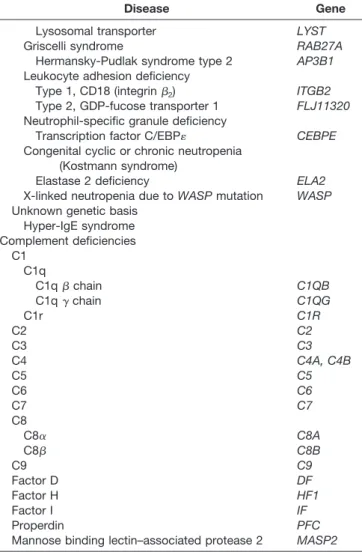
The principal clinical manifestation of immunodeficiency is increased susceptibility to infection. The pattern of organ systems affected and the characteristic pathogens vary with
the type of immune defect (Table 3). Autoimmune disease and malignancy are also often seen in a variety of immuno-deficiencies. A careful family history may provide important clues regarding potential X-linked or autosomal recessive patterns of inheritance.
In evaluating immunodeficiency, it is critical as, much as possible, to document carefully the foci of infections, the organisms, and the response to treatment. This is necessary to distinguish infectious disease from other noninfectious con-ditions such as allergy or to distinguish viral infection from bacterial infection. Any other conditions that may predispose the patient to infection, including anatomic defects, allergy, and metabolic disorders, should be considered wherever ap-propriate
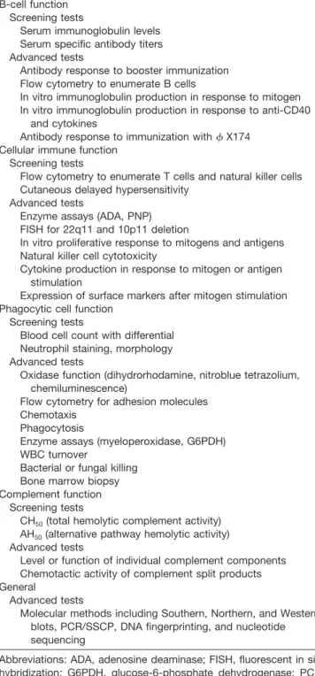
In general, initial evaluation is guided by the clinical presentation (Algorithm 1). Screening tests are applied fol-lowed by advanced tests as indicated (Table 4). This stepwise approach ensures efficient and thorough evaluation of mech-anisms of immune dysfunction that may underlie the clinical presentation, with narrowing of diagnostic options before using costly sophisticated tests that may be required to arrive at specific diagnoses. In addition to global evaluation of immune development via measurement of nonspecific fea-tures such as serum immunoglobulin levels and leukocyte and lymphocyte subpopulations, evaluation of specific immune response is essential. This is most often directed toward evaluation of responses against vaccine antigens, but evalu-ation of responses to natural exposure or infections is also useful.
Wherever uncertainty regarding evaluation occurs, consul-tation with physicians experienced in the diagnosis of immu-nodeficiencies is essential for establishing the most specific and accurate diagnosis as quickly as possible to enable di-rected therapy. Wherever possible, diagnosis at the molecular level is desirable to (1) establish unequivocal diagnosis; (2) permit accurate genetic counseling; (3) allow planning of future pregnancies or their outcomes; (4) better define geno-type-phenotype associations; and (5) identify candidates for gene-specific therapies.
The principal clinical manifestations of humoral immuno-deficiency (Algorithm 2) are recurrent bacterial infections of the upper and lower respiratory tract. Both X-linked and autosomal recessive forms of agammaglobulinemia are asso-ciated with extremely low B-cell counts (absent). The X-linked form (Bruton agammaglobulinemia) is most common. In common variable immunodeficiency (CVID), laboratory evaluation generally shows variable reduction in one or more immunoglobulin classes, impairment of specific antibody re-sponses, and, occasionally, reduction of B-cell counts. Milder antibody deficiencies, such as selective IgA deficiency (SIGAD), IgG subclass deficiency (IGGSD), specific anti-body deficiency (SAD), or transient hypogammaglobuline-mia of infancy (THI), are associated with variably low levels of an immunoglobulin class or subclass in serum, sometimes
Table 2. Continued
Disease Gene
Lysosomal transporter LYST
Griscelli syndrome RAB27A
Hermansky-Pudlak syndrome type 2 AP3B1
Leukocyte adhesion deficiency
Type 1, CD18 (integrin2) ITGB2
Type 2, GDP-fucose transporter 1 FLJ11320
Neutrophil-specific granule deficiency
Transcription factor C/EBP CEBPE
Congenital cyclic or chronic neutropenia (Kostmann syndrome)
Elastase 2 deficiency ELA2
X-linked neutropenia due toWASPmutation WASP
Unknown genetic basis Hyper-IgE syndrome Complement deficiencies C1 C1q C1qchain C1QB C1q␥chain C1QG C1r C1R C2 C2 C3 C3 C4 C4A, C4B C5 C5 C6 C6 C7 C7 C8 C8␣ C8A C8 C8B C9 C9 Factor D DF Factor H HF1 Factor I IF Properdin PFC
Mannose binding lectin–associated protease 2 MASP2
Abbreviations: IFN-␥, interferon-␥; IL, interleukin; MHC, major histo-compatibility complex; SCID, severe combined immunodeficiency; WASP, Wiskott-Aldrich syndrome protein.
Algorithm 1. General approach for the diagnosis of primary immunodeficiency. SCID indicates severe combined immunodeficiency.
Table 3. Primary Immunodeficiency Disorders: Examples of Typical Clinical Presentations
Category of immunodeficiency
and examples Characteristic presentation
Antibody deficiencies
XLA, ARA, CVID, SIGAD, IGGSD, SAD, THI, hypogam
Recurrent sinopulmonary infections with encapsulated bacteria
Cellular deficiencies
IL-12/IFN-␥axis Atypical mycobacterial and salmonella infections
AIRE mutations Mucocutaneous candidiasis and autoimmune endocrinopathy Combined deficiencies
SCID Failure to thrive, diarrhea, opportunistic infection, rash
Wiskott-Aldrich syndrome Thrombocytopenia with bleeding and bruising, eczema, recurrent infection with encapsulated organisms
Ataxia telangiectasia Chronic sinopulmonary disease, cerebellar ataxia, oculocutaneous telangiectasia, malignancy DiGeorge syndrome Hypocalcemic seizures due to hypoparathyroidism, cardiac disease, abnormal facies, infection CD40 ligand deficiency Recurrent, serious pyogenic infections (also opportunistic infections)
Phagocyte defects
Chronic granulomatous disease Deep-seated infection, abscess with granuloma formation
Leukocyte adhesion deficiency Recurrent serious bacterial infections, delayed separation of the umbilical cord; poor wound healing, lack of pus
Hyper-IgE syndrome Chronic dermatitis, recurrent serious infection of lungs with pneumatoceles; skin infections, bone fragility, failure to shed primary teeth
Complement deficiencies
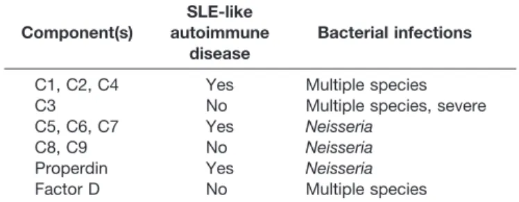
Early classical pathway components Autoimmune disease and bacterial infections Late components Neisserial infection
C3 and regulatory components Recurrent infections with encapsulated bacterial
Abbreviations: AIRE, autoimmune regulator; ARA, autosomal recessive agammaglobulinemia; CVID, common variable immunodeficiency; hy-pogam, hypogammaglobulinemia; IFN-␥, interferon-␥; IGGSD, IgG subclass deficiency; IL-12, interleukin 12; SAD, specific antibody deficiency; SCID, severe combined immunodeficiency; SIGAD, selective IgA deficiency; THI, transient hypergammaglobulinemia of infancy; XLA, X-linked agammaglobulinemia.
accompanied by impaired specific antibody formation. For agammaglobulinemia or CVID, therapy is with gammaglobu-lin, often with the addition of antibiotic prophylaxis. (Table
5). Milder antibody deficiencies are most often managed with antibiotic prophylaxis. In rare cases, gammaglobulin therapy may be applied.
Selective defects of cell-mediated immunity (Algorithm 3) characteristically present with recurrent infections with pathogens that replicate intracellularly, such as mycobacteria or salmonella. Most of the disorders defined at the molecular level involve defects of the interferon-␥ (IFN-␥)/interleukin 12 (IL-12) axis. In the cellular deficiencies with a significant component of natural killer (NK) cell dysfunction, recurrent and/or severe herpesvirus infections may be seen. Laboratory abnormalities may be subtle and often require specialized research tests or molecular genetic analysis for diagnosis. Therapy of these disorders may include anti-infection pro-phylaxis, cytokines (eg, IFN-␥), and/or bone marrow trans-plantation (BMT) (Table 5).
The combined deficiencies of specific immunity (Algo-rithm 3) are somewhat arbitrarily classified as severe com-bined immunodeficiency (SCID) or among a variety of other less severe disorders. Patients with SCID have complete absence of specific immunity and experience the most ex-treme susceptibility to the entire range of possible pathogens, including opportunistic organisms. These children often present initially with chronic diarrhea and failure to thrive. Laboratory abnormalities may include panhypogammaglobu-linemia, lymphopenia or alymphocytosis, and absence of cellular immune function as determined by in vitro stimula-tion tests. The laboratory phenotype often depends on the specific molecular defect (Table 6). A possible diagnosis of SCID is a medical emergency, since these infants may suc-cumb to severe infection at any time, and outcomes are greatly improved by the earliest possible intervention. Initial therapy is supportive and anti-infective with antimicrobials and gammaglobulin. Definitive therapy with BMT should be sought as quickly as possible.
A variety of less severe defects of combined immunodefi-ciency (CID) have been described (Algorithm 3). Most prom-inent among these are Wiskott-Aldrich syndrome (WAS), DiGeorge syndrome (DGS), ataxia-telangiectasia (A-T), nu-clear factor of B essential modifier (NEMO) deficiency, hyper-IgM syndromes (HIM), and X-linked lymphoprolifera-tive disease (XLP). These disorders present with varying degrees of susceptibility to the entire spectrum of organisms, depending on the specific disorder and on other host genetic and environmental factors that are still poorly understood. Many of these diseases have ancillary clinical features that may influence or guide the diagnostic approach. Laboratory abnormalities of specific immune function vary, depending on the specific gene defect, and may include alterations in immunoglobulin levels with impaired specific antibody re-sponses, as well as defects of specific cellular immunity as determined by in vivo and in vitro assays. Therapy is often supportive and anti-infective with drugs and gammaglobulin. BMT has been applied in many of these disorders as well (Table 5).
Table 4. Laboratory Tests for Evaluation of Immunodeficiency B-cell function
Screening tests
Serum immunoglobulin levels Serum specific antibody titers Advanced tests
Antibody response to booster immunization Flow cytometry to enumerate B cells
In vitro immunoglobulin production in response to mitogen In vitro immunoglobulin production in response to anti-CD40
and cytokines
Antibody response to immunization withX174 Cellular immune function
Screening tests
Flow cytometry to enumerate T cells and natural killer cells Cutaneous delayed hypersensitivity
Advanced tests
Enzyme assays (ADA, PNP) FISH for 22q11 and 10p11 deletion
In vitro proliferative response to mitogens and antigens Natural killer cell cytotoxicity
Cytokine production in response to mitogen or antigen stimulation
Expression of surface markers after mitogen stimulation Phagocytic cell function
Screening tests
Blood cell count with differential Neutrophil staining, morphology Advanced tests
Oxidase function (dihydrorhodamine, nitroblue tetrazolium, chemiluminescence)
Flow cytometry for adhesion molecules Chemotaxis
Phagocytosis
Enzyme assays (myeloperoxidase, G6PDH) WBC turnover
Bacterial or fungal killing Bone marrow biopsy Complement function
Screening tests
CH50(total hemolytic complement activity)
AH50(alternative pathway hemolytic activity)
Advanced tests
Level or function of individual complement components Chemotactic activity of complement split products General
Advanced tests
Molecular methods including Southern, Northern, and Western blots, PCR/SSCP, DNA fingerprinting, and nucleotide sequencing
Abbreviations: ADA, adenosine deaminase; FISH, fluorescent in situ hybridization; G6PDH, glucose-6-phosphate dehydrogenase; PCR, polymerase chain reaction; PNP, purine nucleoside phosphorylase; SSCP, single-strand conformation polymorphism; WBC, white blood cell.
Phagocytic cell defects (Algorithm 4) may present with severe pyogenic bacterial and fungal infections of the respi-ratory tract, skin, viscera, and gingivostomatitis. Laborespi-ratory evaluation shows neutropenia, normal numbers, or neutro-philia (in cellular adhesion defects). Functional studies show most often a defect in oxidative metabolism, since chronic granulomatous disease is the most common phagocyte defect. In other disorders, there may be simply severe neutropenia or variable impairment of chemotaxis, phagocytosis, or intracel-lular killing. Therapy is with antibacterial and antifungal prophylaxis and cytokines (IFN-␥) for chronic granulomatous disease (CGD). BMT has also been applied for CGD. The care of patients with other forms of phagocyte defects is primarily anti-infective and supportive. BMT has been ap-plied in some, but experience is limited (Table 5).
Complement deficiencies are the rarest of the primary immunodeficiencies, accounting for less than 1%. Most early classical and alternative pathway complement defects tend to present with either systemic autoimmune disease that resem-bles lupus erythematosus or recurrent respiratory tract bacte-rial infections similar to antibody deficiency (Table 7). De-ficiencies of terminal components may also be associated with recurrent neisserial meningitis. Some patients with low serum levels of mannose-binding lectin (MBL) may be
pre-disposed to bacterial respiratory tract infections, but there may be other host factors that interact to create such suscep-tibility in an individual. There is no specific therapy for complement deficiency. Antibiotic prophylaxis may be con-sidered for recurrent infections (Table 5).
To improve consistency in evaluation and management and to have the best outcomes with respect to patient and family health, education, and planning, it is imperative that diagnosis and therapy are guided overall by individuals with direct experience with a broad range of immunodeficiencies. ALGORITHMS
Annotations to Algorithm 1: General Approach for the Diagnosis of Primary Immunodeficiency
1-1.The patient exhibits symptoms and signs consistent with primary immunodeficiency. It is assumed that immunosup-pressive therapies and other medical conditions potentially resulting in secondary immunodeficiency and other anatomic or biochemical conditions potentially predisposing to infec-tion either have been excluded or are not considered sufficient to explain the observed degree of infectious susceptibility.
1-2. Antibody deficiency is most frequently encountered and commonly presents with sinopulmonary bacterial
infec-Algorithm 2. Diagnosis of humoral immunodeficiency. ARA indicates autosomal recessive agammaglobulinemia; CVID, common variable immunodefi-ciency; HIM, hyper-IgM syndrome; IGGSD, IgG subclass defiimmunodefi-ciency; SAD, specific antibody defiimmunodefi-ciency; SIGAD, selective IgA defiimmunodefi-ciency; THI, transient hypogammaglobulinemia of infancy; and XLA, X-linked agammaglobulinemia.
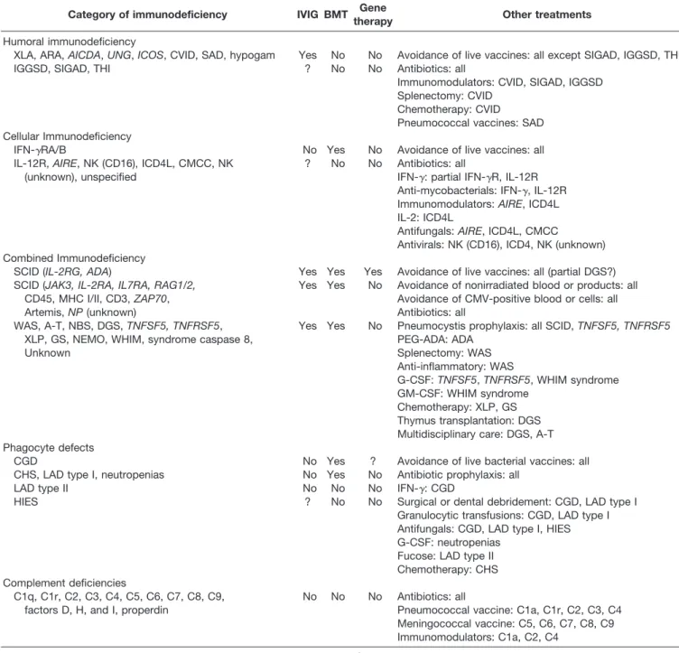
Table 5. Summary of Therapeutic Considerations for Primary Immunodeficiencies and Their Complications
Category of immunodeficiency IVIG BMT Gene
therapy Other treatments
Humoral immunodeficiency
XLA, ARA,AICDA,UNG,ICOS, CVID, SAD, hypogam Yes No No Avoidance of live vaccines: all except SIGAD, IGGSD, THI IGGSD, SIGAD, THI ? No No Antibiotics: all
Immunomodulators: CVID, SIGAD, IGGSD Splenectomy: CVID
Chemotherapy: CVID Pneumococcal vaccines: SAD Cellular Immunodeficiency
IFN-␥RA/B No Yes No Avoidance of live vaccines: all IL-12R,AIRE, NK (CD16), ICD4L, CMCC, NK
(unknown), unspecified
? No No Antibiotics: all
IFN-␥: partial IFN-␥R, IL-12R Anti-mycobacterials: IFN-␥, IL-12R Immunomodulators:AIRE, ICD4L IL-2: ICD4L
Antifungals:AIRE, ICD4L, CMCC
Antivirals: NK (CD16), ICD4, NK (unknown) Combined Immunodeficiency
SCID (IL-2RG, ADA) Yes Yes Yes Avoidance of live vaccines: all (partial DGS?) SCID (JAK3, IL-2RA, IL7RA, RAG1/2, Yes Yes No Avoidance of nonirradiated blood or products: all
CD45, MHC I/II, CD3,ZAP70, Avoidance of CMV-positive blood or cells: all
Artemis,NP(unknown) Antibiotics: all
WAS, A-T, NBS, DGS,TNFSF5, TNFRSF5, Yes Yes No Pneumocystis prophylaxis: all SCID,TNFSF5, TNFRSF5
XLP, GS, NEMO, WHIM, syndrome caspase 8, PEG-ADA: ADA
Unknown Splenectomy: WAS
Anti-inflammatory: WAS
G-CSF:TNFSF5,TNFRSF5, WHIM syndrome GM-CSF: WHIM syndrome
Chemotherapy: XLP, GS Thymus transplantation: DGS Multidisciplinary care: DGS, A-T Phagocyte defects
CGD No Yes ? Avoidance of live bacterial vaccines: all CHS, LAD type I, neutropenias No Yes No Antibiotic prophylaxis: all
LAD type II No No No IFN-␥: CGD
HIES ? No No Surgical or dental debridement: CGD, LAD type I Granulocytic transfusions: CGD, LAD type I Antifungals: CGD, LAD type I, HIES G-CSF: neutropenias
Fucose: LAD type II Chemotherapy: CHS Complement deficiencies
C1q, C1r, C2, C3, C4, C5, C6, C7, C8, C9, No No No Antibiotics: all
factors D, H, and I, properdin Pneumococcal vaccine: C1a, C1r, C2, C3, C4 Meningococcal vaccine: C5, C6, C7, C8, C9 Immunomodulators: C1a, C2, C4
Abbreviations: A-T, ataxia-telangiectasia; ADA, adenosine deaminase; AICDA, activation-induced cytidine deaminase; AIRE, autoimmune regu-lator; ARA, autosomal recessive agammaglobulinemia; BMT, bone marrow transplantation; CGD, chronic granulomatous disease; CHS, Chediak-Higashi syndrome; CMCC, chronic mucocutaneous candidiasis; CMV, cytomegalovirus; CVID, common variable immunodeficiency; DGS, DiGeorge syndrome; G-CSF, granulocyte colony-stimulating factor; GM-CSF, granulocyte-macrophage colony stimulating factor; GS, Griscelli syndrome; HIES, hyper-IgE syndrome; hypogam, hypogammaglobulinemia; ICD4L, idiopathic CD4 lymphocytopenia; ICOS, inducible T-cell costimulator; IFN-␥, interferon-␥; IFN-␥R, interferon-␥receptor; IGGSD, IgG subclass deficiency; IL, interleukin; IL-12R, interleukin 12 receptor; IVIG, intravenous immunoglobulin; LAD, leukocyte adhesion deficiency; MHC, major histocompatibility complex; NBS, Nijmegen breakage syndrome; NEMO, nuclear factor ofB essential modifier; NK, natural killer; NP, nucleoside phosphorylase; PEG, polyethylene glycol; RAG, recombinase activating gene; SAD, specific antibody deficiency; SCID, severe combined immunodeficiency; SIGAD, selective IgA deficiency; THI, transient hypogammaglobulinemia of infancy; TNFSF5, tumor necrosis factor superfamily member 5; TNFRSF5, tumor necrosis factor receptor superfamily member 5; UNG, uracil nucleoside glycosylase; WAS, Wiskott-Aldrich syndrome; WHIM, warts, hypogammaglobulinemia, immuno-deficiency, and myelokathexis; XLA, X-linked agammaglobulinemia.
tions. If these are the only types of infections under consid-eration, screening for antibody deficiency is appropriate.
1-3. Other forms of primary immunodeficiency may present with distinct infectious complications with or without sinopulmonary bacterial disease. Some of these forms of infection are more or less characteristic of specific categories of immunodeficiency (Table 2). Neisserial infections charac-terize terminal complement component deficiencies, ab-scesses and fungal pathogens are seen in phagocyte defects, and mycobacterial, disseminated, or opportunistic infections occur in cellular or combined deficiencies.
1-4. If the clinical presentation is consistent with SCID, then immediate referral for expedited evaluation and treat-ment (BMT) is indicated.
1-5.Successful outcomes depend on timely intervention. 1-6.Suspected antibody deficiency may be evaluated ac-cording to Algorithm 2. Complement deficiency, phagocyte defects, and some combined deficiencies may have a clinical presentation similar to antibody deficiency and should be sought when there is not a definitive diagnosis of such. Depending on the clinical presentation, any of these could be an appropriate subsequent focus of investigation.
Algorithm 3. Diagnosis of cellular and combined immunodeficiencies. ADA indicates adenosine deaminase; CID, combined immunodeficiency; DGS, DiGeorge syndrome; IKBKG, IB kinase␥chain; IL-2Ra, interleukin 2 receptor agonist; MHC, major histocompatibility complex; NK, natural killer; SAP, SLAM-associated protein; SCID, severe combined immunodeficiency; WAS, Wiskott-Aldrich syndrome; and XSCID, X-linked severe combined immunodeficiency.
1-7.Suspected complement deficiency may be evaluated according to Algorithm 5.
1-8.Suspected phagocyte defects may be evaluated using Algorithm 4.
1-9. Suspected cellular or combined immunodeficiencies may be evaluated according to Algorithm 3.
1-10.Depending on the specific characteristics of the in-fections and other medical problems that occur in a given patient, one or all of these immune effector mechanisms may require evaluation. In some cases, no definitive immunologic defect is ascertained. These patients either have an undefined form of compromised immunity or some other medical prob-lem predisposing them to infection.
1-11. Whenever possible, the evaluation and/or manage-ment of suspected primary immunodeficiency should be per-formed by, or in close consultation with, a clinician with experience in this area. At some time during evaluation or after diagnosis is established, referral should be made for further evaluation and/or guidance during therapy.
Annotations to Algorithm 2: Diagnosis of Humoral Immunodeficiency
2-1. The clinical presentation is primarily suggestive of an antibody defect or any evaluation of cellular function is so far normal, and the clinical presentation is at least consistent with a possible antibody deficiency. The initial laboratory examination of humoral immunity consists of measuring the levels of various immunoglobulin isotypes (IgG, IgA, IgM) in serum, as well as a measure of function, or specific antibody production.
2-2. Profound hypogammaglobulinemia with serum IgG levels less than 100 mg/dL in an infant or less than 2 to 3 g/L in an older child or adult should prompt additional evaluation of lymphocyte populations and cellular immune function (2-3)to investigate CID and B-cell count.
2-4.Specific antibody responses may be impaired as a result of a B-cell defect or failure of T-cell help for
antibody production, even if serum immunoglobulin levels are normal or near normal. This situation should also prompt evaluation of lymphocyte subsets and cellular im-munity(2-3).
2-5.Cellular immunity is evaluated either because of severe hypogammaglobulinemia or impaired specific antibody produc-tion (or both). If cellular immunity is abnormal, then the even-tual diagnosis will be a form of CID. If cellular immunity is normal, it is important to determine whether there appears to be a significant impairment of B-cell development(2-7).
2-6. There is no profound hypogammaglobulinemia or demonstrable impairment of specific antibody production. Is there any abnormality of serum immunoglobulins or IgG subclasses? Yes(2-8)or No(2-9).
2-7.Is B-cell count normal? Yes (2-10)or No(2-11). 2-8.All measurements are normal, and alternative expla-nations for recurrent infections should be sought.
2-9.Mild hypogammaglobulinemia in infants, low serum IgA or IgG subclasses, or other poorly defined immunoglob-ulin abnormalities may exist with normal levels of specific antibodies as measured by standard assays. Potential diag-noses include SIGAD, IGGSD, or THI.
2-10.Hypogammaglobulinemia and/or impaired specific an-tibody formation are seen in CVID, SIGAD, IGGSD, SAD, and some forms of HIM, such as activation-induced cytidine deami-nase (AID) or uracil nucleoside glycosylase (UNG) deficiencies. 2-11. Hypogammaglobulinemia or agammaglobulinemia associated with low or absent B-cell counts is seen in X-linked agammaglobulinemia (XLA) or autosomal recessive agammaglobulinemia (ARA) and in CVID.
Annotations to Algorithm 3: Diagnosis of Cellular and Combined Immunodeficiencies
3-1. In this situation, it is appropriate to perform a complete screening evaluation of specific immune function, including measurement of immunoglobulin levels, specific antibody
pro-Table 6. Abnormalities of Lymphocyte Populations in Some Defined SCID Syndromes*
Gene CD4 CD8 B cells Natural
killer cells
Lymph
nodes Thymus Other features Reference(s)
IL2RG 2 2 NL 2 ⫺ ⫺ 18, 326
JAK3 2 2 NL 2 ⫺ ⫺ 329–333
IL2RA 2 2 NL 2 ⫹ ⫹ 334, 335
IL7RA 2 2 NL NL ⫹ ⫺ 336
CD3D 2 2 NL NL ⫹ 337
RAG1, RAG2 2 2 2 NL ⫺ ⫺ May have oligoclonal host T cells
with graft-vs-host disease–like phenotype (Omenn syndrome)
338–340
DCCRE1C 2 2 2 NL ⫺ ⫺ Radiation sensitivity 341, 342
MHCIID 2 NL NL NL Sclerosing cholangitis 343–345
MHCID NL 2 NL NL ⫹ ⫹ Milder phenotype, mainly
respiratory bacterial infections
346–348
ZAP70 NL 2 NL NL ⫹ ⫹ 349–351
ADA 2 2 2 ⫾ ⫾ ⫺ Skeletal abnormalities 352
NP Variable ⫾ ⫾ Central nervous system disease 36, 353
PTPRC 2 2 NL 354, 355
duction, enumeration of lymphocyte subpopulations, evaluation of NK cell cytotoxicity, and measurement of T-cell function.
3-2.If the clinical and laboratory phenotype is consistent with SCID, every effort must be made to expedite definitive therapy (BMT). It is desirable to know the actual molecular defect, but this should not delay therapy.
3-3 through 3-14.The particular form of SCID may often be suspected based on the lymphocyte phenotype (Table 5). If T cells are present, their origin (mother or patient) should be determined.
3-4.If T cells are absent or only of maternal origin(3-6) and B cells are also absent, then one of the alymphocytic SCID syndromes should be considered(3-5). If B cells are present, along with NK cells(3-7),consider IL-7 receptor␣ (IL7RA)mutation or complete DGS(3-8).
3-9.If B cells are present but NK cells absent, consider mutations that involve common␥chain,JAK3, orIL2RA.
3-10. If host T cells are present and there is selective depletion of CD4⫹cells, consider defects of major histocom-patibility complex (MHC) class II expression(3-11).
3-12.If there is selective depletion of CD8⫹cells, consider defects involving MHC class I expression or ZAP70 defi-ciency(3-13).
3-14.Omenn syndrome is associated with a variable host T-cell phenotype, although there is not usually extreme pre-ponderance of one cell type. One should also consider the possibility of a less common (possibly undefined) form of SCID or a severe phenotype of other CID, such as CD40 ligand (CD40L) deficiency.
3-15.If there is at least partial T-cell function, evaluation of NK cell cytotoxicity may partly guide the subsequent evaluation. It has been recently recognized that a few CID syndromes may be associated with depressed NK cytotoxic-ity. These include (but are not limited to) XLP, NEMO deficiency and WAS.(3-16).
3-17.Whether NK function is abnormal or not, there may be characteristic clinical or laboratory features that may sug-gest a particular molecular diagnosis (Table 3).
3-18. The clinical presentation and laboratory evaluation so far is suggestive of 1 or more particular disorders.
Ad-Algorithm 4. Diagnosis of phagocyte defects. CBC indicates complete blood cell count; CGD, chronic granulomatous disease; CHS, Chediak-Higashi syndrome; HIES, hyper-IgE syndrome; and LAD, leukocyte adhesion deficiency.
vanced molecular methods (Table 4) may be applied to detect particular defects.
3-19.If there are no such distinguishing clinical or labo-ratory features or if the suspected diagnosis is proven incor-rect, one should consider(3-20)an undefined CID, an atyp-ical clinatyp-ical presentation of a defined CID, or a severe presentation of a primary humoral immunodeficiency(3-21) (Algorithm 2).
Annotations to Algorithm 4: Diagnosis of Phagocyte Defects
4-1. The clinical presentation is primarily suggestive of a phagocyte defect or evaluation of other immune function is so far normal, and the clinical presentation is at least consistent with a possible phagocyte defect. A complete blood cell count with differential is necessary to show the absolute neutrophil count.
4-2. Marked leukocytosis is observed in most cases of leukocyte adhesion defects and should raise suspicion in the appropriate setting.
4-3.Defects associated with leukocyte adhesion deficiency (LAD) are readily screened by flow cytometry, which may establish the diagnosis(4-4).
4-5.Severe neutropenia may be associated with congenital agranulocytosis or cyclic neutropenia(4-6).
4-7. If leukocyte count is not abnormal and the clinical features are consistent, neutrophil oxidase function may be evaluated by dihydrorhodamine reduction, nitroblue tetrazo-lium, or chemiluminescence. Abnormal oxidase function is indicative of CGD(4-8).
4-9.Chediak-Higashi syndrome (CHS) and specific gran-ule deficiency (SGD) are suspected based on clinical presen-tation and neutrophil appearance under microscopy.
4-10. In the absence of a known syndrome of phagocyte deficiency, it is necessary to establish a functional defect more precisely. These tests include assays of chemotaxis, adhesion, migration, and intracellular killing. If such a func-tional deficit is reproducible, then a diagnosis of a clinically defined or unspecified phagocyte defect may be considered (4-11). Hyper-IgE syndrome (HIES) is usually suspected based on the characteristic clinical presentation. If the pre-sentation is not consistent with this or any of the above, another form of immunodeficiency should be sought(4-12). Annotations to Algorithm 5: Diagnosis of Complement Deficiency
5-1. The clinical presentation is primarily suggestive of a complement deficiency or evaluation of other immune
func-Algorithm 5. Diagnosis of complement deficiency. AH50indicates alternative pathway hemolytic activity; CH50, total hemolytic complement assay.
Table 7. Clinical Associations With Complement Component Deficiencies Component(s) SLE-like autoimmune disease Bacterial infections
C1, C2, C4 Yes Multiple species C3 No Multiple species, severe C5, C6, C7 Yes Neisseria
C8, C9 No Neisseria
Properdin Yes Neisseria
Factor D No Multiple species Abbreviation: SLE, systemic lupus erythematosus.
tion is so far normal, and the clinical presentation is at least consistent with a possible complement deficiency. Two dis-tinct algorithms are presented, depending on whether total hemolytic complement assay (CH50) and alternative pathway hemolytic activity (AH50) are measured sequentially (5a) or simultaneously (5b). The CH50 is available in many clinical laboratories; the AH50is not so widely available (it is avail-able from the Complement Laboratory of the National Jewish Medical Center, Denver, CO). Note that both will be normal in the setting of MBL deficiency
5a-2.Classical pathway function is measured first by the CH50.
5a-3. Following determination of diminished classical pathway function, it is necessary to determine if there is complement consumption.
5a-4. More than one complement component level is di-minished, indicating complement consumption. Another cause of immunodeficiency should be sought.
5a-5.A single complement component level or function is absent, indicative of deficiency of either an early classical pathway component or a terminal pathway component. Note that deficiency of factor H or factor I could lead to a dimin-ished level of C3. The level of each component may be measured by enzyme-linked immunosorbent assay (ELISA), or function may be determined in a lysis assay.
5a-6.The CH50is normal. If complement deficiency is still suspected, function of the alternative pathway is measured by the AH50.
5a-7.Since it has already been determined that the CH50is normal, isolated abnormal AH50is indicative of a defect of a
component of the alternative pathway. Each component may be measured by ELISA or functional assay.
5a-8.CH50and AH50 are normal.
5b-2.CH50and AH50 are measured at the same time. 5b-3.If both are abnormal, this may be due to complement consumption not a primary complement abnormality. Note that deficiency of factor H or factor I could lead to a dimin-ished level of C3.
5b-4. Low levels of multiple complement proteins are indicative of consumption.
5b-5. If there is no complement consumption, simulta-neous abnormality of CH50 and AH50 is indicative of a ter-minal pathway deficiency (ie, C3, C5 to C9).
5b-6. If the CH50 is abnormal and AH50 is normal, this suggests a classical pathway component deficiency (C1, C2, C4)(5b-7).
5b-8.If the AH50is abnormal and the CH50is normal, this is indicative of a defect of a component of the alternative pathway (properdin, factor D)(5b-9). Note that homozygous deficiency of factor B has not been reported.
Annotations to Algorithm 6: General Considerations for Therapy of Primary Immunodeficiency
Four principal general categories of therapy are indicated: BMT or gene therapy, intravenous immunoglobulin (IVIG) or subcutaneous immunoglobulin (SCIG), antimicrobial pro-phylaxis for any pathogen to which the host is susceptible and for which preventive therapy is available, and immunization where appropriate (Table 4).
Algorithm 6. General considerations for therapy of primary immunodeficiency. BMT indicates bone marrow transplantation; CVID, common variable immunodeficiency; IVIG, intravenous immunoglobulin; and SCIG, subcutaneous immunoglobulin.
6-1.For SCID, BMT should be pursued as expeditiously as possible. IVIG or SCIG is indicated before BMT and as necessary afterward for persistent humoral immunodefi-ciency. (Note that the latter could be similar to either6-2or 6-3.) For some diseases, now or in the future, gene therapy is or may be a possibility.
6-2.For XLA, ARA, or CVID, IVIG or SCIG is appropri-ate at the time the diagnosis is established. Many also rec-ommend routine initiation of antibacterial prophylaxis at this time. Some prescribe preventive antibiotics when IVIG or SCIG is inadequate for prevention of infection or when other conditions such as bronchiectasis are present. Immunization may be considered, particularly with inactivated vaccines for which coverage by IVIG or SCIG is not reliable (eg, influ-enza).
6-3. For milder antibody deficiencies (SIGAD, IGGSD, SAD), therapy is often initially with preventive antimicrobi-als and immunization. Depending on all of the clinical and laboratory features, IVIG or SCIG may be considered.
6-4. For specific cellular deficiencies or for non-SCID combined deficiencies, BMT is often considered. BMT may not be appropriate for milder forms or if a suitable donor is not available. Wherever there is significant impairment of specific antibody production (6-5), IVIG or SCIG should be given. Antimicrobial prophylaxis and immunization may also be appropriate, depending on the specific defect.
6-6.For some phagocyte defects (eg, CGD), BMT should be considered. IVIG or SCIG is generally not appropriate for complement or phagocyte defects. Antimicrobial prophylaxis is essential for phagocyte defects and may be considered for complement deficiency. Immunization may be helpful. SUMMARY STATEMENTS
General Considerations
Summary statement 1. Individual immunodeficiencies are rare, but altogether they occur in more than 1 in 2,000 live births. (C)
Summary statement 2. Immunodeficiencies are classified according to the principal immunologic mechanisms that are disrupted. (D)
Summary statement 3. Antibody deficiency is the most common type of primary immunodeficiency. (C)
Summary statement 4.Immunodeficiency usually presents with signs and symptoms of infections that may be repetitive, severe, or refractory to therapy and caused by organisms of low virulence. (C)
Summary statement 5.It is critically important to confirm the precise focus of infection and organism whenever possi-ble. (D)
Summary statement 6. Other conditions that may increase susceptibility to infection should be sought in patients with suspected immunodeficiency. (D)
Summary statement 7.The physician must exercise caution to rule out the possibility of secondary immunodeficiency underlying the patient’s illness. (D)
Summary statement 8. Autoimmune diseases and malig-nancies are complications of many immunodeficiencies. (C) Summary statement 9.Many immunodeficiency disorders have characteristic clinical features. (C)
Summary statement 10.The family history may be a crit-ical diagnostic clue for the presence of immunodeficiency. (C)
Summary statement 11. A stepwise approach is used to evaluate suspected immunodeficiency. (D)
Summary statement 12. Evaluation of specific immune response is essential. (C)
Summary statement 13. Patients suspected of having a primary immunodeficiency require evaluation by a clinical immunologist with experience with these disorders. (D)
Summary statement 14. Wherever possible, immunodefi-ciency should be defined at the molecular genetic level. (D) Summary statement 15.The possibility of a X-linked dis-ease should be considered in female patients when other possibilities have been ruled out. (D)
Summary statement 16. Female carrier status should be determined for all potentially affected female relatives of male patients with X-linked immunodeficiencies. (D)
Summary statement 17. Following diagnosis, it is impor-tant to proceed quickly with preventive and/or replacement therapy. (C)
Summary statement 18. Immunodeficient patients often require more aggressive and prolonged antimicrobial therapy. (C)
Summary statement 19. Antibody replacement therapy is indicated for all disorders with significantly impaired anti-body production. (B)
Summary statement 20.Antibiotics may be needed in ad-dition to immunoglobulin replacement for preventing infec-tion in antibody-deficient patients. (C)
Summary statement 21. Mild antibody deficiencies are treated initially with antibiotic prophylaxis. (C)
Summary statement 22.Immunoglobulin replacement ther-apy may be considered for milder forms of antibody defi-ciency where other therapies have failed or are not tolerated. (D) Summary statement 23.The placement of permanent cen-tral venous access solely for the purpose of IVIG adminis-tration should be discouraged. (F)
Summary statement 24.A role for surgery in the prevention and treatment of infection in immunodeficient patients has not been established. (C)
Summary statement 25. Definitive therapy of cellular or CID requires reconstitution by hematopoietic stem cells. (C) Summary statement 26. Only irradiated, cytomegalovirus (CMV)–negative, lymphocyte-depleted cellular blood prod-ucts should be administered to patients with cellular immu-nodeficiency or CID. (C)
Summary statement 27.No live vaccines should be admin-istered to patients with severely impaired specific immunity. (C)
Summary statement 28. Inactivated or subunit vaccines may be administered to immunocompromised patients. (C)
Summary statement 29. Frequent evaluation by a clinical immunologist with applicable experience is important for patients with immunodeficiencies. (D)
Summary statement 30.Education is important for optimal outcomes for patients and families with immunodeficiency. (D)
Humoral Immunodeficiencies
Summary statement 31.Most patients with XLA present with recurrent bacterial infections, particularly otitis media, sinus-itis, and pneumonia, in the first 2 years of life. (C)
Summary statement 32. The physical examination of pa-tients with XLA usually reveals absent lymph nodes and tonsils. (C)
Summary statement 33. Characteristic laboratory abnor-malities of XLA include agammaglobulinemia and very low or absent B-cell counts. (C)
Summary statement 34.Bruton tyrosine kinase (BTK) pro-tein is absent in most patients with XLA. (C)
Summary statement 35.CertainBTKmutations are associ-ated with variant (milder) phenotypes. (C)
Summary statement 36. Antimicrobial agents are often required in addition to IVIG for therapy of XLA. (C)
Summary statement 37. Chronic enteroviral meningoen-cephalitis in XLA responds to treatment with high doses of IVIG and with the antiviral drug pleconaril. (C)
Summary statement 38.Lung transplantation has been per-formed successfully in patients with XLA. (C)
Summary statement 39. Symptoms, signs, laboratory ab-normalities, and therapy of the agammaglobulinemias due to autosomal gene defects are generally identical to those of XLA. (C)
Summary statement 40.Prominent clinical features of AID or UNG deficiency include bacterial sinopulmonary infec-tions, gastrointestinal infecinfec-tions, and lymphoid hyperplasia. (C)
Summary statement 41.Laboratory evaluation of humoral immunity in AID or UNG deficiency may reveal low IgG, IgA, and IgE levels together with elevated IgM levels. Spe-cific antibody responses may be impaired. (C)
Summary statement 42.IVIG replacement therapy is indi-cated for all patients with AID or UNG deficiency. (C)
Summary statement 43. Inducible T-cell costimulator (ICOS) deficiency is characterized by recurrent respiratory tract bacterial infections and gastrointestinal infections. (C)
Summary statement 44. Patients with ICOS deficiency generally have panhypogammaglobulinemia and impaired specific antibody production, along with reduced B-cell counts. (C)
Summary statement 45.Absence of ICOS expression can be determined by flow cytometric methods. (C)
Summary statement 46. Gammaglobulin replacement and antimicrobial agents are the major elements of therapy for ICOS deficiency. (C)
Summary statement 47.The main clinical features of im-munodeficiency, centromeric instability, and facial anomalies
(ICF) syndrome include abnormal facies and respiratory tract infections. (C)
Summary statement 48.Immunologic abnormalities in ICF syndrome may include hypogammaglobulinemia and mild defects of T-cell function. (C)
Summary statement 49. Characteristic abnormalities of chromosomes 1, 9, and 16 are diagnostic of ICF syndrome. (C)
Summary statement 50. Gammaglobulin replacement is indicated for patients with ICF syndrome and hypogamma-globulinemia. (C)
Summary statement 51.The predominant clinical manifes-tations of CVID are recurrent upper and/or lower respiratory tract infections with encapsulated or atypical bacteria. (C)
Summary statement 52. Gastrointestinal tract disease is common in patients with CVID. (C)
Summary statement 53.Autoimmune diseases occur with increased frequency in patients with CVID. (C)
Summary statement 54.Nonmalignant lymphoproliferative disease is seen frequently in CVID. (C)
Summary statement 55.Hematologic and other malignan-cies occur with increased frequency in patients with CVID. (C)
Summary statement 56.Hypogammaglobulinemia and im-paired specific antibody production are the hallmarks of CVID. (C)
Summary statement 57.T-cell abnormalities are frequently found in patients with CVID. (C)
Summary statement 58.Selected molecular genetic defects should be ruled out in patients who meet diagnostic criteria for CVID, whenever possible. (C)
Summary statement 59. CVID with thymoma may be a distinct syndrome (Good syndrome). (C)
Summary statement 60.Gammaglobulin replacement ther-apy and antimicrobial agents are the mainstays of therther-apy for CVID. (B)
Summary statement 61.Autoimmune, lymphoproliferative, or malignant diseases associated with CVID are treated as they would be in other clinical settings. (C)
Summary statement 62.In patients with Good syndrome, thymomas should be excised. (C)
Summary statement 63.SIGAD is defined as a serum IgA level of less than 0.07 g/L but normal serum IgG and IgM levels in a patient older than 4 years in whom other causes of hypogammaglobulinemia have been excluded. (C)
Summary statement 64.Clinical manifestations of SIGAD include respiratory and gastrointestinal tract infections, atopy, autoimmune diseases, and malignancy. (C)
Summary statement 65. Laboratory evaluation in SIGAD may reveal associated IGGSD and impaired specific antibody formation (C).
Summary statement 66. Atopic disease should be treated aggressively in patients with SIGAD. (C)
Summary statement 67. Aggressive antimicrobial therapy and prophylaxis are often indicated in patients with SIGAD. (C)
Summary statement 68. Rare patients with SIGAD may benefit from IVIG replacement therapy. (C)
Summary statement 69. IGGSD is defined as an ab-normally low level of 1 or more IgG subclasses in patients with normal levels of total IgG and IgM; IgA level may also be low. (C)
Summary statement 70. The diagnosis of IGGSD is con-troversial. (D)
Summary statement 71.Some patients with IGGSD exhibit impaired specific antibody production. (C)
Summary statement 72.The major clinical association with IGGSD is recurrent sinopulmonary bacterial infection. (C)
Summary statement 73.IGGSD may be seen in a variety of primary and secondary immunodeficiencies and with a vari-ety of additional clinical associations. (C)
Summary statement 74.The principles of management of IGGSD include therapy of allergy, preventive antibiotics, and cautious use of gammaglobulin in selected patients. (C)
Summary statement 75.The diagnosis of SAD should be considered in patients older than 2 years with recurrent upper and/or lower respiratory tract infections. (C)
Summary statement 76. SAD is characterized by normal concentrations of IgG, IgA, IgM, and IgG subclasses and ab-normal IgG antibody responses to polysaccharide vaccines. (C) Summary statement 77. Patients with SAD may benefit from additional immunization with conjugate pneumococcal vaccines. (C)
Summary statement 78.The clinical presentation of THI is in infants and young children with recurrent bacterial sinop-ulmonary infections and frequent viral illnesses. (C)
Summary statement 79.In THI, immunoglobulin levels are below the age-specific normal range, specific antibody pro-duction is usually preserved, and cellular immunity is intact. (C)
Summary statement 80.Preventive antibiotic therapy may be indicated for patients with THI. A period of IVIG replace-ment may be considered. (C)
Summary statement 81.Any patient with primary hypoga-mmaglobulinemia and normal cellular immunity who does not fulfill diagnostic criteria for the above disorders has hypogammaglobulinemia of an unspecified type. (D)
Summary statement 82.Management of unspecified hypo-gammaglobulinemia may include antimicrobial therapy and gammaglobulin replacement. (D)
Cellular Immunodeficiencies
Summary statement 83.Clinical manifestations of defects that involve the IFN-␥/IL-12 axis are mainly diseases caused by bacille Calmette-Gue´rin (BCG) or other poorly pathogenic mycobacteria, disseminated tuberculosis, systemic and/or persistent nontyphiSalmonella, or severe herpesvirus infec-tion. (C)
Summary statement 84. Standard screening measures of cellular and humoral immune function are normal in patients with defects of the IFN-␥/IL-12 axis. (C)
Summary statement 85.Markedly increased serum IFN-␥ level can be used as a screening test to prompt further evaluation for IFN-␥receptor (IFN-␥R) defects. (C)
Summary statement 86. Individuals with partial IFN-␥R mutations and IL-12 p40 or IL-12R1 mutations with non-tuberculous mycobacterial disease may benefit from adjunct therapy with subcutaneous interferon gamma. (C)
Summary statement 87. HLA-identical sibling BMT may be considered for therapy of IFN-␥R mutation (C).
Summary statement 88. The principal clinical manifesta-tions of chronic mucocutaneous candidiasis (CMCC) due to autoimmune regulator (AIRE) mutation are immune-mediated destruction of endocrine tissue, chronic candidiasis, and ec-todermal dystrophy. (C)
Summary statement 89.Patients with clinical features con-sistent withAIREmutation should be screened for this defect, when possible. (C)
Summary statement 90.Patients withAIRE mutation may benefit from immunosuppressive therapy. (C)
Summary statement 91.Patients with NK cell deficiency due to mutations of CD16 (Fc␥RIII) may have severe or recurrent herpesvirus disease. (C)
Summary statement 92. Patients with isolated defects of cellular immunity who do not have mutations that affect the IFN-␥/IL-12 axis should be screened for mutation in Fc␥RIII by flow cytometry using anti-CD16 clone B73.1. (C)
Summary statement 93. Patients with recurrent disease caused by herpesviruses associated withFCGR3Amutation may benefit from specific chemoprophylaxis against herpes-viruses. (C)
Summary statement 94.Acquired immunodeficiency syn-drome (AIDS)–like opportunistic infections are often seen in individuals with idiopathic CD4 lymphocytopenia (ICD4L). (C)
Summary statement 95.Laboratory criteria for ICD4L in-clude a CD4⫹T-cell count of less than 300 cells/mm3with no evidence of human immunodeficiency virus (HIV) or other retroviral infection by both serologic and molecular testing. (C)
Summary statement 96.Measurement of adenosine deami-nase (ADA) activity should be considered in patients diag-nosed as having ICD4L. (C)
Summary statement 97. Antimicrobial prophylaxis and IL-2 may be considered for therapy of ICD4L. (C)
Summary statement 98. Patients who present only with recurrent candidal infection of nails, skin, and mucous mem-branes should be considered for the diagnosis of CMCC. (C) Summary statement 99. Laboratory abnormalities in CMCC may include defective cutaneous or in vitro T-cell response toCandidaand low NK cell count and/or function. (C)
Summary statement 100. Antifungal agents are the main-stays of therapy for CMCC. (C)
Summary statement 101. Individuals with severe disease caused by herpesviruses or papillomaviruses who do not have
another defined immunodeficiency should have phenotypic and functional assessments of NK cells performed. (C)
Summary statement 102.Patients with undefined NK cell defects may benefit from chemoprophylaxis against herpes-viruses. (D)
Summary statement 103. Any patient with normal serum immunoglobulin levels and specific antibody production and evidence of impaired cellular immunity who does not fulfill clinical and laboratory diagnostic criteria for any of the above disorders may be considered to have a cellular immunodefi-ciency of an unspecified type. (D)
Summary statement 104.Therapy for unspecified cellular immunodeficiency must be individualized. (D)
Combined Immunodeficiencies
Summary statement 105. Patients with SCID present within the first few months of life with recurrent, persistent, or severe bacterial, viral, or fungal infections and failure to thrive, diarrhea, and rashes (C).
Summary statement 106.A suspicion of SCID should be considered an emergent condition. (C)
Summary statement 107.Physical examination reveals ab-sence of lymphoid tissue and the thymus is radiographically undetectable. (C)
Summary statement 108. Characteristic laboratory abnor-malities may include severe, age-adjusted lymphopenia and panhypogammaglobulinemia, 1 or more reduced or absent major lymphocyte subpopulations, and absent or profoundly reduced T-cell proliferation to mitogens and antigens. (C)
Summary statement 109.Some mutations in genes associ-ated with SCID may lead to atypical (milder) phenotypes. (C) Summary statement 110.Maternal T cells may engraft in some patients with SCID and obscure the peripheral blood lymphocyte phenotype. (C)
Summary statement 111.An established diagnosis of SCID should be considered a medical emergency. (C)
Summary statement 112.Patients with SCID may be im-munologically reconstituted by BMT or gene therapy. (C)
Summary statement 113.Patients with SCID due to IL-2R ␥ chain (common␥ chain) deficiency and ADA deficiency have been successfully treated with gene therapy. (C)
Summary statement 114.Patients with SCID or suspected SCID should receive gammaglobulin replacement therapy. (C)
Summary statement 115.Patients with SCID or suspected SCID should be protected from exposure to infectious agents. (C)
Summary statement 116.Patients with SCID or suspected SCID should receive prophylaxis for Pneumocystis carinii pneumonia (PCP). (C)
Summary statement 117.Early signs of infection should be promptly recognized, and antimicrobial regimens initiated early and for prolonged periods. (C).
Summary statement 118.Patients with SCID due to ADA deficiency may benefit from the administration of polyethyl-ene glycol (PEG) ADA. (C)
Summary statement 119. The classic clinical expressions of WAS are X-linked inheritance, an eczematous skin eruption, petechiae, bruising or bleeding, and recurrent and severe infections, including opportunistic organisms, autoimmune diseases, and Epstein-Barr virus (EBV)–related B-cell lym-phomas. (C)
Summary statement 120. Thrombocytopenia and small platelet size are the most characteristic laboratory abnormal-ities of WAS. (C)
Summary statement 121.Humoral immunologic abnormal-ities in WAS include dysgammaglobulinemia and impaired specific antibody production. (C)
Summary statement 122.Cellular immunologic abnormal-ities in WAS include T lymphocytopenia, impaired in vitro and in vivo T-cell responses, and decreased NK cell activity. (C)
Summary statement 123.A WAS protein (WASP) mutation is expressed in some female patients due to extreme nonran-dom X-chromosome inactivation. (C)
Summary statement 124.WASP is measurable by Western blot or flow cytometry to establish a diagnosis. (C)
Summary statement 125.A molecular diagnosis should be established in every case of WAS for its prognostic value. (C) Summary statement 126. The only curative therapy for WAS is BMT. (C)
Summary statement 127.Before BMT, WAS is managed by a combination of splenectomy, antibiotics, and gamma-globulin replacement. (C)
Summary statement 128. Gait ataxia, oculocutaneous tel-angiectasias, growth retardation, and immune deficiency are the most prominent and consistent clinical features of A-T. (C)
Summary statement 129. Immunologic abnormalities in A-T include low or elevated immunoglobulin levels, IgG subclass deficiencies, impaired specific antibody production, and alterations in lymphocyte populations. (C)
Summary statement 130.Cytogenetic abnormalities, such as chromosomal translocations and chromosome fragility, support a diagnosis of A-T and related disorders. (C)
Summary statement 131. Patients with A-T and related disorders experience an extreme susceptibility to ionizing radiation and radiomimetic drugs and have a high rate of cancer. (C)
Summary statement 132. Elevated levels of oncofetopro-teins are highly characteristic of A-T but not related disor-ders. (C)
Summary statement 133.All children with persistent ataxia should have determination of serum ␣-fetoprotein (AFP) levels. (C)
Summary statement 134.A-T and related disorders should be considered in all children with persistent characteristic neurologic and/or cutaneous manifestations. (D)
Summary statement 135. Patients with A-T and related disorders benefit from a coordinated multidisciplinary ap-proach to management. (D)