R E S E A R C H
Open Access
Can positional MRI predict dynamic
changes in the medial plantar arch? An
exploratory pilot study
Finn Johannsen
1*, Philip Hansen
2, Sandra Stallknecht
1, Michael Skovdal Rathleff
3,4, Stine Hangaard
2,
Janus Damm Nybing
2and Mikael Boesen
2,5Abstract
Background:Positional MRI (pMRI) allows for three-dimensional visual assessment of navicular position. In this exploratory pilot study pMRI was validated against a stretch sensor device, which measures movement of the medial plantar arch. We hypothesized that a combined pMRI measure incorporating both vertical and medial displacement of the navicular bone induced by loading would be correlated with corresponding stretch sensor measurements.
Methods:10 voluntary participants were included in the study. Both pMRI and subsequent stretch sensor measurements were performed in a) supine, b) standing and c) standing position with addition of 10 % body weight during static loading of the foot. Stretch sensor measurements were also performed during barefoot walking.
Results:The total change in navicular position measured by pMRI was 10.3 mm (CI: 7.0 to 13.5 mm). No further displacement occurred when adding 10 % bodyweight (mean difference: 0.7 mm (CI:−0.7 to 2.0 mm),P= 0.29). The total navicular displacement correlated with stretch sensor measurement under static loading conditions (Spearman’s rho = 0.66,P= 0.04) but not with measurements during walking (Spearman’s rho = 0.58,P= 0.08). Conclusions:Total navicular bone displacements determined by pMRI showed concurrent validity with stretch sensor measurements but only so under static loading conditions. Although assessment of total navicular displacement by combining concomitant vertical and medial navicular bone movements would appear
advantageous compared to monoplanar measurement the combined measure did not seem to predict dynamic changes of the medial foot arch during walking, which are among several possible factors depending on different walking patterns.
Keywords:Positional MRI, Validity, Navicular bone, Medial plantar arch
Abbreviations:3D, Three-dimensional; MNP, Medial navicular position; NVH, Navicular height; pMRI, Positional MRI; ST, Standing; ST+ W, Standing + added 10 % bodyweight; SUP, Supine; TPC, Total positional change
* Correspondence:[email protected]
1Institute of Sports Medicine Copenhagen, Bispebjerg Hospital, Building 8, 1.,
Bispebjerg Bakke 23, Copenhagen DK-2400,, Denmark
Full list of author information is available at the end of the article
Background
Assessment of the medial plantar arch posture is import-ant in the clinical work-up and management of symp-tomatic foot disorders in both pediatric [1] and adult patients [2]. Two recent systematic reviews show that both foot posture and dynamic foot function are associ-ated with risk of overuse injury [3, 4]. These reviews show that a pronated foot posture increases the risk of patellofemoral pain and medial tibial stress syndrome while dynamic foot function is associated with patellofe-moral pain, Achilles tendinopathy, and non-specific lower limb overuse injury. Numerous clinical measures exist for characterising the foot posture, albeit there is a lack of consensus in the literature concerning the defin-ition of foot type [5, 6]. Radiographic measurements are often used as gold standard in characterisation of foot posture, although the reproducibility of different radio-graphic plantar arch measures is reported to vary depending on the measurement type [1, 2]. Measure-ment of navicular bone height (NVH) is one such radiographic measure which appears to have high repro-ducibility [1, 7, 8]. However, it is fair to say that conven-tional radiography-based methods for measurement of foot posture have seen only little technical advancement over the last decades.
The navicular bone height (NVH) is generally consid-ered a useful descriptor of the medial plantar arch height [9, 10]. Only few data on the validity of NVH measure-ment are available. A study based on electromagnetic foot motion analysis measurement of NVH in a rather large cohort (n= 106) found NVH to be a valid indicator of dynamic navicular bone movement as well as a
“global” indicator of midfoot and rearfoot components of foot pronation or supination [11]. However, the valid-ity of static NVH measurements in predicting dynamic foot function is a matter of debate [12–15], which could relate to the fact that the navicular bone moves not only vertically but also medially upon loading [11]. In this re-spect NVH measurement in isolation would be insuffi-cient to fully describe the overall navicular bone displacement. In magnetic resonance imaging (MRI) three-dimensional (3D) sequences allow for positional assessment of a structure of interest in any anatomical plane. However, until recently MRI did not offer the possibility to provide true weight-bearing examinations. With the advent of positional MRI (pMRI) it has become possible to perform scanning under physiologic loading of the lower extremity, which allows for assessment of changes in plantar arch configuration from supine to standing position. We have previously found that both NVH and medial navicular bone position (MNP) can be determined repeatedly by pMRI, especially so in stand-ing position [16]. Therefore, pMRI allows for measure-ment of the‘total’ movement of the naviculuar bone i.e.
the resultant of combined vertical and medial displace-ment with loading. Intuitively, it would seem likely that the 3D capabilities of MRI combined with weight-bearing could increase the value of imaging based mea-surements and improve understanding of the mechanical events occurring in the foot during loading. However, since the validity of static measurements in term of pre-dicting dynamic foot function has been criticised it should be assessed whether pMRI suffers from similar limitations. The main purpose was to perform an explora-tory pilot study to examine the concurrent validity of pMRI measurements of total displacement of the navicu-lar bone against a gold standard. For this purpose we chose a recently developed stretch sensor device, which measures the movements of the medial foot arch. The lat-ter method has been shown to be reliable for dynamic measurements during overground walking and valid com-pared to the static navicular drop test [17]. Measurements of loading induced change in navicular position by pMRI were compared to stretch sensor measurements per-formed in a) static conditions and b) dynamically during walking. We also evaluated whether any further change in navicular position occurred with addition of 10 % body-weight in standing position.
Methods Design
The study was designed as a cross-sectional exploratory pilot study and included 10 participants. Analysis of stretch sensor data was performed blinded for the results of pMRI measurements.
Participants
Randomly recruited voluntary subjects from members of staff at the Radiology Department, Frederiksberg Hospital, Copenhagen and Institute of Sports Medicine Copenhagen, Bispebjerg Hospital gave informed consent to participate in the study, which was approved by the local ethics commit-tee (protocol H-2-2012-151). Eligibility criteria were: age 20–50 years; no contraindications to MRI. Exclusion cri-teria were: self-reported foot pain or known foot disorder such as osteoarthritis, inflammatory arthritis or congenital foot deformity. The same cohort participated in a previous study in which reproducibility of pMRI measurements was assessed [16].
MRI procedure
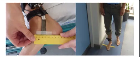
28 ms, FA: 35, FOV: 230×230; Matrix: 256×256, Scan time: 6 m 54 s) sequences. As a precaution to counteract the symptomatic orthostatic hypotension or syncope during scanning a crural pneumatic pumping device was applied to stimulate the venous backflow [18]. The pMRI scanning procedure has previously been described in detail [16]. Briefly, during scanning in supine position (SUP) the talo-crural joint was positioned in a 90° angle to the long axis of the tibia. In the standing position (ST) subjects were positioned in a one-legged stance and instructed to stand with equally distributed pressure on the heel and anterior plantar sole. The non-loaded ex-tremity rested on the housing of the scanner magnet. The foot was oriented parallel to the scanner patient table (Fig. 1). The distance from the table to the medial aspect of the foot was measured allowing the foot to be positioned similarly for stretch sensor measurements. Scanning in the standing position was repeated with addition of 10 % body weight (ST + W) carried by the participant in a backpack.
Image analyses
All image analyses were performed in a commercially available DICOM viewer (Osirix, Pixmeo SARL, Bernex,
Switzerland). We have previously described the measure-ments of NVH and MNP in detail [16]. Briefly, owing to the 3D nature of the MRI sequences the imaging planes could be adjusted in the multiplanar reconstruction (MPR) module of the DICOM viewer in a standardised fashion be-fore actual measurements of NVH and MNP (Fig. 2). Time consumption to perform all measurements of NVH and MNP was below 10 min per subject. One of the co-authors a 3rd year resident radiologist (PH) performed all radio-logical measurements reported in the present study. NVH and NMP were measured in SUP, ST and ST + W.
Stretch sensor measurements
The newly developed strain sensor is based on a dielec-tric electroactive polymer material produced by Danfoss PolyPower. The material acts as an elastic capacitive ma-terial that is strainable in one direction. The sensor mea-sures length by means of change in electrical current. It is mechanically stable, reusable, portable and resistant to perspiration from the foot. Reliability has been tested and demonstrates ICC > 0.76 for barefoot measurements [17]. The stretch sensor was attached to the skin surface as described in previous studies assessing reproducibility of the device [17, 19]: One end was attached≈20 mm behind the medial malleolus and secured by a Velcro strap. The other end was attached with adhesive tape≈ 20 mm behind the prominence of the navicular bone (Fig. 3). Measurements sampled over 15 s were per-formed in supine, standing and standing with 10 % extra bodyweight. In supine position a cushion under the plantar sole was used to stabilize the foot in 90 degrees ankle dorsiflexion. It was ensured that the cushion exerted only very slight pressure on the foot sole. The baseline strain on the sensor upon attachment cannot be controlled for rigorously, therefore measurements are only valid by calculating the difference between standing and supine. In standing position the foot was placed in a position identical to the position during pMRI scanning (Fig. 3). Stretch sen-sor measurements of the dynamic movements of the med-ial plantar arch were performed during barefoot walking. Walking distance was the same for all participants (20 m). Stretch sensor measurements were sampled over two con-tiguous walking sessions. A previous study showed that na-vicular drop varies from step to step [19]. Therefore, stretch sensor data were collected across two walking trials to en-sure an average of more than 20 steps. The normal walking speed of each participant was established and a metronome was used to ensure this pace was consistently demonstrated throughout the gait analysis.
Data analysis
Data were analysed using a custom-written Matlab script. Heel strike for each stance phase was manually determined using data from the accelerometer and Fig. 1Image of a participant in weight-bearing position in the pMRI
gyroscope, which has excellent reliability and validity [19]. Afterwards, a custom written algorithm determined the maximal magnitude of navicular motion for each stance phase. The average of stretch sensor measure-ments over two walking sessions was used for statistical analyses. Stretch sensor measurements were performed immediately after the pMRI scanning.
Blinding procedure
All radiologic measurements of NVH and MNP were performed by one radiologist (PH). The stretch sensor recordings were performed by another co-author (SES). Stretch sensor measurements were assessed by a third co-author (MR) blinded for subject ID and pMRI results.
Calculation of navicular bone position changes
Change (Δ) in NVH and MNP was calculated between SUP and ST (ΔNVHST; ΔMNPST) and between ST and
ST + W (ΔNVHST+W;ΔMNPST+W).
Additionally, “total” positional change of the navicular bone (ΔTPC) was calculated based on the assumption of a combined medial and caudal navicular displacement between SUP and ST. ΔTPC was expected to be better suited than NVH or MNP for direct comparison with stretch sensor measurements, as the stretch sensor mea-sures the resultant of both vertical and medial
displacement, which cannot be individually differentiated by the sensor [17]. ΔTPC was estimated by use of the equation of Pythagoras:
a2þ b2¼ c2
Total positional change of the navicular bone would equal:
ΔTPC ¼√ ΔNVH2þΔMNP2
Statistics
ΔNVH,ΔMNP andΔTPC are presented in mm and re-ported as mean with 95 % confidence intervals (CI). Data were visually assessed for normal distribution by QQ-plots and further examined by a Kolmogorov-Smirnov test. Data on navicular position by pMRI were normally distributed. Changes between scanning posi-tions were assessed using a two-tailed paired student’s t-test. An alpha level of 0.05 was used. Since ΔTPC in essence has a baseline value =0 no student’s t-test was performed for this parameter.
ΔTPC was compared both to the delta values from the stretch sensor measurements under static loading condi-tions between SUP and ST and to the dynamic stretch sensor measurements during walking. Since stretch sen-sor data during walking were non-normally distributed a two-tailed Spearman’s Rho correlation analysis was used.
Results
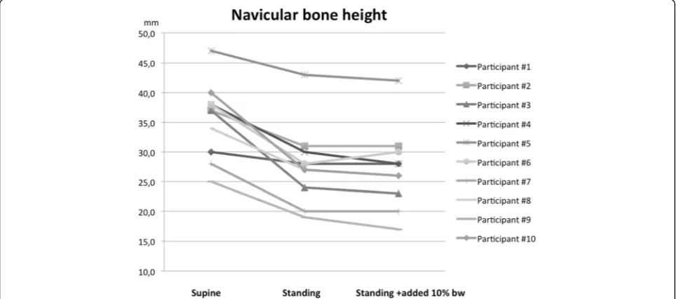
Ten healthy subjects; five females (mean age: 30 years; range: 22–39 years); mean body weight: 58; range: 49–71 kg) and five males (mean age 30 years; range: 24–38; mean body weight 75 kg; range: 63–97 kg) were included in the study. None of the participants displayed signs of orthostatic hypotension. Average values for NVH and MNP in SUP, ST and ST + W are presented as mean (range) ± SD in Table 1.
Between SUP and ST NVH decreased (ΔNVHST:
8.7 mm, CI: 6.5 to 11.0 mm, P< 0.001). No further de-crease in NVH with addition of 10 % BW was found (ΔNVHST+W= 0.7 mm, CI: −0.2 to 1.6 mm, P= 0.12)
Fig. 2Illustration of the measurement of navicular bone height and medial navicular position in images (3D SHARC sequences) obtained by pMRI in supine and standing position respectively. By means of multiplanar reconstruction the imaging planes were adjusted in a highly standardised fashion prior to actual measurements
(Fig. 4). Also a significant medial displacement of the navicular bone position occurred between SUP and ST (ΔMNPST: 4.7 mm, CI: 1.6 to 7.8 mm, P< 0.01)
with no additional medial displacement when adding 10 % body weight (ΔMNPST+W: 0.4 mm, CI: −0.8 to
1.6 mm, P= 0.46).
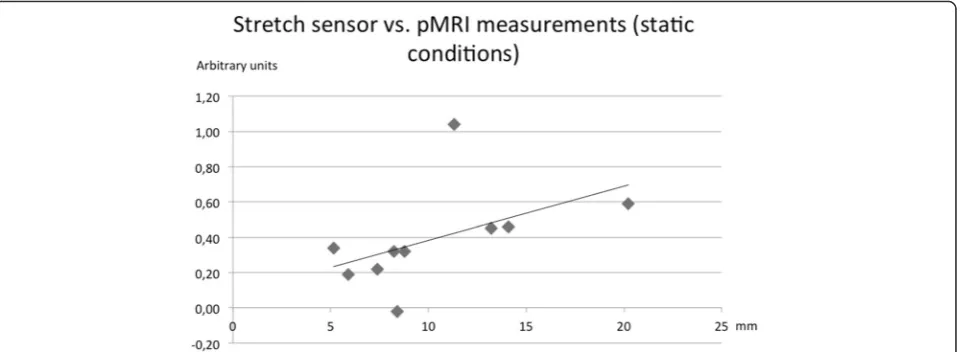
ΔTPC (supine to standing) was 10.3 mm, CI: 7.0 to 13.5 mm. No increase in ΔTPC occurred when adding 10 % bodyweight (mean difference: 0.7 mm,−0.7-2.0 mm, P= 0.29).
ΔTPC was significantly correlated with static stretch sensor measurements (Spearman’s rho = 0.66, P= 0.04) (Fig. 5) but not with measurements during walking (Spearman’s rho = 0.58,P= 0.08).
Raw data for ΔTPC and stretch sensor measurements are provided as Additional file 1.
Discussion
We have demonstrated that the total navicular bone movements distally and medially from supine to stand-ing can be determined by pMRI with acceptable concurrent validity compared to stretch sensor measure-ments under static loading conditions. However, pMRI measurements did not correlate significantly with the dynamic measurements during barefoot walking but cor-related with static stretch sensor measurement. To our
knowledge this is the first study examining and validat-ing loadvalidat-ing induced total movement of the navicular bone measured by pMRI.
We found the mean NVH in the standing position (≈28 mm) to be slightly lower than previously reported values obtained by radiography (≈30-40 mm) as summa-rized by Chang et al. [20]. There are numerous possible contributors to variation in the reported values for NVH. Firstly, numerous methodologies have been ap-plied to determine NVH across studies. One example is skin marker based measurements in which skin move-ment artefacts could cause some degree of error in de-termining the“true” position of the navicular bone [21]. Secondly, in many non-radiographic as well as some radiographic approaches both the height of the plantar sole soft tissues and the bony height of the medial plan-tar arch are included in the measurement [22, 23]. Soft tissue dimensions did not contribute to NVH in the present study, in which measurements solely relied on the bony architecture. Thirdly, the magnitude of foot loading in the ST position is likely to influence NVH to some degree. We opted for a single leg stance during scanning to approximate the loading conditions during walking. Previous radiographic and clinical studies of NVH have applied various loading conditions e.g. bi-pedal vs. unibi-pedal standing, which are likely to influence Table 1Average values for navicular height and medial navicular position measurements presented as mean (range) ± SD
Supine Standing Standing +10%BW
Navicular height 36.4 (25.0-47.0) ± 6.4 mm 27.7 (19.0-43.0) ± 6.7 mm 27.0 (17.0-42.0) ± 6.9 mm Medial navicular position 45.3 (39.0-49.0) ± 3.3 mm 50.0 (45.0-52.0) ± 3.7 mm 50.4 (46.0-54.0) ± 3.1 mm
NVH to some degree. Importantly, we observed no sig-nificant decrease in mean NVH when adding 10 % body-weight during scanning. The total navicular position (ΔTPC) did not change with addition of 10 % extra bodyweight. Obviously, this is reassuring in terms of ap-plying the method in longitudinal studies during which weight gain in participants may occur. The mean change in NVH from supine to standing position was≈9 mm by pMRI, which is slightly higher than previous mean values reported≈7 mm (range 5.3–7.4 mm) [20]. Exist-ing data relatExist-ing to MNP are scarce. We are not aware of studies assessing MNP by conventional radiography. Vinicombe et al. reported a mean medial displacement (“navicular drift”) of 7 mm measured anthropometrically from relaxed position to single limb stand [24], which slightly exceeds the mean change in MNP observed in the present study (≈5 mm). As for NVH soft tissues cov-ering the navicular bone will contribute to anthropomet-ric measurements while pMRI measurements of MNP relied solely on the osseous anatomy. This may explain the small discrepancy to some degree. Comparison of ΔTPC to previous radiologic studies is cumbersome since to our knowledge such data are not readily avail-able. However, using electromagnetic foot motion ana-lysis Cornwall & McPoil have previously reported a total navicular excursion of 7.9 (SD ± 2.5) mm resulting from combined vertical and medial displacement [11], which is somewhat less than the mean displacement (10.3 mm) found in our study. These authors performed measure-ments during dynamic loading conditions during walk-ing as opposed to our static pMRI measurements. Several previous studies have stated that navicular dis-placement measured under static conditions does not correlate well with measurements obtained during loco-motion [14, 15]. Our results are in accordance with such
previous findings. Hence, the notion that measurement of the total movement of the navicular bone could im-prove correlation with measurements during walking could not be confirmed.
with the dynamic movements [14, 15] and we suspected that pMRI measurement of the total navicular move-ment could improve the correlation. A possible explan-ation why this was not the case could among several factors be differences in walking patterns and muscle function [27] as well as variations in walking speed [28], which might influence the dynamic results of the stretch sensor without affecting static measurements. Indeed, it has been stated that the foot is very flexible and has multiple kinematic solutions during locomotion [29]. Also, it should be mentioned that the study sample size is limited and since we have previously found some vari-ation in pMRI measurements of navicular position espe-cially in SUP [16] firm conclusions regarding the validity of pMRI must still be drawn with some caution. The limited sample size was considered a necessary com-promise of resources. Bearing these considerations in mind, it would seem that static measurements should be reserved for describing the medial plantar arch under static loading conditions. However, this is still of obvious clinical importance as for many people the main loading of the feet is occurring in static standing position. Also for evaluation of foot surgery procedures such as plantar fascia release or other interventions to alter foot posture such as insoles static measurements remain relevant. In this regard pMRI is well suited for more elaborate mea-surements of navicular bone position than those feasibly obtainable by conventional radiography. Obviously, many bones other than the navicular bone move during loading and quite likely isolated measurement of navicu-lar position is overly simple to encompass changes in medial plantar arch configuration during locomotion. We do believe that pMRI has potential to provide new insight into the more complex loading induced osseous events of the foot. Time consumption in performing measurements of navicular position by pMRI is relatively low, and in principle multiplanar displacement of any osseous component of the foot can be measured non-invasively, which is a unique feature. It seems attractive to apply pMRI in larger scale studies to examine the as-sociation between complex three-dimensional changes in foot posture and risk of injury as well as the effect of foot orthoses or surgery.
Conclusion
In this exploratory study we have validated pMRI mea-surements of navicular bone displacement and find the method well suited to describe the displacement of the navicular bone under static loading conditions from su-pine to standing. However, although assessment of total navicular displacement by combining concomitant verti-cal and medial navicular position would appear advanta-geous compared to monoplanar measurement the combined measure did not seem to predict dynamic
changes of the medial foot arch during walking, which are among several possible factors depending on differ-ent walking patterns.
Additional file
Additional file 1:Supplementary. (XLSX 30 kb)
Acknowledgements
The Danish Council for Independent Research has kindly provided an unrestricted grant (DFF-4004-00593) supporting Philip Hansen. The study was also kindly supported by the Danish Rheumatism Association
(“Gigtforeningen”) and by the OAK Foundation. Esaote, SpA, Genoa, Italy has provided valuable technical support in optimising the pMRI protocol design. We wish to acknowledge the staff at the Department of Radiology for their efforts in supporting our research.
Funding
The Danish Council for Independent Research, grant no. DFF-4004-00593; Da-nish Rheumatism Association (“Gigtforeningen”), grant no. R116-A2768. The OAK Foundation has provided an unrestricted grant supporting our research.
Availability of data and materials Raw data are provided as Additional file 1.
Authors’contributions
FJ and PH contributed equally to study design, manuscript writing and data interpretation in collaboration with MB. FJ and PH share first authorship. JDN contributed to pMRI scanning of participants and development of the pMRI measurement procedure. SH participated in image data analysis. SS performed stretch sensor measurements, which were subsequently analysed by MSR. All authors read and approved the final manuscript.
Competing interests
Michael Skovdal Rathleff has been part of developing the stretch sensor but the patent and all rights are owned by his employer and he therefore has no financial interests to disclose. The remaining authors declare that they have no conflicts of interest.
Consent for publication Not applicable
Ethics approval and consent to participate
The study was approved by the local ethical committee (De Videnskabsetiske Komiteer–Region Hovedstaden) protocol no. H-2-2012-151. Informed con-sent was obtained from all participants.
Author details
1
Institute of Sports Medicine Copenhagen, Bispebjerg Hospital, Building 8, 1., Bispebjerg Bakke 23, Copenhagen DK-2400,, Denmark.2Department of
Radiology, Copenhagen University Hospital Bispebjerg & Frederiksberg, Nordre Fasanvej 57, vej 4, opg. 5, Frederiksberg DK-2000, Denmark.3Research
Unit for General Practice in Aalborg and Department of Clinical Medicine, Aalborg University, Aalborg, Denmark.4Department of Occupational Therapy
and Physiotherapy, Aalborg University Hospital, Aalborg, Denmark.5The Parker Institute, Department of Rheumatology, Copenhagen University Hospital Bispebjerg & Frederiksberg, Copenhagen, Denmark.
Received: 1 June 2016 Accepted: 16 August 2016
References
1. Metcalfe SA, Bowling FL, Baltzopoulos V, Maganaris C, Reeves ND. The reliability of measurements taken from radiographs in the assessment of paediatric flat foot deformity. Foot (Edinb). 2012;22:156–62. Elsevier Ltd. 2. Younger AS, Sawatzky B, Dryden P. Radiographic assessment of adult
3. Dowling GJ, Murley GS, Munteanu SE, Smith MMF, Neal BS, Griffiths IB, et al. Dynamic foot function as a risk factor for lower limb overuse injury: a systematic review. J Foot Ankle Res. 2014;7:53.
4. Neal BS, Griffiths IB, Dowling GJ, Murley GS, Munteanu SE, Franettovich Smith MM, et al. Foot posture as a risk factor for lower limb overuse injury: a systematic review and meta-analysis. J Foot Ankle Res. 2014;7:55. 5. Barnes A, Wheat J, Milner C. Association between foot type and tibial stress
injuries: a systematic review. Br J Sports Med. 2008;42:93–8. 6. Murphy DF, Connolly DA, Beynnon BD. Risk factors for lower extremity
injury: a review of the literature. Br J Sport Med. 2003;37:13–29. 7. Cavanagh PR, Boulton AJM. The relationship of static foot structure to
dynamic foot function. J Biomech. 1997;30:243–50.
8. McCrory JL, Young MJ, Boulton AJM, Cavanagh PR. Arch index as a predictor of arch height. Foot. 1997;7:79–81.
9. Roth S, Roth A, Jotanovic Z, Madarevic T. Navicular index for differentiation of flatfoot from normal foot. Int Orthop. 2013;37:1107–12.
10. Williams DS, McClay IS. Measurements used to characterize the foot and the medial longitudinal arch : reliability and validity. Phys Ther. 2000;80:864–71. 11. Cornwall MW, McPoil TG. Relative movement of the navicular bone during
normal walking. Foot Ankle Int. 1999;20:507–12.
12. Deng J. Reliability and validity of the sit-to-stand navicular drop test : Do static measures of navicular height relate to the dynamic navicular motion during gait ? J Stud Phys Ther Res. 2010;2:21–8.
13. Cashmere T, Hunt A. Medial longitudinal arch of the foot: stationary versus walking measures. Foot Ankle Int. 1999;20:112–8.
14. Bandholm T, Boysen L, Haugaard S, Zebis MK, Bencke J. Foot medial longitudinal-arch deformation during quiet standing and gait in subjects with medial tibial stress syndrome. J Foot Ankle Surg. 2008;47:89–95. 15. Rathleff MS, Nielsen RG, Kersting UG. Navicula drop test Ad modum Brody.
J Am Podiatr Med Assoc. 2012;102:34–8.
16. Hansen P, Johannsen FE, Hangaard S, Stallknecht SE, Hansen BB, Nybing JD, et al. Navicular bone position determined by positional MRI: a
reproducibility study. Skeletal Radiol. 2016;45:205–11.
17. Christensen BH, Andersen KS, Pedersen KS, Bengtsen BS, Simonsen O, Kappel SL, et al. Reliability and concurrent validity of a novel method allowing for in-shoe measurement of navicular drop. J Foot Ankle Res. 2014;7:12.
18. Hansen BB, Bouert R, Bliddal H, Christensen R, Bendix T, Christensen A, et al. External pneumatic compression device prevents fainting in standing weight-bearing MRI: a cohort study. Skelet. Radiol. 2013;42:1437–42. 19. Kappel SL, Rathleff MS, Hermann D, Simonsen O, Karstoft H, Ahrendt P. A
novel method for measuring in-shoe navicular drop during gait. Sensors (Switzerland). 2012;12:11697–711.
20. Chang Y, Hung W, Wu H, Chiu Y, Hsu H. Measurements of foot arch in standing, level walking, vertical jump and sprint start. Int J Sport Exerc Sci. 2010;2:31–8. 21. Taylor WR, Ehrig RM, Duda GN, Schell H, Seebeck P, Heller MO. On the
influence of soft tissue coverage in the determination of bone kinematics using skin markers. J Orthop Res. 2005;23:726–34.
22. Murley GS, Menz HB, Landorf KB. A protocol for classifying normal- and flat-arched foot posture for research studies using clinical and radiographic measurements. J Foot Ankle Res. 2009;2:22.
23. Menz HB, Munteanu SE. Validity of 3 clinical techniques for the measurement of static foot posture in older people. J Orthop Sports Phys Ther. 2005;35:479–86.
24. Vinicombe A, Raspovic A, Menz HB. Reliability of navicular displacement measurement as a clinical indicator of foot posture. J Am Podiatr Med Assoc. 2001;91:262–8.
25. Coughlin MJ, Kaz A. Correlation of Harris mats, physical exam, pictures, and radiographic measurements in adult flatfoot deformity. Foot Ankle Int. 2009;30:604–12.
26. Menz HB. Alternative techniques for the clinical assessment of foot pronation. J Am Podiatr Med Assoc. 1998;88:119–29.
27. Kameyama O, Ogawa R, Okamoto T, Kumamoto M. Electric discharge patterns of ankle muscles during normal gait. Arch Phys Med Rehabil. 1990;71:969–74. 28. Caravaggi P, Pataky T, Günther M, Savage R, Crompton R. Dynamics of
longitudinal arch support in relation to walking speed: contribution of the plantar aponeurosis. J Anat. 2010;217:254–61.
29. Nester CJ. Lessons from dynamic cadaver and invasive bone pin studies: do we know how the foot really moves during gait? J Foot Ankle Res. 2009;2:18.
• We accept pre-submission inquiries
• Our selector tool helps you to find the most relevant journal
• We provide round the clock customer support
• Convenient online submission
• Thorough peer review
• Inclusion in PubMed and all major indexing services
• Maximum visibility for your research
Submit your manuscript at www.biomedcentral.com/submit