R E S E A R C H A R T I C L E
Open Access
Determination of the differential expression of
mitochondrial long non-coding RNAs as a
noninvasive diagnosis of bladder cancer
Alexis Rivas
1,2, Verónica Burzio
1,2, Eduardo Landerer
3,4, Vincenzo Borgna
1,3, Sebastian Gatica
1, Rodolfo Ávila
1,2,
Constanza López
1, Claudio Villota
1,2, Rodrigo de la Fuente
5, Javiera Echenique
1, Luis O Burzio
1,2and Jaime Villegas
1,2*Abstract
Background:Bladder cancer is a significant cause of morbidity and mortality with a high recurrence rate. Early detection of bladder cancer is essential in order to remove the tumor, to preserve the organ and to avoid metastasis. The aim of this study was to analyze the differential expression of mitochondrial non-coding RNAs (sense and antisense) in cells isolated from voided urine of patients with bladder cancer as a noninvasive diagnostic assay.
Methods:The differential expression of the sense (SncmtRNA) and the antisense (ASncmtRNAs) transcripts in cells isolated from voided urine was determined by fluorescentin situhybridization. The test uses a multiprobe mixture labeled with different fluorophores and takes about 1 hour to complete. We examined the expression of these transcripts in cells isolated from urine of 24 patients with bladder cancer and from 15 healthy donors.
Results:This study indicates that the SncmtRNA and the ASncmtRNAs are stable in cells present in urine. The test reveals that the expression pattern of the mitochondrial transcripts can discriminate between normal and tumor cells. The analysis of 24 urine samples from patients with bladder cancer revealed expression of the SncmtRNA and down-regulation of the ASncmtRNAs. Exfoliated cells recovered from the urine of healthy donors do not express these mitochondrial transcripts. This is the first report showing that the differential expression of these
mitochondrial transcripts can detect tumor cells in the urine of patients with low and high grade bladder cancer. Conclusion:This pilot study indicates that fluorescentin situhybridization of cells from urine of patients with different grades of bladder cancer confirmed the tumor origin of these cells. Samples from the 24 patients with bladder cancer contain cells that express the SncmtRNA and down-regulate the ASncmtRNAs. In contrast, the hybridization of the few exfoliated cells recovered from healthy donors revealed no expression of these mitochondrial transcripts. This assay can be explored as a non-invasive diagnostic tool for bladder cancer.
Background
Bladder cancer (BC) is an important cause of morbidity and mortality, with an estimated 386.000 new cases and 150.000 deaths occurring worldwide in 2008 [1]. Bladder tumors are classified into four categories: papilloma,
low-grade carcinoma, high-grade carcinoma and carcin-oma in situ [2]. About 90% of bladder cancers are urothelial carcinomas and transitional cell carcinomas (TCC) and the rest include squamous cell carcinomas and adenocarcinomas. As many other types of cancer, early detection of BC will allow effective treatments of patients, improving long-term survival.
The “gold standard” in the detection of BC is cystos-copy. This examination, however, is unpleasant, time consuming, expensive and may result in infections and urethral damage [3]. On the other hand, urine cytology * Correspondence:[email protected]
1
Andes Biotechnologies S.A. and Fundación Ciencia para la Vida, 7780272 Santiago, Chile
2
Departamento de Ciencia Biológicas, Facultad de Ciencias Biológicas, Universidad Andrés Bello, 8370146 Santiago, Chile
Full list of author information is available at the end of the article
has high specificity but low sensitivity, especially in low-grade disease [4,5]. To improve the detection of BC cells in voided urine, several tumor markers and tests have been developed [6,7]. One of these tests is based on
fluorescent in situ hybridization (FISH) to detect
chromosomal alterations characteristic of BC [8]. Human cells express a family of mitochondrial long non-coding RNAs (ncRNA) containing stem-loop struc-tures. One of these transcripts, the sense mitochondrial ncRNA or SncmtRNA, is expressed in normal proliferat-ing cells and tumor cells but not in non-dividproliferat-ing cells [9,10]. Experimental evidences suggest that this tran-script plays a regulatory role of the cell cycle [11]. In addition, normal human proliferating cells in culture or in normal human tissues express two antisense tran-scripts, AsncmtRNA-1 and AsncmtRNA-2 [10]. Interest-ingly, the SncmtRNA and the AsncmtRNAs exit the mitochondria and localize to the cytoplasm and the nu-cleus in association with chromatin and nucleoli, sug-gesting that the function of these transcripts take place outside the organelle [12].
The function of the ASncmtRNAs is less clear. How-ever, an interesting observation is that the ASncmtRNAs are down-regulated in tumor cell lines as well as in tumor cells present in different types of human cancer and patients [10]. In situ hybridization of twelve BC bi-opsies from different patients shows expression of the SncmtRNA and down-regulation of the ASncmtRNAs [10]. Since down-regulation of the ASncmtRNAs seems to be independent of the tissue of origin of tumor cells, the differential expression of these transcripts can be ap-plied as a cancer diagnostic method for cells in
suspen-sion. Here, we present a one-tube fluorescence in situ
hybridization protocol applied to cells in suspension (S-FISH), that takes about 60 min to perform and using simultaneously labeled probes for both SncmtRNA and AsncmtRNAs. This method was applied to cells isolated from urine of patients with bladder cancer (BC). In twenty four patients with low and high grade of BC, S-FISH revealed cells expressing the SncmtRNAs and not the ASncmtRNAs, hence corresponding to cancer
cells phenotype. The expression of these transcripts was negative in the few cells isolated from the urine of healthy donors. The differential expression of the SncmtRNA and the ASncmtRNAs in cells isolated from voided urine can be explored as a new non-invasive diagnostic test for BC.
Methods Tumor cell culture
T24 and RT4 cells (human bladder carcinoma) and DU-145 cells (prostate carcinoma) were cultured accord-ing to ATCC recommendations. Cultures were maintained in a humidified incubator at 37°C and 5% CO2. Peripheral
blood mononuclear cells (PBMC) from healthy donors were isolated and stimulated with phytohaemagglutinin (PHA) for 48 h as described before [9,10,13]. Primary renal mixed epithelial cells were obtained from ATCC and cultured according to ATCC guidelines.
Table 1 Distribution of tumor stage and grade among all patients included in this study
Grade and stage
distribution None Grade 1 Grade 2 Grade 3 Total
No tumor 15 15
Ta 11 2 4 17
T1 1 1 1 3
T2-4 3 3
CIS 1 1
Total 15 12 3 9 39
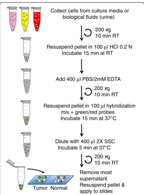
Collect cells from culture media or biological fluids (urine)
200 xg
10 min RT
Resuspend pellet in 100 µl HCl 0.2 N Incubate 15 min at RT
Add 400 µl PBS/2mM EDTA
Resuspend pellet in 100 µl hybridization mix + green/red probes
Incubate 15 min at 37°C
Dilute with 400 µl 2X SSC Incubate 5 min at 37°C
Remove most supernatant Resuspend pellet & apply to slides
200 xg
10 min RT
200 xg
10 min RT
Tumor Normal
S-FISH
All the hybridization steps were performed in
MaxiRe-covery™ tubes of 0.5 ml (Axygen Scientific, US). After
trypsinization (Invitrogen, Carlsbad, US), about 105cells
were recovered by centrifugation at 200 × g for 10 min
at room temperature (RT). The cell pellet was
resus-pended in 100 μl HCl 0,2 N and incubated for 5 min at
RT. Afterwards, the cell suspension was diluted with
400 μl PBS (50 mM sodium phosphate, 150 mM NaCl
and 2 mM EDTA, pH 9.0) and centrifuged again. The sediment was resuspended in 100μl hybridization buffer
(50% formamide, 150μg/ml herring sperm DNA, 4X SSC,
2 mM EDTA) containing 0,5 μM 50-Alexa fluor
488-labeled probe P1 (50 GTTCTTGGGTGGGTGTGGG 30),
complementary to the SncmtRNA and 0,05 μM each of
two 50 Texas Red-labeled probes P2 (50GATAACAGCG
CAATCCTATT 30) and P3 (50 ACCGTGCAAAGGTAG
CATAATCA 30), complementary to the ASncmtRNAs. In
addition, two negative controls corresponding to
mis-match probes P5 for the SncmtRNA (MM: 50 TTTATTT
GATGAGTGTGAG 30), labeled with Alexa fluor 488 and
probe P6 for the ASncmtRNAs (MM: 50GTAAAGATAG
TATAATAATTTATTAATTAAATATA 30), labeled with
Texas Red at the 50end. The labeled probes were obtained from Invitrogen (Carlsbad, CA, USA).
Hybridization was carried out for 15 min to 2 h at 37°C. The final wash was performed by addition of four volumes of stringency buffer (2X SCC + 2 mM EDTA) to the hybridization mix, incubated for 5 min at 37°C and
finally centrifuged at 200 × g for 10 min. The
super-natant was discarded and a small volume of approxi-mately 20 μl of the residual supernatant was left in the tube to resuspend the cells. The cells were finally stained in a solution of 1μg/ml DAPI, deposited onto a positively charged slide (Thermo Scientific, US) and mounted in fluorescent medium (DAKO). Samples were analyzed by fluorescence microscopy on an Olympus BX-51 micro-scope under x600 magnification, with 300-600 ms expos-ition and results were documented with Q-capture Pro software. The positive hybridization control corresponded
to a 50-Texas Red-labeled probe complementary to 18S
rRNA (P4: 50AGTGGACTCATTCCAATTACA 30).
Voided urine
About 30-50 ml voided urine from male and female healthy donors was carried out in agreement with the
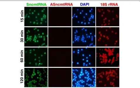
SncmtRNA
ASncmtRNA
DAPI
18S rRNA
15 min
30 min
60 min
120 min
ethical guidelines approved by the Ethical Committee of the Fundacion Ciencia para la Vida. The urine from
healthy donors (50 ml) was loaded with 5×10⁴to 1×10⁵
T24 or DU-145 cells or PHA-stimulated lymphocytes and incubated at 4°C for 24 h. The cells were
sedimen-ted by centrifugation at 700 ×g for 10 min. The
super-natant was discarded, leaving only 5 ml, which were transferred to a Kova tube (Hycor Biomedical Inc., US)
and centrifuged again at 200 ×gfor 10 min at RT. Most
of the supernatant was discarded and the remnant (~1ml) was transferred to a 0,5 ml Maxy-Recovery tube (Axygen Scientific, US), centrifuged at 200 × g and sub-jected to S-FISH as described above.
Twenty four patients diagnosed with BC were recruited at the Urology Unit of the Hospital Barros Luco Trudeau and Clinica Indisa (Santiago, Chile). The urine samples were obtained with informed consent under the Ethical Regulations of the Hospital Barros Luco Trudeau and Clinica Indisa and with the approval of the Ethic Committee of Fundacion Ciencia para la
Vida. The tumor biopsies were graded as reported [14] and the data are summarized in Table 1. The first-morning voided urine (50 ml) was collected and stored at 4°C in a cooler and transported to the laboratory. The
urine was centrifuged at 700 × g for 10 min within 4 h
after collection and S-FISH was performed as described above. Parallel samples were stained with hematoxylin.
Results
Optimization of the S-FISH protocol
A schematic representation of the S-FISH is shown in Figure 1. Briefly, cells were collected by centrifugation from cell culture or from biological fluids such as urine and blood, followed by permeabilization with 0.2 N HCl. After neutralization, the cells were recovered, hybridized with a set of probes labeled with fluorophores, washed and analyzed by fluorescence microscopy (see Methods). To determine the minimum hybridization time needed for the detection of the mitochondrial ncRNAs, T-24 cells (bladder carcinoma cell line) were subjected to
SncmtRNA
ASncmtRNA
DAPI
18S rRNA
A
MM
MM
MM
MM
SncmtRNA
ASncmtRNA
DAPI
18S rRNA
B
S-FISH for different time periods. After 15 minutes of hybridization the fluorescent signal of the SncmtRNA was as strong and specific as longer hybridization times (Figure 2, SncmtRNA). The red signal correspond-ing to the ASncmtRNA was negative at any of the hybridization times tested confirming the tumor pattern of expression of T-24 cells (Figure 2, ASncmtRNAs). The 18S rRNA used as positive control was expressed in all cells and the corresponding red signal was also inde-pendent of hybridization times ranging from 15 to 120 min (Figure 2, 18S rRNA, red). The same results were obtained with the bladder carcinoma cell line RT4 (unpublished data).
Stability of the mitochondrial ncRNAs in urine
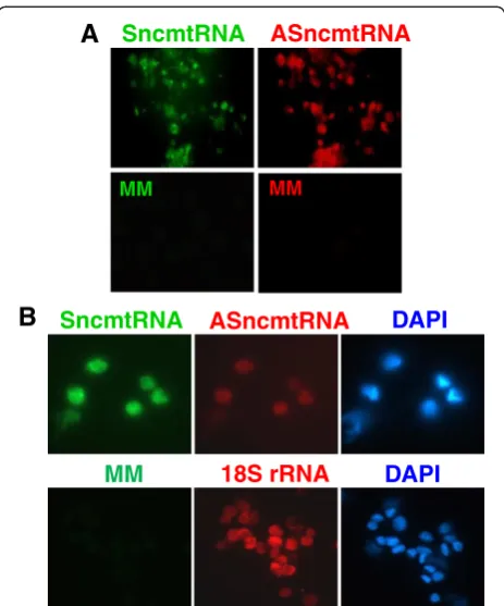
An interesting model to test S-FISH was cancer cells obtained from voided urine of patients with BC. Since the stability of cells and their RNAs in urine is uncertain, we asked whether the urine would affect the stability of the SncmtRNA and the ASncmtRNAs from tumor cells and normal proliferating cells. Fresh urine of healthy
donors was loaded with 5×10⁴ T-24 cells/ml and
main-tained at 4°C for 24 h. Cells were then recovered by cen-trifugation and subjected to S-FISH as described. Analysis of several fields indicated that all cells were positive for the SncmtRNA and the intensity of the fluorescent signal was comparable to that of fresh T-24 cells (compare Figures 2 and 3, SncmtRNA). The red hybridization signal corresponding to the ASncmtRNAs (Figure 3A, ASncmtRNAs) was negative and comparable to the negative green fluorescence of the mismatch (MM) probes for the sense transcript (Figure 3A, panels MM green) or the red fluorescence of the MM probe for the antisense transcripts (Figure 3A, panels MM red). The hybridization signal of 18S rRNA was similar to that of fresh T-24 cells (Figure 3A, 18S rRNA). The same results were obtained with DU-145 cells (prostate car-cinoma cell line) (Figure 3B).
In addition, we determined the stability of the SncmtRNA and the ASncmtRNAs in normal proliferat-ing cells maintained in urine for 24 h at 4°C. Isolated PBMC were activated with PHA and incubated in urine for 24 h previous to S-FISH. PHA-stimulated PBMCs showed a positive signal for both the SncmtRNA and the ASncmtRNAs (Figure 4A) confirming the expression pattern of normal proliferating cells [10]. On the other hand, the hybridization signals were negative with the MM probe to either the SncmtRNA (green fluorescence) and the ASncmtRNAs (red fluorescence) (Figure 3A, MM). Similarly, the same normal renal epithelial cells (DAPI staining) expressing the SncmtRNA were also expressing the ASncmtRNAs (Figure 4B). The green mismatch control was negative (Figure 4B).
Detection of tumor cells in voided urine of patients with BC
Then we asked whether S-FISH can be applied to tumor cells present in voided urine obtained from 24 patients with BC diagnosed by cystoscopy and confirmed by bi-opsy. Table 1 show the grade and stage distribution of the samples. The cells were recovered from urine 4 h after collection and subjected to S-FISH as described be-fore. Then, 25 fields of each sample at 40x magnification were analyzed and recorded. In all 24 urine samples, the S-FISH detects cells expressing the SncmtRNA and down-regulate the ASncmtRNAs. As described before, this expression pattern corresponds to a cancer cells. Figure 5 illustrates S-FISH results obtained with urine cells recovered from four patients with BC. Samples A, B and C correspond to urine cells recovered from patients with grade 3 BC (Figure 5). Samples D corre-sponds to cells obtained from patients with grade 2 (Figure 5). It is important to mention that the cellularity of samples C and D was low. Although the cellularity
SncmtRNA
ASncmtRNA
A
MM MM
SncmtRNA
ASncmtRNA
DAPI
18S rRNA
B
DAPI
MM
of grade 1 BC was low, the S-FISH detected few cells that only express the SncmtRNA. In six urine samples obtained from the 15 healthy donors (Table 1) few cells were recovered. However, the hybridization signals indi-cate that the SncmtRNA and the ASncmtRNAs were down-regulated. The typical debris present in some urine samples did not interfere with the hybridization signal.
Discussion
FISH provides an important tool for conventional cyto-genetics and evaluation of chromosomal abnormalities associated with several malignancies [15]. Some exam-ples of chromosomal abnormalities are found in several diseases, such as BC [16-20], multiple myeloma [21-23], breast cancer [24], hematological malignancies [25-28] and lung cancer [29,30] among others. Different types of tumor require specific sets of probes corresponding to particular chromosomal deletions/translocations charac-teristic of each cancer. Sokolova et al. reported the de-velopment of a FISH assay with high sensitivity and specificity for high grade BC using four labeled probes specific for the pericentromeric regions of chromosomes 3, 7 and 17 and for the detection of the 9p21 deletion
[8]. These results were confirmed in later studies with a large cohort of BC patients [16-20] using several probes combined into a single multiprobe cocktail, to detect polysomy of chromosomes 3, 7 and 17 and homozygous deletion of 9p21 in the urine of BC patients (Urovysion, Abbot Molecular/Vysis, Des Plaines, IL). However, this test has low sensitivity for low-stage and low-grade tumors, which are the main group that recur [3].
The S-FISH assay described here is able to detect the differential expression of the SncmtRNA and the ASncmtRNAs in normal and cancer cells. This a sim-ple protocol that was optimized in three steps includ-ing a sinclud-ingle permeabilization step with HCl, a short hybridization step and a brief washing that basically involves the dilution of the hybridization mix with stringency buffer (Figure 1). The protocol contains only three centrifugation steps in the same tube, min-imizing the manipulation of cells and therefore maxi-mizing RNA preservation and cell recovery. This test is reproducible and has been applied to other normal and tumor cell lines. Hybridization of normal prolifer-ating cells (human umbilical vein endothelial cells, ker-atinocytes and melanocytes) reveals the expression of
SncmtRNA
ASncmtRNA
DAPI
Hematoxylin
A
B
D
C
the SncmtRNA and the ASncmtRNAs. In other human tumor cell lines such as HeLa, 42/95 and SK-MEL-2 (melanoma), Jurkat and HL-60 (leukemia) and MDA-MB-231 (breast carcinoma), S-FISH revealed expres-sion of the SncmtRNA and down-regulation of the ASncmtRNA (unpublished data).
Moreover, S-FISH was able to detect cancer cells in urine from twenty four patients with BC and the results were independent of the grade of BC and the urine cel-lularity (see Figure 5). Taken together, the results suggest that the diagnostic test has a very high positive outcome independent of the grade and the amount of cells recov-ered from urine of patients with BC. In the urine from healthy donors, cells were recovered only from six out of fifteen samples and the S-FISH revealed absence of sig-nal to both transcripts.
Conclusions
Taken together, this pilot study suggests that S-FISH could be used for detection and regular surveillance pro-grams of patients with BC. Interestingly, the results indi-cate that the exfoliated bladder tumor cells from low and high grade BC conserve the expression pattern observed in bladder cancer biopsies: expression of the SncmtRNA and down-regulation of the ASncmtRNAs. In summary, S-FISH may potentially be used as a non-invasive diagnostic test for bladder cancer. However, to validate the test, a large cohort of patients with low-grade and high-low-grade neoplasms should be included together with other urological diseases such as glomer-ulonephritis, infections of the upper urinary track and other benign urinary track diseases.
Competing interests
Authors report no competing interests
Authors’contributions
LOB, VB and JV conceived the experimental plan, analyzed the data, and drafted the manuscript. AR, VB, EL, VB, SG, RA, CL, CV, RF, and JE carried out the experiments. RF, EL and VB reviewed the patients’history and pathological data. All authors read and approved the final manuscript.
Acknowledgements
Supported by Grant 1085210, FONDECYT, Millennium Scientific Initiative N° P-77-09 F, Grants DI-20-11-I, Universidad Andrés Bello, Grant D04I1338, FONDEF, the CCTE-PFB16 Program of Conicyt and Grant 12IDL4-13358, CORFO-INNOVA, Chile
Author details
1Andes Biotechnologies S.A. and Fundación Ciencia para la Vida, 7780272
Santiago, Chile.2Departamento de Ciencia Biológicas, Facultad de Ciencias Biológicas, Universidad Andrés Bello, 8370146 Santiago, Chile.3Facultad de Medicina, Universidad Andrés Bello, 8370146 Santiago, Chile.4Urology Unit, Clínica Indisa, 7520440 Santiago, Chile.5Urology Unit, Hospital Barros Luco Trudeau, 8900085 Santiago, Chile.
Received: 22 March 2012 Accepted: 7 December 2012 Published: 18 December 2012
References
1. Jemal A, Bray F, Center MM, Ferlay J, Ward E, Forman D:Global cancer statistics.CA Cancer J Clin2011,61:69–90.
2. Kaufman DS, Shipley WU, Feldman AS:Bladder cancer.Lancet2009,
374:239–249.
3. Van Tilborg AA, Bangma CH, Zwarthoff EC:Bladder cancer biomarkers and their role in surveillance and screening.Int J Urol2009,16:23–30. 4. Maier U, Simak R, Neuhold N:The clinical value of urinary cytology:
12 years of experience with 615 patients.J Clin Pathol1995,48:314–317. 5. Ross J, Cohen M:Ancilliary methods for the detection of recurrent
urothelial neoplasia.Cancer2000,90:75–86.
6. Msaouel P, Koutsilieris M:Diagnostic value of circulating tumor cells detection in bladder and urothelial cancer: systematic review and meta-analysis.BMC Cancer2011,11:336–349.
7. Ecke T:Focus on urinary bladder cancer markers: a review.Minerva Urol Nefrol2008,60:237–246.
8. Sokolova IA, Halling KC, Jenkins RB, Burkhardt HM, Meyer RG, Seelig SA, King W:The development of a multitarget, multicolor fluorescence in situ hybridization assay for the detection of urothelial carcinoma in urine.
J Mol Diagn2000,2:116–123.
9. Villegas J, Burzio V, Villota C, Landerer E, Martinez R, Santander M, Martinez R, Pinto R, Vera MI, Boccardo E, Villa LL, Burzio LO:Expression of a novel non-coding mitochondrial RNA in human proliferating cells.Nucleic Acids Res2007,35:7336–7347.
10. Burzio VA, Villota C, Villegas J, Landerer E, Boccardo E, Villa LL, Martínez R, Lopez C, Gaete F, Toro V, Rodriguez X, Burzio LO:Expression of a family of noncoding mitochondrial RNAs distinguishes normal from cancer cells.
Proc Natl Acad Sci USA2009,106:9430–9434.
11. Villota C, Campos A, Vidaurre S, Oliveira-Cruz L, Boccardo E, Burzio VA, Varas M, Villegas J, Villa LL, Valenzuela PDT, Socias M, Roberts S, Burzio LO:
Expression of mitocondrial ncRNAs is modulated by high risk HPV oncogenes.J Biol Chem2012,287:21303–21315.
12. Landerer E, Villegas J, Burzio VA, Oliveira L, Villota C, Lopez C, Restovic F, Martinez R, Castillo O, Burzio LO:Nuclear localization of the mitochondrial ncRNAs in normal and cancer cells.Cellular Oncol2011,34:297–305. 13. Dergunova NN, Bulycheva TI, Artemenko EG, Shpakova AP, Pegova AN,
Gemjian EG, Dudnik OA, Zatsepina OV, Malashenko OS:A major nucleolar protein B23 as a marker of proliferation activity of human peripheral lymphocytes.Immunol Lett2002,83:67–72.
14. Babjuk M, Oosterlinck W, Sylvester R, Kaasinen E, Böhle A, Palou-Redorta J, Morgan Roupret M:EAU guidelines on non–muscle-invasive urothelial carcinoma of the bladder, the 2011 update.Eur Urol2011,
59:997–1008.
15. Wolff DJ, Bagg A, Cooley LD, Dewald GW, Hirsch BA, Jacky PB, Rao KW, Rao PN:Guidance for fluorescencein situhybridization testing in
hematologic disorders.J Mol Diagn2007,9:134–143.
16. Halling KC, King W, Sokolova IA, Meyer RG, Burkhardt HM, Halling AC, Cheville JC, Sebo TJ, Ramakumar S, Stewart CS, Pankratz S, O’Kane DJ, Seelig SA, Lieber MM, Jenkins RB:A comparison of cytology and fluorescencein
situhybridization for the detection of urothelial carcinoma.J Urol2000,
164:1768–1775.
17. Kang JU, Koo SH, Jeong TE, Kwon KC, Park JW, Jeon CH:Multitarget fluorescencein situhybridization and melanoma antigen genes analysis in primary bladder carcinoma.Cancer Genet Cytogenet2006,164:32–38. 18. Kipp BR, Tanasescu M, Else TA, Bryant SC, Karnes RJ, Sebo TJ, Halling KC:
Quantitative fluorescencein situhybridization and its ability to predict bladder cancer recurrence and progression to muscle-invasive bladder cancer.J Mol Diagn2009,11:148–154.
19. Colucci G, Floege J, Schena FP:The urinary sediment beyond light microscopical examination.Nephrol Dial Transplant2006,
21:1482–1485.
20. Meiers I, Singh H, Hossain D, Lang K, Liu L, Qian J, Verhest AP, Bostwick DG:
Improved filter method for urine sediment detection of urothelial carcinoma by fluorescencein situhybridization.Arch Pathol Lab Med 2007,131:1574–1579.
21. Stewart AK, Fonseca R:Review of molecular diagnostics in multiple myeloma.Expert Rev Mol Diagn2007,7:453–459.
23. Avet-Loiseau H, Soulier J, Fermand JP, Yakoub-Agh:IFM and MAG groups. Impact of high-risk cytogenetics and prior therapy on outcomes in patients with advanced relapsed or refractory multiple myeloma treated with lenalidomide plus dexaméthasone.Leukemia2010,24:623–628. 24. Pauletti G, Godolphin W, Press MF, Slamon DJ:Detection and quantitation
of HER-2/neu gene amplification in human breast cancer archival material using fluorescencein situhybridization.Oncogene1996,
13:63–72.
25. Escudier SM, Pereira-Leahy JM, Drach JW, Weier HU, Goodacre AM, Cork MA, Trujillo JM, Keating MJ, Andreeff M:Fluorescentin situhybridization and cytogenetic studies of trisomy 12 in chronic lymphocytic leukemia.Blood 1993,81:2702–2707.
26. Hagemeijer A, Buijs A, Smit E, Janssen B, Creemers GJ, Van der Plas D, Grosveld G:Translocation of BCR to chromosome 9: a new cytogenetic variant detected by FISH in two Ph-negative, BCR-positive patients with chronic myeloid leukemia.Genes Chromosomes Cancer1993,8:237–245. 27. Zanardi A, Bandiera D, Bertolini F, Corsini CA, Gregato G, Milani P, Barborini
E, Carbone R:Miniaturized FISH for screening of onco-hematological malignancies.Biotechniques2010,49:497–504.
28. Nelson B, Gupta R, Dewald G, Paternoster S, Rosen S, Peterson L:Chronic lymphocytic leukemia FISH impact on diagnosis panel.American J Clinical Pathol2007,128:323–332.
29. Jiang F, Caraway NP, Nebiyou Bekele B, Zhang HZ, Khanna A, Wang H, Li R, Fernandez RL, Zaidi TM, Johnston DA, Katz RL:Surfactant protein A gene deletion and prognostics for patients with stage I non-small cell lung cancer.Clin Cancer Res2005,11:5417–5424.
30. Li R, Liu Z, Fan T, Jiang F:A novel multiple FISH array for the detection of genetic aberrations in cancer.Lab Invest2006,86:619–627.
doi:10.1186/1471-2490-12-37
Cite this article as:Rivaset al.:Determination of the differential expression of mitochondrial long non-coding RNAs as a noninvasive diagnosis of bladder cancer.BMC Urology201212:37.
Submit your next manuscript to BioMed Central and take full advantage of:
• Convenient online submission
• Thorough peer review
• No space constraints or color figure charges
• Immediate publication on acceptance
• Inclusion in PubMed, CAS, Scopus and Google Scholar
• Research which is freely available for redistribution