Unexpectedly Codepleted of Ficolin-2
Allison M. Brady,aK. Aaron Geno,aAlex G. Dalecki,aXiaogang Cheng,bMoon H. Nahma
Department of Pathologyaand Department of Surgery,bUniversity of Alabama at Birmingham, Birmingham, Alabama, USA
The ficolins are a family of innate pattern recognition molecules that are known to bind acetylated compounds and activate
com-plement through the association of mannose binding lectin (MBL)/ficolin-associated serine proteases (MASPs). Their
impor-tance has more recently become appreciated, as they have been shown to play a role in a variety of disease processes from
infec-tion to autoimmunity. While studying ficolin-2-mediated complement deposiinfec-tion on
Streptococcus pneumoniae
, we found that
sera depleted of C1q or other complement components were also codepleted of ficolin-2 but not ficolin-1, ficolin-3, or MBL. MBL
present in C1q-depleted sera was able to mediate complement deposition on
Saccharomyces cerevisiae
, suggesting the presence
of MASPs. We found that complement was activated on pneumococci in C1q-depleted serum only after opsonization with
exoge-nous recombinant ficolin-2 (rFicolin-2). Also, no complement deposition was observed in C1q-depleted serum when
pneumo-cocci were opsonized with rFicolin-2 mutated at its lysine-57 residue, where MASPs are known to associate. Thus, these depleted
sera are a unique tool to study ficolin-2-mediated complement pathways; however, one should be aware that ficolin-2 is absent
from complement component-depleted sera.
T
he ficolins are a recently described family of innate opsonins,
consisting of ficolin-1, -2, and -3 (also known as M-, L-, and
H-ficolin, respectively). This family of proteins functions as
pat-tern recognition molecules with an affinity for acetylated ligands
(
1
). Ficolin-2 has been recently shown to be important during
pneumococcal pathogenesis (
2–4
) and is known to bind to many
other significant human pathogens (
2
,
5
,
6
). Moreover, ficolin-2
and -3 have been implicated in many different disease pathologies,
such as transplant reperfusion injury (
7
), clearance of apoptotic
cells (
8
,
9
), and even preeclampsia (
10
).
The structure and function of these proteins are similar to
those of the well-characterized mannose binding lectin (MBL).
Like MBL, the ficolins activate the lectin pathway of complement
through the association of their effector serine proteases called
MBL/ficolin-associated serine proteases (MASPs) (
11
). The lectin
pathway shares the same complement proteins as the classical
pathway (
12
). Similar to C1q/r/s of the classical pathway, the
fico-lins and MBL activate their associated MASPs upon binding,
which cleave C4 and C2 to create the C3 convertase (C4b2a) (
11
).
Thus, results of lectin pathway studies can be confounded by the
classical pathway. It is therefore necessary to reduce or eliminate
complement activation through the classical pathway in order to
understand the role of the lectin complement pathway.
As the classical pathway critically depends on C1q, the use of
C1q-depleted serum can eliminate the complications of the
clas-sical pathway. However, while studying the role of the lectin
path-way during pneumococcal disease, we discovered that
commer-cially available C1q-depleted sera also lack ficolin-2, while other
lectin pathway opsonins were still present. Here we describe our
characterization of several commercially available complement
component-depleted sera and how they can be exploited to study
the lectin pathway of complement.
MATERIALS AND METHODS
Strains and reagents.The serotype 11D (SSISP 11D/1) and 19C (SSISP
19C/2) strains used were purchased from Statens Serum Institut (SSI; Copenhagen, Denmark). ThewcjE-null strain JC05 (serotype 11Dx) was
previously described (3). The serotype 20A strain (AMB11) and serotype 20B strain (AMB12) were generated using ATCC 6320 (American Type Culture Collection, Manassas, VA) and CDC5931-06 (Centers for Disease Control and Prevention, Atlanta, GA), respectively, by introducing a streptomycin resistantrpsLallele using a previously described method (13). ThewcjE-null strains AMB13 (serotype 20Ax) and AMB14 (serotype 20Bx) were generated from strains AMB11 and AMB12, respectively, us-ing a previously described protocol (14).Saccharomyces cerevisiaeused for MBL binding assays was a kind gift from the laboratory of Stephen Moser at the University of Alabama at Birmingham (Birmingham, AL). Bacteria were cultured at 37°C and 5% CO2on blood agar plates (BD Biosciences, San Jose, CA) or in Todd-Hewitt broth (BD Biosciences, San Jose, CA) with 0.5% yeast extract (THY). Yeast was cultured in yeast extract-pep-tone-dextrose (YPD; 1% yeast extract, 2% peptone, 2% dextrose) broth or on YPD agar plates (YPD broth supplemented with 2% agar) at 30°C.
Ficolin binding buffer (FBB) contained 1⫻Hanks’ buffered saline solution (Gibco, Life Technologies) with 2.2 mM CaCl2and 0.5% bovine serum albumin (BSA). Gelatin Veronal buffer (GVB) contained 0.15 mM CaCl2, 141 mM NaCl, 0.5 mM MgCl2, 0.1% gelatin, and 5 mM barbital sodium C-IV at pH 7.3.
Complement component-depleted sera were purchased from three manufacturers: CalBiochem (Millipore, Billerica, MA), Quidel (San Di-ego, CA), and Complement Technologies (Tyler, TX), referred to as man-ufacturers A, B, and C, respectively, inTable 1. Normal human serum (NHS) was obtained from a consenting healthy adult volunteer donor at the University of Alabama at Birmingham (protocol approved by the In-stitutional Review Board, University of Alabama at Birmingham).
Binding assays.Ficolin-1 and -2 and MBL binding assays were
per-formed as previously described (15). Briefly, bacteria or yeast was
incu-Received28 May 2014Returned for modification26 June 2014 Accepted10 July 2014
Published ahead of print16 July 2014 Editor:T. S. Alexander
Address correspondence to Moon H. Nahm, [email protected].
Copyright © 2014, American Society for Microbiology. All Rights Reserved. doi:10.1128/CVI.00370-14
on August 17, 2020 by guest
http://cvi.asm.org/
bated with 5% (for ficolin-2 and MBL) or 15% (for ficolin-1) NHS or complement component-depleted sera in FBB for 1 h at 4°C. After a washing, bacteria were stained with an ficolin-1 monoclonal anti-body (MAb; 7G1, catalog number HM2196; Hycult), anti-ficolin-2 MAb (GN5, catalog number HM2091; Hycult), or anti-MBL MAb (3E7, catalog number MA1-40145; Pierce) for 30 min at 4°C, followed by a fluorescein-conjugated goat anti-mouse IgG antibody (Southern Biotech; catalog number 1030-09) for 30 min at 4°C. Stained bacteria were analyzed using FACSCalibur (BD Biosciences, San Jose, CA). Data were analyzed using FCS Express V3 (De Novo Software, Los Angeles, CA).
Determining MBL and ficolin-1, -2, and -3 concentrations.
Concen-trations of MBL and ficolin-1, -2, and -3 in the complement component-depleted sera were determined using commercially available kits for each analyte according to the manufacturer’s instructions. The MBL kit (cata-log number HK323), ficolin-1 kit (cata(cata-log number HK357), ficolin-2 kit (catalog number HK336), and ficolin-3 kit (catalog number HK340) were purchased from Hycult.
Complement deposition on yeast.Yeast were incubated in 100l of
GVB containing 5% NHS or C1q-depleted serum with or without an inhibitor (100 mMD-mannose) for 1 h at 37°C, washed, and stained for 30 min at 4°C for C3 and C4 deposition using anti-human C3 Ab (Pierce; LF-MA0132), followed by phycoerythrin (PE)-labeled goat anti-mouse IgG secondary Ab (Southern Biotech; 1030-09) and fluorescein isothio-cyanate (FITC)-labeled anti-human C4c/C4b antibody (Pierce; PA1-28407). Stained yeast was analyzed using FACSCalibur (BD Biosciences). Data were analyzed using FCS Express V3 (De Novo Software).
Western blotting of ficolin-2.Five microliters of factor-depleted
se-rum or NHS control sese-rum in a total volume of 50l per well was elec-trophoresed on 1.5-mm SDS–12% polyacrylamide gels and blotted onto a nitrocellulose membrane. The membrane was blocked in 5% skim milk in Tris-buffered saline (TBS; 13.7 mM NaCl, 2.15 mM KCl, 10 mM Tris [pH 7.2]) supplemented with 0.05% Tween 20 (TBST) for 1 h. The membrane was then washed and probed with biotinylated anti-ficolin-2 antibody (R&D Systems) diluted 1:1,000 in TBST for 1 h at room temperature before washing and incubation with streptavidin-alkaline phosphatase (Zymed) diluted 1:1,000 in TBST for 30 min at room temperature. Blots were washed and developed in 1 M Tris (pH 8.8) with addition of 1 mg of Nitro Blue Tetrazolium and 5 mg of 5-bromo-4-chloro-3-indolylphos-phate dissolved in 100l of dimethyl sulfoxide. Development was stopped by exchanging buffer with deionized water.
Generation of recombinant wild-type and mutant ficolin-2.Wild-type
ficolin-2 (FCN2) cDNA was purchased from Transomics (TCH1003) and cloned into pcDNA3.1(⫺) via NotI/EcoRI sites (FCN2-pcDNA3.1). To generate the K57 mutants, we performed site-directed mutagenesis. For the K57R construct, ficolin-2 was amplified from the FCN2-pcDNA3.1 using primer set T7 (5=-TAATACGACTCACTATAGGG-3=)/3571 (5=-G TGGTCCTGCCCTCCCAGGAG-3=) and 5571 (5=-CTCCTGGGAGGGC AGGACCAC-3=)/BGH (5=-TAGAAGGCACAGTCGAGG-3=) in
individ-ual reactions. The resulting products were mixed and amplified using primers T7 and BGH and then cloned into pcDNA3.1(⫺) via NotI/EcoRI sites (FCN2K57R-pcDNA3.1). The K57E construct was generated by the same procedure, substituting primers 3572 (5=-GTGGTCCTGCCTCCC CAGGAG-3=) and 5572 (5=-CTCCTGGGGAGGCAGGACCAC-3=) for 3571 and 5571 (FCN2K57E-pcDNA3.1). The FCN2-pcDNA3.1, FCN2K57R-pcDNA3.1, and FCN2K57E-pcDNA3.1 plasmids were transformed into competentEscherichia coliand selected for ampicillin resistance. Plasmids from resulting transformants were isolated and confirmed by sequencing (Heflin Center for Human Genetics, University of Alabama at Birming-ham, BirmingBirming-ham, AL) to verify the sequence or if the desired mutations had been incorporated. Purified plasmids were transfected into CHO-K1 using Lipofectamine 2000 (Invitrogen) per supplier protocol. Cells were cultured in Dulbecco’s modified Eagle’s medium–nutrient mixture Ham’s F-12 liquid medium (Thermo Scientific) with 10% heat-inacti-vated fetal bovine serum (FBS) and selected using 750 mg/liter of Geneti-cin sulfate. Transfected cells were then subcloned.
The concentration of rFicolin-2 present in each of the culture super-natants was determined using a commercially available ficolin-2 enzyme-linked immunosorbent assay (ELISA) kit (catalog number HK336). Wild-type rFicolin-2 was present at 7.25g/ml, the K57R rFicolin-2 was present at 9.35g/ml, and the K57E rFicolin-2 was present at 2.83g/ml. These were comparable to the concentration of ficolin-2 in our NHS (3.6g/ ml). The supernatant control had no detectable levels of ficolin-2 (⬍0.016 g/ml).
Complement deposition with rFicolin-2.Complement deposition
assays were performed as previously described (3). Briefly, bacteria (1⫻ 107CFU/ml) were preincubated in 100l of FBB containing 25% recom-binant ficolin-2 (rFicolin-2) supernatant with or without inhibitors (10 mg/liter of acetylated BSA [acBSA] or 10 mg/liter of BSA) for 1 h at 4°C, washed, and then incubated in 100l of GVB containing 5% C1q-de-pleted serum and 25% rFicolin-2 supernatant with or without inhibitors (10 mg/liter of acBSA or 10 mg/liter of BSA).
RESULTS
Commercially available C1q-, C2-, C3-, C4-, and factor
B-de-pleted sera lack ficolin-2 but not related opsonins.
We have
pre-viously established flow cytometric binding assays to visualize
fi-colin-1, ficolin-2, and MBL binding to their natural microbial
targets (serotype 19C pneumococci, serotype 11A pneumococci,
and
S. cerevisiae
, respectively). We used these binding assays to
investigate the activity of these opsonins in the C1q-depleted sera
from three different vendors. Degrees of ficolin-1 binding to
se-rotype 19C pneumococci were similar for the C1q-depleted sera
and our NHS control (
Fig. 1
). Degrees of MBL binding to
S.
cerevi-siae
(yeast) were also similar for all the C1q-depleted sera and our
NHS control (
Fig. 1
). However, we observed no binding of
ficolin-2 to serotype 11A pneumococci in the C1q-depleted sera with all
three preparations (
Fig. 1
).
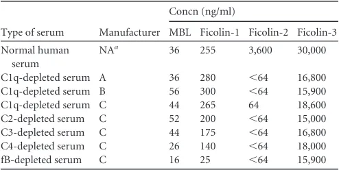
We used commercially available ELISA kits to determine the
presence of ficolin-2 and related opsonins (ficolin-1, ficolin-3, and
MBL) in C1q-depleted sera (
Table 1
). Similar to our binding
re-sults, we observed ficolin-2 at or below our detection limit (
ⱕ
64
ng/ml) in the C1q-depleted sera. Ficolin-1 and MBL were present
at concentrations similar to those in our NHS control. Ficolin-3
was present at concentrations lower than in our NHS control;
however, the concentration of ficolin-3 in the C1q-depleted sera
was similar to the median concentration previously reported for
adults (
16
).
In order to determine if a similar phenomenon occurred in
other commercially available complement component-depleted
sera (C2-, C3-, C4-, and factor B-depleted sera), we determined
the concentrations of ficolin-1, -2, and -3 and MBL in those sera
TABLE 1Opsonin concentrations in commercially available depleted sera
Type of serum Manufacturer
Concn (ng/ml)
MBL Ficolin-1 Ficolin-2 Ficolin-3
Normal human serum
NAa 36 255 3,600 30,000
C1q-depleted serum A 36 280 ⬍64 16,800
C1q-depleted serum B 56 300 ⬍64 15,900
C1q-depleted serum C 44 265 64 18,600
C2-depleted serum C 52 200 ⬍64 15,000
C3-depleted serum C 44 175 ⬍64 16,800
C4-depleted serum C 26 140 ⬍64 18,000
fB-depleted serum C 16 25 ⬍64 15,900
aNA, not applicable.
Brady et al.
on August 17, 2020 by guest
http://cvi.asm.org/
(
Table 1
). We found that all complement component-depleted
sera had little to no detectable ficolin-2 (below our detection limit
of 64 ng/ml). However, ficolin-1, ficolin-3, and MBL were still
present in all of these sera. The factor B-depleted sera had lower
levels of ficolin-1, ficolin-3, and MBL than did NHS. Similar to the
C1q-depleted sera, all of the depleted sera had lower levels of
fico-lin-3 than our NHS control; again, however, they were no
differ-ent than the previously reported median concdiffer-entration (
16
).
Complement component-depleted sera are codepleted of
fi-colin-2.
We previously found that plastic blood collection tubes
contain a ficolin-2-specific inhibitor that sequesters ficolin-2,
ef-fectively inhibiting ficolin-2 binding to pneumococci (
15
). To
in-vestigate if C1q-depleted sera inhibited ficolin-2 binding, we
mixed rFicolin-2 supernatant with C1q-depleted sera and studied
ficolin-2 binding to serotype 11A bacteria using flow cytometry.
Mixing of medium alone with C1q-depleted sera did not show any
ficolin-2 binding, as expected (
Fig. 2A
). Incubation of bacteria
with rFicolin-2 supernatant and C1q-depleted sera (
Fig. 2C
)
showed binding similar to that with rFicolin-2 supernatant alone
(
Fig. 2B
). Thus, C1q-depleted serum does not inhibit ficolin-2
binding in our assay.
Though the inhibitor present in plastic blood collection tubes
affects detection of ficolin-2 by ELISA and flow cytometry,
seques-tered ficolin-2 is still visible by Western blotting (
15
). Therefore,
we assayed NHS and all of the depleted sera for ficolin-2 by
West-ern blotting to determine if the absence of ficolin-2 in the
com-plement component-depleted sera may be due to the type of tube
used during blood collection. Unlike what was observed with sera
collected using plastic blood collection tubes (
15
), Western
blot-ting revealed significantly reduced ficolin-2 compared to our NHS
control for all of the depleted sera (
Fig. 2D
). Taken together, these
data show that ficolin-2 is depleted in complement
component-depleted sera and is likely not due to the type of tube used during
blood collection.
C1q-depleted sera can be used to study MBL-mediated
com-plement deposition.
Upon binding, MBL and the ficolins activate
the lectin pathway of complement activation through the
associ-ation of MASPs. To investigate whether MASPs were still present
in the C1q-depleted sera at concentrations sufficient to support
lectin pathway of complement activation, we investigated
MBL-initiated C3 and C4 deposition on yeast using C1q-depleted sera
or NHS. We observed a high degree of C3 and C4 deposition on
yeast incubated in all three C1q-depleted sera, and this deposition
was similar to C3 and C4 deposition observed on yeast incubated
in NHS (
Fig. 3A
). Addition of 100 mM
D-mannose, which
selec-tively inhibits MBL binding to yeast, inhibited C3 and C4
deposi-tion on yeast in all sera (
Fig. 3B
). Thus, functional MASPs are still
present in the C1q-depleted sera and are capable of activating
complement deposition.
C1q-depleted serum supplemented with recombinant
fico-lin-2 mediates complement deposition on
wcjE
-containing
se-rotypes of
S. pneumoniae
.
Because MASPs appear to be present
in sufficient amounts to activate complement, we next questioned
whether rFicolin-2-opsonized bacteria could mediate
comple-ment deposition in C1q-depleted sera. We have recently shown
that ficolin-2 binds to many pneumococcal serotypes that express
WcjE-mediated O-acetylation (Oac) on their capsular
polysac-charides, and loss of WcjE-mediated Oac in these serotypes leads
to loss of ficolin-2 binding (
3
). Moreover, we found that ficolin-2
mediates complement deposition on serotype 11A pneumococci
(which express WcjE-mediated Oac) but not on serotype 11E
pneumococci (which lack WcjE-mediated Oac) (
3
). Because these
previous studies were performed with only one
wcjE
-containing/
wcjE
-null pair (
3
), we studied ficolin-2-mediated complement
de-position on three other
wcjE
-containing/
wcjE
-null pairs
(sero-types 11D and 11Dx [SSISP 11D/1 and JC08, respectively],
serotypes 20A and 20Ax [AMB11 and AMB13, respectively], and
FIG 1C1q-depleted sera lack ficolin-2 but not related opsonins. Flow cytometry histograms show binding of ficolin-2 to serotype 11A pneumococci (A), MBL toSaccharomyces cerevisiae(B), and ficolin-1 to serotype 19C pneumococci (C) using 5% normal human sera (NHS) or C1q-depleted serum (C1q-dpl) from manufacturers A, B, and C.
on August 17, 2020 by guest
http://cvi.asm.org/
serotypes 20B and 20Bx [AMB12 and AMB14, respectively]) in
the absence of C1q.
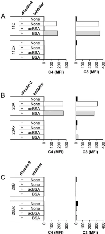
We first opsonized our bacteria with rFicolin-2 or supernatant
control and then incubated them in C1q-depleted sera to allow
MASP association and complement deposition to occur. We then
stained the bacteria for C3 and C4 deposition and analyzed them
using flow cytometry. We observed C3 and C4 deposition on
se-rotype 11D and 20A bacteria but little to no deposition on
sero-type 11Dx and 20Ax bacteria (
Fig. 4A
and
B
). Bacteria incubated
with supernatant control had no C4 and minimal C3 deposition.
Though serotype 20B contains WcjE-mediated Oac of its capsular
polysaccharide, we have previously shown that this serotype is not
bound by ficolin-2 (
3
). The structure of serotype 20B
polysaccha-ride differs from that of serotype 20A polysacchapolysaccha-ride by the
addi-tion of an extra glucose residue (
17
), which may sterically hinder
ficolin-2 recognition of WcjE-mediated O-acetylation on this
polysaccharide. Serotype 20Bx (
wcjE
-null variant of serotype 20B)
has a disrupted
wcjE
and is also not bound by ficolin-2 (data not
shown). Thus, serotypes 20B and 20Bx serve as good negative
controls for ficolin-2-mediated complement deposition, as
nei-ther is bound by ficolin-2. Accordingly, we observed little to no C3
deposition and no C4 deposition on either serotype 20B or 20Bx
FIG 2Complement component-depleted sera are codepleted of ficolin-2. (A to C) Flow cytometry histograms show ficolin-2 binding to serotype 11A pneumococci in 5% C1q-depleted sera with medium control (A), rFicolin-2 supernatant (B), or C1q-depleted sera with rFicolin-2 supernatant (C). Shaded gray areas represent staining with secondary antibody only as a negative control. (D) Western blot for ficolin-2 in normal human sera and depleted sera. Lane 1, NHS; lane 2, C1q-depleted sera from manufacturer A (C1q A); lane 3, C1q-depleted sera from manufacturer B (C1q B); lane 4, C1q-depleted sera from manufacturer C (C1q C); lane 5, C4-depleted sera from manufacturer C (C4); lane 6, C3-depleted sera from manufacturer C (C3); lane 7, C2-depleted sera from manufacturer C (C2); lane 8, factor B-depleted sera from manufacturer C (fB).
FIG 3C1q-depleted sera can activate complement through MBL and therefore still contain MASPs. Shown are flow cytometry dot plots of C4 (xaxis) and C3 (yaxis) deposition onS. cerevisiaeusing 5% normal human serum (NHS) or C1q-depleted serum (C1q-dpl) from manufacturers A, B, and C. (A) Complement deposition using serum without inhibitor. (B) Complement deposition using serum with 100 mMD-mannose to inhibit MBL binding.
Brady et al.
on August 17, 2020 by guest
http://cvi.asm.org/
following the addition of rFicolin-2 (
Fig. 4C
). We have previously
shown that acBSA inhibits ficolin-2 binding (
3
). As expected,
acBSA inhibited C3 and C4 deposition on serotypes 11D and 20A,
while BSA alone did not affect complement deposition (
Fig. 4A
and
B
).
C1q-depleted sera supplemented with lysine-57 ficolin-2
mutants can be used to study the lectin pathway of complement.
Ficolin-2 is known to associate with MASP-1, -2, and -3 through
the lysine-57 (K57) residue of its collagen-like domain, and
mu-tation of this residue alters interaction with the MASPs (
18
).
Mu-tation of K57 to an alanine or glutamic acid strongly inhibits the
interaction of ficolin-2 with MASP-1, -2, and -3 and impaired
lectin complement pathway activation. Mutation of K57 to an
arginine abolished interaction of ficolin-2 with MASP-2 but
al-lowed MASP-1 and -3 to associate (
18
). To investigate the role of
MASPs during ficolin-2-mediated complement deposition in the
C1q-depleted sera, we performed complement deposition
exper-iments using ficolin-2 mutants.
We opsonized serotype 11D and 11Dx bacteria with wild-type
or either mutant form of rFicolin-2 (K57E or K57R) and then
incubated them in C1q-depleted sera. As previously seen (
Fig. 4
),
serotype 11D and 11Dx bacteria incubated with medium only had
little to no complement deposition, indicating that C1q-depleted
serum alone did not activate complement (
Fig. 5
). Also,
opsoniza-tion of serotype 11D with wild-type rFicolin-2 mediated C4 and
C3 deposition that could be inhibited by acBSA but not BSA (
Fig.
5A
). Accordingly, opsonization of serotype 11D bacteria with the
rFicolin-2 K57E mutant, which should not associate with any
MASPs, did not mediate complement deposition. Similarly,
op-sonization of serotype 11D bacteria with the K57R mutant, which
should associate only with MASP-1 and MASP-3, did not mediate
complement deposition (
Fig. 5A
). No C4 deposition and little to
no C3 deposition were observed on serotype 11Dx bacteria
fol-lowing opsonization with any of the rFicolin-2 forms (
Fig. 5B
).
FIG 4Complement deposition using C1q-depleted sera supplemented with rFicolin-2. Shown is complement deposition on wcjE-containing/
wcjE-null isogenic strain pairs in 5% C1q-depleted serum following op-sonization with rFicolin-2 in the absence or presence of inhibitors: acBSA (10 mg/liter) or BSA (10 mg/liter). C4 (left column) and C3 (right column) deposition on serotypes 11D (wcjEcontaining) and 11Dx (wcjEnull) (A), 20A (wcjEcontaining) and 20Ax (wcjEnull) (B), and 20B (wcjEcontaining) and 20Bx (wcjEnull) (C).
FIG 5Complement deposition using C1q-depleted sera supplemented with rFicolin-2 lysine-57 mutants. Shown is deposition of C4 (left) and C3 (right) on serotypes 11D (wcjEcontaining) (A) and 11Dx (wcjEnull) (B) in 5% C1q-depleted serum following opsonization with medium control, wild-type rFicolin-2 (WT rFicolin-2), or mutant rFicolin-2 (K57E or K57R) in the ab-sence or preab-sence of inhibitors acBSA (10 mg/liter) and BSA (10 mg/liter).
on August 17, 2020 by guest
http://cvi.asm.org/
DISCUSSION
Studying the lectin pathway of complement activation can prove
difficult, as it shares common proteins with the classical pathway.
Previous studies have used sera from persons or mice deficient in
specific complement pathway components (
4
,
19
). However,
clinically deficient serum samples are not readily available, and
deficient mouse sera are not a good surrogate, as mouse lectin
pathway components evolved separately from humans (
20
). Thus,
C1-depleted serum should be very useful for separating the
hu-man classical and lectin pathways. However, we show here that
C1q-depleted sera also lack ficolin-2.
Manufacturer labeling of these depleted sera suggests that the
lectin pathway of complement activation is still intact. It is unclear
what specific methods companies use to deplete the sera of specific
complement components; however, we speculate that they use
some form of affinity chromatography. Interestingly, ficolin-2 has
been previously found to bind to cyanogen bromide-activated
Sepharose beads (
6
); thus, it is possible that this may be the reason
ficolin-2 is codepleted. We did find that the MBL-mediated lectin
pathway was still functional in these sera, and presumably the
ficolin-1 and ficolin-3 pathways are still intact, as their effector
proteins are still present. However, we found that the
ficolin-2-mediated lectin pathway is not functional, as these sera lack
fico-lin-2. Moreover, it is unclear if these complement
component-depleted sera are also deficient in complement regulators, like C1
inhibitor (
21
). Thus, manufacturers should improve their
prod-uct labeling. Researchers should be cautious when using these
reagents, as their protein of interest or related proteins may have
been unexpectedly removed. Also, previous studies performed
us-ing these sera should be reevaluated, as their conclusions were
drawn under the assumption that these depleted sera were
suffi-cient in all other components necessary for complement
activa-tion.
We have shown that complement component-depleted sera
are also codepleted of ficolin-2 but not other related opsonins, like
MBL, ficolin-1, and ficolin-3. Thus, these depleted sera (C2-, C3-,
C4-, and factor B-depleted sera) provide a unique tool for
study-ing ficolin-2-mediated complement pathways, as the other
neces-sary factors (i.e., MASPs) appear to be present. For instance,
C2-depleted sera may be used to determine whether ficolin-2 can
mediate the C2 bypass pathway like MBL (
22
,
23
), or factor
B-de-pleted sera could be used to determine the relative contribution of
the alternative pathway during ficolin-2-mediated complement
activation.
Activation of MASPs is required for ficolin-2-mediated
com-plement deposition to occur. Previous studies have shown that
MASP-2 is activated by MASP-1 (
24
,
25
). Following activation,
MASP-2 can cleave C4 and C2 to generate the C3 convertase
(con-sisting of C4bC2a) (
11
). MASP-1 and MASP-3 have been
sug-gested to play a role in alternative pathway activation (
26
).
How-ever, others reported that neither MASP-1 nor MASP-3 is
necessary for alternative pathway activation (
27
). We observed no
C3 activation on whole live bacteria with our K57R mutant, which
should be able to associate with MASP-1 and MASP-3 but not
MASP-2 (
18
), in C1q-depleted sera by flow cytometry (
Fig. 5
).
Although additional studies should be performed, we speculate,
based on our findings (
Fig. 5
), that MASP-1/-3 association is not
sufficient to activate the alternative pathway. Ficolin-2 is also
known to associate with both calreticulin and CD91 through the
same residue as the MASPs (
18
,
28
), and the interaction of
ficolin-2 and CD91 can mediate clearance of apoptotic cells (
28
). Moreover,
calreticulin and CD91 are also known to associate and function as a
cell receptor (
29
,
30
). Thus, complement component-depleted sera
along with rFicolin-2 mutants may be useful tools to study ficolin-2
complement pathways or ficolin-2-mediated phagocytosis (via
calre-ticulin and/or CD91).
ACKNOWLEDGMENTS
This work was supported by the National Institutes of Health (grant num-bers T32 AI007051 to A.M.B. and R56 AI31473 to M.H.N.).
We declare no competing financial interests.
REFERENCES
1.Matsushita M.2010. Ficolins: complement-activating lectins involved in innate immunity. J. Innate Immun.2:24 –32.http://dx.doi.org/10.1159 /000228160.
2.Krarup A, Sorensen UB, Matsushita M, Jensenius JC, Thiel S.2005. Effect of capsulation of opportunistic pathogenic bacteria on binding of the pattern recognition molecules mannan-binding lectin, L-ficolin, and H-ficolin. Infect. Immun.73:1052–1060.http://dx.doi.org/10.1128/IAI .73.2.1052-1060.2005.
3.Brady AM, Calix JJ, Yu J, Geno KA, Cutter GR, Nahm MH.2014. Low invasiveness of pneumococcal serotype 11A is linked to ficolin-2 recogni-tion of O-acetylated capsule epitopes and lectin complement pathway activation. J. Infect. Dis.http://dx.doi.org/10.1093/infdis/jiu195. 4.Ali YM, Lynch NJ, Haleem KS, Fujita T, Endo Y, Hansen S, Holmskov
U, Takahashi K, Stahl GL, Dudler T, Girija UV, Wallis R, Kadioglu A, Stover CM, Andrew PW, Schwaeble WJ.2012. The lectin pathway of complement activation is a critical component of the innate immune re-sponse to pneumococcal infection. PLoS Pathog.8:e1002793.http://dx .doi.org/10.1371/journal.ppat.1002793.
5.Aoyagi Y, Adderson EE, Rubens CE, Bohnsack JF, Min JG, Matsushita M, Fujita T, Okuwaki Y, Takahashi S.2008. L-Ficolin/mannose-binding lectin-associated serine protease complexes bind to group B streptococci primarily through N-acetylneuraminic acid of capsular polysaccharide and activate the complement pathway. Infect. Immun.76:179 –188.http: //dx.doi.org/10.1128/IAI.00837-07.
6.Kilpatrick DC, Chalmers JD.2012. Human L-ficolin (ficolin-2) and its clinical significance. J. Biomed. Biotechnol.2012:138797.http://dx.doi .org/10.1155/2012/138797.
7.Wan QQ, Ye QF, Zhou JD.2013. Mannose-binding lectin 2 and ficolin-2 gene polymorphisms influence the susceptibility to bloodstream infec-tions in kidney transplant recipients. Transplant. Proc.45:3289 –3292. http://dx.doi.org/10.1016/j.transproceed.2013.05.008.
8.Jeannin P, Jaillon S, Delneste Y.2008. Pattern recognition receptors in the immune response against dying cells. Curr. Opin. Immunol.20:530 – 537.http://dx.doi.org/10.1016/j.coi.2008.04.013.
9.Jensen ML, Honore C, Hummelshoj T, Hansen BE, Madsen HO, Garred P.2007. Ficolin-2 recognizes DNA and participates in the clear-ance of dying host cells. Mol. Immunol.44:856 – 865.http://dx.doi.org/10 .1016/j.molimm.2006.04.002.
10. Halmos A, Rigo J, Jr., Szijarto J, Fust G, Prohaszka Z, Molvarec A.2012. Circulating ficolin-2 and ficolin-3 in normal pregnancy and pre-eclampsia. Clin. Exp. Immunol.169:49 –56.http://dx.doi.org/10.1111/j .1365-2249.2012.04590.x.
11. Kjaer TR, Thiel S, Andersen GR.2013. Toward a structure-based com-prehension of the lectin pathway of complement. Mol. Immunol.56:413– 422.http://dx.doi.org/10.1016/j.molimm.2013.05.007.
12. Walport MJ.2001. Complement. Second of two parts. N. Engl. J. Med.
344:1140 –1144.http://dx.doi.org/10.1056/NEJM200104123441506. 13. Calix JJ, Saad JS, Brady AM, Nahm MH.2012. Structural
characteriza-tion ofStreptococcus pneumoniaeserotype 9A capsule polysaccharide re-veals role of glycosyl 6-O-acetyltransferasewcjEin serotype 9V capsule biosynthesis and immunogenicity. J. Biol. Chem.287:13996 –14003.http: //dx.doi.org/10.1074/jbc.M112.346924.
14. Calix JJ, Brady AM, Du VY, Saad JS, Nahm MH.2014. Spectrum of pneumococcal serotype 11A variants results from incomplete loss of cap-sule O-acetylation. J. Clin. Microbiol.52:758 –765.http://dx.doi.org/10 .1128/JCM.02695-13.
Brady et al.
on August 17, 2020 by guest
http://cvi.asm.org/
15. Brady AM, Spencer BL, Falsey AR, Nahm MH.2014. Blood collection tubes influence serum ficolin-1 and ficolin-2 levels. Clin. Vaccine Immu-nol.21:51–55.http://dx.doi.org/10.1128/CVI.00607-13.
16. Sallenbach S, Thiel S, Aebi C, Otth M, Bigler S, Jensenius JC, Schlapbach LJ, Ammann RA.2011. Serum concentrations of lectin-pathway components in healthy neonates, children and adults: mannan-binding lectin (MBL), M-, L-, and H-ficolin, and MBL-associated serine protease-2 (MASP-2). Pediatr. Allergy Immunol.22:424 – 430.http://dx .doi.org/10.1111/j.1399-3038.2010.01104.x.
17. Calix JJ, Porambo RJ, Brady AM, Larson TR, Yother J, Abeygunward-ana C, Nahm MH.2012. Biochemical, genetic, and serological character-ization of two capsule subtypes amongStreptococcus pneumoniaeserotype 20 strains: discovery of a new pneumococcal serotype. J. Biol. Chem.287:
27885–27894.http://dx.doi.org/10.1074/jbc.M112.380451.
18. Lacroix M, Dumestre-Perard C, Schoehn G, Houen G, Cesbron JY, Arlaud GJ, Thielens NM.2009. Residue Lys57 in the collagen-like region of human L-ficolin and its counterpart Lys47 in H-ficolin play a key role in the interaction with the mannan-binding lectin-associated serine pro-teases and the collectin receptor calreticulin. J. Immunol.182:456 – 465. http://dx.doi.org/10.4049/jimmunol.182.1.456.
19. Ma YJ, Doni A, Hummelshoj T, Honore C, Bastone A, Mantovani A, Thielens NM, Garred P.2009. Synergy between ficolin-2 and pentraxin 3 boosts innate immune recognition and complement deposition. J. Biol. Chem.284:28263–28275.http://dx.doi.org/10.1074/jbc.M109.009225. 20. Endo Y, Liu Y, Kanno K, Takahashi M, Matsushita M, Fujita T.2004.
Identification of the mouse H-ficolin gene as a pseudogene and orthology between mouse ficolins A/B and human L-/M-ficolins. Genomics84:737– 744.http://dx.doi.org/10.1016/j.ygeno.2004.07.006.
21. Keizer MP, Kamp AM, Brouwer N, van de Wetering MD, Wouters D, Kuijpers TW.2014. Plasma-derived mannose-binding lectin shows a di-rect interaction with C1-inhibitor. Mol. Immunol.58:187–193.http://dx .doi.org/10.1016/j.molimm.2013.11.022.
22. Tateishi K, Matsushita M.2011. Activation of the alternative comple-ment pathway by mannose-binding lectin via a C2-bypass pathway. Mi-crobiol. Immunol. 55:817– 821. http://dx.doi.org/10.1111/j.1348-0421 .2011.00378.x.
23. Selander B, Martensson U, Weintraub A, Holmstrom E, Matsushita M,
Thiel S, Jensenius JC, Truedsson L, Sjoholm AG.2006. Mannan-binding lectin activates C3 and the alternative complement pathway without in-volvement of C2. J. Clin. Invest.116:1425–1434.http://dx.doi.org/10.1172 /JCI25982.
24. Takahashi M, Iwaki D, Kanno K, Ishida Y, Xiong J, Matsushita M, Endo Y, Miura S, Ishii N, Sugamura K, Fujita T. 2008. Mannose-binding lectin (MBL)-associated serine protease (MASP)-1 contributes to activation of the lectin complement pathway. J. Immunol.180:6132– 6138.http://dx.doi.org/10.4049/jimmunol.180.9.6132.
25. Møller-Kristensen M, Thiel S, Sjoholm A, Matsushita M, Jensenius JC.
2007. Cooperation between MASP-1 and MASP-2 in the generation of C3 convertase through the MBL pathway. Int. Immunol.19:141–149. 26. Iwaki D, Kanno K, Takahashi M, Endo Y, Matsushita M, Fujita T.2011.
The role of mannose-binding lectin-associated serine protease-3 in acti-vation of the alternative complement pathway. J. Immunol.187:3751– 3758.http://dx.doi.org/10.4049/jimmunol.1100280.
27. Degn SE, Jensen L, Hansen AG, Duman D, Tekin M, Jensenius JC, Thiel S.2012. Mannan-binding lectin-associated serine protease (MASP)-1 is cru-cial for lectin pathway activation in human serum, whereas neither MASP-1 nor MASP-3 is required for alternative pathway function. J. Immunol.189:
3957–3969.http://dx.doi.org/10.4049/jimmunol.1201736.
28. Duus K, Thielens NM, Lacroix M, Tacnet P, Frachet P, Holmskov U, Houen G.2010. CD91 interacts with mannan-binding lectin (MBL) through the MBL-associated serine protease-binding site. FEBS J.277:
4956 – 4964.http://dx.doi.org/10.1111/j.1742-4658.2010.07901.x. 29. Ogden CA, deCathelineau A, Hoffmann PR, Bratton D, Ghebrehiwet B,
Fadok VA, Henson PM.2001. C1q and mannose binding lectin engage-ment of cell surface calreticulin and CD91 initiates macropinocytosis and uptake of apoptotic cells. J. Exp. Med.194:781–795.http://dx.doi.org/10 .1084/jem.194.6.781.
30. Vandivier RW, Ogden CA, Fadok VA, Hoffmann PR, Brown KK, Botto M, Walport MJ, Fisher JH, Henson PM, Greene KE.2002. Role of surfactant proteins A, D, and C1q in the clearance of apoptotic cells in vivo and in vitro: calreticulin and CD91 as a common collectin receptor com-plex. J. Immunol. 169:3978 –3986.http://dx.doi.org/10.4049/jimmunol .169.7.3978.