Measure Neutralizing Antibodies against
Clostridium difficile
Binary
Toxin
Jinfu Xie,aMelanie Horton,aJulie Zorman,aJoseph M. Antonello,bYuhua Zhang,bBeth A. Arnold,cSusan Secore,a
Rachel Xoconostle,aMatthew Miezeiewski,eSu Wang,aColleen E. Price,dDavid Thiriot,dAaron Goerke,d*Marie-Pierre Gentile,d* Julie M. Skinner,aJon H. Heinrichsa
Departments of Vaccine Research,aNon-Clinical Statistics,bVaccine Clinical Assays,cand BioProcess Development,dMerck Research Laboratories, West Point, Pennsylvania, USA; Eurofins Laboratories, Lancaster, Pennsylvania, USAe
Clostridium difficile
strains producing binary toxin, in addition to toxin A (TcdA) and toxin B (TcdB), have been associated with
more severe disease and increased recurrence of
C. difficile
infection in recent outbreaks. Binary toxin comprises two subunits
(CDTa and CDTb) and catalyzes the ADP-ribosylation of globular actin (G-actin), which leads to the depolymerization of
fila-mentous actin (F-actin) filaments. A robust assay is highly desirable for detecting the cytotoxic effect of the toxin and the
pres-ence of neutralizing antibodies in animal and human sera to evaluate vaccine efficacy. We describe here the optimization, using
design-of-experiment (DOE) methodology, of a high-throughput assay to measure the toxin potency and neutralizing antibodies
(NAb) against binary toxin. Vero cells were chosen from a panel of cells screened for sensitivity and specificity. We have
success-fully optimized the CDTa-to-CDTb molar ratio, toxin concentration, cell-seeding density, and sera-toxin preincubation time in
the NAb assay using DOE methodology. This assay is robust, produces linear results across serial dilutions of hyperimmune
se-rum, and can be used to quantify neutralizing antibodies in sera from hamsters and monkeys immunized with
C. difficile
binary
toxin-containing vaccines. The assay will be useful for
C. difficile
diagnosis, for epidemiology studies, and for selecting and
opti-mizing vaccine candidates.
C
lostridium difficile
is a Gram-positive bacterium and the
pri-mary cause of hospital-acquired diarrhea.
C. difficile
infection
(CDI) occurs when antibiotic treatment disrupts the normal
bac-terial flora of the intestine, which allows for the colonization of
C.
difficile
bacteria. CDI is characterized by symptoms that range
from mild diarrhea to severe and often life-threatening colitis. The
incidence of more severe CDI has been increasing in recent years
due to the emergence of epidemic hypervirulent strains (1,
2).
Two glucosylating toxins, TcdA and TcdB, are considered the
main virulence factors of
C. difficile
. These two toxins catalyze the
glucosylation of proteins RhoA, Rac, and Cdc42, which leads to
the disruption of actin filaments and collapse of the cell
cytoskel-eton, disruption of other signaling pathways, and eventually, cell
death (3–5). In addition to TcdA and TcdB, hypervirulent strains
have been found to express a binary toxin referred to as
C. difficile
transferase (CDT). Much of what we know of CDT was learned
through a comparison to related binary toxins found in other
clostridial species, such as
Clostridium botulinum
C2 toxin and
Clostridium perfringens
iota toxin (6–8). Binary toxins are
charac-terized as having an enzymatically active “A” component that
causes ADP-ribosylation of globular actin (G-actin) and a cell
binding and translocation “B” component. For
C. difficile
, CDTa
and CDTb are transcribed monocistronically but translated as two
separate proteins. The proposed mechanism of action for the
C.
difficile
binary toxin has been described in the literature (7).
Briefly, the precursor CDTb is cleaved by trypsin, and the
subse-quently activated CDTb protein binds to the cell surface receptor
as either a preformed heptamer or as monomers that subsequently
oligomerize to form heptamers. CDTa binds to the CDTb
hep-tamer on the cell surface. The receptor-binary-toxin complex is
then internalized via receptor-mediated endocytosis into the
en-dosomes. In the acidic environment in the endosome, the CDTb
heptamer then forms a pore, which allows the CDTa enzymatic
component to enter the cell. The CDTa component catalyzes the
ADP-ribosylation of G-actin at Arg
177and prevents actin
polym-erization, causing disruption of the cytoskeleton and fluid loss,
rounding of the cells, and eventually, cell death (8,
9). The cell
surface receptor for
C. difficile
binary toxin has been identified as
lipolysis-stimulated lipoprotein receptor (LSR), which is also the
receptor for two other binary toxins,
C. perfringens
iota toxin and
Clostridium spiroforme
toxin (CST) (10).
Although the exact role of binary toxin in CDI is not clear,
there is increasing evidence of a correlation between CDI severity
and
C. difficile
strains expressing binary toxin. In studies using a
binary toxin-containing TcdA-negative TcdB-negative strain,
re-searchers have shown that binary toxin alone can cause fluid
ac-cumulation in rabbit ileal loop assays and that this strain can
col-Received21 January 2014 Returned for modification13 February 2014
Accepted3 March 2014
Published ahead of print12 March 2014
Editor:R. L. Hodinka
Address correspondence to Jon H. Heinrichs, [email protected]. * Present address: Aaron Goerke and Marie-Pierre Gentile, Genentech, Inc., South San Francisco, California, USA.
J.X. and M.H. contributed equally to the article.
Supplemental material for this article may be found athttp://dx.doi.org/10.1128
/CVI.00038-14.
Copyright © 2014, American Society for Microbiology. All Rights Reserved.
doi:10.1128/CVI.00038-14
on August 17, 2020 by guest
http://cvi.asm.org/
onize hamsters; however, binary toxin alone is not sufficient to
cause disease (11). However, it is postulated that binary toxin
enhances the pathogenesis of
C. difficile
by inducing the
redistri-bution and protrusion of microtubules at intestinal epithelial cell
surfaces, thereby increasing bacterial adherence and colonization
(12). This increase in pathogenesis is supported by
epidemiolog-ical studies showing higher fatality rates in patients infected with
binary toxin-containing strains versus patients infected with
strains that do not produce binary toxin (13,
14). Binary toxin has
also been found to be associated with hypervirulent strains of
C.
difficile
that cause increased CDI severity, higher death rates,
lon-ger hospital stays, and increased recurrence of CDI (1,
2,
15).
However, epidemiological studies also have been performed that
suggest that binary toxin is not a significant predictor of severe
CDI (16,
17).
The typical treatment regimen for CDI is discontinuation of
the antibiotic treatment, which alters the microbiota of the gut,
followed by treatment with a narrow-spectrum antibiotic that
specifically targets the
C. difficile
bacterium (18,
19). More
re-cently, monoclonal antibodies to
C. difficile
toxins A and B have
been shown to prevent CDI onset and reduce the rate of
recur-rence of disease (20). In addition, immunization with TcdA and
TcdB toxins that have been either inactivated recombinantly or by
chemical methods have shown efficacy in preventing disease (21,
22). The efficacy of these vaccines is presumably due to the
pro-duction of antibodies that can neutralize the activities of these
toxins. We have developed a vaccine containing binary toxin in
addition to TcdA and TcdB, which may increase the protective
efficacy of this vaccine for hypervirulent strains expressing binary
toxin (23). Vaccine development requires a functional assay that
can measure neutralizing antibody responses against binary toxin
in animal models and clinical trials. We describe here the
devel-opment and optimization of an assay to measure the cytotoxicity
of
C. difficile
binary toxin and an assay to test the ability of serum
to neutralize this toxin.
MATERIALS AND METHODS
Binary toxin constructs (C. difficileNAP1 strain [GenBank accession no. R20291] sequences) CDTa, 1mCDTa (C2A), glutathioneS-transferase
(GST)_ProCDTb, and CDTb were expressed inEscherichia coli(Table 1). CDTa, 1mCDTa, and GST_ProCDTb were used as reagents in the binary toxin functional assay. The binary toxin constructs 3mCDTa (S345F,
E385Q, E387Q) and ProCDTb were expressed in insect cell expression
sys-tems to improve the yield and reduce the endotoxin level for vaccine products. The three mutations in 3mCDTa were introduced to reduce the enzymatic activity of binary toxin (24). We optimized the binary toxin cytotoxicity assay first. Based on the result, we then optimized the neu-tralization antibody (NAb) assay using control serum samples pooled from monkeys immunized with insect cell-expressed binary toxin.
Fur-thermore, we demonstrated that the optimized NAb assay can be used to measure the neutralizing antibody response in the serum samples of ham-sters immunized withE. coli-expressed binary toxins. The methods of expression, purification, and SDS-PAGE analysis of these toxins can be found in the supplemental material.
The antigens 5mTcdA (W101A, D287A, E514Q, W519A, C700A) and
5mTcdB (W102A, D288A, E515Q, W520A, C698A) used in vaccine
prepara-tion were also expressed in insect cells. The details of expression and purification can be found in International Patent Application publication no. WO2013112867 (23).
Rhesus macaque immunization.The antigens 5mTcdA, 5mTcdB, 3mCDTa, and ProCDTb were used in the vaccine. The 5mTcdB antigen was treated with formaldehyde, followed by dialysis in 50 mM HEPES buffer (pH 7.0, 100 mM NaCl) and sterile filtration (0.22m). Vaccine formulation then involved mixing aluminum adjuvant, antigens, and Iscomatrix adjuvant (CSL Biotherapies, Inc., King of Prussia, PA). Adult rhesus macaques (4 to 10 years of age) were housed at the New Iberia Research Center (NIRC) of the University of Louisiana at Lafayette. They were immunized intramuscularly with 0.5 ml of the vaccine preparations containing 20g of 5mTcdA, 20g of 5mTcdB, 5g of 3mCDTa, and 5
g of ProCDTb in the deltoid muscle on days 0, 7, and 30. Blood samples were collected before each immunization and on day 45. The animals were observed daily for any abnormal clinical signs of illness or distress. Twenty milliliters of hyperimmune serum collected on day 45 from two monkeys was pooled and used as a reagent for assay development. The experimental protocol was approved by the Institutional Animal Care and Use Com-mittee (IACUC) at both Merck & Co., Inc. and the NIRC, and the study was carried out at the NIRC.
Golden Syrian hamster immunization. The antigens 5mTcdA, 5mTcdB, CDTa, and CDTb were used in the vaccine. All four antigens were treated with formaldehyde. Excess formaldehyde was removed by dialysis in 50 mM HEPES buffer (pH 7.0, 100 mM NaCl). The antigens were then mixed with aluminum adjuvant and Iscomatrix adjuvant to produce the vaccines listed inTable 2. Golden Syrian hamsters (male, 90 to 120 g) were obtained from Charles River Laboratories and individually housed in filter-lid cages at Merck’s West Point site. The hamsters in groups 1 to 4 were immunized intramuscularly with 0.2 ml of vaccine, as described inTable 2. The hamsters in group 5 received adjuvant only. The hamsters were immunized on days 0, 21, 42, and 63, and approximately TABLE 1Description of recombinant binary toxin components used in this study
Toxin name Expression system Mutation(s) Expression tag Use in this study
CDTa E. coli None His tag at C terminus NAb assay, hamster immunization
1mCDTa E. coli C2A His tag at C terminus NAb assay
ProCDTb E. coli None GST tag at N terminusa NAb assay
CDTb E. coli None GST tag at N terminusb Hamster immunization
3mCDTa Baculovirus S345F, E385Q, E387Q No Monkey immunization
ProCDTb Baculovirus None No Monkey immunization
aGST_Pro domain was removed by chymotrypsin activation before use in the NAb assay. The activated protein is referred to as a_CDTb. b
GST domain was removed during the protein purification process.
TABLE 2Compositions of vaccines used for hamster immunization
Group
Dose (g/injection) for antigena:
5mTcdA 5mTcdB CDTa CDTb
1 10 10 0 0
2 10 10 0.6 0
3 10 10 0 4.4
4 10 10 0.6 4.4
a5mTcdA and 5mTcdB were immunized in the left leg; CDTa and CDTb were
immunized in the right leg. The hamsters in group 5 (negative-control group) were immunized with adjuvant alone.
on August 17, 2020 by guest
http://cvi.asm.org/
0.5 ml of blood was collected from each hamster via the retroorbital tech-nique immediately prior to each immunization and on day 77. The serum samples were stored in a frozen state. Ten hamsters were used in each group. The hamster experimental protocol was approved by the Institu-tional Animal Care and Use Committee (IACUC) at Merck and Co., Inc.
Activation of GST_ProCDTb.GST-ProCDTb was activated by diges-tion with immobilized chymotrypsin (Princeton Separadiges-tions, Inc., Princeton, NJ) at a 10:1 protein-to-chymotrypsin mass ratio. The GST_ ProCDTb-chymotrypsin mixture was incubated at 37°C with shaking (150 rpm) for 30 min. After incubation, the resin was removed by centrif-ugation for 2 min at 10,000⫻gat room temperature, and the activated GST_ProCDTb (a_CDTb) was collected and stored at⫺70°C until use.
Binary toxin cytotoxicity titration assay.The assay was run over a period of three consecutive days and comprised five steps: (i) cell plating, (ii) serial dilution of toxin and cell intoxication, (iii) cell staining, (iv) data acquisition, and (v) data analysis. For step 1, 50l of a cell suspension of Vero cells at 30,000 cells/ml was seeded into 384-well plates and incubated overnight in a humidified incubator with 5% CO2at 37°C. Vero cells
(ATCC CCL-81) were obtained from ATCC and cultured according to ATCC’s instructions. For step 2, binary toxin mixtures consisting of 1mCDTa and a_CDTb were serially diluted 2-fold in complete culture medium, and 40l was then applied to cells grown in 384-well plates. The cells were incubated in a CO2incubator at 37°C for 20, 24, or 48 h. For
steps 3 and 4, cell staining and data acquisition were performed according to the methods described previously (25). For step 5, the total cell surface area in each well was plotted against the toxin concentration in each dilu-tion. The potency of a test toxin (50% toxic concentration [TC50]) was
determined as the toxin concentration that caused 50% cytotoxicity. The TC50was calculated by linearly interpolating between the consecutive dilutions whose signals bracketed the midpoint signal. The midpoint sig-nal was defined as half of the total cell surface area of the medium-only control wells. For ease of comparison, we describe the TC50for binary
toxin preparations in terms of CDTa concentrations only.
A design-of-experiment methodology was used to further optimize four critical assay parameters, including GST_ProCDTb chymotrypsin activation time, cell-seeding density, toxin intoxication time, and the ratio of CDTa to a_CDTb in the binary toxin. Five cell-seeding densities (500, 750, 1,000, 1,500, and 2,000 cells/well), three toxin intoxication times (20, 24, and 48 h), the GST_ProCDTb chymotrypsin activation time (20, 30, and 40 min), and the CDTa-to-a_CDTb ratio (1:4, 1:7, and 1:10) were evaluated in the optimization experiment based on results from screening experiments. Cell-seeding densities of 1,000, 1,500, and 2,000 cells/well were evaluated at the 20-h and 24-h toxin intoxication times (six combi-nations), and cell-seeding densities of 500, 750, and 1,000 cells/well were evaluated at the 48-h toxin intoxication time (three combinations). The toxin intoxication time was chosen so the cells were between 50% and 90% confluence at the end of the assay. The alternative combinations of cell-seeding density and toxin incubation time resulted in nine 384-well plates. The experiment was performed independently by two analysts across these nine plates. Within each plate, the nine different combina-tions of chymotrypsin activation times (three levels) and CDTa-to-CDTb ratio (three levels) were each tested in two independently prepared 16-point 2-fold dilution series beginning at the 1:2 dilution. The remaining 6 columns of each plate contained a medium-only control and 3 different toxin control arms: CDTa only, a_CDTb only, and CDTa plus GST_ ProCDTb. The toxin control arms were each tested in a single 16-point 2-fold dilution series beginning at the 1:2 dilution.
Cell-based neutralization antibody assay.The NAb assay was run over a period of three consecutive days and comprised, with a few excep-tions, the same five-step procedure used in the cytotoxicity titration assay described above. The exceptions in steps 2 and 5 were that in step 2, 1mCDTa and a_CDTb were combined at a 1:7 CDTa-to-CDTb molar ratio using either 4-, 8-, or 16-fold TC50for 1mCDTa. The control and test
sera were 2-fold serially diluted, followed by incubation with the binary toxin mixture for either 0.5, 1, or 2 h at 37°C. This incubation time is
referred to as toxin preincubation. Forty microliters of the serum-toxin mixture was then applied to cells grown in 384-well plates and incubated in humidified CO2incubators at 37°C for 22 to 24 h. The cells
were fixed, permeabilized, and stained with Alexa Fluor 488 phalloidin, and an image of the monolayer was acquired using the ImageXpress Velos scanning cytometer (25). In step 5, the total cell surface area data were imported into a Microsoft Excel workbook to calculate the neutralization titer. The neutralization titer of a test sample (50% effective dose [ED50])
was defined as the dilution at which cytotoxicity was decreased by 50%. The ED50was calculated by linearly interpolating between the consecutive
dilutions whose signals bracketed the midpoint signal. The midpoint sig-nal was defined as the average of the total cell surface area of medium-only control wells (n⫽16) and the total cell surface area of the toxin-only control wells (n⫽16).
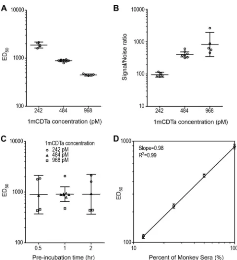
A design-of-experiment methodology was used to further optimize three critical assay parameters, including toxin concentration, cell-seed-ing density, and serum-toxin preincubation time. Three levels (low, me-dium, and high) were chosen for each factor based on preliminary data, cell-seeding density (1,000, 1,500, and 2,000 cells/well), preincubation time (0.5, 1, and 2 h) and toxin concentration (242, 484, and 968 pM). Throughout the testing, GST_ProCDTb chymotrypsin activation was performed for 30 min at 37°C, the molar ratio of CDTa to a_CDTb was 1:7, and the toxin incubation time was fixed at 22 h, based on the cytotox-icity DOE data. The Design-Expert version 7.1.3 software was used to generate a face-centered cube design consisting of 18 runs (one 384-well plate per run) performed in four blocks of four or five runs (see Table S1 in the supplemental material). An identical set of samples was tested on each of the 18 plates. The sample set included medium-only and toxin-only controls, a titration of the toxin, positive and negative hamster and human sera, low- and high-titer positive serum samples from vaccinated monkeys, and three samples created by spiking the high-positive monkey serum into a pool of negative human serum at 12.5%, 25%, and 50% spike levels. The monkey serum spiked into negative human serum was used to assess the matrix effect of human serum in the NAb assay. The toxin titration and the serum samples were each tested in a 16-point 2-fold titration series across the plate rows. The assay conditions were evaluated in terms of the signal-to-noise ratio (ratio of medium-only response to toxin-only response), the toxicity of the toxin (TC50), the level and
vari-ability of the NAb titers (ED50) for the human and monkey samples, and
the linearity of the NAb titers across the range of monkey spikes tested.
Statistical analysis of theC. difficilebinary toxin cytotoxicity or NAb assay optimization experiments.All analyses were performed on the natural log-transformed TC50or ED50data. Differences among the
factor levels in the cytotoxicity assay DOE were assessed using an anal-ysis of covariance (ANCOVA) model in which toxin incubation time and cell-seeding density were treated as categorical factors, and chy-motrypsin activation time and the ratio of CDTa to a_CDTb were treated as continuous factors. The differences among the factor levels in the NAb assay DOE were assessed using a multifactor analysis of variance (ANOVA) model containing all maeffect and two-way in-teraction terms. None of the two-way inin-teraction terms were signifi-cant at the 5% level, and therefore, the interaction terms were dropped from the final model. Statistical significance between the categorical factor levels was assessed based on the differences of the least-squares means.Pvalues of⬍0.05 were considered significant. The precision of the NAb titer was assessed by variance component analysis (VCA) across the set of positive-test and -control samples. All analyses were performed using the MIXED procedure in SAS (version 9.3).
RESULTS
Purification and characterization of CDTa and ProCDTb for
use in neutralizing antibody assays.
We initially sought to
ex-press both components of binary toxin as fully functional proteins
using recombinant production in
E. coli
. CDTa and 1mCDTa (49
kDa) were purified to a purity of
⬎
98% as estimated by
on August 17, 2020 by guest
http://cvi.asm.org/
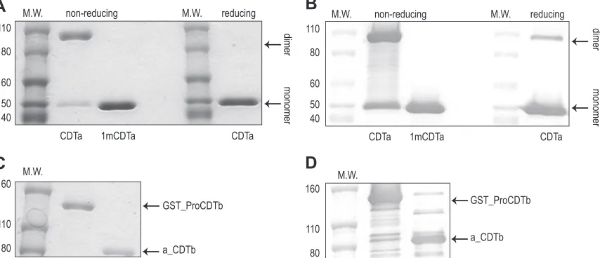
etry on SDS-PAGE gels. Although 80% of CDTa formed a dimer
on nonreduced gels, the CDTa dimer was reduced to a monomer
by adding dithiothreitol (DTT) to the SDS-PAGE loading buffer.
This suggested that the dimerization was a result of disulfide bond
formation, and in an attempt to alleviate this phenomenon, we
engineered a mutation in the molecule that resulted in a change at
position two of cysteine to alanine to yield 1mCDTa. This
muta-tion completely eliminated the propensity of the molecule to
dimerize. Both CDTa and 1mCDTa were recognized by an
anti-CDTa monoclonal antibody, as shown by Western blot (Fig. 1A
and
B), confirming the identities of these two proteins.
To facilitate the solubility of ProCDTb, this protein was
ex-pressed in
E. coli
as a fusion with glutathione
S
-transferase (GST).
This molecule, GST_ProCDTb (121 kDa), was purified to
⬎
95%,
as estimated by densitometry on SDS-PAGE gels. The GST_Pro
domain was removed by incubation with immobilized
chymo-trypsin for 30 min at 37°C (Fig. 1C), and the resulting protein is
referred to as a_CDTb (94 kDa) to indicate that it is in an active
form. Using electrospray ionization–time of flight (ESI-TOF)
mass spectrometry, the activation site of GST_ProCDTb was
identified to be the peptide bond between Met
169and Ser
170. Both
the GST_ProCDTb and a_CDTb were recognized by an
anti-CDTb monoclonal antibody (Fig. 1D), confirming their
identi-ties.
Measurement of binary toxin cytotoxicity.
Next, we
devel-oped a method to evaluate the function of the binary toxin
prep-arations in a Vero cell-based cytotoxicity assay. Vero cells were
used in the cytotoxicity assay, as they have been shown to be
sen-sitive to binary toxin (26). HT29, T84, Caco-2, HepG2, and
IMR-90 cells were also compared to Vero cells to determine
whether these cell lines were sensitive to binary toxin as well (see
supplemental material). Vero cells were selected for use in the
binary toxin NAb assay due to their favorable characteristics for
high-throughput assays, including rapid cell growth and low
vari-ation between cell passages (data not shown).
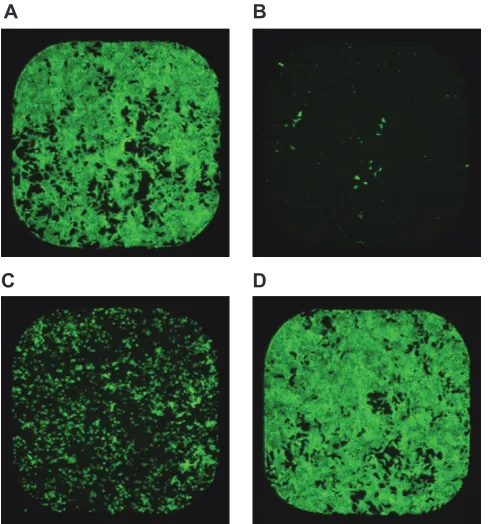
CDTa and a_CDTb need to be combined in a proper molar
ratio in order to have cytotoxic activity. Upon entry into the cell,
CDTa ADP-ribosylates G-actin and depolymerizes the
filamen-tous actin (F-actin) filament (24), and F-actin filaments can be
visualized using Alexa 488 phalloidin staining (Fig. 2A). Vero cells
that had been treated with binary toxin demonstrated
dramati-cally lower levels of F-actin staining (Fig. 2B). Preincubation of
binary toxin with hyperimmune monkey serum protected the
cells from the cytotoxic effect of the toxin (Fig. 2C
and
D),
sug-gesting that this toxicity was specific to binary toxin and was not a
result of nonspecific killing. The changes in F-actin filament
quan-tity can be determined in a rapid manner using an automated
Velos scanning cytometer. The total cell surface area was inversely
correlated with cytotoxicity and was used to calculate the TC
50values of the toxin molecules and the ED
50values of the immune
sera.
Binary toxin cytotoxicity assay optimization.
Several critical
factors were screened in the cytotoxicity assay, such as the
re-quired incubation time of GST_ProCDTb with chymotrypsin to
form a_CDTb, the equivalency of 1mCDTa and CDTa, the
opti-mal 1mCDTa-to-a_CDTb molar ratio, and the cell-seeding
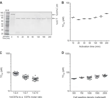
den-sity. GST_ProCDTb was activated by chymotrypsin for 10, 20, 30,
60, 120, 180, or 240 min, with a 10:1 protein-to-chymotrypsin
mass ratio. SDS-PAGE analysis revealed that the GST_Pro
do-main was fully cleaved following incubation with chymotrypsin
for
ⱖ
10 min (Fig. 3A). The TC
50of binary toxin was found to be
similar when GST_ProCDTb was activated for 20, 30, or 60 min.
However, the TC
50was slightly higher when GST_ProCDTb was
activated for 10, 120, 180, or 240 min (
⬍
2-fold higher,
Fig. 3B).
This was further confirmed by the DOE data showing that altering
the GST_ProCDTb activation time from 20 to 40 min had only a
minimal effect on the observed TC
50values (data not shown).
Next, 1mCDTa was evaluated to determine whether it had
ac-tivity similar to that of CDTa in the cytotoxicity assay. We found
that the TC
50for 1mCDTa was only 1.2-fold higher than that
obtained for CDTa, and therefore we used 1mCDTa in the assay to
ameliorate aggregation problems.
FIG 1Identification and characterization of purified CDTa and GST_ProCDTb. (A) SDS-PAGE of CDTa and 1mCDTa. Proteins were stained with Coomassie blue. (B) Western blot of CDTa and 1mCDTa using anti-CDTa monoclonal antibody. (C) SDS-PAGE of GST_ProCDTb and a_CDTb. Proteins were stained with Coomassie blue. (D) Western blot of GST_ProCDTb and a_CDTb using anti-CDTb monoclonal antibody. Some degradation of GST_ProCDTb was seen in the Western blot but not in the Coomassie blue-stained gel. Two micrograms of each protein was loaded onto the SDS-PAGE gels. M.W., molecular weight, in thousands.
on August 17, 2020 by guest
http://cvi.asm.org/
It has been proposed that CDTa binds to a heptamer of CDTb
on the cell surface (7). However, it is not clear how many CDTa
molecules bind to the heptamer of CDTb. Therefore, we evaluated
1mCDTa-to-a_CDTb molar ratios of 5:1, 1:1, 1:4, 1:7, and 1:10 by
altering the amount of a_CDTb while maintaining the 1mCDTa
concentration. The TC
50of 1mCDTa decreased from 1,150 pM at
the 5:1 molar ratio to 56 pM at the 1:7 molar ratio. Molar ratios of
1:7 and 1:10 had similar TC
50values for 1mCDTa (data not
shown). The DOE data confirmed that the TC
50of 1mCDTa
sig-nificantly decreased as the 1mCDTa-to-a_CDTb molar ratio
in-creased from 1:4 to 1:10 (Fig. 3C). A 1:7 molar ratio was selected
for the optimized assay because CDTa is predicted to bind to
a_CDTb heptamer on the cell surface.
Using the 1:7 molar ratio, the TC
50also increased from 44 pM
to 68 pM as the cell-seeding density increased from 500 cells/well
to 2,000 cells/well (Fig. 3D). Despite the marginally higher TC
50at
1,500 cells/well than that at 500 cells/well (1.3-fold), we chose
1,500 cells/well as the optimal density for the assay because it
re-duced the cell intoxication time from 48 h, as required for lower
cell-seeding densities, to 20 to 24 h. The total geometric coefficient
of variation was calculated to be 22.9% for the cytotoxicity assay.
The addition of individual toxin components (1mCDTa alone or
a_CDTb alone) or the combination of 1mCDTa and nonactivated
GST_ProCDTb to the cells did not result in observable toxicity at
concentrations 1,000-fold higher than the TC
50of the active
com-ponents in binary toxin (data not shown).
Optimization of binary toxin NAb assay using
design-of-ex-periment methodology.
Based on the optimized cytotoxicity
as-say, we further optimized the binary toxin NAb assay by
evaluat-ing the joint effects of binary toxin concentration, serum-toxin
preincubation time, and cell-seeding density using DOE
method-ology. The ED
50titer of a monkey control serum sample decreased
from 1,894 to 452 as the binary toxin concentration increased
from 242 pM to 968 pM (Fig. 4A). On the other hand, the
signal-to-noise ratio increased from 92 to 788 as the binary toxin
con-centration increased over this same range (Fig. 4B). To provide for
a suitably rugged assay and to strike an acceptable balance between
assay sensitivity and specificity, the optimized binary toxin
con-centration was selected to be 448 pM, which is the minimum
bi-nary toxin concentration required to cause 100% cytotoxicity.
Varying serum-toxin preincubation times from 30 to 120 min had
only a minimal effect on the ED
50titer (Fig. 4C). Changing the
cell-seeding density from 1,000 cells/well to 2,000 cells/well was
found to have little effect on the ED
50(data not shown).
There-fore, the midpoints of the serum-toxin preincubation times (60
min) and cell-seeding densities (1,500 cells/well) were chosen as
the optimal conditions for the NAb assay. The ED
50correlated
strongly with the concentration of monkey serum spiked into
nor-mal human serum (
R
2⫽
0.99,
Fig. 4D). The total geometric
coef-ficient of variation was 5.3% for the NAb assay.
Measurement of neutralizing antibody titers in hamster
se-rum.
The Syrian hamster challenge model is the most frequently
utilized model for studying
C. difficile
infection. We evaluated the
ability of a vaccine containing formaldehyde-detoxified 5mTcdA
and 5mTcdB with either CDTa, CDTb, or both binary toxin
com-ponents to induce serum antibodies with neutralizing activity. A
method for measuring the NAb titer against TcdA and TcdB was
described previously (25). Here, we determined the NAb titer that
was specific for binary toxin. There were no detectable
neutraliz-ing antibodies for binary toxin in the adjuvant group and the
group that received 5mTcdA and 5mTcdB preparations only.
However, a neutralizing antibody titer was detectable postdose 3
with a vaccine containing CDTa or postdose 2 with a vaccine
con-taining either CDTb or both binary toxin components. The
com-bination of CDTa and CDTb produced a more potent neutralizing
antibody response than that from either protein alone (Fig. 5A).
In addition, we compared the imaging-based assay with the
traditional visual endpoint assay, which relies on microscopic
ex-amination of cell rounding following the intoxication of cell lines.
The ED
50determined using the method employing the scanning
cytometer correlated closely with the ED
50obtained using the
mi-croscopic cytotoxicity determination (
R
2⫽
0.96,
Fig. 5B).
DISCUSSION
Binary toxin-expressing
C. difficile
strains have emerged recently
as the cause of severe outbreaks of CDI. Among these, strains of
the NAP1 type accounted for 67 to 82% of the clinical isolates
identified during CDI outbreaks in Montreal, Canada in 2003 and
2004 (27), and 61% of all clinical isolates from 25 acute health care
facilities in Chicago in 2009 (28). Goldenberg and colleagues (29)
showed that patients infected with
C. difficile
strains containing
binary toxin not only had significantly higher peripheral white
FIG 2Measurement of binary toxin cytotoxicity of Vero cells in a 384-wellplate using an ImageXpress Velos scanning cytometer. Shown are the repre-sentative well images of Vero cells treated with different reagents. (A) Medium only (100%); (B) binary toxin consisting of 484 pM of CDTa with a 1:7 CDTa-to-a_CDTb molar ratio (1%). F-actin filaments were completely depolymer-ized by the binary toxin. (C) Binary toxin in panel B was preincubated with an equal volume of 1- to 640-fold diluted hyperimmune monkey serum. The cells were partially protected from binary toxin cytotoxicity (16%). (D) Binary toxin in panel B was preincubated with hyperimmune monkey serum. The cells were fully protected from binary toxin cytotoxicity (98%). Percentages listed in the figure legend refer to the amount of the cell area that is fluores-cently labeled relative to that of the medium-only well.
on August 17, 2020 by guest
http://cvi.asm.org/
blood cell counts but also had a significantly higher 30-day
all-cause mortality rates (31% versus 14%). These data suggest that
binary toxin may contribute to the pathogenesis of CDI in
hu-mans. A high-throughput functional assay is highly desirable for
the further study of the role of binary toxin in the pathogenesis of
CDI and the role of anti-binary toxin neutralizing antibody in
protection against CDI.
Binary toxin enters the cells through LSR receptor-mediated
endocytosis. LSR is a type I single-pass transmembrane protein
and is mainly expressed in the liver, intestines, kidney, ovaries, and
testes but not in muscle or heart (30,
31). LSR is thought to be
involved in the clearance of triglyceride-rich lipoproteins and in
the organization of tricellular tight junctions (32). To optimize the
binary toxin Nab assay, we screened colon cancer cell lines
(HT-29, Caco-2, and T84), a hepatocarcinoma cell line (HepG2), a
kidney epithelial cell line (Vero), and a fibroblast cell line
(IMR-90) for sensitivity to binary toxin, and we also looked at LSR
ex-pression in cell lysates (see Fig. S1 in the supplemental material).
The results show that the colon cancer cells are the most sensitive
to binary toxin among those tested, which is consistent with the
high LSR protein level shown by the anti-LSR Western blot for
these cell lines.
In addition to the binary toxin complex, we demonstrated that
a_CDTb alone causes cytotoxicity in HT-29 cells (data not
shown). Previously, it was described that the related
C. perfringens
iota toxin b (Ib) alone causes cytotoxicity by necrosis in A431
human epithelial carcinoma cells and A549 human lung
adeno-carcinoma cells (33). It has also been reported that in monolayers
of Caco-2 cells, transepithelial resistance (TER) is decreased by the
formation of pores by Ib oligomer insertion into the cell
mem-brane (34). Papatheodorou et al. (35) have shown by fluorescence
microscopy that a_CDTb can induce the clustering of LSR into
subcompartments of the plasma membrane (lipid rafts), but they
were not able to conclude whether the activated B component
forms a heptamer that subsequently binds to the LSR receptor or
whether the monomer first binds to the LSR receptor and then
oligomerizes. We speculate that a_CDTb causes a high degree of
LSR clustering on the HT-29 cell membrane, which might cause
cytotoxicity through necrosis or disruption of the cell membrane
similar to the cytotoxicity caused by Ib toxin. HT-29 cells were not
selected for the assay because of the toxicity caused by a_CDTb
alone.
To date, it is unknown how many molecules of CDTa bind to
the CDTb heptamer on the cell surface. We demonstrate here that
C
D
B
A
Activation time (min)
TC
50
(pM)
Activation Time (min)
M.W.
0 10 20 30 60 120 180 240
160 110 80 60 50 40 30 20
GST_ProCDTb
a_CDTb
1-to-4 1-to-7 1-to-10 10
100 1000
1mCDTa to a_CDTb molar ratio
TC
50
(pM)
500 750 1000 1500 2000 10
100 1000
Cell seeding density (cells/well)
TC
50
(pM)
10 20 30 60 120 180 240 10
100
FIG 3Optimization of the binary toxin cytotoxicity assay. (A) SDS-PAGE gel of GST_ProCDTb treated with immobilized chymotrypsin for different times from 0 min to 240 min. (B) TC50of CDTa in binary toxin. CDTa was mixed with a_CDTb activated with different chymotrypsin activation times at a 1:7 molar ratio.
The midpoint lines indicate the geometric mean value of two independent tests. (C) TC50of 1mCDTa at 1:4, 1:7, and 1:10 1mCDTa-to-a_CDTb molar ratios in
the DOE. The decreasing trend in TC50with increasing molar ratio was statistically significant (P⫽0.002). The error bars indicate the 95% confidence interval
of the geometric mean. (D) TC50of 1mCDTa in binary toxin with a 1:7 1mCDTa-to-a_CDTb molar ratio at different cell-seeding densities in the DOE. The
increasing trend in TC50with increasing cell-seeding density was statistically significant (P⬍0.001). The midpoint lines indicate the geometric mean values. The
error bars indicate the 95% confidence interval of the geometric mean.
on August 17, 2020 by guest
http://cvi.asm.org/
a 1:7 CDTa-to-a_CDTb molar ratio approaches the lowest TC
50value for binary toxin. This suggests that one CDTa molecule is
bound to the CDTb heptamer and becomes endocytosed into the
endosome. Therefore, 1:7 was determined to be the optimal
CDTa-to-CDTb ratio for binary toxin. One limitation of this
ap-proach is that we have not purified native binary toxin from
C.
difficile
and therefore do not know whether recombinant binary
toxin has the same potency as native binary toxin.
Unlike TcdA and TcdB, there is currently no established
method for purifying binary toxin from
C. difficile
bacterial
cul-ture, likely due to the low expression levels of these proteins (36).
Here, we demonstrate that recombinant CDTa lacking its signal
peptide is fully active, and mutation of the Cys
2to Ala has little
effect on the activity of the protein but eliminates the dimer
formed through disulfide bonding of this residue. However, we
have determined that three mutations at the enzymatic activity
center (S
345F, E
385Q, and E
387Q) (24) eliminate the cytotoxicity of
the binary toxin (S. Secore, S. Wang, J. Zorman, J. Xie, M.
Miezeiewski, M. Horton, R. Xoconostle, B. Wang, C. Lancaster, J.
Wagner, A. Kristopeit, S. C. Wang, S. Christanti, S. Vitelli, R.
Rustandi, I. Rogers, M.-P. Gentile, A. Goerke, J. Skinner, E.
Strable, D. S. Thiriot, J. L. Bodmer, S. Subramanian, and J.
Hei-nrichs, unpublished data). In contrast to CDTa, ProCDTb needs
to be activated by chymotrypsin in order to have activity (26). The
experiments reported here show that the GST_Pro domain is
spe-cifically cleaved off by chymotrypsin at the peptide bond between
Met
169and Ser
170; hence, there is no need to remove the GST tag
before activation. In addition to
E. coli
expression, the insect cell
was also demonstrated to be capable of expressing proteins, which
are suitable for vaccine development.
Although serum anti-TcdA and anti-TcdB IgG titers have been
convincingly shown to correlate with protection against CDI
(likely through toxin neutralization) (20,
37), the role of
anti-binary toxin neutralizing antibodies is still elusive. We show here,
for the first time, a high-throughput assay that measures
neutral-izing antibodies against
C. difficile
binary toxin. We demonstrate
that the serum NAb titer has an inverse correlation with binary
toxin concentration and that the utilization of a binary toxin
con-centration of 8- to 10-fold the TC
50resulted in a robust and
sen-sitive assay. Hamsters immunized with either CDTa or CDTb
alone or with both components of binary toxin generated
neutral-izing antibodies against the binary toxin complex. Combining
CDTa and CDTb had an additive effect in boosting the
neutraliz-ing antibody titer. We speculate that neutralizneutraliz-ing antibodies
tar-geting CDTa might block the binding of CDTa to the CDTb
hep-tamer. Likewise, neutralizing antibodies targeting CDTb might
block the binding of CDTa to CDTb heptamers or might block the
binding of CDTb to the cell surface receptor. Both scenarios might
prevent binary toxin from entering the cell. We have also shown
that the assay was able to detect neutralizing antibodies spiked
into human serum samples that did not contain endogenous
an-tibodies to binary toxin and that the titer obtained correlated very
well with the amount of hyperimmune monkey serum added.
This suggests that this assay may be useful for quantifying
neutral-izing antibodies in human sera.
FIG 4Optimization of binary toxin NAb assay using design-of-experiment methodology. (A) Effect of binary toxin concentration on the NAb titers of the monkey serum. The decreasing trend in the NAb titer of the monkey serum with increasing concentration of binary toxin was statistically significant (P⬍
0.001). (B) Effect of binary toxin concentration on the signal-to-noise ratio. The increasing trend in the signal-to-noise ratio with increasing concentration of binary toxin was statistically significant (P⬍0.001). The midpoint line indicates the geometric mean value. (C) Effect of serum-toxin preincubation time on the NAb titers of monkey serum. No statistically significant difference was observed among the three time points (P⫽0.65). The relatively large spread on monkey serum titers within each preincubation time is due to the effect of toxin concentration. Each point in panels A and C represents the geometric mean titer of duplicate data from one of the DOE runs. The mid-point lines in panels A to C indicate the geometric mean value, and the error bars indicate the 95% confidence interval of the geometric mean. (D) Corre-lation of NAb titer with the concentration of spiked monkey serum in negative human serum with 484 pM of CDTa in the binary toxin NAb assay. Each point represents the geometric mean titer⫾standard deviation of eight runs. The signal-to-noise ratio is the ratio of the medium-only response to the toxin-only response.
FIG 5Measurement of anti-binary toxin NAb titers in hamster serum. (A) Preimmune (Pre), post-dose 1 (PD1), PD2, and PD3 sera were pooled for the testing. Each point at PD4 in the vaccine group represents the geometric mean titer⫾standard deviation of the 10 hamster serum samples. LCT, large clos-tridial toxin, i.e., 5mTcdA and 5mTcdB. (B) Correlation of endpoint titers using a traditional visual endpoint titer assay with NAb titers (ED50) using the
imaging-based assay for anti-binary toxin NAb assay. Each point represents the geometric mean NAb titer of one of the 30 hamster serum samples analyzed independently by two analysts.
on August 17, 2020 by guest
http://cvi.asm.org/
In conclusion, we have developed and optimized
high-throughput binary toxin cytotoxicity and neutralizing antibody
assays that are sensitive, robust, and linear. These assays have
util-ity for quantifying the potency of binary toxin preparations and
neutralizing antibody titers to binary toxin in hamster and
mon-key hyperimmune sera, which are essential for elucidating further
the role of binary toxin in the pathogenesis of CDI. These assays
will also be very useful in epidemiology studies and vaccine
devel-opment for the prevention of
C. difficile
infection.
ACKNOWLEDGMENTS
We thank Martha Brown, Daniel DiStefano, and Kalpit Vora for provid-ing the anti-binary toxin monoclonal antibodies and discussion of the experiments. We also recognize the contributions of Catherine Gallagher, Rachael Grasso, Linda Carangia, and Irene Rogers in the Laboratory An-imal Resources group at Merck, West Point, PA, for their assistance with animal studies. We also thank Bei Wang for help in vaccine formulation, Sianny Christanti and Adam Kristopeit for help in ProCDTb purification, Shyamsundar Subramanian for help in editing, and the staff of the New Iberia Research Center, including Jane Fontenot, for performing the non-human primate studies.
REFERENCES
1.Cartman ST, Heap JT, Kuehne SA, Cockayne A, Minton NP.2010. The emergence of ‘hypervirulence’ inClostridium difficile. Int. J. Med. Micro-biol.300:387–395.http://dx.doi.org/10.1016/j.ijmm.2010.04.008. 2.McDonald LC, Killgore GE, Thompson A, Owens RC, Jr, Kazakova SV,
Sambol SP, Johnson S, Gerding DN.2005. An epidemic, toxin gene-variant strain ofClostridium difficile. N. Engl. J. Med.353:2433–2441.http: //dx.doi.org/10.1056/NEJMoa051590.
3.Giesemann T, Egerer M, Jank T, Aktories K.2008. Processing of Clos-tridium difficiletoxins. J. Med. Microbiol.57:690 – 696.http://dx.doi.org /10.1099/jmm.0.47742-0.
4.Rupnik M, Pabst S, Rupnik M, von Eichel-Streiber C, Urlaub H, Söling HD.2005. Characterization of the cleavage site and function of resulting cleavage fragments after limited proteolysis ofClostridium difficiletoxin B (TcdB) by host cells. Microbiology 151:199 –208.http://dx.doi.org/10 .1099/mic.0.27474-0.
5.Kuehne SA, Cartman ST, Heap JT, Kelly ML, Cockayne A, Minton NP. 2010. The role of toxin A and toxin B inClostridium difficileinfection. Nature467:711–713.http://dx.doi.org/10.1038/nature09397.
6.Barth H, Blocker D, Behlke J, Bergsma-Schutter W, Brisson A, Benz R, Aktories K. 2000. Cellular uptake ofClostridium botulinumC2 toxin requires oligomerization and acidification. J. Biol. Chem.275:18704 – 18711.http://dx.doi.org/10.1074/jbc.M000596200.
7.Barth H, Aktories K, Popoff MR, Stiles BG.2004. Binary bacterial toxins: biochemistry, biology, and applications of commonClostridiumand Ba-cillusproteins. Microbiol. Mol. Biol. Rev.68:373– 402, table of contents. http://dx.doi.org/10.1128/MMBR.68.3.373-402.2004.
8.Aktories K, Reuner KH, Presek P, Bärmann M.1989. Botulinum C2 toxin treatment increases the G-actin pool in intact chicken cells: a model for the cytopathic action of actin-ADP-ribosylating toxins. Toxicon27: 989 –993.http://dx.doi.org/10.1016/0041-0101(89)90149-9.
9.Kaiser E, Kroll C, Ernst K, Schwan C, Popoff M, Fischer G, Buchner J, Aktories K, Barth H. 2011. Membrane translocation of binary actin-ADP-ribosylating toxins fromClostridium difficileandClostridium per-fringensis facilitated by cyclophilin A and Hsp90. Infect. Immun.79:3913– 3921.http://dx.doi.org/10.1128/IAI.05372-11.
10. Papatheodorou P, Carette JE, Bell GW, Schwan C, Guttenberg G, Brummelkamp TR, Aktories K.2011. Lipolysis-stimulated lipoprotein receptor (LSR) is the host receptor for the binary toxinClostridium difficile
transferase (CDT). Proc. Natl. Acad. Sci. U. S. A.108:16422–16427.http: //dx.doi.org/10.1073/pnas.1109772108.
11. Geric B, Carman RJ, Rupnik M, Genheimer CW, Sambol SP, Lyerly DM, Gerding DN, Johnson S.2006. Binary toxin-producing, large clos-tridial toxin-negativeClostridium difficilestrains are enterotoxic but do not cause disease in hamsters. J. Infect. Dis.193:1143–1150.http://dx.doi .org/10.1086/501368.
12. Schwan C, Stecher B, Tzivelekidis T, van Ham M, Rohde M, Hardt WD,
Wehland J, Aktories K.2009.Clostridium difficiletoxin CDT induces for-mation of microtubule-based protrusions and increases adherence of bac-teria. PLoS Pathog. 5:e1000626.http://dx.doi.org/10.1371/journal.ppat .1000626.
13. Bacci S, Molbak K, Kjeldsen MK, Olsen KE.2011. Binary toxin and death afterClostridium difficileinfection. Emerg. Infectious Dis.17:976 – 982.http://dx.doi.org/10.3201/eid/1706.101483.
14. Barbut F, Gariazzo B, Bonné L, Lalande V, Burghoffer B, Luiuz R, Petit JC.2007. Clinical features ofClostridium difficile-associated infections and molecular characterization of strains: results of a retrospective study, 2000 –2004. Infect. Control Hosp. Epidemiol.28:131–139.http://dx.doi .org/10.1086/511794.
15. Barbut F, Decré D, Lalande V, Burghoffer B, Noussair L, Gigandon A, Espinasse F, Raskine L, Robert J, Mangeol A, Branger C, Petit JC.2005. Clinical features ofClostridium difficile-associated diarrhoea due to binary toxin (actin-specific ADP-ribosyltransferase)-producing strains. J. Med. Microbiol.54:181–185.http://dx.doi.org/10.1099/jmm.0.45804-0. 16. Hensgens MP, Kuijper EJ.2013.Clostridium difficileinfection caused by
binary toxin-positive strains. Emerg. Infect. Dis.19:1539 –1540.http://dx .doi.org/10.3201/eid1909.110814.
17. Walk ST, Micic D, Jain R, Lo ES, Trivedi I, Liu EW, Almassalha LM, Ewing SA, Ring C, Galecki AT, Rogers MA, Washer L, Newton DW, Malani PN, Young VB, Aronoff DM.2012.Clostridium difficileribotype does not predict severe infection. Clin. Infect. Dis.55:1661–1668.http: //dx.doi.org/10.1093/cid/cis786.
18. Kelly CP, LaMont JT.2008.Clostridium difficile–more difficult than ever. N. Engl. J. Med.359:1932–1940.http://dx.doi.org/10.1056/NEJMra0707500. 19. Crook DW, Walker AS, Kean Y, Weiss K, Cornely OA, Miller MA, Esposito R, Louie TJ, Stoesser NE, Young BC, Angus BJ, Gorbach SL, Peto TE, Study 003/004 Teams.2012. Fidaxomicin versus vancomycin forClostridium difficileinfection: meta-analysis of pivotal randomized controlled trials. Clin. Infect. Dis.55(Suppl 2):S93–S103.http://dx.doi .org/10.1093/cid/cis499.
20. Lowy I, Molrine DC, Leav BA, Blair BM, Baxter R, Gerding DN, Nichol G, Thomas WD, Jr, Leney M, Sloan S, Hay CA, Ambrosino DM.2010. Treatment with monoclonal antibodies againstClostridium difficiletoxins. N. Engl. J. Med.362:197–205.http://dx.doi.org/10.1056/NEJMoa0907635. 21. Foglia G, Shah S, Luxemburger C, Pietrobon PJ. 2012. Clostridium
difficile: development of a novel candidate vaccine. Vaccine30:4307– 4309.http://dx.doi.org/10.1016/j.vaccine.2012.01.056.
22. Tian JH, Fuhrmann SR, Kluepfel-Stahl S, Carman RJ, Ellingsworth L, Flyer DC.2012. A novel fusion protein containing the receptor binding domains of
C. difficiletoxin A and toxin B elicits protective immunity against lethal toxin and spore challenge in preclinical efficacy models. Vaccine30:4249 – 4258.http://dx.doi.org/10.1016/j.vaccine.2012.04.045.
23. Heinrichs JH, Bodmer JL, Secore SL, Goerke AR, Caro-Aguilar I, Gentile MP, Horton MS, Miezeiewski MR, Skinner JM, Sondermeijer PJA, Subramanian S, van der Heijden-Liefkens KHA, Wang S, Xie J, Xoconostle RF, Zorman JK.August 2013. Vaccines againstClostridium difficilecomprising recombinant toxins. Patent WO2013112867. 24. Gülke I, Pfeifer G, Liese J, Fritz M, Hofmann F, Aktories K, Barth H.
2001. Characterization of the enzymatic component of the ADP-ribosyltransferase toxin CDTa fromClostridium difficile. Infect. Immun. 69:6004 – 6011.http://dx.doi.org/10.1128/IAI.69.10.6004-6011.2001. 25. Xie J, Zorman J, Indrawati L, Horton M, Soring K, Antonello JM,
Zhang Y, Secore S, Miezeiewski M, Wang S, Kanavage T, Skinner JM, Rogers I, Bodmer JL, Heinrichs JH.2013. Development and optimiza-tion of a novel assay to measure neutralizing antibodies against Clostrid-ium difficile toxins. Clin. Vaccine Immunol.20:9.http://dx.doi.org/10 .1128/CVI.00516-12.
26. Sundriyal A, Roberts AK, Ling R, McGlashan J, Shone CC, Acharya KR. 2010. Expression, purification and cell cytotoxicity of actin-modifying binary toxin fromClostridium difficile. Protein Expr. Purif.74:42– 48. http://dx.doi.org/10.1016/j.pep.2010.04.014.
27. Loo VG, Poirier L, Miller MA, Oughton M, Libman MD, Michaud S, Bourgault AM, Nguyen T, Frenette C, Kelly M, Vibien A, Brassard P, Fenn S, Dewar K, Hudson TJ, Horn R, René P, Monczak Y, Dascal A.2005. A predominantly clonal multi-institutional outbreak of Clos-tridium difficile-associated diarrhea with high morbidity and mortality. N Engl. J. Med.353:2442–2449.http://dx.doi.org/10.1056/NEJMoa051639. 28. Black SR, Weaver KN, Jones RC, Ritger KA, Petrella LA, Sambol SP, Vernon M, Burton S, Garcia-Houchins S, Weber SG, Lavin MA, Gerd-ing D, Johnson S, Gerber SI.2011.Clostridium difficileoutbreak strain BI
on August 17, 2020 by guest
http://cvi.asm.org/
is highly endemic in Chicago area hospitals. Infect. Control Hosp. Epide-miol.32:897–902.http://dx.doi.org/10.1086/661283.
29. Goldenberg SD French GL.2011. Lack of association oftcdCtype and binary toxin status with disease severity and outcome in toxigenicClostridium diffi-cile. J. Infect.62:355–362.http://dx.doi.org/10.1016/j.jinf.2011.03.001. 30. Yen FT, Masson M, Clossais-Besnard N, André P, Grosset JM, Bougueleret
L, Dumas JB, Guerassimenko O, Bihain BE.1999. Molecular cloning of a lipolysis-stimulated remnant receptor expressed in the liver. J. Biol. Chem. 274:13390 –13398.http://dx.doi.org/10.1074/jbc.274.19.13390.
31. Mesli S, Javorschi S, Berard AM, Landry M, Priddle H, Kivlichan D, Smith AJ, Yen FT, Bihain BE, Darmon M.2004. Distribution of the lipolysis stimulated receptor in adult and embryonic murine tissues and lethality of LSR⫺/⫺embryos at 12.5 to 14.5 days of gestation. Eur. J. Biochem.271:3103– 3114.http://dx.doi.org/10.1111/j.1432-1033.2004.04223.x.
32. Masuda S, Oda Y, Sasaki H, Ikenouchi J, Higashi T, Akashi M, Nishi E, Furuse M.2011. LSR defines cell corners for tricellular tight junction formation in epithelial cells. J. Cell Sci.124:548 –555.http://dx.doi.org/10 .1242/jcs.072058.
33. Nagahama M, Umezaki M, Oda M, Kobayashi K, Tone S, Suda T, Ishidoh K, Sakurai J.2011.Clostridium perfringensiota-toxin b induces rapid cell necrosis. Infect. Immun.79:4353– 4360.http://dx.doi.org/10 .1128/IAI.05677-11.
34. Richard JF, Mainguy G, Gibert M, Marvaud JC, Stiles BG, Popoff MR. 2002. Transcytosis of iota-toxin across polarized CaCo-2 cells. Mol. Micro-biol.43:907–917.http://dx.doi.org/10.1046/j.1365-2958.2002.02806.x. 35. Papatheodorou P, Hornuss D, Nölke T, Hemmasi S, Castonguay J,
Pic-chianti M, Aktories K.2013.Clostridium difficilebinary toxin CDT induces clustering of the lipolysis-stimulated lipoprotein receptor into lipid rafts. mBio.4(3):e00244 –13.http://dx.doi.org/10.1128/mBio.00244-13. 36. Carman RJ, Stevens AL, Lyerly MW, Hiltonsmith MF, Stiles BG, Wilkins TD.
2011.Clostridium difficilebinary toxin (CDT) and diarrhea. Anaerobe17: 161–165.http://dx.doi.org/10.1016/j.anaerobe.2011.02.005.
37. Kyne L, Warny M, Qamar A, Kelly CP.2000. Asymptomatic carriage of Clos-tridium difficileand serum levels of IgG antibody against toxin A. N. Engl. J. Med.342:390 –397.http://dx.doi.org/10.1056/NEJM200002103420604.