Scholarship@Western
Scholarship@Western
Electronic Thesis and Dissertation Repository
10-26-2017 2:15 PM
Investigating Cognitive Control And Task Switching Using The
Investigating Cognitive Control And Task Switching Using The
Macaque Oculomotor System
Macaque Oculomotor System
Jason L. Chan
The University of Western Ontario
Supervisor Everling, Stefan
The University of Western Ontario
Graduate Program in Neuroscience
A thesis submitted in partial fulfillment of the requirements for the degree in Doctor of Philosophy
© Jason L. Chan 2017
Follow this and additional works at: https://ir.lib.uwo.ca/etd
Part of the Cognitive Neuroscience Commons
Recommended Citation Recommended Citation
Chan, Jason L., "Investigating Cognitive Control And Task Switching Using The Macaque Oculomotor System" (2017). Electronic Thesis and Dissertation Repository. 4988.
https://ir.lib.uwo.ca/etd/4988
This Dissertation/Thesis is brought to you for free and open access by Scholarship@Western. It has been accepted for inclusion in Electronic Thesis and Dissertation Repository by an authorized administrator of
i
Cognitive control is crucial to voluntary behaviour. It is required to select appropriate
goals and guide behaviour to achieve the desired outcomes. Cognitive control is particularly
important for the ability to adapt behaviour to changes in the external environment and internal
goals, and to quickly switch between different tasks. Successful task switching involves a
network of brain areas to select, maintain, implement, and execute the appropriate task.
Uncovering the neural mechanisms of this goal-directed behaviour using lesions, functional
neuroimaging, and neurophysiology studies is central to cognitive neuroscience.
The oculomotor system provides a valuable framework for understanding the neural
mechanisms of cognitive control, as it is anatomically and functionally well characterized. In this
project, pro-saccade and anti-saccade tasks were used to investigate the contributions of
oculomotor and cognitive brain areas to different stages of task processing. In Chapter 2,
non-human primates performed cued and randomly interleaved pro-saccade and anti-saccade tasks
while neural activity was recorded in the superior colliculus (SC). In Chapter 3, non-human
primates performed cued and randomly interleaved pro-saccade and anti-saccade tasks while
local field potential activity was recorded in the SC and reversible cryogenic deactivation was
applied to the dorsolateral prefrontal cortex (DLPFC). In Chapter 4, non-human primates
performed uncued and cued pro-saccade and anti-saccade switch tasks while reversible
cryogenic deactivation was applied to the dorsal anterior cingulate cortex (dACC).
The first study clarifies that macaque monkeys demonstrate similar error rate and reaction
time switch costs to humans performing cued and randomly interleaved pro-saccade and
anti-saccade tasks. These switch costs were associated with switch-related differences in
ii
superior colliculus. In addition, the correlation of gamma power with spike rate in the SC was
attenuated by DLPFC deactivation. Lastly, bilateral dACC deactivation in the third study impairs
saccade performance and increases saccadic reaction times for pro-saccades and
anti-saccades. Deactivation of the dACC also impairs the ability to integrate feedback from the
previous trial.
Overall, these findings suggest unique roles for the dACC, DLPFC, and SC in cognitive
control and task switching. The dACC may monitor feedback to select the appropriate task and
implement cognitive control, the DLPFC may maintain the current task-set and modulate the
activity of other brain areas, and the SC may be modulated by task switching processes and
contribute to the production of switch costs.
KEYWORDS:
Anti-saccade, cortical deactivation, local field potential, oculomotor control, prefrontal cortex,
iii
Jason L. Chan, Michael J. Koval, Thilo Womelsdorf, Kevin Johnston, Stephen G. Lomber,
Stefan Everling
As author of this thesis and the primary author of the three experimental chapters, Jason Chan
was responsible for designing the experiments, collecting the data (Chapter 4), data analysis, and
writing the completed thesis and manuscripts. Michael Koval collected the data for Chapters 2
and 3. Thilo Womelsdorf assisted in data analysis for Chapter 3. Kevin Johnston assisted with
revisions for Chapter 2 and performed the surgical procedures and assisted in data collection for
Chapter 4. Stephen Lomber designed and performed the surgical procedures to implant the
cryoloops for Chapters 3 and 4. Stefan Everling supervised all stages of this thesis and assisted in
iv
“If it seems to neurologists that our present understanding of the brain and the mind of man is hardly more than a beginning of science it may be reassuring to recall that our task is the ultimate one. The problem of neurology is to understand man himself.”
v
Science as an endeavour would not be possible without excellent mentorship,
collaboration, and support from others. First, and without a doubt, I would like to thank Dr.
Stefan Everling for providing me with the opportunity to complete a PhD under his supervision.
His enthusiasm for discovery and innovation and his dedication to scientific rigour are inspiring.
Throughout graduate school, and particularly with the completion of this thesis within the
MD/PhD program, his understanding and patience were greatly appreciated. Overall, his
guidance and support have contributed considerably to my development as an independent and
critical thinker.
Since I was first introduced to the Everling lab in 2010, it has consistently been an
inviting, diverse, dynamic, energetic, and productive environment to work and learn in. Dr.
Kevin Johnston has been a constant presence throughout my academic career and I would like to
thank him for all his timely advice and for keeping me on the right track. Together with Ramina
Adam, Dr. Sahand Babapoor-Farrokhran, Brandon Belbeck, Andree Chartrand, Maryam
Ghahremani, Nicole Hague, Nikoo Hashemi, Sabeeha Hussein, Dr. Michael Koval, Dr. Liya Ma,
Alex Major, Dr. Jessica Phillips, Darren Pitre, Dr. Kevin Skoblenick, and Dr. Susheel
Vijayraghavan, the lab was a great place to be, from engaging in scientific discussions to sharing
a few laughs. Having the Corneil lab, with Dr. Brian Corneil, Dr. Suryadeep Dash, Chao Gu, Kat
Faubert, Dr. Sebastian Lehmann, and Dr. Tyler Peel, as neighbours further enriched the
laboratory experience.
I would like to thank Dr. Thilo Womelsdorf for introducing me to local field potential
analysis and Dr. Steve Lomber for his expertise in reversible cryogenic deactivation. I would
vi
neuroscience without having an idea of what research entails. I would like to thank Drs. Joseph
DeSouza, Martin Paré, and Michael Dorris for introducing me to a career in science and
encouraging my involvement in research.
Throughout graduate school, I have had the opportunity to interact with friends and
colleagues at the University of Western Ontario, across the province and country, and around the
world. I would like to thank them for their insight, perspective, and for keeping life interesting.
In particular, I would like to thank Dr. Aaron Kucyi for his continuing friendship and brilliant
discussions, since we both started on our respective journeys in research ten years ago.
Finally, I would like to thank my family – my parents, grandparents, and brother – for a
lifetime of support. Although there was never any suggestion or expectation that I pursue
graduate school or extended post-secondary education, this would not have been possible
without their continuing support.
For the duration of this work, I was supported by funding from a Canadian Institutes of
vii
ABSTRACT...I
KEYWORDS...II
CO-AUTHORSHIP STATEMENT...III
EPIGRAPH...IV
ACKNOWLEDGEMENTS...V
TABLE OF CONTENTS...VII
LIST OF TABLES...XI
LIST OF FIGURES...XII
LIST OF ABBREVIATIONS...XIV
CHAPTER 1
INTRODUCTION ... 1
1.1 Cognitive Control ... 1
1.1.1 Rule-Guided Behaviour ... 2
1.1.2 Attention ... 3
1.1.3 Working Memory ... 4
1.2 Brain Systems for Cognitive Control ... 5
1.2.1 Prefrontal Cortex ... 8
1.2.1.1 Dorsolateral Prefrontal Cortex ... 9
1.2.1.2 Dorsal Anterior Cingulate Cortex ... 12
1.3 Task Switching ... 18
1.3.1 Task Switching Paradigms ... 18
1.3.2 Task Switching Phenomena... 19
1.3.3 Human and Non-Human Primate Task Switching ... 20
1.3.4 Neural Basis of Task Switching ... 21
1.3.4.1 Task-Set Representation ... 21
1.3.4.2 Task-Set Selection and Interference ... 22
1.3.4.3 Task-Set Implementation ... 23
1.3.4.4 Task and Performance Monitoring ... 24
1.4 Investigating Cognitive Control Using the Oculomotor System ... 24
1.4.1 Oculomotor Neurophysiology ... 25
1.4.1.1 Brainstem ... 25
viii
1.4.1.5 Dorsolateral Prefrontal Cortex ... 29
1.4.1.6 Dorsal Anterior Cingulate Cortex ... 30
1.4.2 Investigating Cognitive Control ... 31
1.5 The Anti-saccade Task ... 31
1.5.1 Neurophysiology ... 33
1.5.2 Task Switching ... 36
1.6 Lesion Studies ... 37
1.6.1 Investigating Brain Function Using Lesions and Reversible Deactivation ... 37
1.6.2 Reversible Cryogenic Deactivation ... 39
1.7 Objectives ... 40
1.7.1 Examine the effects of saccadic task switching on neural activity in the superior colliculus ... 41
1.7.2 Examine the effects of bilateral dorsolateral prefrontal cortex deactivation on superior colliculus local field potential activity... 42
1.7.3 Examine the effects of bilateral dorsal anterior cingulate cortex deactivation on saccadic task switching behaviour ... 42
1.8 References ... 43
CHAPTER 2 NEURAL CORRELATES FOR TASK SWITCHING IN THE MACAQUE SUPERIOR COLLICULUS ... 68
2.1 Introduction ... 68
2.2 Materials and Methods ... 71
2.2.1 Surgical Procedures ... 72
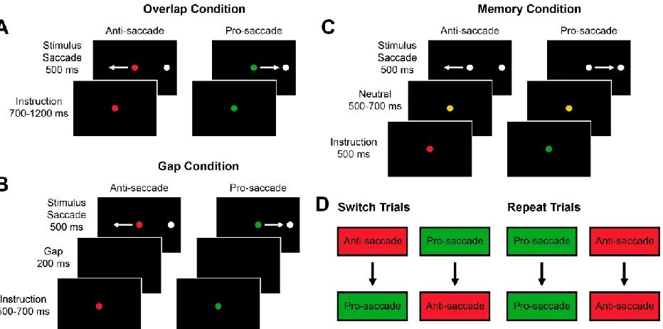
2.2.2 Gap Paradigm ... 72
2.2.3 Memory Paradigm ... 74
2.2.4 Recording Method ... 75
2.2.5 Data Analysis ... 75
2.3 Results ... 78
2.3.1 Switch Costs Present in the Gap and Memory Conditions ... 78
ix
2.3.5 Onset of Motor Activity ... 95
2.3.6 Error Trials ... 97
2.4 Discussion ... 99
2.5 References ... 106
CHAPTER 3 DORSOLATERAL PREFRONTAL CORTEX DEACTIVATION IN MONKEYS REDUCES PREPARATORY BETA AND GAMMA POWER IN THE SUPERIOR COLLICULUS ... 114
3.1 Introduction ... 114
3.2 Materials and Methods ... 116
3.2.1 Surgical Procedures ... 116
3.2.2 Behavioural Task ... 116
3.2.3 Reversible Cryogenic Deactivation ... 118
3.2.4 Recording Method ... 119
3.2.5 Data Analysis ... 120
3.3 Results ... 123
3.3.1 Event-related LFPs Respond to Stimulus Presentation ... 123
3.3.2 LFP Power and Task Performance ... 127
3.3.3 DLPFC Deactivation Reduces Beta and Gamma Power ... 132
3.3.4 DLPFC Deactivation Reduces Correlations Between Spiking Activity and Gamma Power ... 140
3.3.5 LFP Power and SRT ... 140
3.4 Discussion ... 143
3.5 References ... 148
CHAPTER 4 EFFECTS OF BILATERAL DORSAL ANTERIOR CINGULATE CORTEX DEACTIVATION ON COGNITIVELY DEMANDING TASK PERFORMANCE IN MACAQUE MONKEYS ... 156
4.1 Introduction ... 156
x
4.2.3 Reversible Cryogenic Deactivation ... 161
4.2.4 Data Analysis ... 162
4.3 Results ... 164
4.3.1 dACC Deactivation Impairs Anti-saccade Performance ... 164
4.3.2 Performance Following Correct and Erroneous Trials ... 168
4.3.3 dACC Deactivation Increases Pro-saccade and Anti-saccade SRTs ... 170
4.3.4 SRTs Following Correct and Erroneous Trials ... 174
4.3.5 dACC Deactivation and Dropped Trials ... 176
4.4 Discussion ... 178
4.5 References ... 183
CHAPTER 5 DISCUSSION ... 187
5.1 Summary of Main Findings ... 187
5.1.1 Monkeys demonstrate switch costs and switch-related differences in superior colliculus activity ... 188
5.1.2 Bilateral DLPFC deactivation reduces preparatory beta and gamma power in the SC ... 190
5.1.3 Bilateral dACC deactivation impairs feedback integration and cognitively demanding task performance ... 192
5.2 Caveats and Limitations ... 194
5.2.1 Distant effects of reversible cryogenic deactivation ... 194
5.2.2 Non-human primates as an animal model for cognitive control ... 195
5.3 Future Directions ... 196
5.3.1 Switch-related differences in cortical neural activity ... 196
5.3.2 Effects of cortical deactivation on activity in the task switching network ... 197
5.4 Concluding Remarks ... 198
5.5 References ... 198
xi
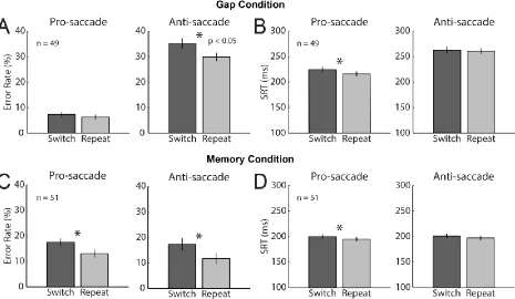
Table 2.1 Gap paradigm switch costs for error rate and SRT ... 79
Table 2.2 Gap condition error rate and SRT switch costs for monkeys A and C ... 80
Table 2.3 Memory paradigm switch costs for error rate and SRT ... 83
Table 2.4 Memory condition error rate and SRT switch costs for monkeys A and B ... 84
Table 3.1 Behavioural effects of DLPFC deactivation ... 124
xii
LIST OF FIGURES
Figure 1.1 Dorsal attention and oculomotor network ... 7
Figure 1.2 Regions of the cingulate cortex ... 13
Figure 1.3 Cingulate motor regions ... 15
Figure 1.4 The anti-saccade task ... 32
Figure 1.5 Task-selective SC activity during the instruction period ... 35
Figure 2.1 Task conditions ... 73
Figure 2.2 Behavior for gap and memory pro- and anti-saccade trials ... 82
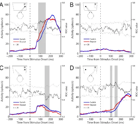
Figure 2.3 SC activity aligned to stimulus onset for the gap pro- and anti-saccade task ... 88
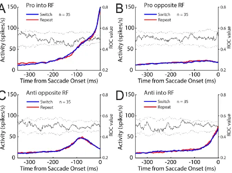
Figure 2.4 SC activity aligned to saccade onset for the gap pro- and anti-saccade task ... 89
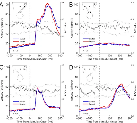
Figure 2.5 SC activity aligned to stimulus onset for the memory pro- and anti-saccade task ... 91
Figure 2.6 SC activity aligned to saccade onset for the memory pro- and anti-saccade task ... 92
Figure 2.7 SC activity on correct memory pro-saccades in which the stimulus was presented into the RF of neurons for individual monkeys ... 93
Figure 2.8 Timing of burst onset in SC neurons with little to no visual activity on switch and repeat trials ... 96
Figure 2.9 SC activity on gap anti-saccade error trials ... 98
Figure 3.1 Experimental setup and experimental paradigm ... 117
Figure 3.2 Event-related LFPs and spike density aligned to stimulus onset... 125
Figure 3.3 ROC time course for event-related LFPs and spike density aligned to stimulus onset ... 126
Figure 3.4 Event-related LFPs and spike density aligned to saccade onset ... 128
Figure 3.5 LFP power spectrograms for correct and error trials ... 130
Figure 3.6 LFP power spectrograms for correct and error trials for frequencies in high gamma ... 131
Figure 3.7 LFP power spectrograms for correct and error trials during the cool period ... 133
Figure 3.8 ROC time course for normalized LFP power in frequency bands ... 134
Figure 3.9 LFP power spectrograms for noncool and cool trials aligned to stimulus onset ... 135
Figure 3.10 LFP power spectrograms for noncool and cool trials aligned to stimulus onset for frequencies in high gamma ... 136
Figure 3.11 Differences in normalized power for cool trials minus precool trials and cool trials minus postcool trials ... 137
Figure 3.12 LFP power spectrograms for noncool and cool trials aligned to fixation cue onset ... 139
Figure 3.13 Proportion of neurons with spike rates that were significantly positively or negatively correlated with LFP power ... 141
Figure 3.14 Relationship between LFP power and SRT ... 142
Figure 4.1 Uncued and cued switch tasks ... 160
Figure 4.2 Effects of bilateral dACC deactivation on uncued switch task performance ... 165
Figure 4.3 Effects of bilateral dACC deactivation on cued switch task performance ... 167
Figure 4.4 Error rates on pro-saccade and anti-saccade trials preceded immediately by a correct or erroneous response during the noncool and cool periods ... 169
Figure 4.5 Effects of bilateral dACC deactivation on uncued switch task SRTs ... 171
xiii
xiv
LIST OF ABBREVIATIONS
ACC Anterior cingulate cortex BOLD Blood oxygen level-dependent CCZ Caudal cingulate zone
CMAd Dorsal cingulate motor area CMAr Rostral cingulate motor area CMAv Ventral cingulate motor area dACC Dorsal anterior cingulate cortex DLPFC Dorsolateral prefrontal cortex ERP Event-related potential EVC Expected value of control FEF Frontal eye field
fMRI Functional magnetic resonance imaging GABA γ-aminobutyric acid
iMLF Interstitial nucleus of the medial longitudinal fasciculus IPS Intraparietal sulcus
LFP Local field potential LIP Lateral intraparietal MCC Midcingulate cortex PCC Posterior cingulate cortex PET Positron emission tomography PFC Prefrontal cortex
PPC Posterior parietal cortex
PPRF Paramedian pontine reticular formation pre-SMA Pre-supplementary motor area
RCZa Anterior rostral cingulate zone RCZp Posterior rostral cingulate zone RF Response field
ROC Receiver operating characteristic RSC Retrosplenial cortex
C
HAPTER1
Introduction
1.1 Cognitive Control
In everyday life, our behaviour is driven by the pursuit of various goals. Consider a writer
who is sitting at a desk working on a lengthy essay as an example. At times, successive sentences
are readily composed. At others, focusing on writing may be difficult as feelings of hunger or the
desire to take a break emerge. Goals can be short-term, such as eating food when hungry, or
long-term, such as finishing the essay. Multiple goals can be elicited by the same external
stimulus and the appropriate goal to pursue depends on the current context. A computer monitor
can prompt writing, or trigger checking incoming emails, reading the news, or watching a movie.
The presence of an immediately impending deadline for the essay or fatigue would likely
determine whether the writer continues working or takes a break. Ultimately, cognitive control is
required to select appropriate goals and guide behaviour to achieve the desired outcomes.
Cognitive control plays a crucial role in our lives, particularly with regard to voluntary
behaviour, and consequently is a central research topic in experimental psychology and cognitive
neuroscience. Notably, cognitive control is not a single unitary process. Instead, it refers to a set
of different cognitive functions that together enable goal-directed behaviour. Cognitive functions
include, but are not limited to, the ability to attend to behaviourally relevant information,
maintain information in working memory, select and inhibit responses, make decisions, plan a
series of steps to achieve a goal, and monitor whether actions have their intended consequences
adapt behaviour to changes in the external environment and internal goals is a hallmark of
cognitive control.
1.1.1 Rule-Guided Behaviour
Much of our behaviour is guided by rules, or learned associations between stimuli,
contexts, actions, and outcomes (Bunge, 2004). Rules vary in their level of abstraction and
enable us to act in an appropriate manner. Simple and concrete rules, or stimulus-response
associations, can be learned and applied quickly. For example, drivers know that a green light
means go whereas a red light means stop. Arbitrary associations between stimuli and responses,
however, are difficult to apply to other situations. Outside of driving, green-go and red-stop do
not readily provide information about which behaviour is appropriate. In contrast, complex and
abstract rules are not constrained by specific stimuli or responses, and can be generalized and
applied to familiar and novel situations (Wallis et al., 2001; Buschman and Miller, 2014). For
example, drivers know that in order to drive a motor vehicle, the engine must be started,
regardless of where the ignition is located or the method used to start the engine. Depending on
the goal, single or multiple concrete or abstract rules may be required to guide the appropriate
behaviour.
A concept related to rules is that of a “task-set.” Each behaviour or task that is performed
is associated with a task-set, or task-specific configuration of mental processes and resources
(Monsell, 2003; Sakai, 2008). As such, a task-set consists of task-relevant information about
stimuli, responses, and rules, as well as the sensory, attentional, and motor processes required for
cognitive control is required to select, maintain, implement, and execute the appropriate rules
and task-sets.
1.1.2 Attention
Attention is integral to cognitive control and its importance in everyday life is generally
understood and accepted. As a phenomenon, William James (1890) described attention as
follows:
“Everyone knows what attention is. It is taking possession by the mind in clear and vivid
form, of one out of what seem several simultaneously possible objects or trains of
thought... It implies withdrawal from some things in order to effectively deal with
others.”
Accordingly, at any given moment, the total amount of attentional and cognitive resources
available for information processing is limited (Kahneman, 1973). Although multiple sensations,
tasks, emotions, and thoughts may occur concurrently, attention selectively allocates resources to
information that is behaviourally relevant, while inhibiting the processing of information that is
behaviourally irrelevant.
How attention is allocated depends on bottom-up and top-down factors (Corbetta and
Shulman, 2002; Knudsen, 2007). Bottom-up or exogenous attention is involuntarily driven by
novel, salient, or unexpected stimuli in the sensory environment. In contrast, top-down or
endogenous attention is voluntarily driven by an individual’s knowledge, experience, and current
goals. To illustrate these factors, take for example a writer working on an essay. If a fire alarm
starts ringing and flashing, attention would be immediately and involuntarily drawn to the alarm
stimulus. Using previous experience and meaning associated with the alarm, the writer may
choose to stop working and focus on evacuating the building. Alternatively, if the writer knew
beforehand that the alarm is a test, attention may instead be maintained on writing in a top-down
manner while ignoring the alarm. Bottom-up and top-down processes interact with each other to
dynamically shift and maintain attention. The top-down allocation of attentional and cognitive
resources, also referred to as executive attention, is a critical component of cognitive control and
particularly important for goal-directed behaviour.
1.1.3 Working Memory
Behaviourally relevant information is temporarily maintained and manipulated online by
working memory, which provides an interface between perception, long-term memory, and
action (Baddeley, 1986; Baddeley, 2003). A key feature of working memory is its ability to
maintain information in the absence of sensory stimulation and motor output. Consequently, it is
critical for the active maintenance of goals and task rules, and for the implementation of
cognitive control and goal-directed behaviour. Notably, there is a close functional relationship
between working memory and attention, especially at the executive level. The central executive
of working memory proposed by Baddeley (1986) to coordinate and process information for the
control of behaviour mirrors the supervisory attentional system proposed by Norman and
Shallice (1986) for attentional control. Furthermore, working memory and attention both have a
limited capacity for information processing and the content of both are often identical, such that
working memory can be considered to represent the contents of attention (Knudsen, 2007).
(2008) described working memory for psychological and neural investigation as “sustained
attention focused on an internal representation.”
1.2 Brain Systems for Cognitive Control
Uncovering the neural mechanisms underlying cognitive control is a central aim of
cognitive neuroscience. Numerous lesion, functional neuroimaging, and neurophysiological
studies have associated cognitive control, including the central executive of working memory
and supervisory attentional system, with the frontal lobes (Stuss and Knight, 2002; Fuster, 2008).
Functional neuroimaging has also shown that broad networks of brain areas are simultaneously
activated with the frontal lobes for cognitive control.
The bottom-up control and top-down control of attention are generally accepted to be
associated with the ventral attention network and dorsal attention network respectively. The
ventral attention network includes the temporal parietal junction (TPJ) and ventral prefrontal
cortex, and is lateralized to the right hemisphere in humans (Corbetta and Shulman, 2002). The
role of these brain areas in the bottom-up control of visual attention is evident when a lesion to
the right hemisphere produces left-sided spatial neglect (Corbetta and Shulman, 2002).
Furthermore, transient activation with functional magnetic resonance imaging (fMRI) is elicited
in the ventral attention network when behaviourally relevant, salient, or unexpected stimuli are
presented, regardless of the modality of the stimuli (Corbetta et al., 2000; Downar et al., 2000).
In non-human primates, neurons in area 7a, which may correspond to the human TPJ (Patel et
al., 2015), respond to stimuli that are behaviourally relevant and presented to previously
unattended locations (Bushnell et al., 1981; Robinson et al., 1995; Steinmetz and Constantinidis,
Whereas the ventral attention network orients attention to salient stimuli in the sensory
environment, the dorsal attention network, which includes the frontal eye field (FEF) and
intraparietal sulcus (IPS), is implicated in the endogenous control of attention (Fig. 1.1)
(Corbetta and Shulman, 2002). With regard to visual attention, sustained activation with fMRI is
elicited bilaterally in these brain areas when attention is covertly directed to a peripheral
location, with stronger activation contralateral to the attended visual field (Kastner et al., 1999;
Corbetta et al., 2000; Hopfinger et al., 2000). In non-human primates, FEF and IPS neurons
increase in activity in anticipation of the onset of a stimulus (Bushnell et al., 1981; Colby et al.,
1996) and encode stimuli features that are behaviourally relevant (Seagraves and Goldberg,
1987; Toth and Assad, 2002). In addition, the FEF and IPS are implicated in working memory
and the control of eye movements (Corbetta and Shulman, 2002). This convergence and
integration of sensory, attentional, and motor information in frontoparietal cortex is critical for
goal-directed behaviour.
More generally, frontoparietal brain areas are implicated in a variety of cognitive
functions, including attention, working memory, task representation, response selection,
inhibition, planning sequences of actions, and decision making (Duncan and Owen, 2000;
Corbetta and Shulman, 2002; Duncan, 2010). The lateral prefrontal cortex, anterior insula,
anterior cingulate cortex (ACC), pre-supplementary motor area (pre-SMA), and IPS are
commonly activated together with fMRI for cognitive tasks and tests of fluid intelligence
(Duncan and Owen, 2000; Duncan, 2010). Accordingly, neurons in these brain areas have
demonstrated the ability to encode task-relevant information and reorganize information rapidly
when the context changes (Asaad et al., 2000; Wallis et al., 2001; Stoet and Snyder, 2004;
control in this multiple-demand network (Duncan, 2010), however, remain unclear. Although
brain areas outside the frontal lobe are certainly involved in cognitive control, the prefrontal
cortex (PFC) has long been recognized to play a particularly important role.
1.2.1 Prefrontal Cortex
The PFC, located in the anterior part of the frontal lobes, is a neocortical region that is
highly developed and expanded in primates, especially humans. Although the entire PFC
receives projections from the mediodorsal nucleus of the thalamus, it can be further subdivided
into distinct regions based on cytoarchitecture and connectivity (Brodmann, 1909; Petrides and
Pandya, 1999; Petrides, 2005; Fuster, 2008; Hutchison and Everling, 2014). These regions are
interconnected and many receive converging inputs from multiple sensory modalities (Pandya
and Kuypers, 1969; Jones and Powell, 1970; Chavis and Pandya, 1975; Petrides, 2005). As a
result, the PFC performs a diverse set of functions that are interrelated and complement each
other. Overall, the PFC receives and sends projections to a variety of cortical sensory and
motor-related areas, and subcortical areas (Pandya and Kuypers, 1969; Jones and Powell, 1970; Miller
and Cohen, 2001; Fuster, 2008), making it well positioned to coordinate neural processes and
integrate information for complex purposeful behaviour.
Within the PFC, the dorsolateral prefrontal cortex (DLPFC) and dorsal anterior cingulate
cortex (dACC) are two areas that are often associated with each other and with cognitive control.
The DLPFC and dACC are highly and reciprocally connected (Barbas and Pandya, 1989; Bates
and Goldman-Rakic, 1993; Morecraft and Van Hoesen, 1993; Paus et al., 2001; Petrides, 2005),
and functional neuroimaging studies have consistently demonstrated co-activation for a variety
cognitive control remain poorly understood, anatomical and physiological studies suggest
distinct contributions for the DLPFC and dACC.
1.2.1.1 Dorsolateral Prefrontal Cortex
Anatomically, the DLPFC consists of Brodmann areas 9, 9/46, and 46 and is defined
from other regions in the PFC by a well-developed granular layer IV, particularly in areas 9/46
and 46 (Petrides and Pandya, 1999; Petrides, 2005). In humans, this corresponds to the superior
frontal gyrus and middle frontal gyrus. In macaque monkeys, the DLPFC is located anterior to
the arcuate sulcus and includes the banks of the principal sulcus, the cortex surrounding the
anterior portion of this sulcus, and the cortex extending dorsally to the midline (Petrides and
Pandya, 1999).
The DLPFC receives converging visual, auditory, and somatosensory inputs from the
occipital, temporal, and parietal cortices (Jones and Powell, 1970; Petrides and Pandya, 1984;
Seltzer and Pandya, 1989), including from multimodal areas such as the superior temporal
sulcus, superior temporal gyrus, cingulate cortex, and retrosplenial cortex (Chavis and Pandya,
1976; Seltzer and Pandya, 1989; Petrides and Pandya, 1999; Petrides, 2005). Through reciprocal
connections with the retrosplenial cortex, orbitofrontal, and medial prefrontal cortex, the DLPFC
also has access to the limbic system for the processing of long-term memory, affect, and
motivation (Morris et al., 1999; Miller and Cohen, 2001; Petrides, 2005). This diverse set of
inputs allows for complex multimodal processing and integration. The DLPFC’s projections to
the supplementary motor area (SMA), pre-SMA, premotor cortex, FEF, ACC, cerebellum, and
and Goldman-Rakic, 1993; Lu et al., 1994) likely enable it to exert cognitive control over
behaviour.
Functionally, the DLPFC is broadly thought to encode representations of rules and goals,
and bias other brain areas to achieve the desired outcome (Miller and Cohen, 2001).
Representations require the maintenance of information and consequently, the DLPFC has long
been implicated in working memory. The role of the DLPFC in working memory has been
extensively studied using a variety of tasks, especially delayed-response tasks where stimulus
information must be retained over a delay period prior to executing an appropriate behavioural
response.
Jacobsen (1935) first demonstrated that bilateral PFC lesions induce delayed-response
impairments in monkeys. Subsequent lesion studies further delineated the anatomical substrate of
delayed-response performance. Unilateral PFC lesions also produce deficits (Warren et al., 1969)
and DLPFC lesions in particular impair the ability to retain spatial information and the
integration of this information over time (Mishkin et al., 1969; Fuster and Alexander, 1970;
Goldman and Rosvold, 1970). In humans, DLPFC lesions increase accuracy errors with
delayed-response tasks (Lewinsohn et al., 1972; Milner et al., 1985; Pierrot-Deseilligny et al., 1991a).
Notably, deficits produced by DLPFC lesions occur regardless of sensory modality.
Studies investigating delayed-response and other memory tasks with neuroimaging
techniques such as fMRI and positron emission tomography (PET) have consistently shown
DLPFC activation (Duncan and Owen, 2000; Curtis and D’Esposito, 2003; Wager and Smith,
2003). This activation increases with the number of items retained in working memory (Jaeggi et
al. 2002; Kirschen et al., 2005). Neuroimaging studies also consistently demonstrate that
and provide support that the DLPFC is part of a larger network underlying working memory.
Neurophysiological investigations provide insight into the specific role of the DLPFC in working
memory.
Early single-unit recordings in the principal sulcus of monkeys performing
delayed-response tasks demonstrated neurons with increased and sustained activity during the delay
period (Fuster and Alexander, 1971; Fuster, 1973). These neurons were termed “memory cells”
because their activity, which bridges the temporal gap between stimulus and response, is thought
to be a neural correlate of working memory. Appropriately, the level of activity during the delay
period is correlated with correct task performance (Fuster, 1973). Visual working memory tasks
preferentially activate memory cells in the inferior convexity of the principal sulcus, whereas
spatial working memory tasks preferentially activate cells in the superior convexity (Fuster et al.,
1982; Wilson et al., 1993). With regard to spatial working memory, neurons are grouped for
distinct locations in visual space and are preferentially activated for the contralateral hemifield
(Funahashi et al., 1989). Unlike receptive fields in visual brain areas, however, memory fields in
the DLPFC are not clearly topographically organized.
Although delay period activity is generally accepted as a neural correlate of working
memory, the information that is represented by this activity remains unclear. DLPFC responses
may represent remembered stimulus information, such as stimulus location, or motor
information, such as the direction of an upcoming movement (Hasegawa et al., 1998;
Constantinidis et al., 2001). DLPFC responses may also be associated with diverse processes
supporting task performance, such as attention, task representation, and task preparation (Asaad
DLPFC signals, the DLPFC is thought to influence neural activity in other brain areas to perform
the appropriate task.
Recent investigations using local field potentials (LFPs), which reflect the average
postsynaptic activity of a population of neurons (Buzaki et al., 2012), further support the
DLPFC’s role in working memory and the implementation of cognitive control. In particular,
oscillations in the beta (12-30 Hz) frequency band have been implicated in working memory
maintenance (Engel and Fries, 2010; Salazar et al., 2012; Spitzer et al., 2014). Task-specific
neural ensembles can be formed in the DLPFC by beta synchronization (Buschman et al., 2012)
and enhanced beta coherence between the DLPFC and other brain areas, such as the posterior
parietal cortex, is thought to facilitate top-down control (Buschman and Miller, 2007; Donner et
al., 2007; Siegel et al., 2012). Overall, both direct neuronal outputs and neuronal oscillations
likely enable the DLPFC to exert control over distant brain areas, and ultimately behaviour.
1.2.1.2 Dorsal Anterior Cingulate Cortex
The cingulate cortex is located immediately above the corpus callosum in the medial wall
of the cerebral hemispheres, and can be divided into four regions based on cytoarchitecture,
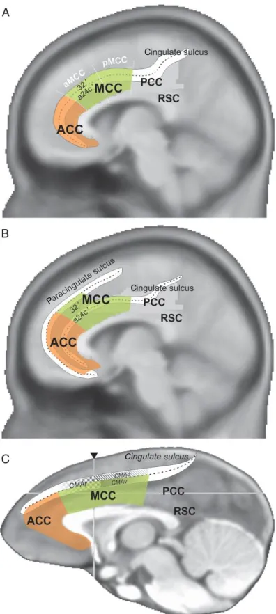
connectivity, and function: the anterior cingulate cortex (ACC), midcingulate cortex (MCC),
posterior cingulate cortex (PCC), and retrosplenial cortex (RSC) (Fig. 1.2) (Vogt et al., 2005;
Vogt, 2009). Although the ACC is variably defined in the literature, here, the dACC will refer to
the MCC and its anterior portion in particular. In macaque monkeys, the dACC consists of
Brodmann area 24 and is located in the cingulate gyrus and sulcus. In humans, the dACC
consists of Brodmann areas 24 and 32’ and corresponds to the cingulate gyrus dorsal to the
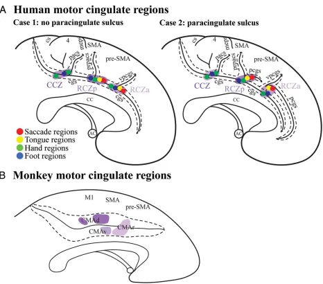
As a whole, the dACC includes the rostral, ventral, and dorsal cingulate motor areas (CMAr,
CMAv, and CMAd), as defined in monkeys by intracortical microstimulation and connectivity,
and the homologous anterior rostral cingulate zone (RCZa), posterior rostral cingulate zone
(RCZp), and caudal cingulate zone (CCZ) respectively in humans (Fig. 1.3) (Picard and Strick,
1996; Amiez and Petrides, 2014). The dACC and cognitive control are particularly associated
with CMAr, which is situated on the dorsal and ventral banks of the cingulate sulcus anterior to
the arcuate sulcus.
The dACC receives input from the temporal cortex, parietal cortex, and insula, and is
highly and reciprocally connected with the DLPFC (Vogt and Pandya, 1987; Barbas and Pandya,
1989; Bates and Goldman-Rakic, 1993). It sends projections to motor areas such as the premotor
cortex, FEF, primary motor cortex, and ventral horn of the spinal cord (Dum and Strick, 1991;
Picard and Strick, 1996; Wang et al., 2004). However, microstimulation of the dACC does not
strongly evoke movement and suggests that it does not play a direct role in motor control
(Luppino et al., 1991; Picard and Strick, 1996).
Instead, the dACC is strongly implicated in cognitive control. Numerous functional
imaging studies have demonstrated dACC activation with a variety of cognitive functions,
including attention, task conflict, action selection, working memory, episodic memory, decision
making, problem solving, reward processing, pain processing, emotion, and motivation (Duncan
and Owen, 2000; Shackman et al., 2011; Shenhav et al., 2013). Nonetheless, precise mechanisms
for dACC function have been difficult to elucidate because of discrepancies between lesion,
functional imaging, and neurophysiological studies, and between species (Fellows and Farah,
2005; Cole et al., 2009).
Early theories of dACC function were based on electrophysiological and functional
imaging studies in humans. Error-related negativity, or a negative deflection in the event-related
potential (ERP) immediately after an erroneous response, has been localized to the ACC and is
thought to be associated with conflict monitoring or performance monitoring (Falkenstein et al.,
1991; Gehring et al., 1995; Botvinick et al., 2001). The conflict monitoring hypothesis proposes
that the dACC detects conflict, defined as the co-activation of two or more competing processes
by a single stimulus, and subsequently signals an increase in cognitive control to resolve the
conflict and improve task performance (Botvinick et al., 2001). In humans, investigations with
fMRI and single-unit recordings have shown that the dACC responds to conflict during tasks that
produce conflict, such as the Stroop task, and predicts adjustments in behaviour (Carter et al.,
2000; MacDonald et al., 2000; Botvinick et al., 2001; Kerns et al., 2004; Davis et al., 2005).
However, studies involving the monkey dACC have been unable to produce evidence of
conflict-related signals.
For example, the countermanding task, where subjects must withhold a planned
movement immediately prior to execution in response to a stop signal, has been shown to
produce conflict-related dACC activation in humans (Curtis et al., 2005), but was not associated
with conflict-related responses in monkeys (Ito et al., 2003; Emeric et al., 2008). Studies
involving other tasks and dACC lesions have also failed to associate the monkey dACC with
conflict monitoring (Nakamura et al., 2005; Mansouri et al., 2007). Rather, monkey studies have
demonstrated that dACC neurons and LFPs signal erroneous responses, rewarded responses, and
unrewarded responses (Shima and Tanji, 1998; Ito et al., 2003; Nakamura et al., 2005; Emeric et
al., 2008). Furthermore, the dACC has been shown to monitor feedback for changes in behaviour
Quilodran et al., 2008; Amiez et al., 2012). Beyond conflict monitoring, both human and monkey
data are consistent with a role of the dACC in performance monitoring.
Another theory of dACC function proposes that the dACC predicts error-likelihood,
which incorporates both conflict and error detection (Brown and Braver, 2005). Activity in the
dACC is thought to be proportional to the likelihood of an error, such that more cognitive control
is recruited for increased task demands. Task demands increase during task switching and task
selectivity in dACC neurons has been shown to be strongest after a task switch, thereby
reflecting the implementation of increased cognitive control (Johnston et al., 2007). Neurons in
the dACC also encode cognitive demand and are modulated by the demands of the previous trial
to mediate behavioural adaptations (Sheth et al., 2012). Thus, the dACC functions to both
monitor performance and implement cognitive control.
To reconcile the diversity of findings regarding the dACC, Shenhav et al. (2013)
proposed that the dACC estimates the expected value of control (EVC) of a task in order to
allocate cognitive control. As such, the dACC functions to monitor and integrate information,
such as task demands, processing capacity, motivation, and positive and negative outcomes, to
determine the EVC. Based on the estimated EVC, the dACC also functions to specify which task
to allocate control to and how much control to allocate to maximize the EVC. In this model of
dACC function, control signals specified by the dACC are implemented by brain areas like the
DLPFC, which are responsible for regulating the information processing required for task
performance. Accordingly, changes in task demand have been shown to increase task selectivity
and LFP power in the dACC prior to more sustained responses in the DLPFC, which are thought
1.3 Task Switching
The ability to flexibly engage in goal-directed behaviour in response to changes in the
external environment and internal goals is a hallmark of cognitive control. Cognitive control is
required to select, maintain, implement, and execute the appropriate task-set. Furthermore, the
act of task switching itself is associated with an increase in cognitive demand. Thus, task
switching paradigms have become an attractive method to investigate cognitive control and task
processing.
1.3.1 Task Switching Paradigms
Jersild (1927) first used a task switching paradigm to examine cognitive control by
asking participants to perform a series of trials where they either repeated a single task or
alternated between two. Original task switching studies were limited to investigating the effects
of task switching on behaviour, but since the development of functional imaging, the number of
task switching experiments has increased dramatically and many variations of task switching
paradigms have been designed (Monsell, 2003).
Although Jersild’s paradigm enabled comparison between task switching and task
repetition (Jersild, 1927; Spector and Biederman, 1976), the method of alternating tasks from
trial to trial required the maintenance of multiple tasks and a task sequence in working memory
and thereby imposed an additional cognitive load (Monsell, 2003). Many contemporary task
switching paradigms avoid this confound by increasing the number of trials before a switch or
signalling when a task switch will occur. Consequently, paradigms may be designed with
alternating blocks of a single task, where the task switch is signalled by a pre-specified number
multiple tasks that are interleaved and cued from trial to trial. Other features of paradigms, such
as preparation time and performance feedback, can also be manipulated to enable specific
aspects of cognitive control to be investigated. Regardless of the variations, all paradigms have
periods where the task either remains the same or changes from one trial to the next.
Performance on or after switch trials can provide insight into cognitive control and task
processing.
1.3.2 Task Switching Phenomena
Task switching is ubiquitously associated with switch costs, or increases in reaction times
and error rates on trials where the task is switched compared to trials where the task is repeated (
Allport et al., 1994; Monsell, 2003). To successfully switch tasks, task-relevant information must
be selected and maintained over task-irrelevant information. Task-set reconfiguration requires
shifting attention to the new task-set while inhibiting the prior task-set. Switch costs may arise
from the time required to complete task-set reconfiguration and accordingly, switch costs can be
reduced if sufficient preparatory time is allowed for reconfiguration (Monsell, 2003).
Interestingly, long preparatory periods do not eliminate switch costs and these residual costs are
thought to be due to the inability to complete task-set reconfiguration before stimulus onset. A
component of reconfiguration may depend on the presence of external stimuli (Rogers and
Monsell, 1995) or reconfiguration may only be successful before stimulus onset on a proportion
of trials in an all-or-none manner (De Jong, 2000).
The persistence of the previous task-set, or task-set inertia, may also contribute to switch
costs. Interference from task-set inertia is particularly evident when switching from a
respectively. Switching to the more practiced and habitual dominant task is counterintuitively
associated with a greater switch cost and can be attributed to increased cognitive control during
performance of the non-dominant task carrying over and interfering with preparation for the
dominant task-set (Allport et al., 1994; Monsell, 2003). Task-set inertia is supported by evidence
that longer periods of time between the performance of the previous task and instruction for the
current task reduce switch costs, which suggests dissipation of the competing previous task-set
(Meiran et al., 2000). In addition, task-set inertia may contribute to residual switch costs.
1.3.3 Human and Non-Human Primate Task Switching
Task switching behaviour and switch costs have been consistently demonstrated in
humans. Non-human primates, particularly macaque monkeys, have been widely used in lesion,
functional neuroimaging, and neurophysiological studies as a model for cognitive control, but
whether they show switch costs is less clear. One comparative study between humans and
monkeys found that monkeys only had switch costs with short intertrial intervals and suggested
that although monkeys experience task-set inertia, the previous task-set dissipates quickly (Stoet
and Snyder, 2003). The absence of a persistent residual switch cost was taken to suggest that
monkeys can complete task-set reconfiguration before stimulus onset. In contrast, another
comparative study found that humans and monkeys had comparable and robust reaction time and
error rate switch costs (Caselli and Chelazzi, 2011). Although studies demonstrate conflicting
results regarding switch costs in non-human primates, monkeys, like humans, are able to perform
complex cognitive tasks and switch between tasks. Thus, the macaque monkey remains a
1.3.4 Neural Basis of Task Switching
1.3.4.1 Task-Set Representation
Successful task switching requires a network of brain areas to select, maintain,
implement, and execute the appropriate task-set. Although a task-set is a psychological construct,
it may be possible for task-sets to be represented in the brain. The neural correlates of a task-set
can be considered task-specific neural activity. Single-unit recordings in monkeys have identified
multiple brain areas with task-specific activity, including the DLPFC (White and Wise, 1999;
Asaad et al., 2000; Wallis et al., 2001; Everling and DeSouza, 2005; Mansouri et al., 2006;
Johnston and Everling, 2006; Johnston et al., 2007), dACC (Johnston et al., 2007), premotor
cortex (Wallis and Miller, 2003), and posterior parietal cortex (PPC) (Stoet and Snyder, 2004;
Kamigaki et al., 2009). Among these brain areas, the DLPFC is thought to be particularly
important for encoding and maintaining task representations, and modulating other brain areas
for task performance (Miller and Cohen, 2001). Task-related information has also been shown to
be represented in the activity of neural populations (Stokes et al., 2013) and LFP activity
(Buschman et al., 2012).
In humans, fMRI is often used to investigate task processing. When a task is performed,
specific areas are more active than others due to the types of stimuli, processing, and responses
that are required. Conventional univariate analysis can identify which brain areas are involved
with task performance, but are unable to distinguish task representations in the same brain areas.
Alternatively, studies using multivariate pattern analysis have demonstrated that task
representations can be identified from blood oxygen level-dependent (BOLD) activity in
frontoparietal cortex (Bode and Haynes, 2009; Woolgar et al., 2011; Chan et al., 2015). Although
stimuli colour and responses, task rule was found to be the most strongly represented feature
(Woolgar et al., 2011). Taken together, the encoding of task representations in the brain can be
demonstrated using a variety of techniques, from single-unit recordings to whole-brain functional
imaging.
1.3.4.2 Task-Set Selection and Interference
In human fMRI studies, preparatory activation during trials where the task is switched is
often compared to activation during trials where the task is repeated to identify brain areas
involved in selecting, establishing, and maintaining task-sets. Although brain areas exclusively
activated by switch trials are consistently absent (Ruge et al., 2013), a network of frontoparietal
brain areas, including the DLPFC, ACC, and PPC, is more strongly activated for switch trials
compared to repeat trials (Sohn et al., 2000; Braver et al., 2003; Liston et al., 2006; Chiu and
Yantis, 2009; Ruge et al., 2013). Switch-related prefrontal activation may be related to the
preparation of response-directed intentional task-sets, whereas parietal activation may be related
to the preparation of stimulus-directed attentional task-sets (Ruge et al., 2013). Increased
processing in these brain areas, as demonstrated by increased activation, may reflect task-set
reconfiguration and the maintenance of the new task-set. Consistent with this, task-related
information in the PFC and PPC has been shown to increase after presentation of the instruction
cue (Bode and Haynes, 2009). This is similar to the presence of task-specific activity in PFC,
ACC, and PPC neurons during the preparatory period (Asaad et al., 2000; Wallis et al., 2000;
Everling and DeSouza, 2005; Johnston et al., 2007; Stoet and Snyder, 2004). While the DLPFC
that a task switch has occurred and selecting the new task (Johnston et al., 2007; Kamigaki et al.,
2009).
Interference from the previous task-set may manifest as task-specific activity that persists
on switch trials. Task-set inertia can be demonstrated with functional imaging by using two tasks
that activate distinct brain areas. When participants were asked to switch between a face
categorization and a word categorization task, activation in brain areas for the irrelevant task was
positively correlated with the reaction time switch cost (Yeung et al., 2006). Thus, at the whole
brain level, processing for the previous task may compete with preparation for the new task.
Unfortunately, single-unit recording studies in monkeys have not yet addressed differences in
neural activity between switch trials and repeat trials and the mechanisms of task-set inertia.
1.3.4.3 Task-Set Implementation
The PFC, with connections to motor areas such as the SMA, pre-SMA, premotor cortex,
FEF, cerebellum, and SC (Goldman and Nauta, 1976; Selemon and Goldman-Rakic, 1988; Lu et
al., 1994), is well situated to implement the tasks it encodes. Human functional imaging and
monkey neurophysiological studies have demonstrated that task-specific activity in the PFC
influences activity in brain areas that are more involved in task execution and this activity is
negatively correlated with reaction times (Johnston and Everling, 2006; Sakai and Passingham,
2006). Unsurprisingly, motor areas such as the premotor cortex and SC encode behavioural
1.3.4.4 Task and Performance Monitoring
Once a task is performed, the outcome of the task must be monitored to determine
whether the task should be repeated or switched. Of the brain areas implicated in task switching,
the dACC may be best positioned to integrate task-related information with task outcomes. The
dACC encodes task-related information (Johnston et al., 2007) and has been shown to respond to
task conflict (Carter et al., 2000; MacDonald et al., 2001; Botvinick et al., 2001), changes in task
demand (Johnston et al., 2007; Sheth et al., 2012), and positive and negative feedback (Shima
and Tanji, 1998; Ito et al., 2003; Nakamura et al., 2005). Integration of monitored information
may enable the dACC to determine the appropriate task to allocate cognitive control to (Shenhav
et al., 2013).
1.4 Investigating Cognitive Control Using the Oculomotor System
Eye movements are integral to human behaviour. In particular, saccadic eye movements,
which involve conjugate, ballistic movements of the eyes, are frequently performed to direct
gaze to objects of interest in the visual world. To generate a saccade, one must decide when to
look, where to look, and whether to look. Consequently, saccades are goal-directed movements
that require cognitive control and are influenced by attention, working memory, inhibition,
decision making, long-term memory, and learning (Hutton, 2008). Sensory input to the
oculomotor system can be precisely manipulated and its output, produced by six distinct
extraocular muscles, is simple compared to movements of other parts of the body and can be
easily and accurately measured. The oculomotor system is well characterized anatomically and
(Liversedge et al., 2011). Thus, the oculomotor system serves as an attractive model for
investigating cognitive control.
1.4.1 Oculomotor Neurophysiology
1.4.1.1 Brainstem
In order to generate saccadic eye movements, position and velocity signals are sent by
motor neurons from the oculomotor nuclei (cranial nerve III), trochlear nuclei (cranial nerve IV),
and abducens nuclei (cranial nerve VI) in the brainstem to the extraocular muscles. Burst
neurons in the paramedian pontine reticular formation (PPRF) and interstitial nucleus of the
medial longitudinal fasciculus (iMLF) produce phasic signals to initiate horizontal and vertical
saccades respectively (Cohen and Henn, 1972; Keller, 1974; Büttner et al., 1977; King and
Fuchs, 1979). Eye position is maintained by tonic activity from the nucleus prepositus
hypoglossi for horizontal saccades and from the interstitial nucleus of Cajal for vertical saccades
(Sparks, 2002). These motor signals are ultimately controlled by the cerebral cortex through the
SC, which projects to contralateral brainstem saccade generators.
1.4.1.2 Superior Colliculus
The SC is a laminated structure in the dorsal midbrain that is critical to oculomotor
control. Functionally, the SC is divided into the superficial layers and the intermediate or deep
layers. These layers contain topographic sensory and motor maps that are similarly aligned,
receives multisensory, motor, and cognitive inputs from various brain areas and is well situated
to integrate information for the control of eye movements and other orienting behaviours.
The superficial layers of the SC are made up of the three dorsal most layers and receive
direct projections from the retina, visual cortex, and FEF (Hubel et al., 1975; Fries, 1984).
Neurons in the superficial layers respond to the appearance of a visual stimulus in their response
field (RF) and produce a topographic map of the contralateral visual hemifield (Schiller and
Koerner, 1971; Cynander and Berman, 1972; Goldberg and Wurtz, 1972). The fovea and
periphery are mapped on to the rostral and caudal SC, respectively, while the upper and lower
visual fields are mapped onto the medial and lateral SC, respectively. In addition, superficial
layer neurons respond to the intensity of a stimulus, but demonstrate minimal preference for the
features of a stimulus. These characteristics implicate the superficial layer of the SC in visual
salience mapping and bottom-up processing (Fecteau and Munoz, 2006). Accordingly, the
superficial layers may influence visual processing through projections to the pulvinar nucleus
and lateral geniculate nucleus of the thalamus (Harting et al., 1978; Stepniewska et al., 2000).
Outputs to the intermediate layers of the SC may facilitate sensorimotor processing (Isa, 2002).
The intermediate layers of the SC are made up of the four deeper layers and receive
inputs from the superficial layers (Isa, 2002), and a variety of cortical and subcortical areas,
including the FEF, DLPFC, supplementary eye field (SEF), ACC, lateral intraparietal (LIP) area,
and substantia nigra pars reticulata of the basal ganglia (Goldman and Nauta, 1976; Leichnetz et
al., 1981; Hikosaka and Wurtz, 1983; Lynch et al., 1985; Stanton et al., 1988b; Shook et al.,
1990). In contrast to the superficial layers, the intermediate layers contain visual, auditory,
Goldberg, 1972; Sparks, 1986; Stein and Stanford, 2008). The map of contralateral saccade
vectors is closely aligned to the map of the contralateral visual hemifield.
Saccades to positions proximal to the fovea are mapped onto the rostral SC, while
saccades to positions distal to the fovea are mapped onto the caudal SC (Schiller and Koerner,
1971; Robinson, 1972; Wurtz and Goldberg, 1972). In the rostral pole of the SC, fixation-related
neurons discharge tonically during visual fixation in the presence and absence of a visual
stimulus and pause for most saccades, and are thought to maintain visual fixation and inhibit
saccade generation (Munoz and Wurtz, 1993a, b; Dorris and Munoz, 1995). Rostral pole neurons
also encode microsaccades, or small-amplitude fixational saccades (Hafed et al., 2009; Hafed
and Krauzlis, 2012). Saccade-related neurons in the remainder of intermediate layers discharge a
burst of action potentials before and during saccades of varying amplitudes and directions
(Wurtz and Goldberg, 1972; Sparks et al., 1976; Munoz and Wurtz, 1995). These neurons can be
subdivided into motor neurons, which only discharge for a saccade, and visuomotor neurons,
which also discharge for stimuli in their RF. In addition, buildup neurons are distinguished from
burst neurons by low-frequency activity prior to the appearance of a stimulus (Munoz and Wurtz,
1995). Buildup activity is thought to be associated with motor preparation and higher-level
processing.
The interaction between visual, motor, and cognitive information in the intermediate
layers is consistent with a role in priority mapping, where visual salience is integrated with the
behavioural relevance of a stimulus (Fecteau and Munoz, 2006). An example of integrating
salience and relevance is saccade target selection, where neurons discriminate a target from
distractors by suppressing distractor-related activity while enhancing target-related activity
by visual stimuli that are associated with reward (Ikeda and Hikosaka, 2003). Taken together, the
SC is well suited to integrating information for the flexible control of behaviour.
Outputs from the intermediate layers to the PPRF and iMLF in the brainstem enable the
generation of saccades (Sparks, 2002). The intermediate layers also send projections to the FEF
through the mediodorsal nucleus of the thalamus (Lynch et al., 1994; Sommer and Wurtz, 2004).
This pathway enables the transmission of a corollary discharge, or an internal copy of the motor
signal, to cortex to enable monitoring of the forthcoming saccade and visual stability.
1.4.1.3 Frontal Eye Field
The FEF is located at the junction of the precentral and superior frontal sulci in humans
and in the anterior bank of the arcuate sulcus in macaque monkeys. It receives inputs from the
SC, substantia nigra pars compacta of the basal ganglia, and dentate nucleus, and is reciprocally
connected with the occipital, temporal, parietal, and prefrontal cortices (Maioli et al., 1984;
Huerta et al., 1987; Lynch et al., 1994). Consistent with a role in saccade generation, the FEF
influences SC activity through direct projections to the SC and basal ganglia (Leichnetz et al.,
1981; Stanton et al., 1988a, b). FEF neurons also project to the brainstem saccade generators
(Stanton et al., 1988b).
FEF neurons, like SC neurons, are topographically organized for saccade vectors and
contralateral visual stimuli. Microstimulation of lateral and medial FEF evokes small- and
large-amplitude saccades, respectively (Robinson and Fuchs, 1969; Bruce et al., 1985). Accordingly,
the lateral FEF projects to the intermediate layers of the rostral SC while the medial FEF projects
to the caudal SC (Stanton et al., 1988b). In addition to saccade-related neurons, the FEF contains
Goldberg, 1985; Seagraves and Goldberg, 1987). Visual responses in the FEF are thought to
facilitate covert visual attention while saccade-related responses are thought to underlie the
orientation of overt visual attention (Thompson et al., 2005). As a key cortical node in the
oculomotor network, the FEF is particularly important for the generation of volitional and
goal-directed saccades (Schall, 2002). Beyond the FEF, cortical oculomotor brain areas are not
directly involved in generating saccades, as discussed below.
1.4.1.4 Posterior Parietal Cortex
The PPC, specifically the medial bank of the posterior IPS in humans and the lateral bank
of the IPS (LIP) in macaque monkeys, has been implicated in oculomotor processing (Grefkes
and Fink, 2005). It receives inputs from various visual areas (Andersen et al., 1990; Baizer et al.,
1991) and sends projections to the FEF and SC (Lynch et al., 1985; Schall et al., 1995).
However, the PPC does not participate directly in saccade generation. PPC lesions do not impair
saccade generation (Lynch and McLaren, 1989) and microstimulation with high currents is
required to evoke saccades (Their and Andersen, 1998). Instead, neural activity in the PPC is
enhanced by attended or behaviourally relevant stimuli (Bushnell et al., 1981; Colby et al., 1996;
Gottlieb et al., 1998). Thus, the PPC is thought to serve as an interface between the visual and
oculomotor systems that mediates visual attention and guides saccadic behaviour.
1.4.1.5 Dorsolateral Prefrontal Cortex
The DLPFC is associated with a diverse set of cognitive functions and has been
neighbouring FEF, microstimulation at low currents does not evoke saccades in the DLPFC
(Bruce et al., 1985). Consistent with a role in working memory, DLPFC neurons are spatially
tuned to visual stimuli and exhibit sustained delay period activity that facilitates saccades to
remembered spatial locations (Funahashi et al., 1989, 1990, 1991). Signals for visual stimuli
location, saccade direction, and oculomotor task have been shown to be sent directly from the
DLPFC to the SC (Johnston and Everling, 2006). Although the DLPFC has long been thought to
suppress saccades by inhibiting the oculomotor system, DLPFC deactivation and
pharmacological manipulation studies suggest that the DLPFC’s influence is excitatory in nature
(Condy et al., 2007; Wegener et al., 2008; Koval et al., 2011; Everling and Johnston, 2013;
Johnston et al., 2014). Thus, erroneous saccades are a result of the DLPFC’s inability to maintain
and implement task rules, rather than a failure to inhibit inappropriate responses.
1.4.1.6 Dorsal Anterior Cingulate Cortex
The dACC, like the DLPFC, has been implicated in the cognitive control of saccades. Its
direct projections to oculomotor brain areas such as the FEF (Wang et al., 2004) and SEF
(Huerta and Kaas, 1990) suggest the existence of cingulate eye fields. However, saccades are
only evoked by dACC microstimulation at a small number of sites (Mitz and Godschalk, 1989)
and visual response latencies in the dACC are considerably longer than in the FEF or SEF
(Pouget et al., 2005). In addition, unilateral dACC deactivation does not affect the reaction times,
velocity, or duration of saccades (Koval et al., 2014). Thus, the dACC is unlikely to be directly
involved in saccade generation. Given the dACC’s strong association with a diversity of
cognitive functions, its connections with oculomotor brain areas likely serve to modulate eye
1.4.2 Investigating Cognitive Control
Saccades, as a goal-directed behaviour, have become a useful model for investigating
cognitive control. Given the close association between saccades and attention, the FEF and PPC
have been shown to be involved in the dorsal attention network (Corbetta and Shulman, 2002).
Furthermore, frontoparietal brain areas in the oculomotor system are implicated in a variety of
cognitive functions, including attention, working memory, task representation, response
selection, response inhibition, planning sequences of actions, and decision making (Duncan and
Owen, 2000; Corbetta and Shulman, 2002; Duncan, 2010). Consequently, oculomotor tasks can
be used in a laboratory setting to study the neural mechanisms of cognitive control. For example,
memory-guided saccade tasks can be used to examine spatial working memory and the
anti-saccade task can be used to examine stimulus-response mapping and task processing.
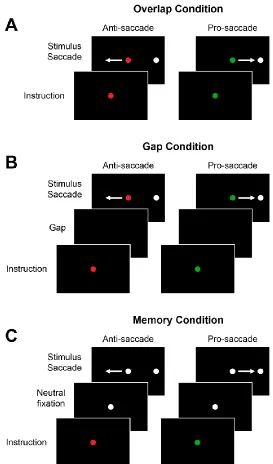
1.5 The Anti-saccade Task
The anti-saccade task, where a saccade is generated away from a peripheral stimulus to
the mirror opposite location, has been extensively used in conjunction with the pro-saccade task,
where a saccade is generated towards a peripheral stimulus, to investigate cognitive control (Fig.
1.4) (Hallett, 1978; Munoz and Everling, 2004). These tasks are particularly useful because they
have distinct stimulus-response associations and behaviour is consistent and comparable between
humans and monkeys. Successful anti-saccade performance first requires the inhibition of the
prepotent response to look at the peripheral stimulus, then the inversion of the stimulus vector to
generate a saccade away from the stimulus. Thus, anti-saccades require additional processing
compared to pro-saccades and are associated with greater reaction times (Fischer and Weber,