Scholarship@Western
Scholarship@Western
Electronic Thesis and Dissertation Repository
11-1-2019 2:00 PM
The role of Xenopus laevis RECK in ECM remodeling and tissue
The role of Xenopus laevis RECK in ECM remodeling and tissue
patterning
patterning
Jessica WillsonThe University of Western Ontario
Supervisor
Damjanovski, Sashko
The University of Western Ontario Graduate Program in Biology
A thesis submitted in partial fulfillment of the requirements for the degree in Doctor of Philosophy
© Jessica Willson 2019
Follow this and additional works at: https://ir.lib.uwo.ca/etd
Part of the Developmental Biology Commons
Recommended Citation Recommended Citation
Willson, Jessica, "The role of Xenopus laevis RECK in ECM remodeling and tissue patterning" (2019). Electronic Thesis and Dissertation Repository. 6645.
https://ir.lib.uwo.ca/etd/6645
This Dissertation/Thesis is brought to you for free and open access by Scholarship@Western. It has been accepted for inclusion in Electronic Thesis and Dissertation Repository by an authorized administrator of
ii
Proper cell-cell and cell-extracellular matrix (ECM) interactions are vital for cell
migration and patterning of the vertebrate embryo. Matrix metalloproteinases (MMPs) and
their inhibitors, reversion-inducing cysteine-rich proteins with Kazal motifs (RECK) and
tissue inhibitors of metalloproteinases (TIMPs), are all differentially expressed during
embryogenesis to regulate such ECM remodeling events and cell interactions. While TIMPs
are a family of 4 secreted proteins that share overlapping substrate specificities of MMPs,
RECK is unique in that it is a membrane-anchored MMP inhibitor that is embryonic lethal in
mice. I used Xenopus laevis as a model organism to investigate the role of RECK as a
regulator of ECM turnover during development. The X. laevis RECK sequence was
compared to a breadth of vertebrate and a few invertebrate RECK amino acid sequences. The
X. laevis RECK amino acid sequence was found to be highly conserved with other RECK
proteins. RECK knockdown in X. laevis embryos resulted in neural tube closure failure and
axial defects, in part due to altered mRNA levels of MT1-MMP, MMP-2, and TIMP-2. Upon
examination of RECK, MT1-MMP, and TIMP-2 protein localization in different tissues
throughout early X. laevis development, I found that all 3 proteins showed highly similar
localization patterns, particularly in the dorsal-ventral differentiation of the neural tube. To
further investigate RECK regulation of MMP activity in vitro, I used X. laevis A6 cells to
knockdown, overexpress, and shed RECK from the cell surface. I demonstrated that changes
in RECK levels (overexpression and cell surface shedding) that may reduce the ability of the
cell to remodel the ECM are compensated for by increases in MT1-MMP and MMP-2 levels
and changes in ERK signaling. Altogether, these results support a role for RECK in the
iii
Summary for Lay Audience
All cells secrete or release materials around themselves, allowing tissues to form. In
multicellular organisms such as ourselves, some of these materials form a network (the
extracellular matrix or ECM) in which the cells anchor themselves to. In adults, the ECM
provides stability and a safe environment in which our cells can carry out their functions -
muscle cells contract, nerve cells communicate, and so on. But the situation is different in a
developing embryo. As animals grow from a fertilized egg, cells need to move. In adults,
when cells move, it is often associated with a disease state, such as cancer cells
metastasizing. Thus, understanding how cell movement is regulated is essential to both
embryogenesis and adults. For cells to move around, the ECM network that surrounds them
often has to be broken down and then remade. The molecules involved in this breakdown are
called matrix metalloproteinases (MMPs), and one of their roles is to cleave the ECM. Their
activity has to be tightly regulated as excessive ECM degradation is detrimental. Therefore,
there are 2 other molecules, tissue inhibitors of metalloproteinases (TIMPs) and
reversion-inducing cysteine-rich protein with Kazal motifs (RECK), whose roles are to control MMPs.
RECK is a protein found on the surface of cells and stops cells from moving by preventing
MMPs from cleaving components of the ECM. RECK has been identified as a crucial player
during embryogenesis, though exactly why RECK is essential to development remains
unclear. My research examined the role of RECK during frog development. I found that
reducing RECK levels in frog embryos caused abnormal development, particularly in the
formation of the spinal cord. These defects were in part due to improper ECM degradation.
Additionally, I found that RECK is associated with MMPs even after cells migrate, at a time
when embryonic cells are beginning to specialize, particularly into nerve cells. Thus, in
iv
breakdown to signal to and help cells function. Overall, my study supports the role for RECK
v
Keywords
RECK; matrix metalloproteinase; tissue inhibitor of metalloproteinase; extracellular matrix;
vi
Dedication
vii
Acknowledgements
First and foremost, I would like to thank my supervisor, Dr. Sashko Damjanovski. I
would not be where I am today without all of your guidance, support, motivation, and
patience. It has been a huge privilege to work in your lab. You have not only been an
amazing mentor, but also a great friend. Thank you for always believing in me.
I would also like to thank my advisory committee, Dr. Susanne Kohalmi and Dr.
Robert Cumming. You have both been a constant source of support and guidance for me
throughout the years.
To all members of the Damjanovski lab, past and present, thank you for all of the
encouragement you have shown me over the years. I would like to especially thank Carlie
Muir and Brad Bork for their constant support and help. Thank you for creating such a fun
and memorable experience in and outside of the lab. You two are both incredibly talented
scientists and I wish you nothing but the very best in your future endeavours.
A huge thanks also goes out to Dr. Kelly’s lab and Dr. Cumming’s lab for their
generosity in providing the use of lab equipment and reagents, all of which have made this
thesis possible.
I would also like to thank all of the staff in the Biology Department - especially Carol
Curtis, Arzie Chant, Diane Gualey, Sherri Fenton, and Hillary Bain - for all of your hard
work behind the scenes to ensure everything went smoothly over the years.
Lastly, I would like to thank my family. To my sister, Cassandra Barber, thank you
for being my confidant throughout all of these years. I’m lucky to call you my best friend. To
my mom, Vicki Barber, thank you for always being there for me and helping me achieve my
goals. I would not be where I am today without all of your love and support. To my husband,
viii
me along this long journey. I will forever be grateful for all of the sacrifices you have made
ix
Table of Contents
Abstract ... ii
Summary for Lay Audience ... iii
Keywords ... v
Dedication ... vi
Acknowledgements ... vii
Table of Contents ... ix
List of Tables ... xiv
List of Figures ... xv
List of Appendices ... xvii
List of Abbreviations ... xviii
Chapter 1 ... 1
1 Global Introduction and Literature Review ... 1
1.1 The Extracellular Matrix ... 2
1.2 Matrix Metalloproteinases ... 3
1.2.1 MT1-MMP ... 3
1.2.2 The Gelatinases ... 6
1.3 Tissue Inhibitors of Metalloproteinases ... 7
1.4 RECK ... 11
1.4.1 Protein Structure ... 11
1.4.2 Tumour Suppression ... 14
1.4.3 MMP Inhibition ... 15
1.5 Regulation of RECK ... 19
1.5.1 Oncogenic Signaling Represses RECK Expression ... 19
x
1.5.3 Glycosylation of RECK ... 20
1.6 RECK in Development ... 20
1.6.1 Angiogenesis ... 21
1.6.2 Neurogenesis ... 24
1.6.3 Neural Crest Cell Migration ... 27
1.6.4 Limb Patterning ... 30
1.7 The X. laevis System ... 31
1.8 Research Project ... 34
1.8.1 Summary ... 34
1.8.2 Hypotheses ... 35
1.8.3 Objectives ... 36
1.9 References ... 38
Chapter 2 ... 46
2 Analysis of Xenopus laevis RECK and Its Relationship to Other Vertebrate RECK Sequences ... 46
2.1 Introduction ... 47
2.2 Materials and Methods ... 49
2.2.1 Animal Care and Rearing ... 49
2.2.2 Cloning X. laevis RECK ... 49
2.2.3 Sequence Analysis ... 50
2.2.4 Immunohistochemistry and Fluorescence Microscopy ... 51
2.2.5 Morpholino Design ... 52
2.2.6 Morpholino Microinjection ... 52
2.2.7 Immunoblot Analysis ... 53
2.2.8 Quantitative Real-Time PCR ... 53
xi
2.3 Results and Discussion ... 54
2.3.1 Sequence Analysis ... 54
2.3.2 RECK Protein Localization ... 61
2.3.3 Functional Analysis ... 64
2.4 Conclusions ... 76
2.5 References ... 78
Chapter 3 ... 81
3 Spatial Analysis of RECK, MT1-MMP, and TIMP-2 Proteins During Early Xenopus laevis Development ... 81
3.1 Introduction ... 82
3.2 Materials and Methods ... 84
3.2.1 Animal Care and Rearing ... 84
3.2.2 Immunohistochemistry and Fluorescence Microscopy ... 84
3.3 Results and Discussion ... 85
3.3.1 RECK Localization During Early X. laevis Development ... 86
3.3.2 MT1-MMP Localization During Early X. laevis Development ... 92
3.3.3 TIMP-2 Localization During Early X. laevis Development ... 98
3.3.4 RECK, MT1-MMP, and TIMP-2 Exhibit Similar Localization Patterns . 99 3.4 Conclusions ... 101
3.5 Supplementary Data ... 103
3.6 References ... 109
Chapter 4 ... 113
4 Modulation of RECK Levels in Xenopus A6 Cells: Effects on MT1-MMP, MMP-2 and pERK Levels ... 113
4.1 Introduction ... 114
4.2 Materials and Methods ... 116
xii
4.2.2 Cell Culture Conditions, Endo-Porter Treatments, Transfections, and
PI-PLC treatments ... 116
4.2.3 Quantitative Real-Time PCR ... 117
4.2.4 Immunoblot analysis ... 118
4.2.5 Gelatin Zymography ... 118
4.2.6 Statistical Analysis ... 119
4.3 Results ... 120
4.3.1 RECK Knockdown in A6 Cells Did Not Alter MT1-MMP or pERK Protein Levels nor MMP-2 Activity Levels ... 120
4.3.2 RECK Overexpression in A6 Cells Caused an Increase in MT1-MMP Protein Levels and Relative Active MMP-2 Levels, and a Decrease in pERK Protein Levels ... 128
4.3.3 Treatment With PI-PLC Reduced Cell-Surface RECK Levels ... 128
4.3.4 PI-PLC Treatment of A6 Cells Caused an Increase in MT1-MMP and Relative Active MMP-2 Levels ... 134
4.3.5 Alteration in RECK Levels or PI-PLC Treatment had Varying Effects on MMP-2, MMP-9, MT1-MMP, and TIMP-2 mRNA Levels ... 134
4.4 Discussion ... 140
4.5 Conclusions ... 145
4.6 References ... 146
Chapter 5 ... 149
5 General Discussion and Conclusions ... 149
5.1 General Overview ... 150
5.1.1 Context and Significance of this Research ... 150
5.1.2 Research Summary and General Conclusions ... 151
5.2 Contributions to the Current Knowledge of ECM Dynamics ... 152
5.2.1 Characterization of X. laevis RECK ... 152
5.2.2 RECK is a Key Regulator During X. laevis Development ... 154
xiii
5.4 Conclusions ... 164
5.5 References ... 166
Appendices ... 169
xiv
List of Tables
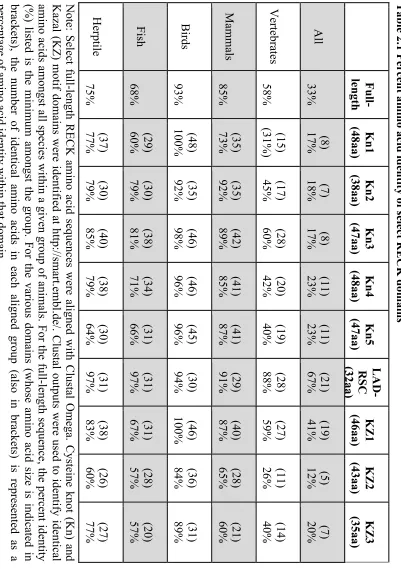
Table 2.1 Percent amino acid identity of select RECK domains ... 58
Table 3.1: Summary of the spatial expression pattern of RECK, MT1-MMP, and TIMP-2
xv
List of Figures
Figure 1.1 MMP domain structure. ... 3
Figure 1.2 Pro-MMP-2 activation. ... 8
Figure 1.3 Domain organization of a typical RECK protein based on the 971 amino acid human protein sequence. ... 11
Figure 1.4 Role of RECK as an MMP inhibitor. ... 16
Figure 1.5 Role of RECK in vascular sprouting. ... 24
Figure 1.6 Role of RECK in neurogenesis. ... 27
Figure 1.7 Schematic representation of X. laevis developmental stages. ... 32
Figure 2.1 X. laevis RECK amino acid sequence ... 56
Figure 2.2 Localization of RECK proteins in late tailbud embryos. ... 61
Figure 2.3 Microinjection of RECK translation-blocking or splice-blocking MO decreased RECK levels in X. laevis embryos. ... 65
Figure 2.4 RECK knockdown embryos displayed reduced survival. ... 67
Figure 2.5 RECK knockdown resulted in developmental defects. ... 69
Figure 2.6 Effect of RECK knockdown on transcript levels. ... 72
Figure 3.1. Localization of RECK and MT1-MMP proteins during early X. laevis development. ... 86
Figure 3.2. Localization of RECK and TIMP-2 proteins during early X. laevis development. ... 89
Figure 3.3. Localization of TIMP-2 and MT1-MMP proteins during early X. laevis development. ... 94
xvi
Figure 4.1. Treatment of A6 cells with RECK MO resulted in decreased RECK protein
levels. ... 120
Figure 4.2. RECK knockdown did not alter MT1-MMP or pERK protein levels or MMP-2
activity levels. ... 123
Figure 4.3. Effect of RECK knockdown on MMP-2, -9, MT1-MMP, and TIMP-2 mRNA
levels. ... 126
Figure 4.4. Transfection of full-length HA-tagged RECK constructs in A6 cells resulted in
increased RECK levels. ... 129
Figure 4.5. RECK overexpression increased MT1-MMP protein and MMP-2 activity levels
and decreased pERK protein levels. ... 131
Figure 4.6. Solubilization of RECK proteins following PI-PLC treatment. ... 135
Figure 4.7 PI-PLC treatment caused an increase in MT1-MMP protein and MMP-2 activity
levels. ... 137
Figure 4.8 Effect of RECK overexpression and PI-PLC treatment on MMP-2, MMP-9,
MT1-MMP, and TIMP-2 mRNA levels. ... 141
Figure 5.1 Schematic representation of overlapping RECK, MT1-MMP, and TIMP-2
proteins in the D-V differentiation of the neural tube during X. laevis development. ... 157
xvii
List of Appendices
Appendix A Copyright Permission ... 169
Appendix B X. laevis RECK full-length coding sequence ... 170
Appendix C Amino acid sequences of the 3 Kazal motifs among different species ... 171
Appendix D The developmental phenotypes of X. laevis embryos ... 172
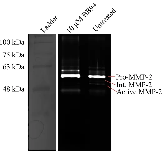
Appendix E Zymography for BB94 treatment ... 173
Appendix F Zymography for MMP-9 detection ... 174
Appendix G UWO Biosafety Certificate ... 175
xviii
List of Abbreviations
ADAM A disintegrin and metalloproteinase
ANOVA Analysis of variance
AP Anterior/posterior
A.U. Arbitrary units
BB94 Batimastat
BMP Bone morphogenetic protein
Bp Base pair
BSA Bovine serum albumin
CD Clusters of differentiation
cDNA Complementary deoxyribonucleic acid
DAPI 4’,6-diamidino-2-phenylindole
DIC Differential interference contrast
Dll Delta-like
DMSO Dimethyl sulfoxide
Dpf Days post fertilization
DQ Dye-quenched
DRG Dorsal root ganglia
D-V Dorsal-ventral
ED Embryonic day
ECM Extracellular matrix
EF1a Elongation factor one alpha
EGF Epidermal growth factor
xix
FBS Fetal bovine serum
Fra Fos-related antigen
GDE Glycerophosphodiester phosphodiesterase
GFP Green fluorescent protein
GPI Glycosylphosphatidylinositol
HA Hemagglutinin
HDAC Histone deacetylase
HRP Horseradish peroxidase
ICD Intracellular domain
IHC Immunohistochemistry
MAPK Mitogen-activated protein kinase
MMP Matrix metalloproteinase
MMR Marc’s modified ringers
MO Morpholino
mRNA Messenger ribonucleic acid
MT Membrane type
PBS Phosphate-buffered saline
pERK Phosphorylated extracellular signal-regulated kinase
PI-PLC Phosphatidylinositol-specific phospholipase C
PVDF Polyvinylidene fluoride
qPCR Quantitative polymerase chain reaction
Rap Ras-related protein
RECK Reversion-inducing cysteine-rich protein with Kazal motifs
xx
RLM-RACE RNA Ligase Mediated Rapid Amplification of cDNA Ends
SDS Sodium dodecyl sulphate
SE Standard error
SEM Standard error of the mean
SF Serum-free
Shh Sonic hedgehog
shRNA Short hairpin ribonucleic acid
SMART Simple Modular Architecture Research Tool
Sp Specificity protein
Src Proto-oncogene tyrosine-protein kinase
STAT3 Signal transducer and activator of transcription factor 3
TBST Tris-buffered saline with Tween 20
TIMP Tissue inhibitor of metalloproteinase
TRE 12-O-tetradecanoylphorbol-13-acetate-responsive element
VEGF Vascular endothelial growth factor
Chapter 1
1
Global Introduction and Literature Review
Portions of this chapter have been published as a review article: “Willson, J.A. and
Damjanovski, S. (2014). Vertebrate RECK in development and disease. Trends in Cell
and Molecular Biology, 9: 95-105”. (Reprint permission in Appendix A). The text has
been modified from the original manuscript to adhere to formatting guidelines for this
1.1
The Extracellular Matrix
The extracellular matrix (ECM) is the non-cellular network of interacting
macromolecules that surrounds cells in metazoans. It is primarily composed of
proteoglycans, which are large hydrophilic molecules, and large fibrous proteins, such as
collagens and fibronectin, that provide structural support to cells. These fibrous proteins
may contain important signaling domains, such as the epidermal growth factor (EGF)
domain of laminin and heparin-II domain of fibronectin, which influence cell behaviour
(Daley et al., 2008). There are 2 main types of ECM in animal tissues: basement
membrane and interstitial matrix. The basement membrane is a thin layer of defined
ECM that underlies epithelial cells, providing a barrier between epithelia and
mesenchyme, whereas the interstitial matrix is a vast network of ECM surrounding
sparsely packed cells that make up connective tissue, such as cartilage and bone (McKee
et al., 2019). The amount and composition of ECM is specific to each type of tissue and
is crucial for the overall integrity and function of that tissue.
The ECM not only provides a scaffolding network for cells, but it also sequesters
a variety of signaling molecules including cytokines and growth factors, that once
released, regulate cell growth, survival, migration, and differentiation (Daley et al.,
2008). Changes in cell-cell and cell-ECM contact and the release of these sequestered
proteins occur through cleavage and remodeling of the ECM, and as such, these
processes are crucial for proper development and maintenance of multicellular organisms
(Daley et al., 2008). The integrity and breakdown of the ECM is regulated mainly
through the combined action of a group of extracellular proteases called matrix
1.2
Matrix Metalloproteinases
There are 24 vertebrate MMPs, each with their own, but overlapping, ECM
substrate specificities. MMPs are present in all metazoans and their expression is crucial
in guiding many embryonic cell signaling and migration events (Visse and Nagase,
2003). MMPs are classified into 2 categories based on their localization and structure:
secreted MMPs, which are soluble in the ECM, and membrane type (MT)-MMPs, which
are tethered to the cell membrane. The 3 common domains of MMPs are an N-terminal
signal sequence that targets them for secretion, a pro-domain that inhibits catalytic
activity, and a catalytic domain with endopeptidase activity (summarized in Fig. 1.1). All
MMPs are synthesized as inactive zymogens and become activated with the removal of
their pro-domain. MT-MMPs reach the cell surface in an active form, as they have their
pro-domain cleaved intracellularly in the Golgi by furin-like proteases, whereas secreted
MMPs are activated in the ECM by other active MMPs already present (Sternlicht and
Werb, 2001). Once active, the common, but not only function of all MMPs, is to cleave
and remodel the ECM through selective degradation of ECM proteins (Vu and Werb,
2000).
1.2.1
MT1-MMP
A well-characterized member of the MMP family is MT1-MMP. Unlike secreted
MMPs, which can diffuse freely though the ECM, MT-MMPs are anchored to the cell
surface and their function is therefore restricted to the pericellular space (Seiki, 2002). Of
the many MMP null mice, only MT1-MMP null mice die shortly after birth due to severe
skeletal defects (Holmbeck et al., 1999). MT1-MMP can cleave major components of the
Figure 1.1MMP domain structure.
The various domains of human MMPs are shown. SP=signal peptide, Pro=pro-domain,
Cat=catalytic domain, F=fibronectin II repeats, PEX=hemopexin domain,
TM=transmembrane domain, C=cytoplasmic domain, GPI=Glycosylphosphatidylinositol
anchor, Cys=cysteine array, Ig=immunoglobulin-like domain. The flexible hinge region
SP Pro Cat Cys Ig
SP Pro Cat
SP Pro Cat
PEX
SP Pro Cat F F F
SP Pro Cat TM C
SP Pro Cat GPI
MMP-7, -26
MMP-1, -3, -8, -10, -11, -12, -13, -19, -20, -21, -27, -28
MMP-2, -9
MT1-MMP (MMP-14), MT2-MMP (MMP-15), MT3-MMP (MMP-16), MT5-MMP (MMP-24)
MT4-MMP (MMP-17), MT6-MMP (MMP-25)
MMP-23
PEX
PEX
such as clusters of differentiation 44 (CD44) and activate pro-MMP-2 (Gifford and Itoh,
2019). These non-proteolytic functions may in fact help to explain its embryonic
necessity.
One of the confounding properties of MT1-MMP is its ability to cause an increase
in cell migration even when it is in a proteolytically inactive form (Bonnans et al., 2008;
Hara et al., 2011). These studies suggest that MT1-MMP may influence cell migration
beyond a role that relies solely on ECM degradation. In fact, recent studies have shed
light on a new mechanism of MT1-MMP involving extracellular signal-regulated (ERK)
activation (Cepeda et al., 2016; Cepeda et al., 2017; Takino et al., 2010; Willson et al.,
2018). The mitogen-activated protein kinase (MAPK)/ERK pathway is a signaling
cascade that is initiated by an extracellular mitogen, which subsequently leads to the
activation of ERK and the transcription of various genes involved in growth, survival,
differentiation, and development (Morrison, 2012). These multifunctional properties
reveal the importance of MT1-MMP in both cell-ECM interactions as well as cellular
behaviour.
1.2.2
The Gelatinases
The gelatinases, MMP-2 and MMP-9, have been identified as 2 potent secreted
proteases due to their ability to cleave gelatin, but more importantly collagen type IV, a
major component of basement membranes (Khasigov et al., 2003). Since cleavage of the
basement membrane is an important step during cell migration, MMP-2 and MMP-9 have
been implicated to play important roles during both development and cancer progression.
Although the severity of defects that arise from knocking out only one MMP gene may be
family, defects have still been observed in single MMP knockout mice. For example,
MMP-2 null mice display craniofacial defects (Mosig et al., 2007), and MMP-9 null mice
exhibit defects in endochondral bone formation (Vu et al., 1998), both of which rely on
extensive ECM degradation and cell migration.
In adults, ECM remodeling is limited, and any disturbances in MMP levels can
contribute to the onset and progression of diseases (Visse and Nagase, 2003). While the
dysregulation of MMPs is associated with a number of disease states that involve cell
migration, including atherosclerosis (Lin et al., 2014), fibrosis (Giannandrea and Parks,
2014), and arthritis (Burrage et al., 2006), a major focus has been on their role in cancer,
particularly the importance of MMP-2 and MMP-9, whose upregulation have been
associated with tumour progression and metastasis (Bergers et al., 2000; Bernhard et al.,
1994; Jodele et al., 2005; Mook et al., 2004). Therefore, as it is crucial that MMP activity
be tightly regulated, there are a number of different endogenous inhibitors that control
MMP activity, including tissue inhibitors of metalloproteinases (TIMPs) (Visse and
Nagase, 2003).
1.3
Tissue Inhibitors of Metalloproteinases
TIMPs, endogenous inhibitors of MMPs, are a small family of secreted proteins.
All 4 TIMPs have been identified in mammals, but not all 4 are found in all vertebrates.
For example, only TIMP-1, TIMP-2, and TIMP-3 have been identified in frog.
Depending on the type, TIMPs are either expressed constitutively or in a tissue-specific
manner, and each are regulated at the transcriptional level through cytokine and growth
factor signaling (Murphy, 2011). For example, TIMP-2 is constitutively expressed,
2004). Overall, each TIMP has overlapping abilities to inhibit members of the MMP
family. TIMPs contain structurally and functionally distinct N- and C-terminal domains.
The N-terminal domain binds to MMPs with a 1:1 molar stoichiometry and inhibits MMP
catalytic activity (Gomis-Ruth et al., 1997), whereas the C-terminal domain functions
independently by binding to cell surface receptors and inducing cell signaling cascades
(Visse and Nagase, 2003).
One of the best described non-MMP-inhibitory functions of TIMPs involves
TIMP-2. As previously mentioned in Section 1.2.1, an important function of MT1-MMP
is its ability to activate pro-MMP-2 (Itoh et al., 2001). The mechanism involves binding
of the C-terminal domain of TIMP-2 to the hemopexin-like domain of pro-MMP-2, while
the N-terminal domain of that same TIMP-2 molecule binds the catalytic site of
MT1-MMP and inhibits its proteolytic activity. This complex then allows an adjacent
active MT1-MMP molecule to cleave the pro-domain of MMP-2, which releases it from
the complex in an intermediate form. The intermediate form of MMP-2 then
autocatalyzes into its active form (Fig. 1.2) (Itoh et al., 2001). This ability of MT1-MMP
to activate MMP-2 and thus increase ECM remodeling therefore further potentiates cell
migration.
Due to their importance in mediating ECM turnover, the balance between MMPs
and TIMPs is crucial for proper development to occur. For example, overexpression of
TIMP-1, TIMP-2 or TIMP-3 during early Xenopus laevis development all resulted in
severe developmental defects (Nieuwesteeg et al., 2012; 2014). Until recently, TIMPs
were thought to be the main MMP inhibitors, however, another protein has been
Figure 1.2 Pro-MMP-2 activation.
The C-terminal domain of TIMP-2 binds to the hemopexin-like domain of pro-MMP-2,
while the N-terminal domain of TIMP-2 binds the catalytic site of a MT1-MMP. An
adjacent active MT1-MMP protein then cleaves the pro-domain of MMP-2, thus
releasing MMP-2 in its intermediate form. The intermediate form of MMP-2 then
undergoes autocatalytic cleavage to generate an active form of MMP-2. (Based on Itoh et
Pro-MMP-2
TIMP-2
Active MMP-2
N C
Extracellular matrix
Intermediate MMP-2
protein with Kazal motifs (RECK) encodes a membrane-anchored protein and regulates
ECM remodeling by inhibiting MMP activity (Takahashi et al., 1998).
1.4
RECK
1.4.1
Protein Structure
RECK was originally discovered by screening a human fibroblast cDNA library
for genes that induced flat cell morphology (reversion-inducing clones) when expressed
in transformed mouse fibroblasts (Takahashi et al., 1998). Mammalian RECK proteins
are 971 amino acids in length and weigh approximately 110 kDa (Takahashi et al., 1998).
Many vertebrate and invertebrate RECK sequences cloned to date are highly conserved at
the amino acid level and share the domains characteristic of RECK (Willson and
Damjanovski, 2014). RECK proteins contain hydrophobic regions flanking both ends of
the protein. The N-terminal region encodes a signal peptide sequence and the C-terminal
region encodes a glycosylphosphatidylinositol (GPI)-anchoring signal sequence (Fig. 1.3)
(Takahashi et al., 1998). RECK was confirmed as a GPI-anchored protein by treatment
with phospholipase C, an enzyme that selectively cleaves GPI-anchored proteins from the
surface of cells (Takahashi et al., 1998). RECK proteins are also rich in cysteine residues
(9%) and contain 5 repeats of a putative cysteine knot motif located near their N-terminus
(Fig. 1.3) (Takahashi et al., 1998).
As the name suggests, RECK proteins contain Kazal motifs, which are
serine-protease inhibitor-like domains (Fig. 1.3) (Rimphanitchayakit and Tassanakajon,
2010). There are 3 Kazal motif domains in RECK proteins. Evidence shows that these
Kazal motifs play an important role in MMP inhibition (Chang et al., 2008). The middle
Figure 1.3 Domain organization of a typical RECK protein based on the 971 amino
acid human protein sequence.
Mammalian RECK proteins contain a signal sequence at the N-terminus and a GPI
anchor signal sequence at the C-terminus (which are not present in the mature protein).
The N-terminal region of RECK contains 5 putative cysteine knot motifs. The C-terminal
region contains 3 Kazal motifs (MMP inhibitory domains) and 2 epidermal growth
Signal sequence
Cysteine knot motif
EGF-like repeat
Kazal motif
GPI anchor sequence
et al., 1998). EGF domains are common to membrane-bound and secreted proteins and
have the potential to bind to EGF receptors and stimulate mitosis (Wouters et al., 2005).
1.4.2
Tumour Suppression
Since its discovery in transformed fibroblasts, RECK quickly became recognized
as a tumour suppressor protein, and studies were carried out to determine if there was a
correlation between RECK expression and tumour cell invasiveness. In normal adult
tissues, RECK mRNA is relatively highly expressed, however, RECK expression is low
and sometimes undetectable in many tumour-derived cell lines (Takahashi et al., 1998).
Takahashi et al. (1998) was the first to determine the anti-invasive properties of RECK
by generating stable RECK-expressing fibrosarcoma cells and showing that invasiveness
significantly decreased in RECK-expressing cells versus control. Soon after, various
human tumours were analyzed to determine the relationship between RECK expression
and prognosis, including breast (Span et al., 2003), lung (Takenaka et al., 2005),
pancreatic (Masui et al., 2003), colorectal (Takeuchi et al., 2004), and prostate (Ohl et al.,
2005) cancer. These studies concluded that high RECK expression in tumours correlated
with better prognosis and survival rate.
Given that loss of RECK is associated with tumour progression, Walsh et al.
(2015) examined if loss of RECK is sufficient to transform normal cells. They knocked
down RECK in normal human mammary epithelial cells and examined cell
transformation using xenograft assays. RECK knockdown was not sufficient to induce
transformation (Walsh et al., 2015). Therefore, although reconstitution of RECK in
transformed cells reduces invasiveness, loss of RECK does not lead to malignant
1.4.3
MMP Inhibition
RECK was originally identified as a tumour suppressor due to its ability to
regulate MMP activity (Takahashi et al., 1998). Numerous in vitro studies have shown
that RECK can negatively regulate MMP-2, MMP-9, and MT1-MMP activity (Fig. 1.4)
(Chang et al., 2008; Matsuzaki et al., 2018; Oh et al., 2001; Simizu et al., 2005;
Takahashi et al., 1998). The pioneering study was conducted by Takahashi et al. (1998)
who saw a decrease in the amount of pro-MMP-9 in serum collected from fibrosarcoma
cells transfected with RECK. This result only occurred when RECK was
membrane-bound. When RECK was solubilized, pro-MMP-9 levels in the serum did not change
(Takahashi et al., 1998). Oh et al. (2001) expanded on these results and reported that
levels of active MMP-2 also decreased in RECK-expressing fibrosarcoma cells. They also
showed that RECK can directly interact with MT1-MMP and inhibit its proteolytic
activity.
Since RECK proteins contain Kazal motifs, which are protease-inhibitor domains,
it is no surprise that evidence suggests these domains play a role in MMP inhibition.
Chang et al. (2008) generated recombinant proteins containing all 3 Kazal motifs (K123)
or only the last 2 Kazal motifs (K23). Using human lung cancer cells, they showed that
K23 recombinant proteins reduced pro-MMP-9 and active MMP-2 and MMP-9 levels.
Moreover, K23 recombinant proteins bound directly to MMP-9. Surprisingly, K123 did
not show any MMP inhibition, however, they attributed this result to protein misfolding
(Chang et al., 2008). Their results also showed that RECK does not need to be
membrane-bound in order for inhibition of pro-MMP-9 to occur (Chang et al., 2008),
Figure 1.4 Role of RECK as an MMP inhibitor.
RECK is a membrane-anchored protein and has been shown to directly inhibit
MT1-MMP, MMP-2, and MMP-9 activity. RECK can also inhibit the secretion of
pro-MMP-9, although the mechanism is unknown. RECK has also been shown to inhibit
MMP-9 transcription by preventing Fra-1 and c-Jun from binding to the TRE-1 binding
site on the MMP-9 promoter region, leading to less secreted pro-MMP-9. (Based on
RECK
Cytosol
Extracellular matrix
Nucleus
MMP-2
MT1-MMP
MMP-9
MMP-9
TRE-1
c-Jun Fra-1
pro-MMP-9 levels using solubilized RECK proteins (Takahashi et al., 1998).
An important factor in the progression of tumour growth and metastasis is the
branching of new vascular networks from pre-existing ones, termed angiogenesis
(Hoeben et al., 2004). With respect to tumour progression, not only does RECK decrease
invasiveness, but RECK has also been shown to suppress tumour angiogenesis. When
RECK-expressing tumour cells were inoculated into mice, RECK+-tumoured mice lived
longer and displayed a reduction in angiogenic sprouting, as was seen by laminin staining
versus the mice inoculated with control tumours (Oh et al., 2001).
As a GPI-anchored protein, RECK does not contain an intracellular domain,
however, many GPI-anchored proteins are involved in transducing cell-signaling
cascades by binding to neighboring receptors (Mayor and Riezman, 2004). Takagi et al.
(2009) were the first to discover that RECK decreases MMP-9 transcription.
Overexpression of RECK in fibrosarcoma cells caused a decrease in MMP-9 mRNA, but
not MMP-2 mRNA. Although they also saw a decrease in the amount of MMP-9 present
in the serum from RECK-expressing fibrosarcoma cells, they attributed this result to the
decrease in MMP-9 mRNA, not inhibition of pro-MMP-9 secretion. They discovered that
RECK overexpression suppressed the binding of Fos-related antigen 1 (Fra-1) and c-Jun
transcription factors to the 12-O-tetradecanoylphorbol-13-acetate-responsive element-1
(TRE-1) binding site within the MMP-9 promoter region in fibrosarcoma cells. However,
the mechanisms underlying this process are currently unknown. For example, there is no
evidence to suggest that RECK translocates to the nucleus. Instead, RECK may act on the
surface of cells by interacting with other cell surface receptors (Takagi et al., 2009).
inhibit its catalytic activity, but it may also modulate the endocytic pathway of
MT1-MMP. A study performed by Miki et al. (2007) examined whether RECK
influences the clearance of MT1-MMP and CD13, another membrane protease, from the
cell surface. They found that when RECK was present, it complexed with MT1-MMP
and CD13 and caused preferential endocytosis of these proteins using a novel endocytic
pathway that was caveolae-independent. This study highlights the multifaceted properties
of RECK as a cell surface protein.
1.5
Regulation of RECK
1.5.1
Oncogenic Signaling Represses
RECK
Expression
The MAPK/ERK pathway plays a crucial role in cell proliferation and
differentiation, however, aberrant activity of this pathway can lead to malignant
transformation (Kohno and Pouyssegur, 2006). For example, activated RAS causes a
decrease in ECM proteins and receptors and an increase in MMPs, such as MT1-MMP
(Howard et al., 1978; Plantefaber and Hynes, 1989; Thant et al., 1997). RECK was
discovered as a downstream target of oncogenic signaling, as reconstitution of RECK in
ras-transformed fibroblasts induced reversion (Takahashi et al., 1998). As such, studies
have been carried out to determine how RECK is regulated at the transcriptional level by
the MAPK/ERK pathway. There is a specificity protein 1 (Sp1)-binding site within the
RECK promoter region that has been identified as a repressor of RECK transcription
(Sasahara et al., 1999). Activation of ERK causes phosphorylation of Sp1, which is then
recruited to the Sp1 binding site along with histone deacetylase 1 (HDAC) and results in
1.5.2
TIMP-2 Induces
RECK
Expression
TIMPs are multi-faceted proteins. Not only do TIMPs inhibit MMPs, but they can
also bind to cell surface receptors and induce cell-signaling cascades (Li et al., 1999; Oh
et al., 2004). Of importance is the upregulation of RECK expression by TIMP-2. TIMP-2
was shown to bind to α3β1 integrins on the surface of human endothelial cells, causing
inactivation of proto-oncogene tyrosine-protein kinase (Src) through changes in paxillin
phosphorylation (Oh et al., 2004). Alteration of paxillin phosphorylation regulates the
activation of the small GTPase Ras-related protein 1 (Rap1), which ultimately results in
increased expression of RECK through unknown mechanisms (Oh et al., 2004).
1.5.3
Glycosylation of RECK
RECK has also been shown to be regulated at the post-translational level. RECK
contains 5 putative glycosylation sites at the N-terminal portion of the protein (Takahashi
et al., 1998). Glycosylation of 3 of the 5 potential asparagines of RECK are required to
suppress tumour cell invasion (Simizu et al., 2005). Simizu et al. (2005) generated RECK
mutants where they replaced asparagines with glutamines. RECK mutant constructs were
transfected into fibrosarcoma cells to measure the amount of MMP-2 and MMP-9 levels
in the media. Glycosylation of Asn297 was required to inhibit MMP-9 secretion and
Asn352 was required to inhibit the activation of pro-MMP-2. They also correlated
glycosylation of RECK with tumour cell invasion and found that glycosylated Asn86,
Asn297, and Asn352 residues were required to suppress fibrosarcoma cell invasion (Simizu
et al., 2005).
1.6
RECK in Development
such, MMPs are expressed, and studies have found that aberrant upregulation of MMPs
has been associated with developmental defects and death (Page-McCaw et al., 2007).
Therefore, to maintain an appropriate level of MMP activity, MMP inhibitors are also
crucial for proper development to occur. Although TIMP-1 or TIMP-2 deficiency in
mouse embryos has little effect on development (Caterina et al., 2000; Nothnick et al.,
1997), most likely due to functional redundancy between TIMPs, RECK is necessary for
mouse development. Oh et al. (2001) were the first to report that RECK knockout in mice
resulted in embryonic lethality.
1.6.1
Angiogenesis
When the RECK gene was knocked out in mice, about 2/3 of RECK-/- embryos
died halfway through embryogenesis at embryonic day (ED) 10.5, and none survived past
ED11.5 (Oh et al., 2001). RECK-/- embryos had overt phenotypes, which included
abdominal hemorrhaging, disorganized vascularization, disrupted organogenesis, and
smaller body sizes (Oh et al., 2001). Although vascular networks were present in RECK
-/-embryos, histological examination of these tissues indicated abnormally large and
deformed blood vessels, which indicated defects in angiogenesis rather than
vasculogenesis (Oh et al., 2001).
Due to its characterized role in MMP inhibition, RECK-/- mice were analyzed to
determine if there was aberrant upregulation of MMPs in vivo. Oh et al. (2001) plated
ED10.5 cells and collected the serum. MMP-2 activity was elevated in RECK-/- embryos,
however, since pro-MMP-9 is not expressed until ED11, MMP-9 activity was not
detected (Oh et al., 2001). Collagen IV and laminin were also disrupted in areas
These results suggest that loss of RECK caused aberrant upregulation of MMPs and
disruption of the basal lamina. This is supported by the fact that RECK, MMP-2, and
MT1-MMP expression patterns overlap in the areas surrounding the neural tube.
Furthermore, a similar phenotype was seen in collagen 1-/- mice, which also died due to
ruptured blood vessels (Löhler et al., 1984). A partial rescue of the RECK mutant phenotype was obtained by generating RECK-/-/MMP-2-/-double mutants. These embryos
had larger body sizes and improved vascular integrity, however, they still died half a day
later (ED11.5) (Oh et al., 2001). On the other hand, RECK-/-/MT1-MMP-/-mutants did not
rescue the RECK mutant phenotype (Oh et al., 2001).
Chandana et al. (2010) tried a conditional RECK knockout by silencing the RECK
gene using tamoxifen-inducible treatment at ED11, however, mice still showed lethality.
Conditional RECK-/- mutants had large and deformed blood vessels as well as large
cavities in the brain vasculature at ED15.5 (Chandana et al., 2010). This phenotype was
also apparent when Prendergast et al. (2012) knocked down RECK in zebrafish using
Morpholinos (MO). RECK knockdown embryos had impaired vascular integrity and
intracranial hemorrhaging 48 hours post-fertilization (Prendergast et al., 2012). This
suggests that RECK function is conserved throughout development and essential
particularly with respect to vascular development.
Since RECK was implicated to play a role in angiogenesis during development,
Chandana et al. (2010) decided to examine the importance of RECK in maternal vascular
remodeling in mice. The uterus of a pregnant mouse is one of the most active sites of
angiogenesis in adult mice (Abrahamsohn and Zorn, 1993; Cross et al., 1994; Dey et al.,
remodeling in the uterus and are required for proper ECM remodeling (Curry and Osteen,
2003; Wang and Dey, 2006). However, it is unclear how some blood vessels are
protected while others undergo extensive vascular sprouting. RECK was implicated to
play a role in vascular remodeling in maternal tissues. RECK is expressed in cells
associated with remodeling blood vessels in the mouse implantation chamber (Chandana
et al., 2010). When they knocked down RECK expression within the implantation
chamber using short hairpin RNA (shRNA), cavities and tissue slits were present instead
of organized and compact blood vessels (Chandana et al., 2010). They also saw decreased
collagen IV surrounding the deformed blood vessels (Chandana et al., 2010). This
phenotype is very similar to the vascular defects seen in RECK-/- embryos and supports
the role of RECK as a key regulator during angiogenic sprouting (Chandana et al., 2010).
Mechanisms of RECK action during angiogenesis have been proposed by Chandana et al.
(2010). The most likely scenario suggests that RECK helps mark and protect the blood
vessels that are preserved during angiogenic sprouting by inhibiting surrounding MMPs.
However, RECK-/- embryos cannot be rescued by MMP null mutations, although this may
be due to functional redundancy between MMPs. Instead, RECK may affect intracellular
signaling pathways by protecting ECM proteins and other cell surface receptors
(Chandana et al., 2010). Angiogenesis occurs when vascular endothelial growth factor
(VEGF) and Notch signaling regulate endothelial tip cell formation (Adams and Alitalo,
2007; Carmeliet, 2005; Roca and Adams, 2007). Delta-like 4 (Dll4), a ligand of Notch
signaling, has been reported to reduce tip cell formation and thus vascular sprouting
(Phng and Gerhardt, 2009). Interestingly, the vascular defects seen in RECK-/- embryos
signaling (Krebs et al., 2000; Xue et al., 1999). RECK has been shown to activate Notch
signaling during cortical neurogenesis by protecting Dll4 from being shed from the
membrane (Muraguchi et al., 2007). Therefore, RECK deficiency may suppress Notch
signaling during embryonic and maternal angiogenesis and cause excessive sprouting
(Fig. 1.5). However, this mechanism of action does not solely explain the large cavities
formed in RECK-deficient tissues. The biggest unanswered question that remains is why
RECK is necessary for embryonic angiogenesis but suppresses tumour angiogenesis.
Although it was recently discovered that RECK suppresses angiogenesis by
downregulating signal transducer and activator of transcription 3 (STAT3) (Walsh et al.,
2015), no link has been made to whether or not this process occurs during developmental
angiogenesis.
1.6.2
Neurogenesis
Notch signaling is a highly conserved pathway that is crucial during development to
ensure cell fate specification, stem cell maintenance, and tissue patterning (Roca and
Adams, 2007). In the mammalian central nervous system, Notch ligands, Dll and Jagged,
are expressed on the surface of neural cells and bind to Notch present on the surface of
neighboring neural precursor cells (Roca and Adams, 2007). Once activated, Notch
signaling maintains neural precursor cells in an undifferentiated state (Roca and Adams,
2007). Notch signaling is regulated by a disintegrin and metalloproteinase-10
(ADAM-10) (Yang et al., 2006). ADAM-10 proteases are expressed throughout
development in various neural cells in the mammalian central nervous system and inhibit
Notch signaling by cleaving Notch ligands from the surface of neural cells to allow
Figure 1.5 Role of RECK in vascular sprouting.
During vascular sprouting in tip cells, VEGF and Notch signaling modulate the selective
formation of tip cells. VEGF signaling in tip cells leads to high levels of Dll4. Dll4 binds
to Notch on neighboring stalk cells, which induces Notch signaling and attenuates
VEGFR signaling. RECK has been proposed to regulate vascular sprouting by
maintaining Notch signaling, however the mechanism is currently unknown. (Based on
Stalk cell
ICD
VEGFR signaling (maintenance of stalk cells)
Dl
l4
N
ot
ch
Tip cell
RECK VEGFR
2002).
RECK-/- mice embryos not only had vascular defects, but they also had premature
differentiation of neural precursor cells (Muraguchi et al., 2007). This phenotype was a
result of impaired Notch signaling in the central nervous system (Muraguchi et al., 2007).
They demonstrated that RECK regulated Notch signaling by directly inhibiting
ADAM-10 in neural precursor cells. They confirmed this by showing a direct interaction
between RECK and ADAM-10 by co-immunoprecipitation as well as by
co-electroporating shRNA targeting ADAM-10 and RECK in the central nervous system
of ED12.5 wildtype embryos and rescuing the phenotype induced by RECK depletion
(Muraguchi et al., 2007).
A more recent study expanded on these results and reported that RECK was
regulated within this pathway by glycerophosphodiester phosphodiesterase 2 (GDE2)
(Park et al., 2013). GDE2 is a 6-transmembrane protein that is crucial for the
differentiation of spinal motor neurons by inhibiting Notch signaling (Rao and
Sockanathan, 2005; Sabharwal et al., 2011; Yan et al., 2009). Interestingly, GDE2 and
RECK mRNA expression colocalize in differentiating motor neurons during neurogenesis
in chicks (Park et al., 2013). Using chick spinal cord extracts, they determined that GDE2
cleaves RECK within the GPI-anchor. The release of RECK prevents inhibition of
ADAM-10, which then sheds Dll from the surface and attenuates Notch signaling
(Fig. 1.6) (Park et al., 2013).
1.6.3
Neural Crest Cell Migration
Neural crest cells are a transient population of migratory cells that differentiate
Figure 1.6 Role of RECK in neurogenesis.
RECK maintains Notch signaling in neural progenitor cells by inhibiting the proteolytic
activity of ADAM-10 on the surface of neural cells. ADAM-10 sheds Notch ligands from
the surface, preventing the intracellular domain (ICD) of Notch to translocate to the
nucleus and turn on target genes that maintain neural progenitor cells. The presence of
RECK prevents shedding of Notch ligands and maintains Notch signaling. In addition,
GDE2 regulates RECK by cleaving its GPI-anchor. This prevents inhibition of
ADAM-10 and thus attenuates Notch signaling. (Based on Muraguchi et al., 2007; Park
Extracellular matrix
Cytosol
RECK
Dl
l4
N
ot
ch
RECK
ICD GDE2
ADAM-10
ICD Neural progenitor cell
muscle, sensory ganglia, and craniofacial cartilage and bone (Le Douarin and Kalcheim,
1999). RECK knockdown in zebrafish embryos was also reported to have defects in
neural crest migration (Prendergast et al., 2012). Prendergast et al. (2012) began their
study by screening zebrafish mutants that were unable to form the dorsal root ganglia
(DRG) by looking at neurogenin expression and found that this phenotype was a result of
a mutation in the RECK gene. In the absence of RECK, neural crest cells exhibited
aberrant migratory behaviour and were unable to migrate to the appropriate position for
differentiation into DRG. They confirmed this phenotype by also knocking down RECK
expression using MOs (Prendergast et al., 2012). RECK mutants displayed increased
MMP activity, as shown by taking protein extracts from embryos and incubating them
with dye-quenched (DQ) gelatin, a substrate that allows for quantification of MMP
activity (Prendergast et al., 2012). However, this phenotype could not be rescued by the
treatment of MMP inhibitors (Prendergast et al., 2012). This study was also the first to
determine that RECK function is cell autonomous. Cells taken from wildtype embryos
and transplanted into RECK knockdown embryos were able to form DRG, but they
couldn’t induce the host cells (RECK knockdown cells) to form DRG (Prendergast et al.,
2012).
1.6.4
Limb Patterning
When the RECK gene was knocked out in mice, embryos did not survive past
ED11 (Oh et al., 2001). Therefore, although studies have focused on the role of RECK
early on during development, little is understood regarding developmental processes
occurring later in embryogenesis. Therefore, Yamamoto et al. (2012) generated
development. The most obvious phenotype displayed in these mutants was limb
abnormalities. Further histological examination showed poor chondrocyte condensation
in forelimb buds (Yamamoto et al., 2012). To determine potential morphogenetic
signaling molecules affected by low RECK expression in mice such as Wnt7a signaling
and sonic hedgehog (Shh) signaling, in situ hybridization was performed. These signaling
pathways were all attenuated in low-RECK mutants (Yamamoto et al., 2012).
Interestingly, Wnt7a-/- embryos displayed very similar limb abnormality phenotypes,
whereas deficiencies in the other signaling pathways (such as Shh or bone morphogenetic
protein (BMP)) yielded distinctly different phenotypes (Adamska et al., 2004; Chiang et
al., 1996; Selever et al., 2004). Indeed, Wnt7a expression was greatly reduced in
low-RECK mutant forelimb buds. They came to the conclusion that RECK is required for
Wnt7a signaling in the forelimb bud (Yamamoto et al., 2012).
1.7
The
X. laevis
System
The African clawed frog, X. laevis, has been widely used in the developmental biology
field since the 1950s (Schmitt et al., 2014). The X. laevis system provides a number of
advantages as a model organism, including the ease of in vitro fertilization and rearing as
well as their resilience to experimental manipulation. Moreover, development occurs
rapidly, and each stage has been well-characterized (Nieuwkoop and Faber, 1956).
X. laevis embryos are large (approximately 1 mm in diameter) and therefore
developmental stages are easily recognizable (Fig. 1.7). Development begins at
fertilization (stage 1), followed by the blastula stage (stage 9), at which time zygotic
transcription is turned on. Within the first 24 hours post fertilization, gastrulation occurs
Figure 1.7 Schematic representation of X. laevis developmental stages.
(a) Development begins with a fertilized single cell egg. Stage 9 (blastula stage) marks
the onset of zygotic transcription. Gastrulation occurs between stages 10 and 12 to form
the 3 germ layers (ectoderm, mesoderm, and ectoderm) and the embryonic axes.
Neurulation occurs between stages 14 and 20 to form the neural tube. Organogenesis and
differentiation are predominant in the tailbud stages (stages 21-28) and beyond. By stage
37 the embryo has hatched, and by stage 41 embryos have developed into feeding
tadpoles. (b) Schematic representation of an early tailbud embryo to denote the
embryonic axes. Dorsal refers to back and ventral refers to front (belly). Anterior refers to
endoderm) as well as the embryonic axes, followed by neurulation, which occurs between
stages 14 and 20 to form the neural tube (neural crest cell migration begins during these
stages as well). Organogenesis and differentiation begin at the tailbud stages, and by 3
days post fertilization (dpf), a functional free-swimming tadpole has developed (Fig. 1.7).
Thus, X. laevis has been a fundamental tool in studying cell cycle, cell metabolism, cell
behaviour, and cell migration during development (Hardwick and Philpott, 2015).
For a long time, investigators have taken advantage of the parallels that exist
between embryonic development and cancer metastasis. Many signaling pathways that
are crucial for normal development, such as BMP, Shh, and Notch pathways, become
unregulated during cancer metastasis (Bailey et al., 2007). ECM remodeling is yet
another example of an event that is highly dynamic during both development and cancer
metastasis. Therefore, using X. laevis as a model organism to study ECM remodeling
during development can assist in understanding why this process becomes uncontrolled
during tumour progression.
In vitro cell culture work can also be conducted using A6 cells, a well-established
cell line derived from X. laevis kidney tissue. These cells behave like typical polarized
epithelial cells and can be instrumental in studying cell signaling pathways and cell
behaviour in vitro (Mimori-Kiyosue et al., 2007), pathways which can be difficult to
discern in a multicellular embryo.
1.8
Research Project
1.8.1
Summary
ECM remodeling is a highly dynamic process that is important for the
primarily through MMPs, and their activity is modulated by their endogenous inhibitors,
TIMPs and RECK. Numerous studies have shown that perturbing the delicate balance
that exists between MMPs and their inhibitors can lead to devastating consequences to
the integrity and function of tissues. For example, uncontrolled ECM remodeling can
contribute to the onset and progression of cancer metastasis. For these reasons, it is
essential to understand the functions and interplay of these proteins and how they control
ECM remodeling to ensure proper development and tissue homeostasis.
The importance of RECK during development is becoming more evident, as
studies have already identified a role for RECK during angiogenesis, neural crest cell
migration, and limb patterning. Not only does RECK inhibit MMP function at many
levels, there is also emerging evidence suggesting that RECK may play a more important
role in regulating intracellular signaling. However, most research characterizing RECK
function has been done in vitro in tumour-derived cell lines. My research focused on the
functional characterization of RECK during X. laevis development, a well-established
developmental model. By disrupting endogenous RECK levels, I attempted to elucidate
the molecular mechanisms that are critical for balancing the proteolytic activity of MMPs
with the role of RECK required for proper ECM remodeling and tissue patterning during
development.
1.8.2
Hypotheses
1) Previous studies have revealed similar biological functions of RECK in
vertebrates, therefore, I hypothesize that all domains of vertebrate RECK peptide
2) Since RECK transcripts are localized to dorsal axial structures in X. laevis
embryos, I hypothesize that alteration of RECK levels will cause axial and neural
tube defects in X. laevis embryos resulting from excessive ECM remodeling.
3) Previous studies have demonstrated that RECK is an inhibitor of MT1-MMP,
therefore, I hypothesize that X. laevis RECK will colocalize with MT1-MMP in
dorsal axial structures, including the neural tube and head region.
4) Given that RECK has an established role as an MMP inhibitor, I hypothesize that
knockdown/shedding of RECK in X. laevis A6 cells will result in increased
MT1-MMP and MMP-2 levels, whereas overexpression of RECK will result in
reduced MT1-MMP and MMP-2 levels.
1.8.3
Objectives
The overarching goal of this research was to characterize the role of X. laevis
RECK during development. I previously cloned the mature X. laevis RECK sequence,
which lacked the N-terminal signal sequence and C-terminal GPI-anchor signal sequence,
as part of my Master’s thesis, and performed a preliminary sequence comparison analysis
of RECK. However, that study was limited by the number of available annotated
sequences at the time. For this Doctoral study, I finished cloning the full-length X. laevis
RECK gene and performed a detailed sequence comparison of RECK with more diverse
species to determine evolutionary conservation. This research had the following
objectives:
1) To perform a comparison of amino acid sequence identity of X. laevis RECK to
2) To knock down RECK in X. laevis embryos using a MO approach. Following
RECK knockdown, to examine embryos for any subsequent morphological
changes and developmental defects and correlate these defects with alterations in
mRNA levels of developmental marker genes as well as ECM-remodeling genes
(Chapter 2).
3) To perform immunohistochemistry (IHC) on transverse sections of embryos to
determine the localization patterns of RECK, MT1-MMP, and TIMP-2 during
X. laevis neurulation and organogenesis (Chapter 3).
4) To disrupt endogenous levels of RECK in X. laevis A6 cells using a combination
of overexpression, knockdown, and phosphatidylinositol-specific phospholipase C
(PI-PLC) treatment experiments and examine the effects on MT1-MMP, MMP-2
1.9
References
Abrahamsohn, P.A., Zorn, T.M. (1993). Implantation and decidualization in rodents. J Exp Zool. 266: 603-628.
Adams, R.H., Alitalo, K. (2007). Molecular regulation of angiogenesis and lymphangiogenesis. Nat Rev Mol Cell Biol. 8: 464-478.
Adamska, M., MacDonald, B.T., Sarmast, Z.H., Oliver, E.R. Meisler, M.H. (2004). En1 and Wnt7a interact with Dkk1 during limb development in the mouse. Dev Biol. 272: 134-144.
Bailey, J.M., Singh, P.K., Hollingsworth, M.A. (2007). Cancer metastasis facilitated by developmental pathways: Sonic hedgehog, Notch, and bone morphogenic proteins. J Cell Biochem. 102: 829-839.
Bergers, G., Brekken, R., McMahon, G., Vu, T.H., Itoh, T., Tamaki, K., Tanzawa, K., Thorpe, P., Itohara, S., Werb, Z., Hanahan, D. (2000). Matrix metalloproteinase-9 triggers the angiogenic switch during carcinogenesis. Nat Cell Biol. 2: 737-744.
Bernhard, E.J., Gruber, S.B., Muschel, R.J. (1994). Direct evidence linking expressing of matrix metalloproteinase 9 (92-kDa gelatinase/collagenase) to the metastatic phenotype in transformed rat embryo cells. Proc Natl Acad Sci USA. 91: 4293-4297.
Bonnans, C., Chou, J., Werb, Z. (2008). Remodelling the extracellular matrix in development and disease. Nat Rev Mol Cell Biol. 15: 786-801.
Burrage, P. S., Mix, K.S., Brinckerhoff, C.E. (2006). Matrix metalloproteinases: role in arthritis. Front Biosci. 11: 529-543.
Carmeliet, P. (2005). Angiogenesis in life, disease and medicine. Nature. 438: 932-936.
Caterina, J.J., Yamada, S., Caterina, N.C., Longenecker, G., Holmback, K., Shi, J., Yermovsky, A.E., Engler, J.A., Birkedal-Hansen, H. (2000). Inactivating mutation of the mouse tissue inhibitor of metalloproteinases-2 (Timp-2) gene alters proMMP-2 activation. J Biol Chem. 275: 26416-26422.
Cepeda, M.A., Evered, C.L., Pelling, J.H., Damjanovski, S. (2017). Inhibition of MT1-MMP proteolytic function and ERK1/2 signalling influences cell migration and
invasion through changes in MMP-2 and MMP-9 levels. J Cell Commun Signal. 11:
167-179.
Chandana, E.P.S., Maeda, Y., Ueda, A., Kiyonari, H., Oshima, N., Yamamoto, M., Kondo, S., Oh, J., Takahashi, R., Yoshida, Y., Kawashima, S., Alexander, D.B., Kitayama, H., Takahashi, C., Tabata, Y., Matsuzaki, T., Noda, M. (2010). Involvement of the Reck tumor suppressor protein in maternal and embryonic vascular remodeling in mice. BMC Dev Biol. 10: 84.
Chang, C.K., Hung, W.C., Chang, H.C. (2008). The Kazal motifs of RECK protein inhibit MMP-9 secretion and activity and reduce metastasis of lung cancer cells in vitro and in vivo. J Cell Mol Med. 12: 2781-2789.
Chang, H.C., Liu, L.T., Hung, W.C. (2004). Involvement of histone deacetylation in ras-induced down-regulation of the metastasis suppressor RECK. Cell Signal. 16: 675-679.
Chiang, C., Litingtung, Y., Lee, E., Young, K.E., Corden, J.L., Westphal, H., Beachy, P.A. (1996). Cyclopia and defective axial patterning in mice lacking Sonic hedgehog gene function. Nature. 383: 407-413.
Cross, J.C., Werb, Z., Fisher, S.J. (1994). Implantation and the placenta: key pieces of the development puzzle. Science. 266: 1508-1518.
Curry, T.E. Jr., Osteen, K.G. (2003). The matrix metalloproteinase system: changes, regulation, and impact throughout the ovarian and uterine reproductive cycle. Endocr Rev. 24: 428-465.
Daley, W.P., Peters, S.B., Larsen, M. (2008). Extracellular matrix dynamics in development and regenerative medicine. J Cell Sci. 121: 255-264.
Dey, S.K., Lim, H., Das, S.K., Reese, J., Paria, B.C., Daikoku, T., Wang, H. (2004). Molecular cues to implantation. Endocr Rev. 25: 341-373.
Giannandrea, M., Parks, W.C. (2014). Diverse functions of matrix metalloproteinases during fibrosis. Dis Model Mech. 7: 193-203.
Gifford, V., Itoh, Y. (2019). MT1-MMP-dependent cell migration: proteolytic and non-proteolytic mechanisms. Biochem Soc Trans. 47: 811-826.
Gomis-Ruth, F.X., Maskos, K., Betz, M., Bergner, A., Huber, R., Suzuki, K., Yoshida, N., Brew, K., Bourenkov, G.P., Bartunik, H., Bode, W. (1997). Mechanism of inhibition of the human matrix metalloproteinase stromelysin-1 by TIMP-1. Nature. 221: 77-81.
Hara, T., Mimura, K., Seiki, M., Sakamoto, T. (2011). Genetic dissection of proteolytic and non-proteolytic contributions of MT1-MMP to macrophage invasion. Biochem