T H E BEHAVIOR OF X-RAY INDUCED CHROMOSOMAL ABERRATIONS I N ALLIUM ROOT TIP CELLS
KARL SAX
Arnold Arboretum, Harvard University, Jamaica Plain, Massachusetl-r
Received January 13, 1941
H E R E is considerable evidence that the initial types of chromosomal
T
aberrations induced by X-rays have differential survival rates in sub- sequent cell generations. The initial aberrations include translocations and inversions, which involve only rearrangement of chromosome segments, and alterations which involve losses of chromosome segments-dicentrics, rings, interstitial deletions, and terminal deletions. Although the dicentric and ring chromosomes must constitute a large proportion of the initial aberrations, only inversions and translocations were found to survive in successive cell generations in Crepis plants which had been X-rayed a t the time of seed germination (GERASSIMOVA 1937) or in the progeny of X-rayed seedlings (LEWITSKY 1940). The deficient types of spontaneous chromosomal aberrations also are eliminated in later cell generations in Allium (NICHOLS 1941). On the other hand, the dicentric chromosomes do persist in the endosperm of Zea (MCCLINTOCK 1939; CLARK and COPELAND1940), and ring chromosomes survive during the entire somatic cycle of Zea (MCCLINTOCK 1938).
The failure of dicentric and ring chromosomes to persist in the somatic development of Crepis and Allium may be explained in several ways. The deficient types of aberrations may be lethal or deleterious, or the ends of broken rings and bridges may heal so that the chromosomes divide nor- mally. The various chromosomal aberrations were examined in successive divisions of onion root tip cells in order to determine the cause of the elimination of certain types of aberrations. This work also permitted the analysis of several other problems including the duration of the nuclear cycle in root tips, differential sensitivity, the primary and secondary ef- fects of X-rays, and the effect of delayed germination after irradiation.
Onion bulbs and seeds were subjected to irradiation both before and after germination. Root tips were fixed in alcohol-acetic, and the Feulgen technique was used in staining. The analysis of division figures was based largely on anaphase stages, but satisfactory “metaphase” figures were ob- tained by treating the roots with a 0.1 percent colchicine solution for 24
hours before fixation. Aberration frequency was based on the number of chromosomal alterations per hundred cells.
CHROMOSOMAL ABERRATIONS I N ROOT TIP CELLS
Two different types of effects are produced in cells by X-ray treatments. The first is the “primary” effect (MARQUARDT 1938), which results in the
419 clumping and partial fusion of chromosomes and a temporary inhibition of nuclear development. At the higher X-ray doses nuclear division is sup- pressed for one or two days. With lower doses there is little retardation of nuclear activity, but the clumping and fusion of chromosomes is found for several hours after irradiation.
The primary effect of X-rays is evident at metaphase in less than an hour after raying. The chromosomes are more closely associated on the metaphase plate and appear to be more contracted. At anaphase the sister chromatids separate with difficulty resulting in bridges and occa- sional fragments. The chromatids of such a bridge may ultimately sep- arate, but with a high frequency of bridge formation normal segregation of sister chromatids is prevented, and broken bridges and released frag- ments may produce deficient daughter nuclei. With a low X-ray dosage the primary effect persists for about three hours, but a t higher doses the primary effect lasts considerably longer. According to MARSHAK and HUD-
SON (1937) the aberrations of this type produced in onion root tips are directly related to the X-ray dosage, since the dosage curve is linear.
The “secondary” effect of X-rays involves the production of the typical chromosomal aberrations. These types of aberrations are the same as those induced in Tradescantia miscrospore chromosomes (SAX 1940). When growing root tips are rayed, only chromatid aberrations are found during the first 24 hours following irradiation. Terminal deletions of one or both chromatids are the most frequent types of alterations, although exchanges do occur between the chromatids of different chromosomes. The terminal deletion of both sister chromatids followed by lateral fusion of broken ends gives rise to the single chromatid bridges which are readily detected at anaphase.
Chromosomes rayed in the resting stage usually result in only chromo- some aberrations as a result of breaks before the chromosomes are ef- fectively split into sister chromatids. Occasionally, however, irradiation of dry seeds or dormant bulbs produces a few chromatid aberrations. In the case of the seeds there is evidence that these may be of spontaneous origin. Spontaneous aberrations in dormant bulbs are rare, and the chromatid aberrations observed must be attributed either to the occurrence of the effective split in the late resting stage or to the delay in fusion of broken ends of chromosomes until prophase is initiated and the sister chromatids are differentiated. It seems more probable that breaks may remain open for many hours or perhaps indefinitely in the completely dormant rest- ing nucleus, and that fusions occur only with the initiation of nuclear activity. Some of the fusions may be delayed until the chromsomes are effectively split, resulting in occasional chromatid aberrations.
4 2 0 KARL SAX
versions can be recognized only in extreme cases in the Allium chromo- somes, and the small interstitial deletions are often not easily detected. Only the dicentric and ring chromosomes have been included in the analy- ses. The ratio of dicentric to ring chromosomes is about'9: I. If union of broken ends were a t random, the ratio should be about 30:1. The differ- ence between observed and expected frequencies is of the same order in Tradescantia microspores (SAX 1940). The discrepancy is attributed to spatial limitations in unions of broken ends of chromosomes.
The dicentric chromosomes may separate freely, form a criss-cross bridge, or become interlocked a t anaphase. About a third of the dicen- trics separate freely, and of the dicentric bridges a t anaphase about one- fourth are interlocked. The ring chromosomes may separate freely, form a single large ring, or become interlocked. The acentric fragments associ- ated with these aberrations usually lie free in the cytoplasm and are not included in the daughter nuclei.
The second and subsequent nuclear divisions following irradiation also contain chromosomal aberrations. The secondary divisions can be dis- tinguished from the primaries by the absence or disintegration of the acen- tric fragments. Secondary rings and dicentrics may persist owing to the free separation of these types of aberrations a t the primary-division or by fusions of ends of broken bridges or rings. The relative proportions of primaries and secondaries in certain experiments and the fact that some of the secondary bridges are long and interlocked suggests that fusion of broken ends may be involved, but the evidence does not exclude the pos- sibility that all are due to freely separating primary dicentrics. The occa- sional occurrence of secondary chromatid bridges does seem to indicate that fusions can occur between the ends of sister chromatids following breakage of the primary bridge. Occasionally two single and apparently independent bridges are found among chromosome secondaries, suggesting that the break in a dicentric bridge is followed by fusion between sister chromatids. The occurrence of secondary chromatid bridges must mean that the breaks induced a t the preceding anaphase remain open during the resting stage, or that the anaphase chromosome is more effectively split than those in the resting stage.
Although secondary dicentric and ring chromosomes are found, their relative frequency is low, and they are rarely found after several cell gen- erations. In one series of observations the proportion of primary and sec- ondaries was 364 to 125, while for rings the proportion of primary and
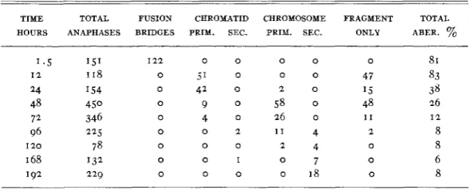
secondary dicentrics was 35 to 15 in the same material. The frequencies of all classes of aberrations for an eight day period are shown in table I.
from 1 2 to 24 hours, followed by the primary chromosome aberrations. The secondary chromatid and chromosome aberrations are found on the fourth day and persist with about the same frequency during the next four days. As indicated above, the ratio of dicentric and ring chromosomes is about the same for both primary and secondary aberrations.
TABLE I
Chromosomal aberrations indzrced in onion root t i p cells. Roots rayed. Dose 30w.
TIME TOTAL FUSION CHROMATID CHROMOSOME FRAGMENT TOTAL HOURS ANAPHASES BRIDGES PRIY. SEC. PRIY. SEC. ONLY ABER. %
I 2 2 0 0 0 0
0 5' 0 0 0
0 41 0 2 0
0 9 0 5 8 0
0 4 0 16 o
0 0 2 1 1 4
0 0 0 2 4
0 0 1 0 7
0 0 0 o 18
0
47 15 48
1 1 2
0
0
0
81 83 38 26
8 8 6 8 I 2
I n another experiment dormant onion bulbs were used so that only rest- ing cells were rayed. The bulbs were germinated, and root tips were fixed at various stages of growth. The results are shown in table 2 . Only the
dicentric bridges observed at anaphase were included in the analysis. The frequency of total aberrations declined as the roots elongated, and ulti- mately no bridges were found at anaphase in the longest roots. Other ex- periments confirm these results. Both onion bulbs and Tradescantia plants examined several weeks or months after irradiation have shown no bridge formation at anaphase in somatic cells.
TABLE 2
Chromosomal aberrations induced in onion root tip cells. Dormant bulbs. Dose 12001.
ROOT TOTAL DICENTRIC BRIDGES TOTAL LENGTH ANAPHASES PRIMARY SECONDARY ABER. %
I cm. 377
2-3 cm. 427 6-8 cm. 242 1-13 cm. 72 2-25 cm. 87
0 46
5 33
23 21
8 I 1
0 0
4 2 2 KARL SAX
somes the released acentric fragments usually are large because breaks are more frequent in the proximal end of the chromosome arm. Thus, the healing of broken ends of broken bridges of dicentrics should result in chromosomes with shortened arms. No apparent deficiencies were ob- served in the chromosomes after a number of cell generations, indicating that the cells carrying dicentric and ring chromosomes usually are elimi- nated. The failure of such cells to survive can be attributed to the large deficiencies usually associated with the formation of rings and dicentrics. Apparently these large deficiencies, even in the heterozygous condition, inhibit the growth rate so that deficient cells are unable to compete with normal cells or with cells with balanced chromosomal alterations.
Some support of the deleterious effect of deficiencies in somatic tissue is found in X-ray treatment of seedling plants. About 50 species of various ornamental plants were rayed in the seedling stages. We had hoped to produce gross chromosome deficiencies which might result in phenotypic changes of horticultural value due to unbalanced genoms. The seedlings were grown in long narrow flats so that differential dosage could be given in a single treatment. At one end of the flat the seedlings received an X-ray dosage of 5ooor and a t the other end 1500'. The maximum dose killed or greatly retarded the seedlings of most species. In each case seedlings were selected which were most severely affected, but still viable. Most of these grew slowly and were aberrant in many respects for several months, but a t the end of the growing season the great majority appeared to be quite normal in their gross morphological characters. Presumably the more aberrant cells were eliminated and only the more normal ones or those with balanced genoms took part in the later development of the irradiated seedlings.
The survival of dicentric chromosomes and the continuation of bridge formation in the developing endosperm of Zea might be attributed to the decreased deleterious effect of deficiencies in the triploid tissue. In order to test this hypothesis, a triploid Tradescantia was irradiated and the fre- quencies of aberrations were determined a t intervals. The frequency of primary bridges was 2 2 percent; a t the end of a month there were still
8 percent of secondary dicentrics, but a t the end of seven weeks all divi- sion figures appeared to be normal, and no aberrations could be found.
THE EFFECTS OF DIFFERENTIAL DOSAGE AND TREATMENT
somes is more difficult owing to greater variability and to technical dif- ficulties in detecting the types of aberrations produced. The data for one of the experiments are shown in table 3 . The dosage was increased by in- creasing the time of exposure. Although the dosage curve is not so con- sistent as those determined for Tradescantia microspore chromosomes, it appears that the aberration frequency increases approximately as the
1.5 power of the dosage.
The exponent of 1.5 in the dosage curve equation for root tip chromo- somes suggests that the time-intensity factor is operative. The time-in- tensity factor has been used only with onion seeds, but with rather sur-
TABLE 3
Frequency of chromosomal aberrations in onion root tips following various X - r a y dosage. Roots rayed. Fixed four days ajler raying.
DOSE IN TOTAL DICENTRICS TOTAL
r-UNITS ANAPHASES AND RINGS ABER %
1 5 0
300
600
8
30
68
4 9
27
4 2 4 KARL SAX
The data presented in table 4 also show that the onion bulbs are much more sensitive to X-rays than are the dry onion seeds. Although the seeds were given nearly three times the dosage given the bulbs, the aberration frequency was higher in the root tips developing from the bulbs. Consider- ing the differential dosage, the bulbs were nearly ten times as sensitive as the dry seeds, as measured by aberration frequency. In another experiment where the dry seeds and growing roots were given the same X-ray dose, the aberration frequency of the dry seed was 3 percent compared with 2 3
percent for the rayed roots. A comparison of dry seeds and dormant bulbs
TABLE 4
The eject of storage after X-ray treatment of onion seeds (r6oor) and bulbs (6007).
-
MATERIAL TREATMENT TOTAL ANAPHASES
PERCENTAGE ABERRATIONS
Seed I day
8 days Control
301 1 7 -3= 14%
3 20 3
299 31-3=28%
Bulbs March 6 216 23%
February 26 I 26 21 %
given equal X-ray doses showed about a I : IO difference in aberration fre-
quency.
The duration of the nuclear cycle in Allium root tips may be determined from the data presented in table I . The X-ray treatment does cause a
temporary cessation of nuclear activity, but after recovery from the “primary” effect the nuclear activity seems to be normal. The time lapse between successive anaphases is about three to four days as indicated by the time between the appearance of primary and secondary aberrations.
The survival of certain monosomics and other deficiencies in the hetero- zygous condition suggests that irradiation of gametes or zygotes would be more effective than irradiation of seedlings in producing plants with un- balanced genoms. At sub-lethal doses some of the seedling cells will not be altered, and those cells will grow faster and replace the cells with large chromosome deficiencies. A heterozygous deficiency in the zygote would not be subject to such competition and would survive in development if not cell lethal.
SUMMARY
Irradiation of Allium root tip cells with X-rays produces a “primary” effect which results in fusion bridges. The “secondary” effect produces breaks usually followed by the union of broken ends to produce rings, di- centrics, inversions, translocations, and interstitial deletions. Terminal deletions are produced when a break is not followed by union of broken ends of sister chromatids.
The ring and dicentric chromosomes tend to persist for several cell gen- erations but are soon eliminated, and only cells with normal or balanced genoms survive. The elimination of ring and dicentric chromosomes ap- pears to be due to the deleterious effect of the deletions associated with these aberrations so that they are unable to compete with normal or bal- anced cells.
Delayed germination of irradiated onion seed increases the frequency of chromosomal aberrations due to a physiological effect similar to the effects of age or heat, but delayed germination of irradiated onion bulbs has no effect on aberration frequency.
LITERATURE CITED
CLARK, F. J., and F. C. COPELAND, 1940 Chromosome aberrations in the endosperm of maize. GERASSIMOVA, HELEN, 1937 Interspecific translocations in Crepis. Bull. Inst. Genet. 1 1 : 143-1 78. GUSTAFSSON, A., 1937 The different stability of chromosomes and the nature of mitosis. Hereditas
12: 281-335.
LEWITSKY, C. A., 1940 A cytological study of the progeny of X-rayed Crepis capillaris Wallr. Cytologia 11: 1-29.
MCCLINTOCK, BARBARA, I 938 The production of homozygous deficient tissues with mutant characteristics by means of the aberrant mitotic behavior of ring-shaped chromosomes. Genetics 23: 315-376.
1939 The behavior of successive nuclear divisions of a chromosome broken a t meiosis. Proc. Nat. Acad. Sci. 25: 405-416.
Amer. J. Bot. 27: 247-251.
MARQUARDT, H., 1938 Die Rontgenpathologie der Mitose. Z. Bot. 32: 401-482.
MARSHAK, A., and J. C. HUDSON, 1937 Measurement of roentgen-ray dosage by determining the NICHOLS, C., 1941 Spontaneous chromosome aberrations in Allium. Genetics 26: 89-100. SAX, K., 1940 Analysis of X-ray induced chromosomal aberrations in Tradescantia. Genetics 25:
effect of radiation on chromosomes. Radiology 29: 669-675.